Article

The Effect of Ni2+ Ions Substitution on Structural, Morphological, and Optical Properties in CoCr2O4 Matrix as Pigments in Ceramic Glazes

1
Faculty of Chemistry and Chemical Engineering, Babeş-Bolyai University, 11 Arany Janos Street, 400028 Cluj-Napoca, Romania
2
Faculty of Physics, Babes-Bolyai University, 1 Kogalniceanu Street, 400084 Cluj-Napoca, Romania
3
National Institute for Research and Development of Isotopic and Molecular Technologies, 67-103 Donath Street, 400293 Cluj-Napoca, Romania
4
Electron Microscopy Center, Faculty of Biology and Geology, Babes-Bolyai University, 400006 Cluj-Napoca, Romania
*
Author to whom correspondence should be addressed.
Materials 2022, 15(24), 8713; https://doi.org/10.3390/ma15248713
Submission received: 11 November 2022 / Revised: 30 November 2022 / Accepted: 4 December 2022 / Published: 7 December 2022

Abstract

:
The structural, morphological, and optical properties of Ni2+ ions substitution in CoCr2O4 matrix as ceramic pigments were investigated. The thermal decomposition of the dried gel was performed aiming to understand the mass changes during annealing. The X-ray diffraction (XRD) studies reveal a spinel-type Face–Centered Cubic structure and a secondary Cr2O3 phase when x ≤ 0.75 and a Body–Centered Tetragonal structure when x = 1. Fourier Transform Infrared Spectroscopy (FT–IR) indicated two strong absorption bands corresponding to the metal–oxygen stretching from tetrahedral and octahedral sites, characteristic of spinel structure. Ultraviolet–Visible (UV–Vis) spectra exhibited the electronic transitions of the Cr2+ Cr3+ and Ni2+ ions. From the UV–Vis data, the CIE color coordinates, (x, y) of the pigments were evaluated. The morphology was examined by Scanning Electron Microscopy (SEM) and Transmission Electron Microscopy (TEM) showing the agglomeration behavior of the particles. The stability, coloring properties and potential ceramic applications of studied pigments were tested by their incorporation in matte and glossy tile glazes followed by the application of obtained glazes on ceramic tiles. This study highlights the change in pigment color (from turquoise to a yellowish green) with Ni2+ ions substitution in the CoCr2O4 spinel matrix.

1. Introduction

The color of ceramic tiles, followed by the design and the technical properties, is one of the most appreciated characteristics by the customer. Thereby, there is a real need in diversifying the ceramic color palette. In addition, the use of the newer technologies for obtaining easier, faster, and qualitative colored models on tiles keeps the research in the field of ceramic pigments open. Nowadays, the demand is still on in finding new materials suitable for the challenge created by the digital decoration ink-jet printing for ceramic tiles. This application allows the use of colloidal suspensions of pigments to improve decorative features of ceramic parts and other ceramic tiles [1].
The ceramic pigments are inorganic materials which have high thermal and chemical stability. These properties allow them to be subjected to the processing conditions without losing their coloring characteristics.
Spinel compounds have a huge contribution in the field of ceramic pigments to obtain different colors like red, pink, brown, gray, or blue.
Spinels with the general formula AB2O4 represent a large family of inorganic compounds which are now used in different industrial branches as ceramic pigments [2,3,4,5], magnetic materials [6,7], catalytic materials [8], supercapacitors [9,10], due to their excellent chemical, thermal, mechanical, magnetic properties, and high melting point. Although spinels have a complex composition, they are intrinsically colored and present several advantages, when compared to the metal oxides as colorants in ceramics and enamels.
The properties of the oxide materials and consequently of spinels, are strongly correlated with the synthesis method and thermal behavior, which affects the interspersion of the components and thus defines the morphology [11]. This also influences the coloring properties of pigments.
In general, the synthesis methods employed depend especially on the final use of the material and are based on the relation between structure—morphology and properties. Consequently, there are different methods utilized in the synthesis of spinel structures, the conventional ones (co-precipitation and hydro/solvo-thermal) and non-conventional ones (sol-gel and derivatives or auto-combustion) [12,13,14]. Among them, the sol-gel synthesis offers great versatility to prepare low-cost materials with a rigorous control from the compositional and microstructural point of view. The sol-gel method is based on inorganic polymerization reactions and so, starting from molecular precursors an oxide can be easily obtained via hydroxylation-condensation reactions [1,12].
Spinel pigments can be obtained by the addition of chromophores, transition metal ions into inert matrices such as oxides or by calcination of metal-organic complexes [15]. They have the general formula AB2O4 where the A-sites are tetrahedral coordinated occupied by divalent ions as A2+ (like Co, Ni, Mn, Zn), and the B-sites are octahedral coordinated and occupied by trivalent ions B3+ (like Cr or Al) [16].
In the literature, there are studies regarding spinel oxides such as CoCr2O4, MgCr2O4, Co(1−x)MgxCr2O4 [17,18] and NiCr2O4 [19] which have been synthesized by different methods and are investigated for different applications due to their thermal stability, as previously noted.
Nowadays, for the blue pigment, cobalt-based materials are used [1]. There are few drawbacks for the use of cobalt, i.e., abundance, extraction, geopolitical instabilities, and the need to be used in the electronic or other industries.
In the present context, the aim of this work is to synthesize and to investigate new competitive pigments for novel technologies with as little cobalt as possible. Considering that, we studied the nickel substitution in the cobalt chromite matrix. For this investigation, an easy to use and environmentally friendly modified sol-gel method was employed for the synthesis of Co(1−x)NixCr2O4 (x = 0, 0.25, 0.5, 0.75 and 1).
Structural, morphological, and optical properties were studied as a function of the nickel substitution in the spinel matrix focusing on the application as ceramic pigments.
The proposed modified sol-gel method involves metallic salts combined with the sucrose and pectin as organic precursors. The method was successfully utilized in the synthesis of other spinel oxide compounds with nanometric dimensions [20].
More than obtaining these chromite spinel pigments through an easy, cost-effective method for the first time, the aim of this work was to test their potential applicability in the ceramic tile industry. For this purpose, all synthetized pigments were embedded in two different types of glazes to also test their efficacity as ceramic pigments. The novelty of this work is to investigate the viability of all synthesized pigments to be successfully used in matte and glossy glazes applied on tiles. The coloring properties were successfully demonstrated for all samples embedded in both types of glazes.

2. Materials and Methods

2.1. Materials

The following precursors were used for the synthesis of pigments: Co(NO3)2·6H2O (99.5% purity, Merck, Darmstadt, Germany Merk), Ni(CH3COO)2·4H2O (99.5% purity, Merck, Darmstadt, Germany) and (NH4)2Cr2O7 (99.5% purity, Merck, Darmstadt, Germany). All the reagents used in this experiment are of high analytical grade and they were used as purchased, without further purification. HNO3 (65%, Merck, Darmstadt, Germany) was used as a pH regulator. The sucrose and pectin used to support the condensation reaction were commercial food grade.

2.2. Synthesis of Pigments

Concentrated precursor solutions were prepared using the metal salts (Co(NO3)2·6H2O, Ni(CH3COO)2·4H2O and (NH4)2Cr2O7) dissolved in distilled water. Separately, another solution containing the sucrose with the ratio sucrose: grams of final oxide (wt.:wt.) of 2:1 was prepared. The sol-gel process starts when mixing the precursor solutions with sucrose solution and pectin to form a gel. The addition of sucrose and pectin to the precursor solution containing the metal cations forms a polymer matrix in which the metal cations are distributed through the polymeric network structure [20]. This mechanism, and the role played by sucrose and pectin in the formation of the oxide structures, is discussed in more detail in reference [20].
The hydrolysis process occurs during the vigorous magnetic stirring (1000 RPM) of the metal solutions under strict temperature control (at 40–45 °C) and with a pH correction to around 1–1.5. After stirring, the obtained sol is left to age for 24 h at 80 °C to ensure the formation of the gel lattice, with the elimination of the water present in the pores and the final formation of a porous structure. The thermal treatment of the dried gels was performed in an electric furnace, in porcelain crucibles. The furnace temperature had an increase rate of 300 °C/h, with an isotherm plateau of 30 min, at 1000 °C.

2.3. Analysis Methods

The mass loss and the phase transformations occurring during the heating of the dried gel were investigated by thermal analyses (TG-DTA) employing a SDTQ600 TA Instruments (TA Instruments New Castle, DE, USA) thermal analyzer.
The thermal analysis was performed on a sample of about 10 mg placed in an alumina crucible and non-isothermally heated from 30 °C to 1000 °C at a heating rate of 10 °C/min in dynamic flow-air.
The structural characterization was carried out at room temperature by powder X-ray diffraction using a Bruker D8 Advance AXS diffractometer (Karlsruhe, Germany) with Cu Κα radiation in the 20–80° 2θ region. Crystallite size, cell arrangements and phase fractions were calculated by Rietveld refinement analysis using FullProf Suit Software (FullProff suite July–2017).
The FT-IR absorption spectra were recorded with a JASCO FTIR 6200 spectrometer in the 400–1500 cm−1 spectral range, with a standard resolution of ±2 cm−1.
Scanning Electron Microscopy (SEM, Chiyoda, Tokyo, Japan) and Transmission Electron Microscopy (TEM, Chiyoda, Tokyo, Japan) were performed employing a combined electron scanning (SE, Chiyoda, Tokyo, Japan) and transmission (TE) Hitachi HD–2700 electron microscope (Chiyoda, Tokyo, Japan) operated at a maximum acceleration voltage of 200 kV. The energy–dispersive X-ray spectrometry (EDX) was used to obtain the images with the nickel distribution in the glossy ceramic glaze.
The absorption spectra of the powder samples were obtained using a Perkin–Elmer Lambda 45 UV/Vis spectrometer (Waltham, MA, USA) with integrating sphere using the pellet technique. The powder samples mixed with BaSO4 were uniaxial press in a pellet matrix using a load force of 10 tons/cm2 to form transparent disks with diameter of 13 mm, and 2 mm thick. The spectra were recorded in the 200–850 nm wavelength range, with the wavelength accuracy ±2 nm.
The stability of pigments was checked by incorporating them in both matte and glossy tile glaze, using a 2% pigment addition. Thermal treatment for glaze melting was performed at 1200 °C for 6 h at maximum temperature. The glazes were characterized using a color spectrophotometer spectro–guide series (BYK Gardner, Los Angeles, CA, USA).

3. Results

3.1. Thermal Analysis

The thermal decomposition of dried precursor gel was investigated to understand the details of the decomposition process, but most importantly, to determine the temperature at which nucleation and crystallization takes place. This is imperative from a technological point of view and the potential application of the studied pigments in the industry. The thermogravimetric (TG) and differential thermal analysis (DTA) thermograms (20–1000 °C) of the CoCr2O4 dried gel are presented in Figure 1.
The decomposition of the dried gel follows two steps:
  • the removal of the residual water (the absorbed and the coordinated water) with a mass loss of about 5.09% (30–61 °C),
  • the weight loss of the organic fragment with formation of volatile compounds, with a total mass loss of approximately 9.13% (61–1000 °C).
A significant mass loss takes place up to 400 °C, after which no significant losses are recorded. Overall, the total mass loss at 1000 °C was 15.02% and is assigned to the drying process of the gel, in which much of the organic precursor has been removed. Prior to 900 °C, a small mass loss of 1.03% is attributed to the volatile compounds formed and remained inside the material pores. The DTA curve presents a big exothermic peak which unfolds in the 20–800 °C range confirm the two listed decomposition steps. The peak corresponding to the elimination of the organic fragments can be described by the following temperatures: Tonset = 318.35 °C, Tpeak = 331.02 °C and Tend = 352.76 °C.
The dried gels were subjected to thermal treatment at 1000 °C for 30 min to ensure the compound crystallization.

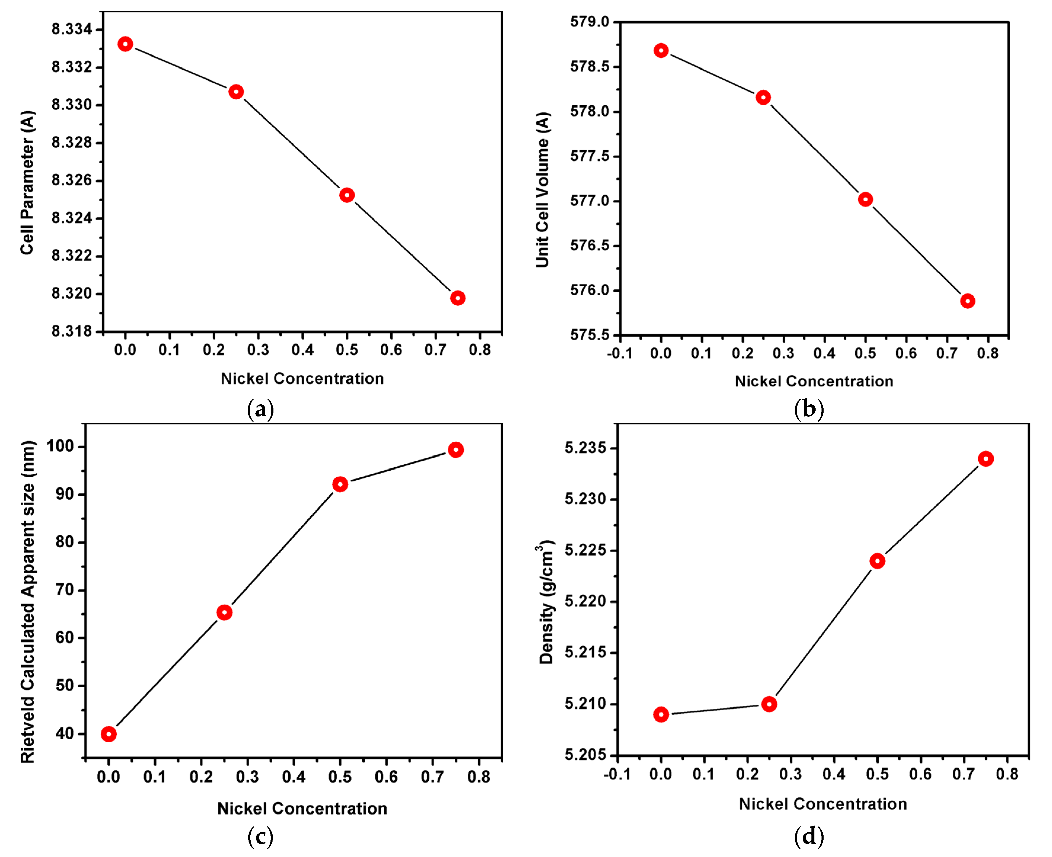
3.2. Structural Characterization of Spinels

Figure 2 presents the XRD patterns of the Ni2+ ions substitution in CoCr2O4 spinel matrix after the thermal treatment in air atmosphere at 1000 °C for 30 min.
Both NiCr2O4 and CoCr2O4 oxides crystallize into normal spinel structures in the AB2O4 form with Co2+ and Ni2+ ions occupying the tetrahedral A sites and Cr3+ ions occupying octahedral B sites.
The XRD patterns present only intense and sharp peaks characteristic to the crystalline phases.
The Co(1−x)NixCr2O4 spinel (when x ≠ 1) crystallizes in Face-Centered Cubic structure and is indexable to the space group Fd-3m (no. 227). This is consistent with the standard values of Face-Centered Cubic phase (00-022-1084 PDF files). On the other hand, NiCr2O4 crystalizes in the Body-Centered Tetragonal structure, the space group I41/amd (no. 141). So, a transition from the cubic spinel (when x < 1) to a Body Centered Tetragonal structure (when x = 1) is observed.
For samples with x < 1 the XRD data analysis indicated the formation of two crystalline phases: a well crystallized phase Co(1−x)NixCr2O4 spinel with similar structure to CoCr2O4 (00-022-1084 PDF files) as the major phase, and Cr2O3 (00-038-1479 PDF files) as a secondary phase. Crystallinity dependent properties of crystals are attributed to their crystallite size. Larger crystallites develop sharper peaks on the XRD pattern for each crystal plane. The width of a peak is related to its crystallite size [21]. The formation of Cr2O3 impurity phase is common during the synthesis process of NiCr2O4 and is often reported in the literature [22,23].
The XRD peaks at 2θ values observed in the NiCr2O4, pattern, match the (112), (200), (103), (211), (202), (004), (220), (312), (106), (321), (224) and (400) crystalline planes of the Body-Centered Tetragonal spinel structure (01-088-0109 PDF files). No other secondary peaks were detected, suggesting that the Ni2+ substitution occurred.
The Rietveld refinement fittings were carried out to investigate the changes in the crystal structure by using FullProf software. The experimental recorded data points are represented with black circles, the structural model fit is represented with a red solid line, the structural model fit is represented by the blue solid line and the vertical green bars represent the Bragg positions. Up until the best possible convergence, the refinement was continued. The good fit is evidenced in Figure 3 where the small variance in the difference curve can be observed.
The average crystallite sizes, the lattice parameters and the average strain and standard deviation calculated with the FullProf software are listed in Table 1 and Table 2. Thereby, the average crystallite sizes increase with the increase in concentration of Ni2+ ions in Co(1−x)NixCr2O4 spinel starting from 39.9 nm when x = 0 and reaching 99.42 nm when x = 0.75. The average crystallite size of NiCr2O4 (when x = 1) was 58.98 nm.
The evolution of the average crystallite size, cell parameter, density, and unit cell volume as function of nickel concentration are represented in Figure 4. Thereby, the lattice constant value of Co(1−x)NixCr2O4 spinel when x ≤ 0.75 structure was measured to be 8.33 Å, and it is in concordance with the literature data [24]. This large value of the lattice constant was assigned to the disordering of cations in the spinel structure of CoCr2O4 and due to the exchange of tetrahedral A-site Co2+ ions with octahedral B-site Cr3+ ions [2].
When the Ni2+ content is increased, the lattice parameter and volume unit decrease (Figure 4a,b). This decrease is assigned to the difference of ionic radius of Ni2+ and Co2+, i.e., with nickel having a smaller radius. Similarly, the density increases with the increase of nickel concentration (Figure 4d).
Considering the targeted application as pigments, the crystallite size plays a determining role in the coloration capacity as being conditioned/size dependent on the specific surface.

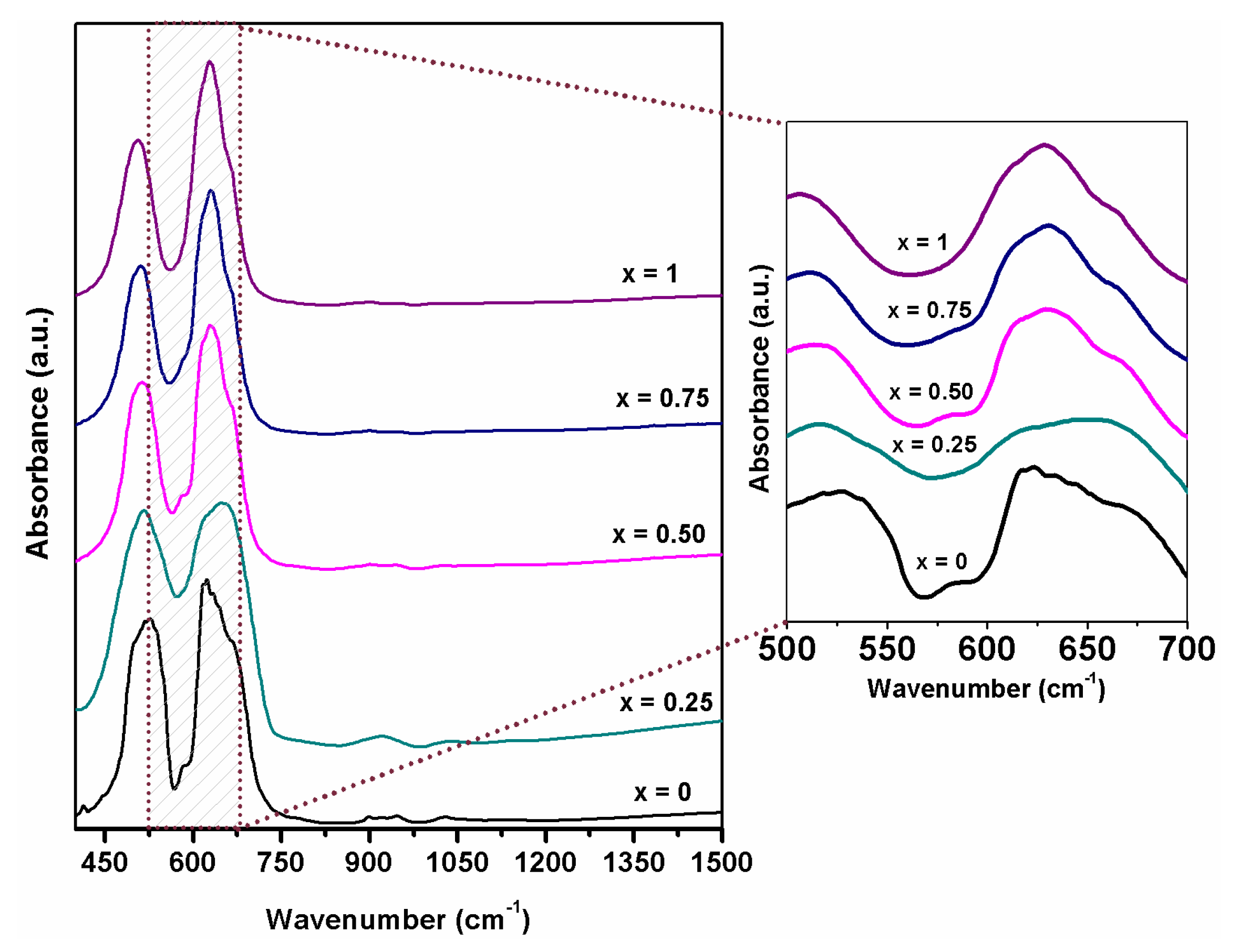
3.3. FT–IR Spectroscopy

The FT–IR spectra of Co(1−x)NixCr2O4 (x = 0, 0.25, 0.5, 0.75 and 1) samples are presented in Figure 5. Two strong absorption bands centered at 516, 620 cm−1 with a shoulder at 666 cm−1 are observed. These absorption bands are the spinel characteristic peaks and depend on the vibration of the cations at the B site [25,26,27].
As can be observed in FTIR spectra, the peak centered at 525 cm−1 from the sample with x = 0 shifts to a lower frequency, at 504 cm−1, with the increasing nickel content x = 1 (in NiCr2O4). The crystal theory of the field stabilization energy advises that the Cr3+ and Co2+ are incorporated into the octahedral and tetrahedral interstices site and compared to the Cr3+ and Ni2+, which by occupying the octahedral interstices site own a smaller ligand gravity and splitting energy of d orbital due to the smaller amount of electric charge will cause the peak to shift to lower frequencies [25,26,28]. The second peak at 620 cm−1 also shifts towards lower frequencies with the increase of nickel content and has the same origin as the peak from 516 cm−1 [25]. Minor changes in the observed band shape can be assigned to the particle size, a similar behavior being observed in CoCr2O4 samples presented in the literature [29].

3.4. Morphological Characteristics

SEM and TEM micrographs of Co(1−x)NixCr2O4 samples, with x = 0, 0.25, 0.5, 0.75 and 1, are presented in Figure 6. All samples have the tendency to agglomerate their small particles generating irregular and larger aggregates with various rhombohedral-like shapes. Additionally, no significant differences in the spinel microstructure with the increase of Ni2+ ion content was observed, suggesting a similar morphology for all compositions. The estimated crystallite sizes, according to TEM analysis, confirm the calculated values from XRD.

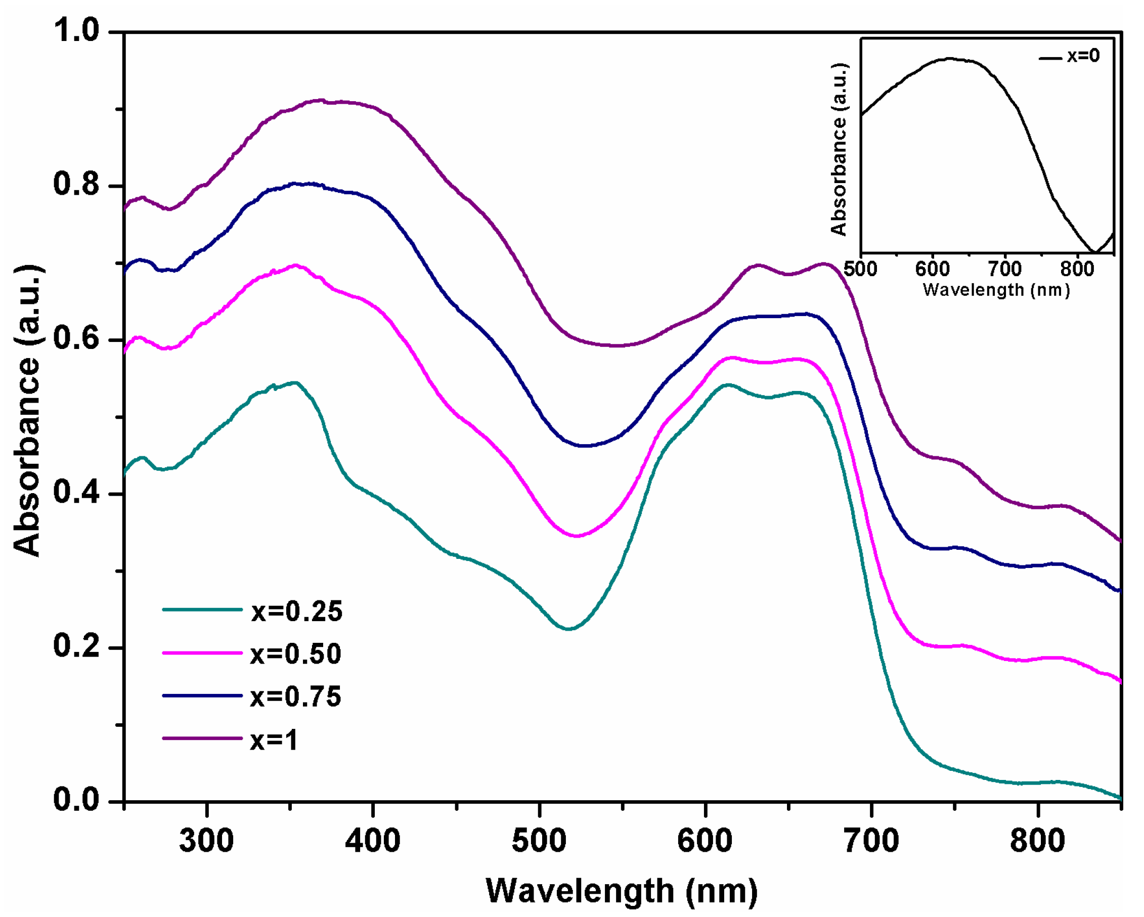
3.5. UV-Vis Spectroscopy

The Ni2+ substitution of Co2+ in CoCr2O4 matrix was investigated by UV-VIS spectroscopy. The optical absorption spectra of the Co(1−x)NixCr2O4 (x = 0, 0.25, 0.50, 0.75 and 1) recorded in the wavelength range from 250–850 nm are presented in Figure 7. The recorded spectra of the synthesized nanoparticles are very similar to the spectra of CoCr2O4 [29] and, also of NiCr2O4 [29] as reported in the literature. The band around 259 nm can be attributed to the charge-transfer transitions between O2− and Cr3+ [17,29].
The incorporation of nickel ions in the CoCr2O4 matrix leads to the broadening of the band centered around 350 nm, band assigned to a charge transfer of Ni2+ cation [25,30] and to a decrease in intensity of the bands in the 574–700 nm wavelength range. Additionally, the absorption bands of Co(1−x)NixCr2O4 samples centered at 574 nm, 612 nm, and 660 nm shifts with 10 nm to higher wavelengths and the band centered at 760 nm shifts to lower wavelengths with the increase of Ni2+ content.
The shoulder present in all the spectra at around 460 nm is attributed to the intrinsic d-d transition of Cr3+ ions [31,32,33]. A decrease in the intensity of the absorption bands at wavelengths higher than 500 nm was observed with the increase of Ni2+ ions in the matrix. The absorption bands at about 574 nm, 612 nm are frequently observed in cobalt chromite oxides and can be attributed to the spin-allowed electronic transition 4A2(F)→4T1(P) of Co+2 ions in A site. The absorption band from 612 nm it is also assigned to the transition 4A2g4T2g of Cr3+ ion at the B site [17,25]. The band from 660 nm and 750 nm corresponds to 4A24T2 and 3A23T1 transitions and are due to the d-d transition of Ni2+ ions in the tetrahedral coordinated O2− environment [20,32]. The bands from about 750 nm and 820 nm become more visible with the increase of Ni2+ ions in the matrix and confirm the incorporation of Ni2+ in tetrahedral coordination [34].

3.6. CIE Diagram of the Obtained Pigments

The UV-Vis data were used to determinate the CIR color coordinates, (x, y) of the pigment samples. The obtained color parameters are listed in Table 3 and represented in the CIE diagram illustrated in Figure 8. As can observed, the color coordinates slightly vary from (0.3865, 0.3295) when x = 0.25 to (0.3118, 0.2762) when x = 1 and ranging in between for x = 0.5 and 0.75. Increasing the nickel concentration in the matrix leads to a shift from a pink-turquoise to a greenish-blue color. This observation is correlated with the literature data [10] and the UV-Vis absorption spectra described previously.
The pigments were embedded in both matte and glossy tile glazes to test their applications in the ceramic tile industry. Thereby, the homogeneous glasses were prepared by incorporating spinel nanopowders into ceramic glazes which were further applied on ceramic substrates and were finally subjected to thermal treatments.
Figure 9 presents a more comprehensive view of the pigments before and after their embedding. As can be observed, the pigment powders exhibit bluish tones that tend to become greener with the progression of Ni2+ substitution up to a yellowish green with the full Co2+ replacement.
The color of pigment powders and those embedded in glazes, in terms of color parameters (L* = lightness, a* = red-green axe values, b* = yellow-blue axe values, G* = gloss) are presented in Table 4. A major difference that can be observed is that of the ‘L’ parameter, that ranges between 31.09 and 33.49 for the matte glaze and from 40.69 to 45.72 for the glossy one. The gloss parameter (‘G’) is lower for matte glazes (50.5–58.1) and higher for glossy ones (83.8–86.8), due to the nature of the glaze itself. This can be explained by the fact that the latter reflects light due to its smoother surface, whereas the former tends to scatter it. Therefore, pigments embedded in glossy glazes will naturally become ‘brighter’ in nature.
The a* and b* coordinates of the glazes increase with increasing nickel substitution in pigment which impacts the color by his chromophore capacity and also by the impact on particle size. Generally, this ranging can also be correlated with different synthesis methods, with different factors such as synthesis conditions (temperature, stoichiometry, and pH) or particle size. For example, the growth in b* value coordinate was correlated with the increase in particle size in CoAl2O4 for blue pigment [1]. In glazes, b* values are highly reduced to a range of −6.7 and −0.4, also with greener hues [1,35].
An increase in a* (red-green) and b* (yellow-blue) values can also be observed with an increase in Ni substitution. Higher a* values tend to lead to yellowish-brown hues and higher b* values to reddish-brown ones. This can be better observed by the color progression in Figure 9. Once embedded in the glazes, the color of the pigment becomes darker and more intense due to thermal treatment.
The experimental results indicate that the synthesized Co(1−x)NixCr2O4 spinel (0.25 ≤ x ≤ 1) oxide nanopowders can be successfully used as ceramic pigments due to their coloration capacity and thermal resistance. The thermal and chemical stability of the Co(1−x)NixCr2O4 spinel pigment in the glazes is high enough to obtain a uniform color distribution of the product and is not affected by melting in the firing process.

3.7. EDX Mapping

The nickel substitution in the spinel was evidenced after embedding in glossy ceramic tile by using the Energy-Dispersive X-ray Spectrometry (EDX) from SEM. Figure 10 presents the EDS element layered image including Ni Kα1 of the ceramic glossy tile cross-section as a function of nickel substitution in the spinel. A uniform distribution of the nickel in the cross-section of the glaze is observed for all samples and the partial and total substitution of nickel is evidenced in Figure 10.

4. Conclusions

The paper reports structural, morphological, and optical properties of new ceramic pigments-based spinel structure, obtained by sol-gel route. The structural characterization is consistent with the substitution of chromophores Co2+ ions with the Ni2+ ions in the CoCr2O4 matrix. The XRD analysis revealed the formation of Co(1−x)NixCr2O4 spinel-type Face-Centered Cubic structure as a principal phase and Cr2O3 as a secondary one when x ≤ 0.75. A decrease in unit cell parameter and the unit cell volume was achieved with an increase of x. When x = 1, NiCr2O4 crystallizes in the Body-Centered Tetragonal spinel as a single phase when compared to lower ‘x’ values when a secondary phase appears. The increase of nickel substitution in the matrix has a pronounced increase in the size of the crystallites from 39.9 nm (for x = 0) to 99.42 nm (for x = 0.75). FT-IR spectra confirmed the spinel structure formation and the elimination of all the organic fragments. The UV-Vis absorption spectra presented the bands corresponding nickel ions located at the A site and the chromium ions located at the B sites. Adjusting the nickel content in the CoCr2O4 matrix the color of the pigment can be easily controlled, as it can also be seen in the CIE diagram chromaticity. SEM and TEM microscopy confirmed evidence of the powder morphology and the tendency of nanoparticles to agglomerate. The CIELab coordinates of the pigments embedded in glossy and matte tile glazes reveal the color ranging of the two glazes. Additionally, the elemental EDX distribution of Ni Kα1 confirms the homogeneous and uniform distribution of the pigment in the glossy glaze after firing. The obtained pigments can be successfully applied in glaze tiles and ceramics.

Author Contributions

Conceptualization and methodology, validation, investigation, data curation, supervision, F.G.; writing—original draft preparation, R.A.M.; validation, investigation, data curation, supervision, writing—review and editing, R.A.M., A.A., R.A.B. and M.Z., investigation, data curation, L.B.T. All authors have read and agreed to the published version of the manuscript.

Funding

This research received no external funding.

Institutional Review Board Statement

Not applicable.

Informed Consent Statement

Not applicable.

Data Availability Statement

Not applicable.

Acknowledgments

This work was partially supported by a grant of the Ministry of Research, Innovation and Digitization: Nucleu-Program, project number PN 19 35 01 01.

Conflicts of Interest

The authors declare no conflict of interest.

References

  1. Enríquez, E.; Reinosa, J.; Fuertes, V.; Fernández, J. Advances and challenges of ceramic pigments for inkjet printing. Ceram. Int. 2022, 48, 31080–31101. [Google Scholar] [CrossRef]
  2. Mindru, I.; Gingasu, D.; Marinescu, G.; Patron, L. Design de Nanomateriale Oxidice cu Structura Spinelica. De la Sinteza la Aplicatii, 1st ed.; Matrixrom: Bucharest, Romania, 2018; pp. 1–148. (In Romanian) [Google Scholar]
  3. Livage, J.; Sanchez, C.; Henry, M.; Doeuff, S. The chemistry of the sol-gel process. Solid State Ionics 1989, 32–33, 633–638. [Google Scholar] [CrossRef]
  4. Ilosvai, A.M.; Dojcsak, D.; Váradi, C.; Nagy, M.; Kristály, F.; Fisercz, B.; Vanyorek, L. Sonochemical Combined Synthesis of Nickel Ferrite and Cobalt Ferrite Magnetic Nanoparticles and Their Application in Glycan Analysis. Int. J. Mol. Sci. 2022, 23, 5081. [Google Scholar] [CrossRef] [PubMed]
  5. Chen, T.-W.; Tamilalagan, E.; Chen, S.M.; Akilarasan, M.; Maheshwaran, S.; Liu, X. An Ultra-Sensitive Electrochemical Sensor for the Detection of Carcinogen Oxidative Stress 4-Nitroquinoline N-Oxide in Biologic Matrices Based on Hierarchical Spinel Structured NiCo2O4 and NiCo2S4; A Comparative Study. Int. J. Mol. Sci. 2020, 21, 2373. [Google Scholar] [CrossRef] [PubMed]
  6. Younis, M.; Saleem, M.; Atiq, S.; Naseem, S. Magnetic phase transition and magneto-dielectric analysis of spinel chromites: MCr2O4 (M = Fe, Co and Ni). Ceram. Int. 2018, 44, 10229–10235. [Google Scholar] [CrossRef]
  7. Dippong, T.; Levei, E.A.; Deac, I.G.; Petean, I.; Cadar, O. Dependence of Structural, Morphological and Magnetic Properties of Manganese Ferrite on Ni-Mn Substitution. Int. J. Mol. Sci. 2022, 23, 3097. [Google Scholar] [CrossRef]
  8. Zhou, X.; Zhang, X.; Zou, C.; Chen, R.; Cheng, L.; Han, B.; Liu, H. Insight into the Effect of Counterions on the Chromatic Properties of Cr-Doped Rutile TiO2-Based Pigments. Mater 2022, 15, 2049. [Google Scholar] [CrossRef]
  9. Tena, M.Á.; Mendoza, R.; Trobajo, C.; García-Granda, S. Cobalt Minimisation in Violet Co3P2O8 Pigment. Materials 2022, 15, 1111. [Google Scholar] [CrossRef] [PubMed]
  10. Mohanty, P.; Prinsloo, A.R.E.; Doyle, B.P.; Carleschi, E.; Sheppard, C.J. Structural and magnetic properties of (Co1–xNix)Cr2O4 (x = 0.5, 0.25) nanoparticles. AIP Adv. 2018, 8, 056424. [Google Scholar] [CrossRef] [Green Version]
  11. Anju, Y.R.S.; Pötschke, P.; Pionteck, J.; Krause, B.; Kuritka, I.; Vilcáková, J.; Škoda, D.; Urbánek, P.; Machovský, M.; Masar, M.; et al. CuxCo1−xFe2O4 (x = 0.33, 0.67, 1) Spinel Ferrite Nanoparticles Based Thermoplastic Polyurethane Nanocomposites with Reduced Graphene Oxide for Highly Efficient Electromagnetic Interference Shielding. Int. J. Mol. Sci. 2022, 23, 2610. [Google Scholar] [CrossRef]
  12. Novikov, V.A.; Xanthopoulou, G.G.; Amosov, A.P. Solution Combustion Synthesis of Nanostructured NiCr2O4 Spinel and Its Catalytic Activity in CO Oxidation. Int. J. Self-Propagating High-Temp. Synth. 2021, 30, 246–250. [Google Scholar] [CrossRef]
  13. Wang, C.; Zhou, E.; He, W.; Deng, X.; Huang, J.; Ding, M.; Wei, X.; Liu, X.; Xu, X. NiCo2O4-Based Supercapacitor Nanomaterials. J. Nanomater. 2017, 7, 41. [Google Scholar] [CrossRef] [PubMed] [Green Version]
  14. Xu, X.; Gao, J.; Hong, W. Ni-based chromite spinel for high-performance supercapacitors. RSC Adv. 2016, 6, 29646–29653. [Google Scholar] [CrossRef]
  15. Dippong, T.; Levei, E.A.; Goga, F.; Petean, I.; Avram, A.; Cadar, O. The impact of polyol structure on the formation of Zn0.6Co0.4Fe2O4 spinel-based pigments. J. Sol-Gel Sci. Technol. 2019, 92, 736–744. [Google Scholar] [CrossRef]
  16. Lazau, I.; Pacurariu, C.; Ecsedi, Z.; Ianos, R. Metode Neconvenţionale Utilizate în Sinteza Compuşilor Oxidici, 1st ed.; Politehnica: Timisoara, Romania, 2006; pp. 1–500. (In Romanian) [Google Scholar]
  17. Chavarriaga, E.; Lopera, A.; Bergmann, C.; Alarcónd, J. Effect of the substitution of Co2+ by Mg2+ on the color of the CoCr2O4 ceramic pigment synthesized by solution combustion. Bol. Soc. Esp. Ceram. Vidr. 2020, 59, 176–184. [Google Scholar] [CrossRef]
  18. Grazenaite, E.; Pinkas, J.; Beganskiene, A.; Kareiva, A. Sol–gel and sonochemically derived transition metal (Co, Ni, Cu, and Zn) chromites as pigments: A comparative study. Ceram. Int. 2016, 42, 9402–9412. [Google Scholar] [CrossRef]
  19. Enhessari, M.; Salehabadi, A.; Khanahmadzadeh, S.; Arkat, K.; Nouri, J. Modified Sol-Gel Processing of NiCr2O4 Nanoparticles; Structural Analysis and Optical Band Gap. High Temp. Mater. Process. 2017, 36, 121–125. [Google Scholar] [CrossRef]
  20. Suciu, C.; Hoffmann, A.C.; Vik, A.; Goga, F. Effect of calcination conditions and precursor proportions on the properties of YSZ nanoparticles obtained by modified sol–gel route. J. Chem. Eng. 2008, 138, 608–615. [Google Scholar] [CrossRef]
  21. Abdullah, M.M.; Rajab, F.M.; Al-Abbas, S.M. Structural and optical characterization of Cr2O3 nanostructures: Evaluation of its dielectric properties. AIP Adv. 2014, 4, 027121. [Google Scholar] [CrossRef]
  22. Mohanty, P.; Sheppard, C.J.; Prinsloo, A.R.E.; Roos, W.D.; Olivi, L.; Aquilanti, G. Effect of cobalt substitution on the magnetic properties of nickel chromite. J. Magn. Magn. Mater. 2018, 451, 20–28. [Google Scholar] [CrossRef]
  23. Ptak, M.; Maczka, M.; Gągor, A.; Pikul, A.; Macalik, L.; Hanuza, J. Temperature-dependent XRD, IR, magnetic, SEM and TEM studies of Jahn–Teller distorted NiCr2O4 powders. J. Solid State Chem. 2013, 201, 270–279. [Google Scholar] [CrossRef]
  24. Castiglioni, G.L.; Minelli, G.; Portab, P.; Vaccari, A. Synthesis and Properties of Spinel-Type Co–Cu–Mg–Zn–Cr Mixed Oxides. J. Solid State Chem. 2000, 152, 526–532. [Google Scholar] [CrossRef]
  25. Gao, Y.; Chang, H.; Wu, Q.; Wang, H.-Y.; Pang, Y.-B.; Liu, F.; Zhu, H.-J.; Yun, Y.-h. Optical properties and magnetic properties of antisite-disordered Ni1−xCoxCr2O4 spinels. Trans. Nonferrous Met. Soc. China 2017, 27, 863–867. [Google Scholar] [CrossRef]
  26. Wang, Z.; Saxena, S.; Lazor, P.; O’Neill, H. An in-situ Raman spectroscopic study of pressure induced dissociation of spinel NiCr2O4. J. Phys. Chem. Solids 2003, 64, 425–431. [Google Scholar] [CrossRef]
  27. Ahmadyari-Sharamin, M.; Hassanzadeh-Tabrizi, S. Polyacrylamide gel synthesis, characterization, and optical properties of Co1−xNixCr2O4 spinel nanopigment. J. Sol-Gel Sci. Technol. 2021, 99, 534–545. [Google Scholar] [CrossRef]
  28. Han, A.; Ye, M.; Zhang, Z.; Liao, J.; Li, N. Crystal structure and optical properties of CoCr2O4–NiCr2O4 solid solutions prepared by low-temperature combustion synthesis method. Adv. Mater. Res. 2013, 616–618, 1877–1881. [Google Scholar] [CrossRef]
  29. Mączka, M.; Ptak, M.; Kurnatowska, M.; Hanuza, J. Synthesis, phonon and optical properties of na-nosized CoCr2O4. Mater. Chem. Phys. 2013, 138, 682–688. [Google Scholar] [CrossRef]
  30. El-Kemary, M.; Nagy, N.; El-Mehasseb, I. Nickel oxide nanoparticles: Synthesis and spectral studies of interactions with glucose. Mater. Sci. Semicond. Process. 2013, 16, 1747–1752. [Google Scholar] [CrossRef]
  31. Liang, S.-t.; Zhang, H.-l.; Luo, M.-t.; Luo, K.-j.; Li, P.; Xu, H.-b.; Zhang, Y. Colour performance investigation of a Cr2O3 green pigment prepared via the thermal decomposition of CrOOH. Ceram. Int. 2014, 40, 4367–4373. [Google Scholar] [CrossRef]
  32. Singh, J.; Kumar, R.; Verma, V.; Kumar, R.K. Role of Ni2+ substituent on the structural, optical and magnetic properties of chromium oxide (Cr2-x NixO3) nanoparticles. Ceram. Int. 2020, 46, 24071–24082. [Google Scholar] [CrossRef]
  33. Li, L.; Yan, Z.F.; Lu, G.Q.; Zhu, Z.H. Synthesis and structure characterization of chromium oxide prepared by solid thermal decomposition reaction. J. Phys. Chem. B 2006, 110, 178–183. [Google Scholar] [CrossRef] [PubMed]
  34. Tripathi, V.K.; Nagarajan, R. Influencing Optical and Magnetic Properties of NiCr2O4 by the Incorporation of Fe(III) for Cr(III) Following Epoxide Gel Synthesis. J. Electron. Mater. 2019, 48, 1139–1146. [Google Scholar] [CrossRef]
  35. Costa, G.; Ribeiro, M.; Hajjaji, W.; Seabra, M.; Labrincha, J.; Dondi, M.; Cruciani, G. Ni-doped hibonite (CaAl12O19): A new turquoise blue ceramic pigment. J. Eur. Ceram. Soc. 2009, 29, 2671–2678. [Google Scholar] [CrossRef]
Figure 1. The TG and DTA curves for CoCr2O4 dried gel.
Materials 15 08713 g001
Figure 2. XRD patterns for Co(1−x)NixCr2O4 pigments annealed at 1000 °C in air.
Materials 15 08713 g002
Figure 3. Rietveld analysis of powder-diffraction pattern of (a) CoCr2O4 (b) Co0.75Ni0.25Cr2O4 (c) Co0.5Ni0.5Cr2O4 (d) Co0.25Ni0.75Cr2O4 (e) NiCr2O4.
Materials 15 08713 g003
Figure 4. Variation of (a) cell parameter, (b) unit cell volume with the nickel concentration, (c) Rietveld Calculated Apparent size and density (d).
Materials 15 08713 g004
Figure 5. FT-IR spectra of Co(1−x)NixCr2O4 (x = 0, 0.25, 0.5, 0.75 and 1).
Materials 15 08713 g005
Figure 6. SEM and TEM images of (a) CoCr2O4 (b) Co0.75Ni0.25Cr2O4 (c) Co0.5Ni0.5Cr2O4 (d) Co0.25Ni0.75Cr2O4 (e) NiCr2O4.
Materials 15 08713 g006
Figure 7. UV–Vis spectra of Co(1−x)NixCr2O4 pigment (x = 0, 0.25, 0.5, 0.75 and 1).
Materials 15 08713 g007
Figure 8. Chromaticity diagram. The place of the pigment nanopowders.
Materials 15 08713 g008
Figure 9. The Co(1−x)NixCr2O4 pigment Nano powders before after their embedding in matte and glossy glaze.
Materials 15 08713 g009
Figure 10. EDX element layered image including Ni Kα1 of the ceramic glossy tile cross-section as a function of nickel substitution in the chromite spinel (Co(1−x)NixCr2O4) where (a) x = 0.25, (b) x = 0.5, (c) x = 0.75 and (d) x = 1.
Materials 15 08713 g010
Table 1. Calculated crystallographic parameters.
Ni Concentration (%)Average Apparent Size and Standard Deviation (nm)Cell Parameter a (Å)Cell Parameter c (Å)Average Maximum Strain and Standard Deviation
039.902 (9.847)8.33325 1.9764 (0.0007)
0.2565.478 (26.594)8.33072 1.9764 (0.0007)
0.592.199 (52.736)8.32525 1.9520 (0.0005)
0.7599.428 (61.333)8.31979 1.9764 (0.0007)
158.983 (13.616)5.840128.423495.3902 (0.0046)
Table 2. Calculated phase distribution.
Ni Concentration (%)Mass (%)
Phase 1
Mass (%)
Phase 2 (Cr2O3)
Density Phase 1
(g/cm3)
Density Phase 2
(g/cm3)
090.91 (2.25)9.09 (0.85)5.2095.229
0.2593.22 (2.23)6.78 (0.74)5.2095.210
0.597.12 (2.32)2.88 (0.50)5.2245.248
0.7595.36 (2.00)4.65 (0.71)5.2345.250
1--5.241-
Table 3. Calculated values of the pigment nanopowder color.
Sample
Co(1−x)NixCr2O4
(% Ni)
Color Coordinates (x, y)
xyRepresentation in the CIE Diagram
x = 0.250.38650.3295dark circle
x = 0.500.35850.2957pink
x = 0.750.33610.2801purple
x = 10.31180.2762blue
Table 4. The CIELab coordinates of pigments embedded in opaque tile glazes.
PigmentMatte GlazeGlossy Glaze
CIELab CoordonatesL*a*b*GL*a*b*G
Co0.75Ni0.25Cr2O432.32−9.78−3.250.540.69−10.635.6586.1
Co0.5Ni0.5Cr2O431.31−6.671.257.642.03−9.450.1583.8
Co0.25Ni0.75Cr2O431.09−4.073.8258.143.34−6.634.9886.7
NiCr2O431.71−0.626.7452.745.72−1.6311.1086.8
Publisher’s Note: MDPI stays neutral with regard to jurisdictional claims in published maps and institutional affiliations.

