Article

Mutation and Microsatellite Instability (MSI) Affect the Differential Gene Expression of Folic Acid and 5-Flourouracil Metabolism-Related Genes in Colorectal Carcinoma

1
Institute for Population and Precision Health, Biological Sciences Division, University of Chicago, Chicago, IL 60637, USA
2
Department of Pathology, Jahurul Islam Medical College, Kishoregonj 2336, Bangladesh
3
Department of Public Health Sciences, Biological Sciences Division, University of Chicago, Chicago, IL 60637, USA
*
Author to whom correspondence should be addressed.
Curr. Oncol. 2025, 32(12), 661; https://doi.org/10.3390/curroncol32120661
Submission received: 27 October 2025 / Revised: 21 November 2025 / Accepted: 25 November 2025 / Published: 26 November 2025
(This article belongs to the Special Issue Molecular Integrative Genomics in Cancer)

Simple Summary

5-fluorouracil (5-FU) is a chemotherapy that is used in combination with other biologics to treat colorectal cancer (CRC). Folic acid increases the potency of 5-FU; however, certain patient characteristics impact the response to this treatment. This study aims to identify the characteristics that significantly affect the genes that are related to 5-FU and folic acid metabolism by utilizing data from gene expression assays on CRC samples. The significant relationships discovered in this population can help guide the development of tailored therapy for these subgroups of CRC patients.

Abstract

In colorectal carcinoma (CRC), 5-fluorouracil (5-FU) remains the cornerstone of adjuvant systemic therapy, with folic acid (FA) serving as an essential adjunct. Expression of genes related to the metabolism and action of 5-FU and FA can be influenced by patient- and tumor-specific biological factors. In this study, we explore differential gene expression profiles of 180 genes representing 14 different gene sets associated with different 5-FU and FA metabolism processes, at both gene and pathway levels across clinical and molecular subgroups. In 71 patients with CRC, paired tumors and normal colonic tissues were analyzed. In CRC tissue, several gene sets (including Cell Cycle Checkpoint, Oxidative Stress Response, and Signaling Pathway, etc.) were upregulated, while three gene sets (Apoptotic, Tumor Suppressor, and Endoplasmic Reticulum Stress) were downregulated. Kirsten rat sarcoma virus (KRAS), tumor protein p53 (TP53), and microsatellite instability (MSI) status impacted gene expression across molecular subgroups. At the individual gene level, among cell cycle genes, the BUB3 mitotic checkpoint protein (BUB3) was upregulated in MSI tumors compared to MSS, whereas SMAD family member 4 (SMAD4) was downregulated in MSS tumors compared to MSI. DNA fragmentation factor alpha (DFFA) was downregulated in MSI and upregulated in MSS. Notably, thymidylate synthetase (TYMS) was more upregulated in MSI tumors (1.65-fold; 95% CI: 1.27–2.13) compared to MSS (1.19-fold; 95% CI: 1.02–1.39). Dysregulation of these genes across these factors will broaden our understanding of 5-FU-based treatment in CRC. Furthermore, targeting dysregulated pathways could form the basis for improved precision therapies tailored to CRC subtypes.

Graphical Abstract

1. Introduction

In 2022, an estimated 1.93 million new colorectal cancer (CRC) cases were diagnosed, and 0.94 million CRC-related deaths were reported [1]. According to the American Cancer Society (ACS), CRC is the second most common cause of cancer-related mortality in the United States, projected to account for 52,900 deaths in 2025 [2]. For patients with early CRC without evidence of distant metastasis, surgical intervention remains the primary treatment approach. Adjuvant chemotherapy using 5-fluorouracil-based (5-FU) combinations has been shown to reduce the risk of disease recurrence in stage III and high-risk stage II CRC [3]. For metastatic CRC (mCRC), treatment typically involves chemotherapy with 5-FU-based combinations alongside biologics such as bevacizumab or cetuximab and, in some cases, immunotherapy [4].
For treating CRC, 5-FU is the most widely used chemotherapeutic agent. It is a synthetic fluorinated pyrimidine (FP) analog of uracil administered yearly to over 2 million cancer patients worldwide [5,6]. Due to its structural similarity to pyrimidine, 5-FU is an antimetabolite that readily incorporates into DNA and RNA instead of thymine or uracil. This incorporation triggers cell cycle arrest during the S phase, suppresses proliferation, and activates apoptotic pathways in cancer cells [5,7]. The efficacy of 5-FU depends on multiple factors, including its molecular targets, cellular transport mechanisms, the body’s detoxification capacity, and functional DNA damage repair pathways [8,9,10,11,12]. Its effectiveness is also influenced by intact apoptotic processes, nucleotide synthesis pathways essential for DNA replication, cell adhesion properties, and epithelial–mesenchymal transition (EMT) regulators [13]. In addition, folic acid (FA) plays a critical role in enhancing the therapeutic effectiveness of 5-FU by facilitating the inhibition of thymidylate synthase, which is a key enzyme required for DNA synthesis [14]. When administered alongside 5-FU, folate is converted into active reduced folate forms, such as 5,10-methylene tetrahydrofolate, which stabilizes the ternary complex formed between thymidylate synthase and the active metabolites of 5-FU [15]. This stabilization prolongs the binding of 5-FU to the enzyme, thereby amplifying its ability to block DNA synthesis and induce cytotoxic effects in rapidly dividing cancer cells. Without folate, this complex is less stable, and the antitumor efficacy of 5-FU is markedly reduced [14]. Thus, folic acid supplementation, commonly given as leucovorin (folinic acid), is essential for maximizing the potency of 5-FU and improving treatment outcomes in CRC patients. Moreover, biological factors significantly impact 5-FU response, including patient age, gender, tumor site, histological grade, disease stage, Kirsten rat sarcoma virus (KRAS) mutation status, microsatellite instability (MSI), telomere length, and tumor-infiltrating lymphocytes (TILs) [4,16,17,18,19,20,21,22].
MSI is a form of genomic instability characterized by length alterations in short, tandemly repeated DNA sequences (microsatellites) due to insertion or deletion errors during DNA replication. Under normal conditions, the DNA mismatch repair (MMR) system—comprising MLH1, MSH2, MSH6, and PMS2—detects and corrects such errors. Deficiency of DNA mismatch repair (MMR-D) results in error accumulation and the MSI phenotype [23]. MSI occurs in approximately 15% of CRC cases, most often sporadically, although 2–4% are associated with Lynch syndrome, which is an autosomal dominant disorder caused by germline MMR gene mutations [24]. The prevalence of MSI is stage-dependent, being higher in early-stage CRC (≈20% in stages I–II, 12% in stage III) and lower in metastatic disease (4–5%) [25]. MSI-High (MSI-H)/MMR-D CRCs display distinctive features, including a bimodal age distribution, female predominance, proximal colon location, and KRAS wild-type status [26]. Moreover, MSI-H tumors are associated with a more favorable prognosis in early-stage CRC and with limited benefit from adjuvant 5-FU therapy in early-stage disease [27]. Overall, MSI is an important biomarker for CRC with significant diagnostic, prognostic, and predictive value [25].
Therefore, understanding the interactions between MSI and other variables such as age, sex, and disease stage is essential, not only for gaining deeper insight into tumor biology, but also for generating hypotheses that can inform precision medicine approaches based on molecular findings from clinical samples. In this study, we aimed to (a) determine the differential expression of FA and 5-FU-related genes in CRC; (b) assess whether the differential expression of gene pathways related to 5-FU and FA metabolism is influenced by different molecular features of the tumor, including MSI and mutational status and the key clinical and pathological variables; and (c) explore whether such molecular profiling can enhance our understanding of CRC pathogenesis and provide a molecular basis for 5-FU resistance, ultimately supporting more informed selection of patients for tailored therapeutic strategies.

2. Materials and Methods

The tissue samples from 71 CRC patients (male = 43 and female = 28) used in this study were collected from the Department of Pathology, Bangabandhu Sheikh Mujib Medical University (BSMMU), Dhaka, Bangladesh, at different times, spanning from December 2009 to May 2016. All consecutive patients who were referred to for surgical intervention for sporadic CRC during each collection period were included. None of them knew about distal metastasis. None of them received any chemotherapy and/or radiotherapy prior to surgery. There were no other exclusion criteria. These patients were included in our previous studies [28,29,30,31]. A surgical pathology fellow collected all the surgically resected specimens directly from the operating room. For each patient, fresh specimens were collected from resected tumors (referred to as CRC in this article) and surrounding normal-appearing colon tissue (referred to as non-lesional), 5–10 cm from the tumor. For each tissue sample, one part was preserved as fresh frozen, and the other part was preserved in RNAlater stabilization solution (Invitrogen by Fisher Scientific, Waltham, MA, USA) and kept frozen for gene expression study. Samples were shipped on dry ice to the University of Chicago and stored at −80 °C until extraction. We collected samples from a total of 165 CRC patients. For the first 71 consecutive patients, we generated gene expression data using microarray platforms in the past. We have included the first 71 CRC patients in this study. Patient characteristics are shown in Supplementary Table S1. RNA was extracted from tissue preserved in RNAlater using the RiboPure kit (Qiagen, Germantown, MD, USA) following the manufacturer’s recommended protocol. DNA was extracted from the fresh frozen tissue using the Puregene Core kit (Qiagen, Germantown, MD, USA).

2.1. Gene Selection and Functional Relevance

To investigate the 5-FU response in CRC, we selected a comprehensive panel of genes spanning metabolism, apoptosis, cell cycle, DNA repair, drug transport, metastatic plasticity, stress adaptation, metabolic reprogramming, signaling cascades, and tumor suppression (for a complete list of the 180 gene names, gene symbols, and categories, please see the Supplementary Table S2). Genes implicated in 5-FU metabolism (DPYD, TYMS, UCK2, UMPS, UPP1) were prioritized due to their pivotal role in regulating drug activation, degradation, and therapeutic efficacy, as dysregulation directly influences sensitivity and resistance [32]. In parallel, apoptotic regulators (AKT1, APAF1, BAX, BBC3, BCL2 family members, BID, BIRC5, CASP3, CASP7, CASP8, CASP9, CYCS, DAPK1, DFFA, FAS/FASLG, GADD45A, MAPK1, MCL1, NOXA1, PIK3CA, RASSF1, TNFRSF10A, TNFRSF10B, TNFSF10, TP53) were included, as they govern intrinsic and extrinsic apoptotic pathways whose imbalance promotes tumor cell survival and chemoresistance [33]. Their imbalance allows tumor cells to evade programmed cell death and contributes to chemo-resistance [34]. Similarly, cell cycle checkpoint regulators (ATR, AURKA, AURKB, BUB1, BUB3, CCNB1, CCND1, CDC2, CDC20, CDC25C, CDK4, CDK6, CDKN1A, CDKN2A, CHEK1, CHEK2, E2F1, MAD2L1, PLK1, RB1, TTK, WEE1) were selected as they safeguard G1/S, G2/M, and spindle assembly transitions, with dysregulation driving unchecked proliferation [35,36]. DNA repair genes (ATM, BRCA1, BRCA2, BRIP1, ERCC1, ERCC4, ERCC5, MLH1, MSH2, MSH6, PARP1, PARP2, PMS2, RAD50, RAD51) were incorporated due to their essential role in mismatch repair, homologous recombination, and nucleotide excision repair; mutations, particularly MMR, underpin MSI and altered therapeutic responses [23,37,38,39,40,41,42].
Drug transport genes (ABCB1, ABCC family members, ABCG2, SLC1A5, SLC29A1, SLC38A1, SLC39A4, SLC7A5) were added as they encode influx and efflux transporters, with overexpression driving multidrug resistance [43]. Genes associated with epithelial–mesenchymal transition (EMT) and invasion (CDH1, CDH2, ENO1, FLNA, SNAI2, ZEB1, ZEB2) were included for their role in metastatic spread through loss of adhesion and enhanced motility [44,45]. Stress adaptation was addressed by incorporating endoplasmic reticulum (ER) stress response genes (ATF4, ATF6, DDIT3, DNAJB9, DNAJC3, EIF2AK3, ERN1, HERPUD1, HSPA5, XBP1), which regulate the unfolded protein response and survival under proteotoxic stress [46,47], and heat shock response genes (DNAJA1, DNAJB1, HSF1, HSP90AA1, HSP90AB1, HSPA1A, HSPA1B, HSPB1, HSPB8), which encode chaperones supporting protein homeostasis and therapy resistance [48]. Oxidative stress response genes (GCLC, GCLM, GPX family, HMOX1, KEAP1, NFE2L2, NQO1, PRDX family, SOD1, SOD2, TXN, TXNRD1) were selected for their central role in redox balance and tumor adaptation to ROS induced by chemotherapy [49].
In addition, metabolic regulators of folate and one-carbon metabolism (ALDH1L1, ALDH1L2, BHMT, CBS, CHDH, CTH, DHFR, FOLH1, FOLR1, FPGS, GGH, MAT1A, MTHFR, MTR, MTRR, PEMT, PLD1, PLD2, RFC1, SHMT1) were integrated to capture their influence on nucleotide biosynthesis, methylation, and 5-FU/folate therapy efficacy [50,51,52]. Similarly, genes involved in trans-sulfation and pyrimidine metabolism (NFE2L2, RBP7, RRM2, SLC7A9) were included for their role in regulating nucleotide pools and redox states [53,54,55,56]. Several signaling pathway genes (APEX1, AXIN1, AXIN2, BRAF, CSNK1A1, CTNNB1, EGFR, GSK3B, MAP2K1, MAP2K2, MAPK14, MAPK3, PIK3CA, RAC3, RNF43, WNT3A, WNT5A) were also selected as key nodes of MAPK, PI3K, and Wnt/β-catenin cascades commonly dysregulated in CRC [57]. Finally, core tumor suppressors (APC, PTEN, RUNX3, SMAD4, TP53) were incorporated for their central role as gatekeepers of proliferation and differentiation, whose dysregulation is related to chemoresistance in CRC [58,59,60,61]. We also included previously reported resistance-associated genes (CCL22, CHGB, CSH2, FABP7, GALP, HSPA8, ICAM2, ICOS, LTBR, RABEP2, RARB, RELA), as prior evidence has identified their role in conferring intrinsic or acquired resistance to 5-FU [62].
We extracted the gene expression data for these selected 180 genes (described above) from our previous studies [29,31,63], where we used the Illumina HT12 v4 BeadChip (Illumina Inc., San Diego, CA, USA) for the gene expression experiment. Tumor and normal samples from the same individuals were processed on the same chip. For cRNA synthesis, the Illumina® TotalPrep RNA Amplification Kit (Ambion, a part of Life Technologies Corporation, Carlsbad, CA, USA) was used.

2.2. MSI Detection

For MSI detection, we used PCR followed by a high-resolution melt (HRM) analysis method. We used three MSI markers—BAT25, BAT26, and CAT25, as described in earlier studies [64]. BAT25 and BAT26 are the most widely used quasi-monomorphic mononucleotide repeats in the Bethesda panel for the identification of MSI [64]. CAT25 was described by Findeisen et al. as displaying a quasi-monomorphic repeat pattern in normal tissue [64]. An earlier study confirmed the efficacy of the CAT25 marker [65]. The thermocycling conditions included 95 °C for 2 min for enzyme activation, followed by 5 cycles of denaturation at 95 °C for 15 s, annealing starting at 60 °C for 30 s, extension at 72 °C for 30 s, and an additional 33 cycles of denaturation at 95 °C for 15 s, annealing at 53 °C for 30 s, and extension at 72 °C for 30 s. Before the HRM step, the products were heated to 95 °C for 1 min and cooled to 40 °C for 1 min, to allow heteroduplex formation. HRM was carried out, and the data were collected over the range from 60 °C to 95 °C, with a temperature increment of 0.2 °C/s at every 0.05 s [30,66]. A total of 18 tumor samples showed MSI for BAT25 or BAT26 markers, and all were confirmed by CAT25.

2.3. Statistical Analysis
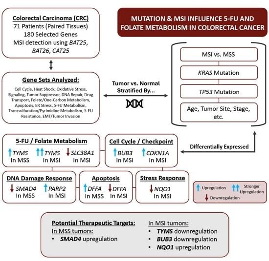
For categorical variables, we used the chi-square test. For continuous variables, a t-test or one-way analysis of variance (ANOVA) was used. For gene expression data, we used the Partek Genomics Suite (v7.0) (https://www.partek.com/partek-genomics-suite/, accessed on 22 July 2025). Fold change (FC) with 95% confidence intervals (CIs) is reported. Different clinicopathological variables like MSI status, KRAS mutation status, TP53 mutation status, as well as clinical and pathological factors such as patient age (<40 years versus >40 years), tumor location (left- versus right-sided CRC), and disease stage were used as categorical variables in different ANOVA models. To determine if a given factor (e.g., MSI status) significantly influences the magnitude of differential expression between CRC and normal tissue, we introduced an interaction term Tissue × factor in the ANOVA model(s), where the p-value of the interaction term indicates if the difference between the magnitudes of differential expression of the gene significantly differs by the presence or absence of that given factor. For multiple testing correction, we used the FDR.
In the GO enrichment analysis, we tested whether a list of differentially expressed genes fell into a Gene Ontology category more often than expected by chance [67]. We used a chi-square test to compare the “number of significant genes from a given category/total number of significant genes” vs. “number of genes on chip in that category/total number of genes on the microarray chip”.
The gene set ANOVA is a mixed model ANOVA that compares expression levels of a “set of genes” instead of an individual gene in different groups (https://www.partek.com/partek-genomics-suite/, accessed on 22 July 2025). The result is expressed at the level of the “gene set” category by averaging the member genes’ results.

3. Results

The characteristics of the 71 CRC patients are summarized in Supplementary Table S1. Paired comparisons between CRC tissues and their corresponding non-lesional tissues were conducted to evaluate differential gene expression profiles. The results of this paired analysis of 380 probes covering all 180 genes, including fold changes and 95% confidence intervals, revealed that 105 probes were at least 1.2-fold differentially expressed in either direction at the FDR 0.05 level, and are presented in Supplementary Table S3. To account for multiple testing, false discovery rates (FDR) and adjusted p-values are also provided.
In the next step, instead of a single gene-level comparison, we asked if the 14 sets of genes (sharing similar biological pathways, e.g., apoptotic genes), on average, were differentially expressed in CRC tissue compared to corresponding normal tissue from the same patient (see Table 1). We used Gene set ANOVA for this analysis. We found that 11 out of the tested 14 sets of genes were dysregulated (upregulated = 8 and downregulated = 3) with a statistically significant p-value (<0.05) in either direction, with FDR 0.05 (see Table 1). Examples from among the upregulated gene sets are “Cell Cycle Checkpoint”, “Heat Shock Response”, “Oxidative Stress Response”, “Signaling pathway”, and examples from downregulated gene sets include “Tumor suppressor”, “Apoptotic gene”, “Endoplasmic Reticulum (ER) Stress”. It may be noted that 11 of these gene sets were also picked up by the enrichment analysis.
Considering the clinical significance of KRAS and TP53 mutations as well as MSI status in CRC, we examined whether these molecular features of the tumor affect the magnitudes of differential expression.
To assess whether KRAS mutation status modifies the extent of differential gene expression between CRC and non-lesional tissue, we incorporated an interaction term—“tissue type (1 = CRC, 0 = normal) × KRAS mutation status (1 = mutant, 0 = wild-type)”—into the ANOVA model. This allowed us to determine whether the presence of a KRAS mutation significantly altered the magnitude of differential expression. Our analysis identified three upregulated gene sets (see Table 2) for which the average differential expression between CRC and non-lesional tissues was significantly more pronounced in KRAS-mutated tumors (ANOVA interaction p < 0.05, Table 2).
Similarly, to examine the impact of TP53 mutation status, we introduced a similar interaction term—“tissue type (1 = CRC, 0 = normal) × TP53 mutation status (1 = mutant, 0 = wild-type)”—into the model. This allowed us to determine whether TP53 mutations significantly alter gene expression patterns between CRC and normal tissues. We found that for three gene sets, the magnitude of differential expression was significantly greater in the presence of TP53 mutations (ANOVA interaction p < 0.05, Table 3).
We further investigated the influence of another key molecular marker—MSI status—on the differential expression of different gene sets. We identified significant alterations in multiple gene sets when comparing CRC to normal tissues. Notably, gene sets related to Cell Cycle Checkpoint control and DNA Repair Mechanisms were slightly more upregulated in the presence of MSI, indicating an enhanced cellular response to genomic instability. Conversely, Oxidative Stress Response-related genes were more upregulated, and Tumor Suppressor genes were more markedly downregulated in MSS patients; see Table 4.
Finally, to assess the influence of broader clinicopathological factors—including age at diagnosis (early-onset < 40 years vs. late-onset ≥ 40 years), tumor grade (high vs. low), peri-neural invasion (present vs. absent), sex (male vs. female), presence of signet ring cells (present vs. absent), cancer stage (I, II, or III), telomere shortening (yes vs. no), and tumor infiltration (yes vs. no)—on gene expression differences between CRC and non-lesional tissue, we introduced an interaction term into the ANOVA model: tissue type (CRC = 1, normal = 0) × mutation status or presence/absence of the factor (1 = present, 0 = absent). This allowed us to assess whether these factors modify the extent of differential gene expression. Our analysis revealed that gene sets were differentially expressed depending on these variables; detailed results are presented in Supplementary Tables S4–S11.

3.1. Association of MSI Status on Differential Expression of Genes

Considering the clinical importance of MSI in CRC, we also examined the individual gene-level data (in addition to gene set level, described earlier), whether the expression of folate- and 5-FU-related genes differ between MSI and MSS tumors, based solely on gene expression profiles, without assessing actual chemotherapy response.
In folic acid metabolism-related genes, differential expressions vary significantly by the presence or absence of MSI (at FDR 0.05 level) and are shown in Table 5. The fold changes (FC) with 95% CI for each gene are presented. We also included the location (right-sided/left-sided tumor) into account. The GO Enrichment analysis (see Figure 1) showed that the list of genes was enriched in genes related to cell cycle genes (BUB3, CDKN1A, SMAD4); apoptosis (e.g., DFFA, TNFRSF10B, ATF4, etc.); Folate & One carbon metabolism (TYMS, SHMT1); p53 signaling pathway (TNFRSF10B, CDKN1A), etc.
Among the cell cycle-related genes, BUB3 (see Figure 2A) was significantly upregulated only in MSI, whereas SMAD4 was significantly downregulated only in MSS CRC (see Figure 2B). PARP2 and RRM2 were upregulated in both MSS and MSI CRCs, but the magnitude of upregulation was more marked in MSI tumors compared to MSS tumors (ANOVA interaction p < 0.001; see Figure 2C and Figure 2D, respectively).
In the apoptotic genes group, FAS was downregulated in both MSS and MSI tumors, with a stronger effect in MSS [FC = −1.58 (95% CI −1.74 to −1.44) in MSS patients compared to FC = −1.32 (95% CI −1.13 to −1.33) in MSI patients, ANOVA interaction p = 0.001]. TNFRSF10B was significantly upregulated in MSI (FC = 1.40, 95% CI 1.25 to 1.55, p = 6.09 × 10⁻⁹). DFFA showed subtype-specific regulation, which is upregulated in MSS (FC = 1.10, 95% CI 1.01 to 1.21, p = 0.07), but downregulated in MSI (FC = −1.22, 95% CI −1.45 to −1.03, p = 0.01).
Among the 5-FU metabolism genes, TYMS was significantly more upregulated in MSI CRC than in MSS tumors, (FC = 1.65, 95% CI 1.27–2.13 in patients with MSI tumors vs. FC 1.19 95% CI 1.02–1.39 in patients with MSS tumor; ANOVA interaction p = 1.01 × 10⁻⁶) even after adjustment for tumor location (see Figure 3A). Similarly, PEMT expression was higher in MSI tumors (FC = 1.23, 95% CI 1.11–1.38) than in MSS tumors (FC = 1.08, 95% CI 1.01–1.16; ANOVA interaction p = 0.002). In contrast, the one-carbon metabolism-related gene SLC38A1 was significantly more downregulated in MSI tumors compared to MSS (MSI: FC = −1.59, 95% CI −1.91 to −1.33 vs. MSS: FC = −1.05, 95% CI −1.17 to −1.06; ANOVA interaction p = 2.2 × 10⁻⁴), independent of tumor location. CDKN1A was weakly upregulated in both MSI and MSS CRC, though the effect was more pronounced in MSI tumors (FC = 1.14, 95% CI 1.06–1.22; p < 0.001; see Figure 3B). Notably, FOLR1 expression was specifically higher in MSS CRC compared to MSI tumors across both probes (ILMN_1661733 and ILMN_1698608) (Figure 3C,D).
In the stress response pathway, ATF4 and SOD1 were significantly upregulated in MSI compared to MSS CRCs. For ATF4, expression was elevated in MSI tumors (FC = 1.27, 95% CI 1.10–1.45) versus MSS (FC = 1.65, 95% CI 1.27–2.13; ANOVA interaction p = 1.43 × 10⁻⁴) (See Table 5). Similarly, SOD1 expression was higher in MSI tumors (FC = 1.20, 95% CI 1.04–1.40) compared to MSS (FC = 1.04, 95% CI −1.05–1.14; ANOVA interaction p = 8.6 × 10⁻⁴). Notably, MAPK3, a key component of the MAPK/ERK pathway, was markedly downregulated in MSI tumors compared to MSS (MSI: FC = −2.02, 95% CI −2.46 to −1.67 vs. MSS: FC = −1.52, 95% CI −1.70 to −1.35; ANOVA interaction p = 3.75 × 10⁻⁴), with a statistically significant interaction. In contrast, NQO1 showed divergent expression patterns, was downregulated in MSI tumors (FC = −1.38, 95% CI −1.92 to −1.67) but upregulated in MSS tumors (FC = 1.68, 95% CI 1.38–2.04; ANOVA interaction p = 2.22 × 10⁻⁶).

3.2. Association of MSI Status and KRAS Mutation Status on Differential Expression of Genes

Considering the clinical importance of MSI and KRAS mutation in CRC, we also looked for interactions between these two molecular markers. Based on the presence or absence of these two markers, the patients were divided into four groups: (a) MSS and KRAS wild (n = 38); (b) MSS and KRAS mutant (n = 15); (c) MSI and KRAS wild (n = 13) and (d) MSI and KRAS mutant (n = 5). Table 6 shows the magnitude of differential expressions (tumor vs. normal) of the genes that were significantly different among these four categories of CRC patients. From a 5-FU therapeutic point-of-view, the result suggests that (a) CRC patients with MSI, and more specifically MSI with KRAS mutation may benefit from therapy that can reduce TYMS or BUB3 expression; and from therapy that can enhance NQO1 expression; and (b) CRC patients with MSS irrespective of KRAS mutation status may benefit from therapy that may increase SMAD4 expression.

3.3. Association of Age of Onset (<40 Years and >40 Years) on Differential Expression of Genes

At the individual gene level, the age of onset (EOCRC vs. LOCRC) significantly influenced (at p < 0.05) the magnitude of differential expression (CRC vs. normal tissue) of 29 out of 180 folic acid-related genes tested (see Supplementary Table S12). GO enrichment analysis of the pathways that involve this list of genes is presented in Supplementary Figure S1. Among these genes, MSH2 was upregulated only in LOCRC (FC = 1.10, 95% CI 1.05–1.15), but not in EOCRC (FC −1.00, 95% CI −1.06–1.06), ANOVA interaction p = 0.0001) (Figure 4). Notably, PLD1, MAPK3, CHGB, and TNFSF10 were strongly downregulated in the older patients, whereas BIRC5, ENO1, and CDK4 were markedly upregulated. In contrast, among early-onset CRC (<40 years), GADD45A was downregulated while HSPA1A was significantly upregulated (see Supplementary Table S12).
Enrichment analysis revealed that younger CRC patients (<40 years) exhibited significantly higher enrichment scores for folate biosynthesis (ES = 5.93), antifolate resistance (ES = 9.22), and colon cancer signaling (ES = 12.32) compared with older patients (>40 years) (Supplementary Figure S1). This indicates the preferential activation of these pathways in the younger CRC patients.

4. Discussion

In CRC, 5-FU remains the backbone of chemotherapy in both adjuvant and palliative settings [68]. Genetic variations in genes regulating enzymes involved in 5-FU and folate metabolism play pivotal roles in shaping treatment response and clinical outcomes [69]. In this study, we tested the differential expression of a large number of genes related to folic acid that comprehensively cover different gene sets implicated in 5-FU resistance and folate metabolism. We also investigated whether their differential expressions vary by key clinical, pathological, and molecular genomic features, including age, sex, MSI status, KRAS mutation, TP53 status, and telomere shortening in non-metastatic CRC. Our analysis revealed significant upregulation of cell cycle checkpoint genes, heat shock response genes, oxidative stress response genes, and signaling pathway genes, while tumor suppressor genes were consistently downregulated in CRC tissues compared with adjacent non-lesional tissues. Importantly, MSI tumors demonstrated distinct expression profiles compared with MSS tumors, particularly across pathways involving the p53 signaling pathway, cell cycle regulation, DNA damage response, apoptosis, stress signaling, and one-carbon metabolism. These findings reflect fundamental biological divergence between the two CRC subtypes and are supported by previous research and carry significant implications for the development of precise therapeutic strategies in CRC. Our major limitation is the lack of follow-up data, and, therefore, we could not test the effect of gene expression pattern of the tumor on disease outcome or therapeutic implications. Also, we did not perform any cell line study to address the mechanistic relationships. The study does not show causal relationships; rather, it shows some clinically relevant associations.
We acknowledge the fact that both (a) possible inhibition of antitumor effector responses (related to immune checkpoint inhibition) and (b) epigenetic factors are important aspects from a therapeutic point of view for CRC. But, in this study, we intentionally focused only on Folic acid and 5-FU-related genes and their differential expression. In fact, using the same patients, in a recent study, we have shown the interaction of MSI status and differential expression of Inflamed T-cell-related genes, which suggested that the MSS patients were less likely to benefit from immune checkpoint inhibitor (ICI) therapy compared to patients with MSI [63]. Similarly, in another previous study, we also described the interaction between MSI and epigenetic alteration (methylation) in these CRC patients, which suggested an opportunity for potential use of certain immune checkpoint inhibitors (CTLA4 and HAVCR2 inhibitors) in CRC with MSI [70].

4.1. Folate and One-Carbon Metabolism

In paired analyses, distinct expression patterns of 5-FU metabolism and folate cycle-related genes were observed between MSI and MSS tumors. Our data clearly showed more pronounced upregulation (CRC tissue compared to normal tissue) of TYMS in patients with MSI tumors compared to patients with MSS tumors, even after taking the location of the tumor into account. This partly explains the lesser effectiveness of 5-FU therapy in MSI patients. A previous study also suggested that thymidylate synthase (TYMS), the primary target of 5-FU [71], was more strongly upregulated in MSI tumors. TYMS overexpression is a well-recognized mechanism of 5-FU resistance, as it outcompetes fluorodeoxyuridylate (FdUMP) binding [72,73]. This may indicate that MSI tumors are relatively less sensitive to 5-FU due to an enhanced nucleotide biosynthesis capacity [74]. Another gene, phosphatidylethanolamine methyltransferase (PEMT), which regulates membrane phospholipid metabolism and methyl group flux [75], was also upregulated in MSI tumors. Emerging evidence suggests that PEMT dysregulation may alter membrane fluidity and influence drug uptake and efflux pathways, thereby potentially modulating chemosensitivity [76]. However, this requires further investigation. Conversely, solute carrier family 38 member 1 (SLC38A1), an amino acid transporter critical for glutamine uptake [77], was more downregulated in MSI tumors. Since glutamine availability fuels nucleotide biosynthesis and maintains redox balance, SLC38A1 suppression could limit metabolic support for proliferation [78]. This may, paradoxically, sensitize MSI tumors to 5-FU under certain contexts, although compensatory pathways could offset this effect, warranting further exploration. Interestingly, serine hydroxy-methyltransferase 1 (SHMT1), which links serine/glycine metabolism to folate-mediated one-carbon metabolism [79], was downregulated in MSS tumors. Its reduced expression may disrupt one-carbon flux, potentially impairing DNA synthesis and repair [80]. This observation is consistent with prior clinical findings that MSS tumors often exhibit relatively higher 5-FU responsiveness compared with MSI tumors [81]. Overall, these gene expression differences may explain why MSI tumors are relatively resistant to 5-FU, whereas MSS tumors may remain more sensitive, emphasizing the need for further studies to guide subtype-specific therapies.

4.2. Cell Cycle Regulation and Checkpoint Control

Among the 180 genes analyzed, 38 were associated with cell cycle checkpoint regulation, and these were generally upregulated in CRC tissues compared with adjacent non-lesional tissues (see Table 1). When analyzed according to MSI and MSS status, adjusted for tumor location (right vs. left), BUB3, CDKN1A (p21), and RRM2 were significantly more upregulated in MSI tumors than in MSS tumors (see Table 5). CDKN1A, a well-established transcriptional target of p53, is both necessary and sufficient for p53-mediated transcriptional repression [82] and has been implicated in 5-FU resistance [83]. In MSI CRC, CDKN1A upregulation suggests enhanced checkpoint fidelity and stronger p53-dependent growth arrest, which may allow tumor cells to repair DNA damage induced by 5-FU, potentially contributing to relative chemoresistance [84]. Our analysis also showed that CRCs harboring mutant TP53 exhibited overall upregulation of signaling pathway-related genes (Table 3), likely reflecting activation of alternative compensatory mechanisms in the absence of intact p53. These findings support the idea that enhanced p53-CDKN1A signaling in MSI tumors may reduce 5-FU sensitivity, whereas MSS tumors with lower CDKN1A expression may be more susceptible to 5-FU-induced cytotoxicity, consistent with prior evidence linking altered p53 pathway activity to chemoresistance in MSI CRC [85]. Importantly, the differential expression of checkpoint regulators between MSI and MSS tumors remained significant after adjusting for tumor site, highlighting MSI status as an independent determinant of CDKN1A-mediated 5FU sensitivity. In our analysis, we observed contrasting roles for RRM2 and BUB3 in MSI CRC. According to Zuo et al., drug-resistant tumor cells often exhibit amplification of the RRM2 gene and its promoter, resulting in elevated transcriptional activity and increased DNA synthesis [86]. Consistently, we found RRM2 overexpression in MSI tumors, suggesting that it may contribute to chemoresistance by enhancing replication potential and promoting survival under 5-FU-induced stress. In contrast, BUB3, a spindle assembly checkpoint (SAC) regulator transcriptionally controlled by YY2, enforces checkpoint fidelity. Hyperactivation of the YY2/BUB3 axis delays mitosis, increases chromosomal instability beyond tolerable thresholds, and induces tumor cell death, thereby enhancing drug sensitivity [87]. These findings indicate that RRM2 overexpression may drive resistance, whereas BUB3 hyperactivation may sensitize tumors to therapy. The opposing functional effects of these two genes in MSI CRC warrant further investigation to determine which pathway predominates.

4.3. DNA Damage Response and Repair

The cytotoxic effect of 5-FU is mediated primarily by inhibiting TYMS, resulting in thymidine depletion, uracil misincorporation, DNA replication stress, and subsequent strand breaks [88]. PARP2 plays a pivotal role in repairing 5-FU-induced single-strand breaks through the base excision repair (BER) pathway and subsequently engages the downstream HR/NHEJ repair mechanism [13]. In our cohort, PARP2 was significantly overexpressed in MSI CRC, which may represent a compensatory response to increased DNA damage repair demands. Another important gene, SMAD4, a central mediator of the TGF-β pathway and regulator of 5-FU sensitivity, also showed subtype-specific expression patterns. In our analysis, SMAD4 was significantly downregulated in MSS tumors, whereas MSI tumors demonstrated a mild, non-significant upregulation. Previous studies have shown that SMAD4 knockout or downregulation promotes 5-FU resistance in CRC models [89], suggesting that SMAD4 loss in MSS tumors may contribute to reduced drug sensitivity, consistent with experimental evidence [90]. Similarly, PTEN, a key tumor suppressor that regulates PI3K/AKT signaling, displayed distinct expression profiles across subtypes. Although PTEN mutations are more frequently reported in MSI CRC [91], we observed overall PTEN downregulation in both groups, with more pronounced loss in MSS tumors. This aligns with recent findings that circPTEN is downregulated in CRC [92]. PTEN is also a functional target of several microRNAs that mediate chemoresistance. For instance, miRNA-17-5p directly suppresses PTEN following chemotherapy, promoting multidrug resistance [93]. Likewise, miRNA-193-3p downregulation has been shown to restore PTEN expression, reduce proliferation, and enhance apoptosis, thereby reversing 5-FU resistance [94]. In another study, miRNA-141-3p overexpression suppressed PTEN in resistant CRC cells, whereas its inhibition restored PTEN and improved sensitivity to 5-FU and oxaliplatin [95]. These findings, together with our observations, suggest that targeting PARP2 with PARP inhibitors, restoring SMAD4-mediated apoptotic pathways, or counteracting PTEN loss through inhibition of the PI3K/AKT pathway may represent promising strategies to enhance treatment efficacy in CRC.

4.4. Apoptosis Regulation

Among the apoptotic gene sets studied, DFFA, TNFRSF10B (DR5), and FAS exhibited differential expression patterns depending on MSI status in CRC. DFFA, an inhibitory subunit of the DNA fragmentation factor complex (DFF), was downregulated in MSI CRC. Under normal conditions, DFFA is cleaved by caspase-3 to release DFFB, which facilitates endonuclease-mediated DNA fragmentation—a terminal step of chemotherapy-induced apoptosis, including that triggered by 5-FU. Reduced DFFA expression may therefore impair apoptotic execution despite upstream caspase activation, potentially contributing to partial 5-FU resistance in MSI CRC. This finding aligns with reports that MSI tumors, while often associated with a favorable prognosis, can display relative chemoresistance to 5-FU-based therapies [96]. In contrast, MSI CRC also showed significant upregulation of DR5, consistent with preserved p53-dependent apoptotic signaling [97]. Elevated DR5 expression relative to adjacent non-lesional tissues suggests enhanced sensitivity to 5-FU-induced apoptosis, in line with previous evidence [98,99,100]. Conversely, FAS (CD95) was significantly downregulated in MSS CRC, reflecting impaired extrinsic apoptotic signaling in this subgroup. As a critical mediator of the FAS/FASL pathway, and a transcriptional target of p53 [101], FAS loss has been linked to 5-FU resistance in colon carcinoma models [102]. The contrasting patterns of DR5 upregulation in MSI, FAS suppression in MSS, and DFFA downregulation in MSI highlight a complex balance between apoptosis initiation and execution. While these alterations may offset each other at the bulk level, they remain therapeutically relevant, suggesting that enhancing DR5 in MSI, restoring FAS in MSS, or overcoming DFFA defects could help tailor and improve 5-FU-based treatment responses.

4.5. Stress Response Pathways

In the studied samples, oxidative stress-related genes were generally upregulated in both MSI and MSS CRC, though more prominently in MSS tumors (Table 4). In MSI tumors, ATF4 and SOD1 were consistently upregulated, whereas MAPK3 was strongly downregulated. Interestingly, NQO1 displayed a biphasic pattern, being downregulated in MSI but upregulated in MSS. These findings indicate that oxidative stress responses differ according to MSI status. The oxidative stress pathway is increasingly recognized as a key determinant of chemotherapeutic sensitivity [103], and 5-FU is known to induce reactive oxygen species (ROS) that drive apoptosis in cancer cells [104]. Upregulation of ATF4, a component of the PERK–ATF4 pathway linked to 5-FU resistance [105], along with SOD1 overexpression, may reflect adaptive stress responses in MSI CRC, although SOD1 is also frequently upregulated in early-stage CRC [106]. In contrast, MAPK3 downregulation in MSI tumors may shift the balance between apoptosis and autophagy toward resistance [7]. Notably, MSS tumors exhibited stronger upregulation of NQO1, which likely functions as a compensatory antioxidant mechanism to counteract chemotherapy-induced ROS, thereby limiting apoptosis and conferring relative resistance [107,108]. Taken together, the combination of MAPK3 downregulation and reduced NQO1 expression in MSI tumors may impair ROS neutralization, resulting in greater oxidative damage and enhanced 5-FU sensitivity. Conversely, MSS tumors, through more robust antioxidant defenses, may mitigate ROS-mediated cytotoxicity and thus exhibit relative drug resistance. These results suggest that differential oxidative stress responses between MSI and MSS CRC contribute to their divergent therapeutic outcomes. Importantly, targeting oxidative stress regulators—such as inhibiting NQO1 or ATF4 in MSS tumors, or modulating MAPK3 and ROS balance in MSI tumors—may provide opportunities for tailored therapeutic strategies to enhance 5-FU efficacy in a subtype-specific manner.

5. Conclusions

In conclusion, MSI colorectal tumors exhibited TYMS and PARP2 upregulation and DFFA downregulation. These alterations may be linked to reduced 5-FU sensitivity through enhanced nucleotide biosynthesis, impaired apoptotic execution, and increased DNA repair activity. FAS expression was decreased in both MSI and MSS tumors, though the downregulation was more pronounced in MSS, suggesting a greater impairment of extrinsic apoptotic signaling in this subgroup. Additionally, MSS tumors show downregulation of SMAD4, which is partially offset by compensatory upregulation of NQO1 and GSK3B. This distinct expression may explain subgroup differences in treatment response and suggest potential avenues for tailored therapy, such as TYMS or PARP inhibition in MSI tumors and restoration of apoptotic signaling or targeting compensatory pathways in MSS tumors.

Supplementary Materials

The following supporting information can be downloaded at https://www.mdpi.com/article/10.3390/curroncol32120661/s1, Figure S1: Enrichment analysis of differentially expressed genes, which are influenced by age of onset of CRC; Table S1: Patient Characteristics; Table S2: List of FA and 5-FU-related genes tested in this study. Table S3: Significant Paired Analysis Results from 380 Probes Covering 180 Genes; Table S4: Pathways with significant differential expression in relation to age of onset; Table S5: Pathways with significant differential expression in relation to grade; Table S6: Pathways with significant differential expression in relation to PN Invasion; Table S7: Pathways with significant differential expression in relation to sex; Table S8: Pathways with significant differential expression in relation to signet ring presence; Table S9: Pathways with significant differential expression in relation to CRC stage; Table S10: Pathways with significant differential expression in relation telomere status; Table S11: Pathways with significant differential expression in relation to TIL status; Table S12: Differential expression of 29 folic acid-related genes in early vs. late onset CRC.

Author Contributions

Conceptualization, M.R.I., F.J., M.G.K. and H.A.; methodology, F.J. and M.R.; formal analysis, M.G.K.; investigation, F.J., M.R., D.V. and A.A.; resources, H.A.; data curation, A.A. and D.V.; writing—original draft preparation, M.R.I., F.J. and M.G.K.; writing—review and editing, D.V., A.A. and H.A.; supervision, M.G.K. and H.A.; project administration, H.A. and M.G.K.; funding acquisition, H.A. All authors have read and agreed to the published version of the manuscript.

Funding

This research was partly funded by NIH funds P20CA210305 and P30ES027792.

Institutional Review Board Statement

This study was conducted in accordance with the Declaration of Helsinki and approved by the “Biological Sciences Division, University of Chicago Hospital Institutional Review Board”, Chicago, IL, USA (approval code: 10-264-E; approval date: 18 May 2010).

Informed Consent Statement

Informed consent was obtained from all subjects involved in the study.

Data Availability Statement

All the supporting data are presented in the tables presented in the main manuscript and Supplementary Materials.

Acknowledgments

We acknowledge the support and help of all the patients included in this study. We thank Zahidul Haq, late M Kamal, Rupash Paul and Mustafizur Rahman for their help and support. We thank the University of Chicago Research, Bangladesh (URB) staff for the handling and shipping of all the study material to the University of Chicago molecular genomics laboratory.

Conflicts of Interest

The authors declare no conflicts of interest. The funders had no role in the design of the study; in the collection, analyses, or interpretation of data; in the writing of the manuscript; or in the decision to publish the results.

Abbreviations

The following abbreviations are used in this manuscript:
5-FU5-Fluorouracil
CRCColorectal Cancer
MSIMicrosatellite Instability
MSSMicrosatellite Stable
FAFolic Acid
CIConfidence Interval
ACSAmerican Cancer Society
mCRCMetastatic Colorectal Cancer
DNADeoxyribonucleic Acid
RNARibonucleic Acid
EMTEpithelial–Mesenchymal Transition
TILTumor-Infiltrating Lymphocyte
MMRMismatch Repair
MMR-DMismatch Repair Deficiency
MSI-HMSI-High
BSMMUBangabandhu Sheikh Mujib Medical University
EREndoplasmic Reticulum
HRMHigh Resolution Melt
PCRPolymerase Chain Reaction
ANOVAAnalysis of Variance
FCFold Change
GOGene Ontology
FDR False Discovery Rate
KRASKristen Rat Sarcoma Virus
EOCRCEarly-Onset CRC
LOCRCLate-Onset CRC
PEMTPhosphatidylethanolamine Methyltransferase
FdUMPFluorodeoxyuridylate
TYMSThymidylate Synthase
SHMT1Serine Hydroxymethyltransferase 1
SLC38A1Solute Carrier Family 38 Member 1
BERBase Excision Repair
DFFDNA Fragmentation Factor Complex

References

  1. Kandel, A.; Thida, A.M.; Preet, M. A Review of the Early Detection of Colon Cancer and the Role of Circulating Tumor DNA. Cureus 2025, 17, e84394. [Google Scholar] [CrossRef] [PubMed] [PubMed Central]
  2. Abdelgadir, O.; Kuo, Y.F.; Khan, M.F.; Okorodudu, A.O.; Cheng, Y.W.; Dong, J. Mortality Outcome Associated with Specific KRAS, NRAS, and BRAF Hot-Spot Mutations in Metastatic Colorectal Cancer Patients: A Retrospective Cohort Study. Diagnostics 2025, 15, 590. [Google Scholar] [CrossRef] [PubMed] [PubMed Central]
  3. Chan, G.H.J.; Chee, C.E. Making sense of adjuvant chemotherapy in colorectal cancer. J. Gastrointest. Oncol. 2019, 10, 1183–1192. [Google Scholar] [CrossRef] [PubMed] [PubMed Central]
  4. Gmeiner, W.H.; Okechukwu, C.C. Review of 5-FU resistance mechanisms in colorectal cancer: Clinical significance of attenuated on-target effects. Cancer Drug Resist. 2023, 6, 257–272. [Google Scholar] [CrossRef] [PubMed] [PubMed Central]
  5. Długosz-Pokorska, A.; Pięta, M.; Janecki, T.; Janecka, A. New uracil analogs as downregulators of ABC transporters in 5-fluorouracil-resistant human leukemia HL-60 cell line. Mol. Biol. Rep. 2019, 46, 5831–5839. [Google Scholar] [CrossRef] [PubMed]
  6. Sethy, C.; Kundu, C.N. 5-Fluorouracil (5-FU) resistance and the new strategy to enhance the sensitivity against cancer: Implication of DNA repair inhibition. Biomed. Pharmacother. 2021, 137, 111285. [Google Scholar] [CrossRef] [PubMed]
  7. Blondy, S.; David, V.; Verdier, M.; Mathonnet, M.; Perraud, A.; Christou, N. 5-Fluorouracil resistance mechanisms in colorectal cancer: From classical pathways to promising processes. Cancer Sci. 2020, 111, 3142–3154. [Google Scholar] [CrossRef] [PubMed] [PubMed Central]
  8. Barathan, M.; Zulpa, A.K.; Ng, S.L.; Lokanathan, Y.; Ng, M.H.; Law, J.X. Innovative Strategies to Combat 5-Fluorouracil Resistance in Colorectal Cancer: The Role of Phytochemicals and Extracellular Vesicles. Int J Mol Sci. 2024, 25, 7470. [Google Scholar] [CrossRef] [PubMed] [PubMed Central]
  9. Ghafouri-Fard, S.; Abak, A.; Tondro Anamag, F.; Shoorei, H.; Fattahi, F.; Javadinia, S.A.; Basiri, A.; Taheri, M. 5-Fluorouracil: A Narrative Review on the Role of Regulatory Mechanisms in Driving Resistance to This Chemotherapeutic Agent. Front. Oncol. 2021, 11, 658636. [Google Scholar] [CrossRef] [PubMed] [PubMed Central]
  10. LaCourse, K.D.; Zepeda-Rivera, M.; Kempchinsky, A.G.; Baryiames, A.; Minot, S.S.; Johnston, C.D.; Bullman, S. The cancer chemotherapeutic 5-fluorouracil is a potent Fusobacterium nucleatum inhibitor and its activity is modified by intratumoral microbiota. Cell Rep. 2022, 41, 111625. [Google Scholar] [CrossRef] [PubMed] [PubMed Central]
  11. Seiple, L.; Jaruga, P.; Dizdaroglu, M.; Stivers, J.T. Linking uracil base excision repair and 5-fluorouracil toxicity in yeast. Nucleic Acids Res. 2006, 34, 140–151. [Google Scholar] [CrossRef] [PubMed] [PubMed Central]
  12. Showalter, S.L.; Showalter, T.N.; Witkiewicz, A.; Havens, R.; Kennedy, E.P.; Hucl, T.; Kern, S.E.; Yeo, C.J.; Brody, J.R. Evaluating the drug-target relationship between thymidylate synthase expression and tumor response to 5-fluorouracil. Is it time to move forward? Cancer Biol. Ther. 2008, 7, 986–994. [Google Scholar] [CrossRef] [PubMed] [PubMed Central]
  13. Azwar, S.; Seow, H.F.; Abdullah, M.; Faisal Jabar, M.; Mohtarrudin, N. Recent Updates on Mechanisms of Resistance to 5-Fluorouracil and Reversal Strategies in Colon Cancer Treatment. Biology 2021, 10, 854. [Google Scholar] [CrossRef] [PubMed] [PubMed Central]
  14. Zsigrai, S.; Kalmár, A.; Barták, B.K.; Nagy, Z.B.; Szigeti, K.A.; Valcz, G.; Kothalawala, W.; Dankó, T.; Sebestyén, A.; Barna, G.; et al. Folic Acid Treatment Directly Influences the Genetic and Epigenetic Regulation along with the Associated Cellular Maintenance Processes of HT-29 and SW480 Colorectal Cancer Cell Lines. Cancers 2022, 14, 1820. [Google Scholar] [CrossRef] [PubMed] [PubMed Central]
  15. Danenberg, P.V.; Gustavsson, B.; Johnston, P.; Lindberg, P.; Moser, R.; Odin, E.; Peters, G.J.; Petrelli, N. Folates as adjuvants to anticancer agents: Chemical rationale and mechanism of action. Crit. Rev. Oncol. Hematol. 2016, 106, 118–131. [Google Scholar] [CrossRef] [PubMed]
  16. Garg, M.B.; Lincz, L.F.; Adler, K.; Scorgie, F.E.; Ackland, S.P.; Sakoff, J.A. Predicting 5-fluorouracil toxicity in colorectal cancer patients from peripheral blood cell telomere length: A multivariate analysis. Br. J. Cancer 2012, 107, 1525–1533. [Google Scholar] [CrossRef] [PubMed] [PubMed Central]
  17. Gmeiner, W.H. Fluoropyrimidine Modulation of the Anti-Tumor Immune Response-Prospects for Improved Colorectal Cancer Treatment. Cancers 2020, 12, 1641. [Google Scholar] [CrossRef] [PubMed] [PubMed Central]
  18. Jang, H.Y.; Kim, D.H.; Lee, H.J.; Kim, W.D.; Kim, S.Y.; Hwang, J.J.; Lee, S.J.; Moon, D.H. Schedule-dependent synergistic effects of 5-fluorouracil and selumetinib in KRAS or BRAF mutant colon cancer models. Biochem. Pharmacol. 2019, 160, 110–120. [Google Scholar] [CrossRef] [PubMed]
  19. Jover, R.; Zapater, P.; Castells, A.; Llor, X.; Andreu, M.; Cubiella, J.; Balaguer, F.; Sempere, L.; Xicola, R.M.; Bujanda, L.; et al. The efficacy of adjuvant chemotherapy with 5-fluorouracil in colorectal cancer depends on the mismatch repair status. Eur. J. Cancer 2009, 45, 365–373. [Google Scholar] [CrossRef] [PubMed]
  20. Klingbiel, D.; Saridaki, Z.; Roth, A.D.; Bosman, F.T.; Delorenzi, M.; Tejpar, S. Prognosis of stage II and III colon cancer treated with adjuvant 5-fluorouracil or FOLFIRI in relation to microsatellite status: Results of the PETACC-3 trial. Ann. Oncol. 2015, 26, 126–132. [Google Scholar] [CrossRef] [PubMed]
  21. Thrall, M.M.; Wood, P.; King, V.; Rivera, W.; Hrushesky, W. Investigation of the comparative toxicity of 5-FU bolus versus 5-FU continuous infusion circadian chemotherapy with concurrent radiation therapy in locally advanced rectal cancer. Int. J. Radiat. Oncol. Biol. Phys. 2000, 46, 873–881. [Google Scholar] [CrossRef] [PubMed]
  22. Zalcberg, J.; Kerr, D.; Seymour, L.; Palmer, M. Haematological and non-haematological toxicity after 5-fluorouracil and leucovorin in patients with advanced colorectal cancer is significantly associated with gender, increasing age and cycle number. Tomudex International Study Group. Eur. J. Cancer 1998, 34, 1871–1875. [Google Scholar] [CrossRef] [PubMed]
  23. Richman, S. Deficient mismatch repair: Read all about it (Review). Int. J. Oncol. 2015, 47, 1189–1202. [Google Scholar] [CrossRef] [PubMed] [PubMed Central]
  24. Chen, W.; Frankel, W.L. A practical guide to biomarkers for the evaluation of colorectal cancer. Mod. Pathol. 2019, 32 (Suppl. S1), 1–15. [Google Scholar] [CrossRef] [PubMed]
  25. Battaglin, F.; Naseem, M.; Lenz, H.J.; Salem, M.E. Microsatellite instability in colorectal cancer: Overview of its clinical significance and novel perspectives. Clin. Adv. Hematol. Oncol. 2018, 16, 735–745. [Google Scholar] [PubMed] [PubMed Central]
  26. Gutierrez, C.; Ogino, S.; Meyerhardt, J.A.; Iorgulescu, J.B. The Prevalence and Prognosis of Microsatellite Instability-High/Mismatch Repair-Deficient Colorectal Adenocarcinomas in the United States. JCO Precis. Oncol. 2023, 7, e2200179. [Google Scholar] [CrossRef] [PubMed] [PubMed Central]
  27. Tomasello, G.; Ghidini, M.; Galassi, B.; Grossi, F.; Luciani, A.; Petrelli, F. Survival benefit with adjuvant chemotherapy in stage III microsatellite-high/deficient mismatch repair colon cancer: A systematic review and meta-analysis. Sci. Rep. 2022, 12, 1055. [Google Scholar] [CrossRef] [PubMed] [PubMed Central]
  28. Jasmine, F.; Rahaman, R.; Dodsworth, C.; Roy, S.; Paul, R.; Raza, M.; Paul-Brutus, R.; Kamal, M.; Ahsan, H.; Kibriya, M.G. A genome-wide study of cytogenetic changes in colorectal cancer using SNP microarrays: Opportunities for future personalized treatment. PLoS ONE 2012, 7, e31968. [Google Scholar] [CrossRef] [PubMed] [PubMed Central]
  29. Kibriya, M.G.; Jasmine, F.; Pekow, J.; Munoz, A.; Weber, C.; Raza, M.; Kamal, M.; Ahsan, H.; Bissonnette, M. Pathways Related to Colon Inflammation Are Associated with Colorectal Carcinoma: A Transcriptome- and Methylome-Wide Study. Cancers 2023, 15, 2921. [Google Scholar] [CrossRef] [PubMed] [PubMed Central]
  30. Kibriya, M.G.; Raza, M.; Jasmine, F.; Roy, S.; Paul-Brutus, R.; Rahaman, R.; Dodsworth, C.; Rakibuz-Zaman, M.; Kamal, M.; Ahsan, H. A genome-wide DNA methylation study in colorectal carcinoma. BMC Med. Genom. 2011, 4, 50. [Google Scholar] [CrossRef] [PubMed] [PubMed Central]
  31. Kibriya, M.G.; Raza, M.; Kamal, M.; Haq, Z.; Paul, R.; Mareczko, A.; Pierce, B.L.; Ahsan, H.; Jasmine, F. Relative Telomere Length Change in Colorectal Carcinoma and Its Association with Tumor Characteristics, Gene Expression and Microsatellite Instability. Cancers 2022, 14, 2250. [Google Scholar] [CrossRef] [PubMed] [PubMed Central]
  32. Wang, Y.; Wei, Q.; Chen, Y.; Long, S.; Yao, Y.; Fu, K. Identification of Hub Genes Associated with Sensitivity of 5-Fluorouracil Based Chemotherapy for Colorectal Cancer by Integrated Bioinformatics Analysis. Front. Oncol. 2021, 11, 604315. [Google Scholar] [CrossRef] [PubMed] [PubMed Central]
  33. Pistritto, G.; Trisciuoglio, D.; Ceci, C.; Garufi, A.; D’Orazi, G. Apoptosis as anticancer mechanism: Function and dysfunction of its modulators and targeted therapeutic strategies. Aging 2016, 8, 603–619. [Google Scholar] [CrossRef] [PubMed] [PubMed Central]
  34. Neophytou, C.M.; Trougakos, I.P.; Erin, N.; Papageorgis, P. Apoptosis Deregulation and the Development of Cancer Multi-Drug Resistance. Cancers 2021, 13, 4363. [Google Scholar] [CrossRef] [PubMed] [PubMed Central]
  35. Fulcher, L.J.; Batley, C.; Sobajima, T.; Barr, F.A. Time as a danger signal promoting G1 arrest after mitosis. Trends Cell Biol. 2025, in press. [Google Scholar] [CrossRef] [PubMed]
  36. Kato, S.; Okamura, R.; Adashek, J.J.; Khalid, N.; Lee, S.; Nguyen, V.; Sicklick, J.K.; Kurzrock, R. Targeting G1/S phase cell-cycle genomic alterations and accompanying co-alterations with individualized CDK4/6 inhibitor-based regimens. JCI Insight 2021, 6, e142547. [Google Scholar] [CrossRef] [PubMed] [PubMed Central]
  37. Godet, I.; Gilkes, D.M. BRCA1 and BRCA2 mutations and treatment strategies for breast cancer. Integr. Cancer Sci. Ther. 2017, 4, 1–7. [Google Scholar] [CrossRef] [PubMed] [PubMed Central]
  38. Hsieh, P.; Yamane, K. DNA mismatch repair: Molecular mechanism, cancer, and ageing. Mech. Ageing Dev. 2008, 129, 391–407. [Google Scholar] [CrossRef] [PubMed] [PubMed Central]
  39. Manandhar, M.; Boulware, K.S.; Wood, R.D. The ERCC1 and ERCC4 (XPF) genes and gene products. Gene 2015, 569, 153–161. [Google Scholar] [CrossRef] [PubMed] [PubMed Central]
  40. Miyashita, K.; Shioi, S.; Kajitani, T.; Koi, Y.; Shimokawa, M.; Makiyama, A.; Oda, S.; Esaki, T. More subtle microsatellite instability better predicts fluorouracil insensitivity in colorectal cancer patients. Sci. Rep. 2024, 14, 27257. [Google Scholar] [CrossRef] [PubMed] [PubMed Central]
  41. Wright, W.D.; Shah, S.S.; Heyer, W.D. Homologous recombination and the repair of DNA double-strand breaks. J. Biol. Chem. 2018, 293, 10524–10535. [Google Scholar] [CrossRef] [PubMed] [PubMed Central]
  42. Zhang, Y.; Rohde, L.H.; Wu, H. Involvement of nucleotide excision and mismatch repair mechanisms in double strand break repair. Curr. Genom. 2009, 10, 250–258. [Google Scholar] [CrossRef] [PubMed] [PubMed Central]
  43. Song, Y.K.; Kim, M.J.; Kim, M.S.; Lee, J.H.; Chung, S.J.; Song, J.S.; Chae, Y.J.; Lee, K.R. Role of the efflux transporters Abcb1 and Abcg2 in the brain distribution of olaparib in mice. Eur. J. Pharm. Sci. 2022, 173, 106177. [Google Scholar] [CrossRef] [PubMed]
  44. Nie, F.; Sun, X.; Sun, J.; Zhang, J.; Wang, Y. Epithelial-mesenchymal transition in colorectal cancer metastasis and progression: Molecular mechanisms and therapeutic strategies. Cell Death Discov. 2025, 11, 336. [Google Scholar] [CrossRef] [PubMed] [PubMed Central]
  45. Yeung, K.T.; Yang, J. Epithelial-mesenchymal transition in tumor metastasis. Mol. Oncol. 2017, 11, 28–39. [Google Scholar] [CrossRef] [PubMed] [PubMed Central]
  46. Chen, X.; Shi, C.; He, M.; Xiong, S.; Xia, X. Endoplasmic reticulum stress: Molecular mechanism and therapeutic targets. Signal Transduct. Target. Ther. 2023, 8, 352. [Google Scholar] [CrossRef] [PubMed] [PubMed Central]
  47. Hetz, C.; Papa, F.R. The Unfolded Protein Response and Cell Fate Control. Mol. Cell 2018, 69, 169–181. [Google Scholar] [CrossRef] [PubMed]
  48. Somu, P.; Mohanty, S.; Basavegowda, N.; Yadav, A.K.; Paul, S.; Baek, K.H. The Interplay between Heat Shock Proteins and Cancer Pathogenesis: A Novel Strategy for Cancer Therapeutics. Cancers 2024, 16, 638. [Google Scholar] [CrossRef] [PubMed] [PubMed Central]
  49. Arfin, S.; Jha, N.K.; Jha, S.K.; Kesari, K.K.; Ruokolainen, J.; Roychoudhury, S.; Rathi, B.; Kumar, D. Oxidative Stress in Cancer Cell Metabolism. Antioxidants 2021, 10, 642. [Google Scholar] [CrossRef] [PubMed] [PubMed Central]
  50. Afzal, S.; Jensen, S.A.; Vainer, B.; Vogel, U.; Matsen, J.P.; Sørensen, J.B.; Andersen, P.K.; Poulsen, H.E. MTHFR polymorphisms and 5-FU-based adjuvant chemotherapy in colorectal cancer. Ann. Oncol. 2009, 20, 1660–1666. [Google Scholar] [CrossRef] [PubMed]
  51. Locasale, J.W. Serine, glycine and one-carbon units: Cancer metabolism in full circle. Nat. Rev. Cancer 2013, 13, 572–583. [Google Scholar] [CrossRef] [PubMed] [PubMed Central]
  52. Thorn, C.F.; Marsh, S.; Carrillo, M.W.; McLeod, H.L.; Klein, T.E.; Altman, R.B. PharmGKB summary: Fluoropyrimidine pathways. Pharmacogenet Genom. 2011, 21, 237–242. [Google Scholar] [CrossRef] [PubMed] [PubMed Central]
  53. Feng, H.; Yu, J.; Xu, Z.; Sang, Q.; Li, F.; Chen, M.; Chen, Y.; Yu, B.; Zhu, N.; Xia, J.; et al. SLC7A9 suppression increases chemosensitivity by inducing ferroptosis via the inhibition of cystine transport in gastric cancer. eBioMedicine 2024, 109, 105375. [Google Scholar] [CrossRef] [PubMed] [PubMed Central]
  54. Tang, D.; Kang, R. NFE2L2 and ferroptosis resistance in cancer therapy. Cancer Drug Resist. 2024, 7, 41. [Google Scholar] [CrossRef] [PubMed] [PubMed Central]
  55. Yu, Y.; Xu, Z.; Zhou, H.; Xu, R.; Xu, J.; Liu, W.; Wu, Y.; Qiu, Y.; Zhang, G.; Huang, X.; et al. RBP7 functions as a tumor suppressor in HR + breast cancer by inhibiting the AKT/SREBP1 pathway and reducing fatty acid. Cancer Cell Int. 2024, 24, 118. [Google Scholar] [CrossRef] [PubMed] [PubMed Central]
  56. Zhan, Y.; Jiang, L.; Jin, X.; Ying, S.; Wu, Z.; Wang, L.; Yu, W.; Tong, J.; Zhang, L.; Lou, Y.; et al. Inhibiting RRM2 to enhance the anticancer activity of chemotherapy. Biomed. Pharmacother. 2021, 133, 110996. [Google Scholar] [CrossRef] [PubMed]
  57. Yuan, S.; Tao, F.; Zhang, X.; Zhang, Y.; Sun, X.; Wu, D. Role of Wnt/β-Catenin Signaling in the Chemoresistance Modulation of Colorectal Cancer. Biomed. Res. Int. 2020, 2020, 9390878. [Google Scholar] [CrossRef] [PubMed] [PubMed Central]
  58. Hsu, C.P.; Kao, T.Y.; Chang, W.L.; Nieh, S.; Wang, H.L.; Chung, Y.C. Clinical significance of tumor suppressor PTEN in colorectal carcinoma. Eur. J. Surg. Oncol. 2011, 37, 140–147. [Google Scholar] [CrossRef] [PubMed]
  59. Li, X.L.; Zhou, J.; Chen, Z.R.; Chng, W.J. P53 mutations in colorectal cancer—Molecular pathogenesis and pharmacological reactivation. World J. Gastroenterol. 2015, 21, 84–93. [Google Scholar] [CrossRef] [PubMed] [PubMed Central]
  60. Wu, Y.; Xue, J.; Li, Y.; Wu, X.; Qu, M.; Xu, D.; Shi, Y. Expression, clinical significance and correlation of RUNX3 and HER2 in colorectal cancer. J. Gastrointest. Oncol. 2021, 12, 1577–1589. [Google Scholar] [CrossRef] [PubMed] [PubMed Central]
  61. Zhang, L.; Shay, J.W. Multiple Roles of APC and its Therapeutic Implications in Colorectal Cancer. J. Natl. Cancer Inst. 2017, 109, djw332. [Google Scholar] [CrossRef] [PubMed] [PubMed Central]
  62. Huang, X.; Ke, K.; Jin, W.; Zhu, Q.; Zhu, Q.; Mei, R.; Zhang, R.; Yu, S.; Shou, L.; Sun, X.; et al. Identification of Genes Related to 5-Fluorouracil Based Chemotherapy for Colorectal Cancer. Front. Immunol. 2022, 13, 887048. [Google Scholar] [CrossRef] [PubMed] [PubMed Central]
  63. Kibriya, M.G.; Jasmine, F.; Khamkevych, Y.; Raza, M.; Kamal, M.; Bissonnette, M.; Ahsan, H. Association of Microsatellite Instability and Gene Expression Profile in Colorectal Carcinoma and Potential Implications for Therapy. Medicina 2024, 60, 348. [Google Scholar] [CrossRef] [PubMed] [PubMed Central]
  64. Morifuji, M.; Hiyama, E.; Murakami, Y.; Imamura, Y.; Sueda, T.; Yokoyama, T. Fluorescent-based BAT-26 analysis for distinct screening of microsatellite instability in colorectal cancers. Int. J. Oncol. 2003, 22, 807–813. [Google Scholar] [CrossRef] [PubMed]
  65. Deschoolmeester, V.; Baay, M.; Wuyts, W.; Van Marck, E.; Van Damme, N.; Vermeulen, P.; Lukaszuk, K.; Lardon, F.; Vermorken, J.B. Detection of microsatellite instability in colorectal cancer using an alternative multiplex assay of quasi-monomorphic mononucleotide markers. J. Mol. Diagn. 2008, 10, 154–159. [Google Scholar] [CrossRef] [PubMed] [PubMed Central]
  66. Janavicius, R.; Matiukaite, D.; Jakubauskas, A.; Griskevicius, L. Microsatellite instability detection by high-resolution melting analysis. Clin. Chem. 2010, 56, 1750–1757. [Google Scholar] [CrossRef] [PubMed]
  67. Downey, T. Analysis of a multifactor microarray study using Partek genomics solution. Methods Enzymol. 2006, 411, 256–270. [Google Scholar] [CrossRef] [PubMed]
  68. Vodenkova, S.; Buchler, T.; Cervena, K.; Veskrnova, V.; Vodicka, P.; Vymetalkova, V. 5-fluorouracil and other fluoropyrimidines in colorectal cancer: Past, present and future. Pharmacol. Ther. 2020, 206, 107447. [Google Scholar] [CrossRef] [PubMed]
  69. Ulrich, C.M.; Rankin, C.; Toriola, A.T.; Makar, K.W.; Altug-Teber, Ö.; Benedetti, J.K.; Holmes, R.S.; Smalley, S.R.; Blanke, C.D.; Lenz, H.J. Polymorphisms in folate-metabolizing enzymes and response to 5-fluorouracil among patients with stage II or III rectal cancer (INT-0144; SWOG 9304). Cancer 2014, 120, 3329–3337. [Google Scholar] [CrossRef] [PubMed] [PubMed Central]
  70. Jasmine, F.; Haq, Z.; Kamal, M.; Raza, M.; da Silva, G.; Gorospe, K.; Paul, R.; Strzempek, P.; Ahsan, H.; Kibriya, M.G. Interaction between Microsatellite Instability (MSI) and Tumor DNA Methylation in the Pathogenesis of Colorectal Carcinoma. Cancers 2021, 13, 4956. [Google Scholar] [CrossRef] [PubMed] [PubMed Central]
  71. Rose, M.G.; Farrell, M.P.; Schmitz, J.C. Thymidylate synthase: A critical target for cancer chemotherapy. Clin. Color. Cancer 2002, 1, 220–229. [Google Scholar] [CrossRef] [PubMed]
  72. Ahn, J.Y.; Lee, J.S.; Min, H.Y.; Lee, H.Y. Acquired resistance to 5-fluorouracil via HSP90/Src-mediated increase in thymidylate synthase expression in colon cancer. Oncotarget 2015, 6, 32622–32633. [Google Scholar] [CrossRef] [PubMed] [PubMed Central]
  73. Nishizawa, N.; Kurasaka, C.; Ogino, Y.; Sato, A. Regulation of 5-fluorodeoxyuridine monophosphate-thymidylate synthase ternary complex levels by autophagy confers resistance to 5-fluorouracil. FASEB Bioadv. 2023, 5, 43–51. [Google Scholar] [CrossRef] [PubMed] [PubMed Central]
  74. Jo, W.S.; Carethers, J.M. Chemotherapeutic implications in microsatellite unstable colorectal cancer. Cancer Biomark. 2006, 2, 51–60. [Google Scholar] [CrossRef] [PubMed] [PubMed Central]
  75. Vance, D.E. Physiological roles of phosphatidylethanolamine N-methyltransferase. Biochim. Biophys Acta 2013, 1831, 626–632. [Google Scholar] [CrossRef] [PubMed]
  76. Sharom, F.J. Complex Interplay between the P-Glycoprotein Multidrug Efflux Pump and the Membrane: Its Role in Modulating Protein Function. Front. Oncol. 2014, 4, 41. [Google Scholar] [CrossRef] [PubMed] [PubMed Central]
  77. Bhutia, Y.D.; Ganapathy, V. Glutamine transporters in mammalian cells and their functions in physiology and cancer. Biochim. Biophys. Acta 2016, 1863, 2531–2539. [Google Scholar] [CrossRef] [PubMed] [PubMed Central]
  78. Zhou, F.F.; Xie, W.; Chen, S.Q.; Wang, X.K.; Liu, Q.; Pan, X.K.; Su, F.; Feng, M.H. SLC38A1 promotes proliferation and migration of human colorectal cancer cells. J. Huazhong Univ. Sci. Technolog Med. Sci. 2017, 37, 30–36. [Google Scholar] [CrossRef] [PubMed]
  79. Monti, M.; Guiducci, G.; Paone, A.; Rinaldo, S.; Giardina, G.; Liberati, F.R.; Cutruzzolá, F.; Tartaglia, G.G. Modelling of SHMT1 riboregulation predicts dynamic changes of serine and glycine levels across cellular compartments. Comput. Struct. Biotechnol. J. 2021, 19, 3034–3041. [Google Scholar] [CrossRef] [PubMed] [PubMed Central]
  80. Macfarlane, A.J.; Perry, C.A.; McEntee, M.F.; Lin, D.M.; Stover, P.J. Shmt1 heterozygosity impairs folate-dependent thymidylate synthesis capacity and modifies risk of Apc(min)-mediated intestinal cancer risk. Cancer Res. 2011, 71, 2098–2107. [Google Scholar] [CrossRef] [PubMed] [PubMed Central]
  81. de la Chapelle, A.; Hampel, H. Clinical relevance of microsatellite instability in colorectal cancer. J. Clin. Oncol. 2010, 28, 3380–3387. [Google Scholar] [CrossRef] [PubMed] [PubMed Central]
  82. Löhr, K.; Möritz, C.; Contente, A.; Dobbelstein, M. p21/CDKN1A mediates negative regulation of transcription by p53. J. Biol. Chem. 2003, 278, 32507–32516. [Google Scholar] [CrossRef] [PubMed]
  83. Kho, P.S.; Wang, Z.; Zhuang, L.; Li, Y.; Chew, J.L.; Ng, H.H.; Liu, E.T.; Yu, Q. p53-regulated transcriptional program associated with genotoxic stress-induced apoptosis. J. Biol. Chem. 2004, 279, 21183–21192. [Google Scholar] [CrossRef] [PubMed]
  84. De Angelis, P.M.; Svendsrud, D.H.; Kravik, K.L.; Stokke, T. Cellular response to 5-fluorouracil (5-FU) in 5-FU-resistant colon cancer cell lines during treatment and recovery. Mol. Cancer 2006, 5, 20. [Google Scholar] [CrossRef] [PubMed] [PubMed Central]
  85. Song, B.; Yang, P.; Zhang, S. Cell fate regulation governed by p53: Friends or reversible foes in cancer therapy. Cancer Commun. 2024, 44, 297–360. [Google Scholar] [CrossRef] [PubMed] [PubMed Central]
  86. Zuo, Z.; Zhou, Z.; Chang, Y.; Liu, Y.; Shen, Y.; Li, Q.; Zhang, L. Ribonucleotide reductase M2 (RRM2): Regulation, function and targeting strategy in human cancer. Genes. Dis. 2024, 11, 218–233. [Google Scholar] [CrossRef] [PubMed] [PubMed Central]
  87. Hosea, R.; Duan, W.; Meliala, I.T.S.; Li, W.; Wei, M.; Hillary, S.; Zhao, H.; Miyagishi, M.; Wu, S.; Kasim, V. YY2/BUB3 Axis promotes SAC Hyperactivation and Inhibits Colorectal Cancer Progression via Regulating Chromosomal Instability. Adv. Sci. 2024, 11, e2308690. [Google Scholar] [CrossRef] [PubMed] [PubMed Central]
  88. Zhang, N.; Yin, Y.; Xu, S.J.; Chen, W.S. 5-Fluorouracil: Mechanisms of resistance and reversal strategies. Molecules 2008, 13, 1551–1569. [Google Scholar] [CrossRef] [PubMed] [PubMed Central]
  89. Zhang, B.; Leng, C.; Wu, C.; Zhang, Z.; Dou, L.; Luo, X.; Zhang, B.; Chen, X. Smad4 sensitizes colorectal cancer to 5-fluorouracil through cell cycle arrest by inhibiting the PI3K/Akt/CDC2/survivin cascade. Oncol. Rep. 2016, 35, 1807–1815. [Google Scholar] [CrossRef] [PubMed][Green Version]
  90. Zhang, B.; Chen, X.; Bae, S.; Singh, K.; Washington, M.K.; Datta, P.K. Loss of Smad4 in colorectal cancer induces resistance to 5-fluorouracil through activating Akt pathway. Br. J. Cancer 2014, 110, 946–957. [Google Scholar] [CrossRef] [PubMed] [PubMed Central]
  91. Shin, K.H.; Park, Y.J.; Park, J.G. PTEN gene mutations in colorectal cancers displaying microsatellite instability. Cancer Lett. 2001, 174, 189–194. [Google Scholar] [CrossRef] [PubMed]
  92. Li, C.; Li, X. circPTEN suppresses colorectal cancer progression through regulating PTEN/AKT pathway. Mol. Ther. Nucleic Acids. 2021, 26, 1418–1432. [Google Scholar] [CrossRef] [PubMed] [PubMed Central]
  93. Fang, L.; Li, H.; Wang, L.; Hu, J.; Jin, T.; Wang, J.; Yang, B.B. MicroRNA-17-5p promotes chemotherapeutic drug resistance and tumour metastasis of colorectal cancer by repressing PTEN expression. Oncotarget 2014, 5, 2974–2987. [Google Scholar] [CrossRef] [PubMed] [PubMed Central]
  94. Jian, B.; Li, Z.; Xiao, D.; He, G.; Bai, L.; Yang, Q. Downregulation of microRNA-193-3p inhibits tumor proliferation migration and chemoresistance in human gastric cancer by regulating PTEN gene. Tumour Biol. 2016, 37, 8941–8949. [Google Scholar] [CrossRef] [PubMed]
  95. Jin, Y.Y.; Chen, Q.J.; Xu, K.; Ren, H.T.; Bao, X.; Ma, Y.N.; Wei, Y.; Ma, H.B. Involvement of microRNA-141-3p in 5-fluorouracil and oxaliplatin chemo-resistance in esophageal cancer cells via regulation of PTEN. Mol. Cell Biochem. 2016, 422, 161–170. [Google Scholar] [CrossRef] [PubMed]
  96. Sargent, D.J.; Marsoni, S.; Monges, G.; Thibodeau, S.N.; Labianca, R.; Hamilton, S.R.; French, A.J.; Kabat, B.; Foster, N.R.; Torri, V.; et al. Defective mismatch repair as a predictive marker for lack of efficacy of fluorouracil-based adjuvant therapy in colon cancer. J. Clin. Oncol. 2010, 28, 3219–3226. [Google Scholar] [CrossRef] [PubMed] [PubMed Central]
  97. Nyiraneza, C.; Jouret-Mourin, A.; Kartheuser, A.; Camby, P.; Plomteux, O.; Detry, R.; Dahan, K.; Sempoux, C. Distinctive patterns of p53 protein expression and microsatellite instability in human colorectal cancer. Hum. Pathol. 2011, 42, 1897–1910. [Google Scholar] [CrossRef] [PubMed]
  98. Akpinar, B.; Bracht, E.V.; Reijnders, D.; Safarikova, B.; Jelinkova, I.; Grandien, A.; Vaculova, A.H.; Zhivotovsky, B.; Olsson, M. 5-Fluorouracil-induced RNA stress engages a TRAIL-DISC-dependent apoptosis axis facilitated by p53. Oncotarget 2015, 6, 43679–43697. [Google Scholar] [CrossRef] [PubMed] [PubMed Central]
  99. Babinčák, M.; Jendželovský, R.; Košuth, J.; Majernik, M.; Vargova, J.; Mikulášek, K.; Zdráhal, Z.; Fedoročko, P. Death Receptor 5 (TNFRSF10B) Is Upregulated and TRAIL Resistance Is Reversed in Hypoxia and Normoxia in Colorectal Cancer Cell Lines after Treatment with Skyrin, the Active Metabolite of Hypericum spp. Cancers 2021, 13, 1646. Cancers 2021, 13, 1646. [Google Scholar] [CrossRef] [PubMed] [PubMed Central]
  100. Nazim, U.M.; Rasheduzzaman, M.; Lee, Y.J.; Seol, D.W.; Park, S.Y. Enhancement of TRAIL-induced apoptosis by 5-fluorouracil requires activating Bax and p53 pathways in TRAIL-resistant lung cancers. Oncotarget 2017, 8, 18095–18105. [Google Scholar] [CrossRef] [PubMed] [PubMed Central]
  101. Maecker, H.L.; Koumenis, C.; Giaccia, A.J. p53 promotes selection for Fas-mediated apoptotic resistance. Cancer Res. 2000, 60, 4638–4644. [Google Scholar] [PubMed]
  102. Xiao, W.; Ibrahim, M.L.; Redd, P.S.; Klement, J.D.; Lu, C.; Yang, D.; Savage, N.M.; Liu, K. Loss of Fas Expression and Function Is Coupled with Colon Cancer Resistance to Immune Checkpoint Inhibitor Immunotherapy. Mol. Cancer Res. 2019, 17, 420–430. [Google Scholar] [CrossRef] [PubMed] [PubMed Central]
  103. Jiang, H.; Zuo, J.; Li, B.; Chen, R.; Luo, K.; Xiang, X.; Lu, S.; Huang, C.; Liu, L.; Tang, J.; et al. Drug-induced oxidative stress in cancer treatments: Angel or devil? Redox Biol. 2023, 63, 102754. [Google Scholar] [CrossRef] [PubMed] [PubMed Central]
  104. Chun, K.S.; Joo, S.H. Modulation of Reactive Oxygen Species to Overcome 5-Fluorouracil Resistance. Biomol. Ther. 2022, 30, 479–489. [Google Scholar] [CrossRef] [PubMed] [PubMed Central]
  105. Shi, Z.; Yu, X.; Yuan, M.; Lv, W.; Feng, T.; Bai, R.; Zhong, H. Activation of the PERK-ATF4 pathway promotes chemo-resistance in colon cancer cells. Sci. Rep. 2019, 9, 3210. [Google Scholar] [CrossRef] [PubMed] [PubMed Central]
  106. Irawan, B.; Labeda, I.; Lusikooy, R.E.; Sampetoding, S.; Kusuma, M.I.; Uwuratuw, J.A.; Syarifuddin, E.; Faruk, M. Association of superoxide dismutase enzyme with staging and grade of differentiation colorectal cancer: A cross-sectional study. Ann. Med. Surg. 2020, 58, 194–199. [Google Scholar] [CrossRef]
  107. Shah, M.A.; Rogoff, H.A. Implications of reactive oxygen species on cancer formation and its treatment. Semin. Oncol. 2021, 48, 238–245. [Google Scholar] [CrossRef] [PubMed]
  108. Xu, X.; Wang, C.; Zhang, P.; Gao, X.; Guan, W.; Wang, F.; Li, X.; Yuan, J.; Dou, H.; Xu, G. Enhanced Intracellular Reactive Oxygen Species by Photodynamic Therapy Effectively Promotes Chemoresistant Cell Death. Int. J. Biol. Sci. 2022, 18, 374–385. [Google Scholar] [CrossRef] [PubMed] [PubMed Central]
Figure 1. Enrichment Scores for pathways in patients with MSI.
Curroncol 32 00661 g001
Figure 2. Differential gene expression in CRC versus adjacent normal tissue, stratified by MSI status and adjusted for tumor location. Tumor tissues are shown in blue and normal tissues in red. Expression profiles are shown for BUB3 (ILMN_2386100) (A); SMAD4 (ILMN_1741477) (B); PARP2 (ILMN_1757995) (C); and RRM2 (ILMN_1678669) (D).
Curroncol 32 00661 g002
Figure 3. Differential gene expression in CRC versus adjacent normal tissue, stratified by MSI status and adjusted for tumor location. Tumor tissues are shown in blue and normal tissues in red. Expression profiles are shown for TYMS (ILMN_1806040) (A), CDKN1A (ILMN_1787212) (B), and FOLR1 (ILMN_1661733 and 1698608) (C,D).
Curroncol 32 00661 g003
Figure 4. Differential expression for MSH2 ILMN_1737413 in patients < 40 years of age and >40 years of age.
Curroncol 32 00661 g004
Table 1. Differential expression of gene sets between CRC tissue and adjacent non-lesional tissues.
GO Descriptionp-ValueFDR (p-Value)Fold-Change(95% CI)
Cell Cycle Checkpoint Genes8.94 × 10−2171.25 × 10−2151.27(1.25–1.29)
Heat Shock Response Genes3.04 × 10−532.13 × 10−521.32(1.27–1.37)
Oxidative Stress Response Genes5.43 × 10−332.54 × 10−321.11(1.09–1.13)
Signaling pathway4.09 × 10−231.43 × 10−221.08(1.06–1.10)
Tumor suppressor gene5.08 × 10−211.42 × 10⁻²⁰−1.16(−1.20–−1.13)
DNA Repair Mechanisms1.17 × 10−152.73 × 10−151.04(1.03–1.05)
Drug Transport3.46 × 10−106.91 × 10−101.07(1.05–1.10)
Folate & One carbon metabolism6.65 × 10−81.16 × 10−71.04(1.02–1.05)
Apoptotic gene1.78 × 10−62.77 × 10⁻⁶−1.02(−1.03–−1.01)
Endoplasmic Reticulum (ER) Stress0.0103820.014534−1.03(−1.06–−1.01)
5FU Metabolism0.0169430.0215641.04(1.01–1.08)
Transsulfuration & Pyrimidine metabolism0.055170.0643651.06(−1.00–1.13)
known 5FU-resistance genes0.3635360.39151.01(−1.01–1.03)
EMT & tumor invasion0.5784790.5784791.01(−1.03–1.05)
Table 2. Differential gene sets expression between KRAS Mutant and KRAS Wild CRC.
Gene SetInteraction
p-Value
FDR p-ValueKRAS MutantKRAS Wild
Fold Change(95% CI)Fold Change(95% CI)
Cell Cycle Checkpoint Genes1.09 × 10−81.53 × 10−71.33(1.30 to 1.37)1.24(1.22 to 1.26)
Heat Shock Response Genes4.57 × 10−43.20 × 10−31.45(1.36 to 1.55)1.26(1.21 to 1.31)
Oxidative Stress Response Genes2.65 × 10−21.24 × 10−11.14(1.11 to 1.18)1.09(1.07 to 1.11)
Table 3. Differential gene sets expression between TP53 Mutant and TP53 Wild CRC.
Gene SetInteraction
p-Value
FDR p-ValueTP53 MutantTP53 Wild
Fold Change(95% CI)Fold Change(95% CI)
Oxidative Stress Response Genes1.69 × 10−112.36 × 10−101.21(1.17 to 1.25)1.05(1.03 to 1.08)
Endoplasmic Reticulum (ER) Stress1.28 × 10−58.98 × 10−51.05(1.00 to 1.10)−1.07(−1.11 to −1.04)
Signaling pathway2.61 × 10−31.22 × 10−21.12(1.09 to 1.15)1.06(1.04 to 1.08)
Table 4. Differential gene sets expression between MSI and MSS CRC.
Gene SetInteraction
p-Value
FDR p-ValueCRC with MSSCRC with MSI
Fold Change(95% CI)Fold Change(95% CI)
Cell Cycle Checkpoint Genes3.72 × 10−155.21 × 10−141.26(1.24 to 1.29)1.27(1.23 to 1.30)
Oxidative Stress Response Genes5.30 × 10−103.71 × 10−91.11(1.09 to 1.13)1.08(1.05 to 1.12)
Tumor suppressor gene1.37 × 10−43.82 × 10⁻⁴−1.19(−1.24 to −1.15)−1.08(−1.14 to −1.02)
DNA Repair Mechanisms4.96 × 10−27.71 × 10−21.04(1.03 to 1.05)1.05(1.03 to 1.07)
Table 5. Differential expression of folic acid metabolism-related genes between MSI and MSS in CRC.
Probeset IDGene Interaction p-ValueFDR (p-Value)CRC MSI vs. Normal MSICRC MSS vs. Normal MSS)
Fold-Change(95% CI)p-ValueFold-Change(95% CI)p-Value
ILMN_2386100BUB33.45 × 10−70.00011.31(1.14–1.50)0.0002331.06(−1.02–1.16)0.13
ILMN_1806040TYMS1.01 × 10−60.00011.65(1.27–2.13)0.0001811.19(1.02–1.39)0.02
ILMN_1720282NQO12.22 × 10−60.0002−1.38(−1.92–1.01)0.051.68(1.38–2.04)6.20 × 10⁻⁷
ILMN_2385220DFFA6.92 × 10−60.0005−1.22(−1.45–−1.03)0.011.1(−1.01–1.21)0.07
ILMN_1699265TNFRSF10B1.67 × 10−50.0011.69(1.42–2.02)2.17 × 10−81.17(1.06–1.30)0.002
ILMN_2331010TNFRSF10B5.03 × 10−50.0021.4(1.25–1.55)6.09 × 10−91.16(1.09–1.24)5.29 × 10⁻⁷
ILMN_1741477SMAD48.71 × 10−50.0041.02(−1.15–1.20)0.8−1.28(−1.41–−1.16)1.22 × 10⁻⁶
ILMN_2358457ATF40.0001430.0051.27(1.10–1.45)0.000971.23(1.13–1.34)4.13 × 10⁻⁶
ILMN_1769911SLC38A10.0002020.01−1.59(−1.91–−1.33)1.25 × 10⁻⁶−1.05(−1.17–1.06)0.37
ILMN_1757995PARP20.0002980.011.27(1.15–1.39)1.68 × 10−61.13(1.07–1.19)3.50 × 10⁻⁵
ILMN_2402341MAPK30.0003750.01−2.02(−2.46–−1.67)3.22 × 10⁻¹¹−1.52(−1.70–−1.35)4.32 × 10⁻¹¹
ILMN_1787212CDKN1A0.0004970.011.14(1.06–1.22)0.0002161.05(1.01–1.09)0.01
ILMN_1667260MAPK30.0006650.01−2.05(−2.51–−1.67)1.17 × 10⁻¹⁰−1.55(−1.75–−1.37)4.89 × 10⁻¹¹
ILMN_1743784SHMT10.0007820.011.02(−1.07–1.12)0.641.001(−1.05–1.06)0.97
ILMN_1662438SOD10.000860.011.2(1.04–1.40)0.011.04(−1.05–1.14)0.36
ILMN_1701134PTEN0.0010770.02−1.2(−1.33–−1.07)0.001−1.22(−1.31–−1.15)7.77 × 10⁻⁹
ILMN_2319077FAS0.0011650.02−1.32(−1.56–−1.13)0.0007−1.58(−1.74–−1.44)1.64 × 10⁻¹⁶
ILMN_1811933SHMT10.0013830.02−1.07(−1.33–1.15)0.51−1.13(−1.28–1.01)0.06
ILMN_1779376GSK3B0.0019340.031.03(−1.10–1.18)0.611.19(1.10–1.28)2.04 × 10⁻⁵
ILMN_1727855PEMT0.0020.0421.23(1.11–1.38)0.00021.08(1.01–1.16)0.015
ILMN_1678669RRM20.0020.0421.32(1.13–1.55)0.00071.25(1.13–1.37)1.03 × 10⁻⁵
Table 6. Differential expression of genes in relation to KRAS mutation status and microsatellite instability status in CRC vs. normal tissue.
Gene SymbolInteraction
p-Value
MSS in CRC vs. NormalMSI in CRC vs. Normal
KRAS Wild KRAS Mutant KRAS Wild KRAS Mutant
FC
(95% CI)
FC
(95% CI)
FC
(95% CI)
FC
(95% CI)
BUB31.10 × 10−51.05
(−1.06 to 1.16)
1.11
(−1.05 to 1.30)
1.27
(1.08 to 1.49)
1.49
(1.12 to 1.99)
NQO12.24 × 10−51.51
(1.20 to 1.91)
2.16
(1.51 to 3.09)
−1.53
(−2.24 to −1.05)
−1.08
(−2.10 to 1.81)
TYMS2.94 × 10−51.17
(−1.03 to 1.40)
1.27
(−1.04 to 1.68)
1.58
(1.17 to 2.12)
2.02
(1.19 to 3.42)
TNFRSF10B2.00 × 10−41.11
(−1.02 to 1.26)
1.35
(1.11 to 1.63)
1.65
(1.35 to 2.02)
1.86
(1.30 to 2.66)
DFFA3.00 × 10−41.10
(−1.03 to 1.24)
1.10
(−1.10 to 1.32)
−1.20
(−1.46 to 1.02)
−1.36
(−1.93 to 1.04)
TNFRSF10B5.00 × 10−41.14
(1.06 to 1.23)
1.23
(1.09 to 1.38)
1.40
(1.24 to 1.59)
1.39
(1.12 to 1.73)
GSK3B7.00 × 10−41.12
(1.02 to 1.23)
1.38
(1.20 to 1.59)
1.11
(−1.05 to 1.28)
−1.22
(−1.58 to 1.07)
SMAD41.10 × 10−3−1.25
(−1.40 to −1.11)
−1.36
(−1.62 to −1.14)
−1.05
(−1.27 to 1.15)
1.29
(−1.08 to 1.80)
Disclaimer/Publisher’s Note: The statements, opinions and data contained in all publications are solely those of the individual author(s) and contributor(s) and not of MDPI and/or the editor(s). MDPI and/or the editor(s) disclaim responsibility for any injury to people or property resulting from any ideas, methods, instructions or products referred to in the content.

