Abstract
Congenital left ventricular diverticula are rare cardiac malformations. Few data are available regarding their prevalence, which is estimated to be around 0.04% and 0.7%, according to retrospective series. In our case a diverticular apical lesion of the left ventricle was incidentally found on an urgent thoracic computed tomography scan, performed to rule out abdominal aortic rupture. A thorough evaluation with echocardiography, coronary angiography, ventriculography and cardiac magnetic resonance imaging was performed. After discussion in our heart team, which took into consideration the lack of cardiac symptoms, the small size of the diverticulum and the absence of thrombus, we decided on a conservative option.
Case Description
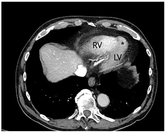
A 79-year-old male, with known ischaemic heart disease, was admitted to our emergency department because of acute abdominal pain and clinical suspicion of a ruptured abdominal aortic aneurism, which was confirmed by a computed tomography (CT) scan. His past history included hypercholesterolaemia treated with statins and silent myocardial ischaemia that was documented on prior myocardial scintigraphy. Emergent endovascular treatment of the aneurysm was successfully performed. The CT scan showed as an incidental finding the presence of a small left apical aneurysm (17 mm in its long axis and 7 mm at the base) (Figure 1).

Figure 1.
Thorax computed tomography. * = aneurysmal cavity; LV = left ventricle; RV = right ventricle.
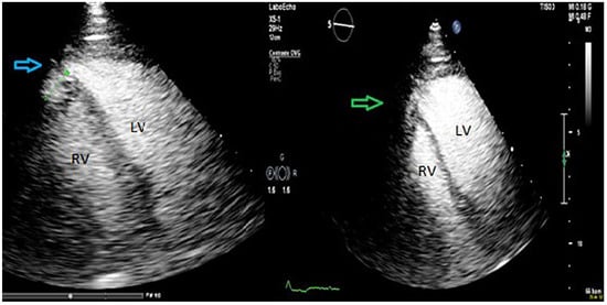
Two months after the intervention the patient was admitted for further work-up. Clinical examination was normal. The electrocardiogram (ECG) showed sinus rhythm, with normal axis and right bundle branch block. The laboratory findings, including high sensitive troponin I, were normal. At transthoracic echocardiography, the left ventricle was normal sized, with no evidence of ventricular septal defects (Figure 2). Injection of transpulmonary echocardiographic contrast agent (SonoVue, Bracco Suisse SA, Swissmedic, Manno, Switzerland) confirmed the presence of a small apical cavity with a 6 mm neck localised at the septo-apical level with extension under the apex of the right ventricle. The walls of this cavity showed a trabecular appearance with preserved contractility. There was a systolic expansion of a maximum length of 15 mm, without evidence of communication between the two ventricles on colour and pulsed-wave Doppler.

Figure 2.
Transthoracic echocardiography showed cavity expansion in systole (blue arrow) and size reduction during diastole (green arrow). LV = left ventricle; RV = right ventricle.
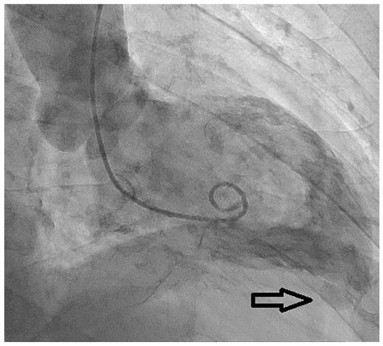
The coronary angiogram showed chronic total occlusion of the mid-right coronary artery, which was not treated as there was only slight ischaemia on scintigraphy and large epicardial collaterals from the left anterior descending artery (LAD). An intermediate lesion on the LAD was not haemodynamically significant after instantaneous wave-free ratio measurement (IFR 0.94). Left ventriculography confirmed the presence of an apical lesion compatible with a left ventricular diverticulum (Figure 3).

Figure 3.
Left ventriculogram: the examination showed the presence of a left ventricular apical diverticular excrescence (black arrow) without interventricular communication.
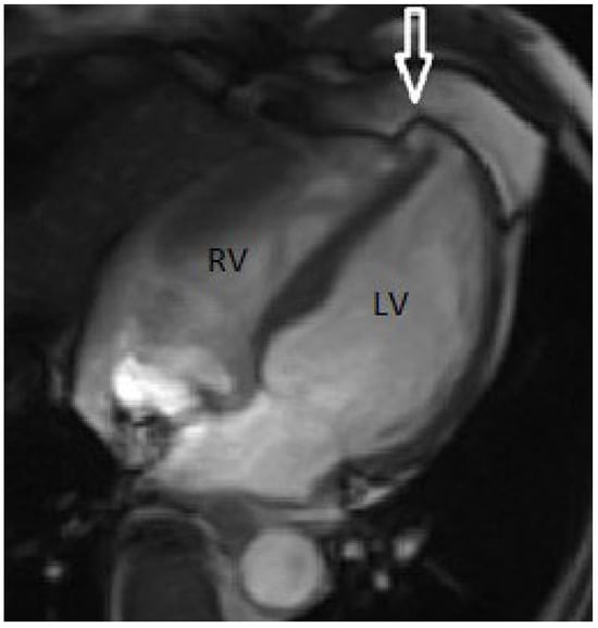
To clarify the nature of this finding, cardiac magnetic resonance imaging (MRI) was performed (Figure 4), which showed a small aneurismal cavity with a narrow neck and an extension of 6 mm at the septo-apical level, with extension under the right ventricular apex, no evidence of communication between the cavities and absence of scar.

Figure 4.
Magnetic resonance imaging: evidence of a small diverticular pouch (white arrow) at the apex of the left ventricle with narrow walls, not associated with late enhancement and without presence of interventricular communication. LV = left ventricle; RV = right ventricle.
From the multi-modality images our final diagnosis was a congenital diverticulum. After discussion within the heart team, given the absence of clinical symptoms, the small size of the cavity, and the absence of thrombus and of communication with the right ventricle, we decided for a conservative approach with clinical follow-up.
Discussion
Congenital diverticula are rare cardiac malformation. This entity was first described in 1816, often in association with other cardiac, vascular or thoraco-abdominal abnormalities (about 70% of the cases). It is usually a developmental anomaly starting during the fourth embryonic week. They can be found in all heart chambers, but most are seen in left ventricle, rarely in the right ventricle or in both ventricles [1]. In the typical presentation they are characterised by a finger or hook-like appendix coming out from the wall of the left ventricle, which contracts in synchrony with the heart chambers. Usually they are made of histologically normal heart wall tissue and the communication with the ventricular cavity is small (narrow neck). Some authors classify diverticula into two categories: fibrous types and muscular types. The first are characterised by the absence of myocardial layer. Other authors consider a diverticulum without a myocardial layer to be a pseudo-diverticulum and to make a distinction between the two entities they generally recommend a late enhancement cardiac MRI. A classification according to localisation (apical vs non-apical) is also used [2].
The embryological development of diverticula starts in the fourth embryogenic week. There are different theories about the way they develop. Some authors explain the formation of the diverticula by a partial stop in the development of the embryonic ventricle [4]. The “traction theory” postulates a developmental failure in embryogenesis with abnormal attachment of the heart tube to the structures of the yolk sac. These structures are stretched out during subsequent phases of development [5].
Congenital outpouchings include, apart from diverticula, left ventricular aneurysms, hernias, clefts and crypts.
Congenital left ventricular aneurysms are generally characterised by a thin wall and present a wide connection between the cavity and the ventricle. Histological assessment shows fibrotic and non-contractile tissue. Cardiac hernias are very rare findings that are defined as myocardium protruding through a pericardial defect. Myocardial crypts were described for the first time at autopsy of patients with hypertrophic cardiomyopathy; they are narrow, deep invaginations of congenital origin within the myocardium localised most often in the basal posterior septum and inside left ventricle free wall. They are generally described as fissures related to disarray of myocardial fibres. The definition of myocardial clefts is similar and the terms “cleft” and “crypt” are often used interchangeably in the literature (Table 1).

Table 1.
Classification of congenital left ventricular outpouchings. Modified and adapted from Vincenti and Müller [6].
Data from prospective studies on the incidence and prevalence of congenital aneurysms and diverticula are not available. In a large retrospective study including 43,000 consecutive echocardiograms in adult patients the prevalence was 0.04% [1]. In another large retrospective study of 12,271 patients, the prevalence of ventricular outpouchings was 0.76% [7]. In the literature, however, the type of ventricular outpouching is often mixed and in many series there is not a clear differentiation between them.
Congenital left ventricular aneurysms and diverticula can often be seen in association with other congenital anomalies, including vascular and extra-cardiac structural anomalies. Most frequently they are associated with ventricular septal defects, atrial septal defects, tricuspid atresia, patent foramen ovale, cardiac malposition (dextro-rotation or -position) and tetralogy of Fallot. Left ventricular diverticula seen in combination with diaphragmatic defects in the sternal part, abdominal wall defects, sternal defects and absence of inferior pericardial membrane are a defined entity called pentalogy of Cantrell or Cantrell’s syndrome [8].
Generally, left ventricular diverticula are asymptomatic and, as in our case, they are found incidentally during some diagnostic test done for other reasons. Sometimes association with rhythmic anomalies, systemic embolic events, typical angina and exertion dyspnoea have been described. Rupture of the aneurysm or the diverticulum are very rare and often fatal events.
Laboratory tests are of limited diagnostic value. Chest radiography is generally normal, but sometimes changes in cardiac silhouette might be seen. Typical or pathognomonic ECG changes are unknown, but many series show a high prevalence in association with left ventricular aneurysms and diverticula of first degree trio-ventricular block, bundle branch block, and intraventicular conduction delay; an association with right bundle branch block (RSR pattern in V1 and V2, QRS >120 ms), as in our case, has also been documented.
Transthoracic echocardiography, in particular with contrast, is an important tool to clarify the diagnosis, as it is a noninvasive test and widely available. As in our case, a Doppler flow study can be useful to exclude a communication between the two ventricles. Cardiac MRI has a crucial role in the diagnostic pathway as it is important to differentiate the disorder from ventricular dysplasia. MRI also allows tissue characterisation, possibly showing fibrotic areas after gadolinium injection. In addition, it may allow evaluation of other underlying cardiac anomalies. Coronary angiography remains an important diagnostic tool; on one hand it is crucial for the exclusion of coronary arteries disease, on the other hand, contrast ventriculography can show typical changes and the possible presence of contractility anomalies, the size and the possible communication between the left and the right ventricle.
Because these findings are very rare and there are only small series or case reports in the literature, clear recommendations about therapy are very difficult to derive, in particular given the lack of guidelines for management. Some authors recommend surgery even in asymptomatic patients in order to prevent serious complications. Congenital left ventricular aneurysms and diverticula can both be treated either conservatively or surgically, treatment modalities being determined by clinical presentation and specific findings in each patient. In some series, surgical treatment of left ventricular aneurysms included in three quarters of cases aneurysmectomy, either alone or combined with correction of accompanying congenital heart defects in 18.1%. Isolated repair of left ventricular diverticula was less frequent (51.9%), mainly owing to the higher prevalence of accompanying congenital defects in this group. There is not a clear consensus in all authors about anticoagulation therapy. Careful follow up is generally the strategy of choice in apical muscular diverticula with preserved contractilty, whereas in patients with large hypo- or akinetic aneurysms or fibrous diverticula, especially after systemic embolisation, therapeutic oral anticoagulation is more frequently administered [9].
Generally in asymptomatic patients with a small and contracting diverticulum, as in our case, a non-surgical strategy with strict follow up and without anticoagulation therapy seems reasonable.
Data about morbidity and mortality of patients with congenital left ventricular aneurysms or diverticula are lacking, because cohorts are too small and follow up periods are not homogeneous. Predictive factors are not known, and we also do not know whether size, multiple sites and intra-aneurysmatic pressures might have a prognostic impact. From a pathophysiological point of view, we could postulate a higher probability of rupture when the wall is thin and fibrous, but this suggestion is not supported by the literature.
Disclosure Statement
No financial support and no other potential conflict of interest relevant to this article was reported.
References
- Mayer, K.; Candinas, R.; Radounislis, C.; Jenni, R. Kongenitale linksventrikuläre Aneurysmen und Divertikel: Klinik, Diagnostik und Verlauf [Congenital left ventricular aneurysms and diverticula: clinical findings, diagnosis and course]. Article in German. Schweiz. Med. Wochenschr. 1999, 129, 1249–1256. [Google Scholar] [PubMed]
- Nam, K.H.; Kwon, J.Y.; Son, G.H.; Cho, N.H.; Park, Y.W.; Kim, Y.H. Prenatally diagnosed left ventricular diverticulum with thoracoabdominal wall defect: a case and review of the literature. J. Perinatol. 2010, 30, 760–762. [Google Scholar] [CrossRef] [PubMed]
- Soslow, J.H.; Parra, D.A.; Bichell, D.P.; Dodd, D.A. Left ventricular hernia in a pediatric transplant recipient: case report and review of the literature. Pediatr. Cardiol. 2009, 30, 55–58. [Google Scholar] [CrossRef] [PubMed]
- Van Mierop, L.; Kutschke, L. Embryology of the heart. In Hurst’s the Heart, 8th ed.; McGraw-Hill: New York, NY, USA, 1994. [Google Scholar]
- Albrecht, G. Beitrag zur Morphologie und formalen Genese kongenitaler Herzdivertikel [Morphology and formal etiology of congenital heart diverticula]. Zentralbl Allg. Pathol. 1972, 116, 42–47. [Google Scholar] [PubMed]
- Vincenti, G.M.; Müller, H. A pulsatile mass in the epigastric region. Poster DGK: Deutsche Gesellschaft für Kardiologie Nürnberg 07.10.2010. Clin. Res. Cardiol. 2010, 99 (Suppl. 2). [Google Scholar]
- Ohlow, M.A.; Secknus, M.A.; Geller, J.C.; von Korn, H.; Lauer, B. Prevalence and outcome of congenital left ventricular aneurysms and diverticula in an adult population. Cardiology 2009, 112, 287–293. [Google Scholar] [CrossRef] [PubMed]
- Cantrell, J.R.; Haller, J.A.; Ravitch, M.M. A syndrome of congenital defects involving the abdominal wall, sternum, diaphragm, pericardium, and heart. Surg. Gynecol. Obstet. 1958, 107, 602–614. [Google Scholar] [PubMed]
- Malakan Rad, E.; Awad, S.; Hijazi, Z.M. Congenital left ventricular outpouchings: a systematic review of 839 cases and introduction of a novel classification after two centuries. Congenit. Heart Dis. 2014, 9, 498–511. [Google Scholar] [CrossRef] [PubMed]
© 2019 by the authors. Attribution - Non-Commercial - NoDerivatives 4.0.