DG-PET/CT enables direct visualisation of inflammatory activity and may represent a useful tool for diagnosis, risk stratification and therapy monitoring in patients with pericarditis.
A 65-year-old man with a previous history of periaortitis was admitted to the emergency department of our hospital because of fever and intermittent chest pain, increasing in intensity and duration.
Inflammatory markers (erythrocyte sedimentation rate, C-reactive protein and leucocyte count) were abnormally increased.
The patient underwent a fluorine-18 fluorodeoxyglucose positron emission tomography / computed tomography (FDG-PET/CT) searching for the origin of this inflammatory syndrome.
PET images showed several areas of increased radiotracer uptake around the heart, corresponding to a mild pericardial effusion on CT images (Figure 1). This “ring of fire” sign on PET/CT images represents increased metabolic activity around the heart and suggested the presence of acute pericarditis.
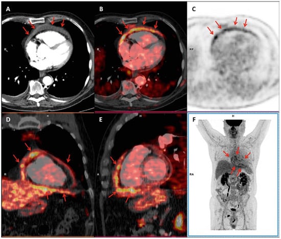
Figure 1.
The patient underwent a fluorine-18 fluorodeoxyglucose positron emission tomography / computed tomography (FDG-PET/CT) searching for the origin of this inflammatory syndrome.
Based on these FDG-PET/CT findings, the patient was treated with corticosteroids and colchicine with amelioration of symptoms and normalisation of serum inflammatory markers.
FDG-PET/CT offers valuable information in the assessment of cardiovascular inflammation [1,2,3]. As it enables direct visualisation of inflammatory activity, FDG-PET/CT may represent a useful tool for diagnosis, risk stratification and therapy monitoring in patients with pericarditis.
Conflicts of Interest
No financial support and no other potential conflict of interest relevant to this article was reported.
Ethical Approval
This article has been written in accordance with the ethical standards laid down in the 1964 Declaration of Helsinki and its later amendments.
References
- Slart, R.H.J.A. Writing group; Reviewer group; Members of EANM Cardiovascular; Members of EANM Infection & Inflammation; Members of Committees, SNMMI Cardiovascular; Members of Council, PET Interest Group; Members of ASNC; EANM Committee Coordinator. FDG-PET/CT(A) imaging in large vessel vasculitis and polymyalgia rheumatica: joint procedural recommendation of the EANM, SNMMI, and the PET Interest Group (PIG), and endorsed by the ASNC. Eur J Nucl Med Mol Imaging. 2018, 45, 1250–69. [Google Scholar] [PubMed]
- Colombo, J.; Elzi, L.; Treglia, G.; Perren, A. Light in the dark: (18)F-FDG PET/CT in Staphylococcus aureus bacteremia of unknown origin. Intensive Care Med. 2018, 44, 488–9. [Google Scholar] [CrossRef] [PubMed]
- Kircher, M.; Lapa, C. Novel Noninvasive Nuclear Medicine Imaging Techniques for Cardiac Inflammation. Curr Cardiovasc Imaging Rep. 2017, 10, 6. [Google Scholar] [CrossRef] [PubMed]

© 2018 by the author. Attribution - Non-Commercial - NoDerivatives 4.0.