Case report
A 63-year-old hypertensive male presented with atypical chest discomfort and breathlessness on exertion. Chest X-ray showed a “snowman”-like appearance of the cardiac silhouette (Figure 1). There was no evidence of chamber enlargement and the pulmonary vascularity was normal. In the inferior mediastinum, double density within the cardiac margins was noticed. Contrast computed tomogram of chest revealed a large saccular descending thoracic aortic aneurysm (Figure 2) extending from the subcarinal area to the gastro-oesophageal junction. The aneurysm was eroding the vertebra posteriorly and was filled with thrombus (Figure 3). The spinal cord was unaffected and there was no neurological deficit. The head of the snowman was formed by the arch of aorta and the body was formed by the globular descending thoracic aortic aneurysm. Although he was advised simultaneous aneurysm resection and vertebral column stabilisation surgery, he refused. His medical therapy was optimised and it included betablocker, ACE inhibitor and oral anticoagulant. He continues to have NYHA class II symptoms without any increase in size of aneurysm at two years of follow-up.
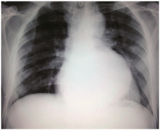
Figure 1.
Chest X-ray PA view showing “Snowman”-like appearance of the cardiac silhouette.
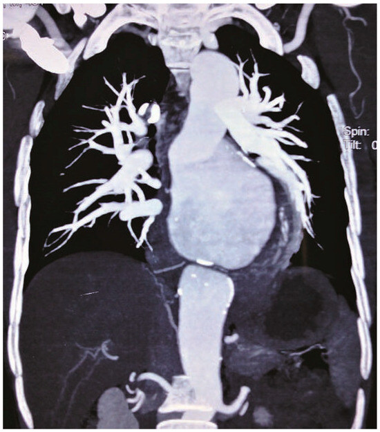
Figure 2.
CT chest demonstrating a large saccular aneurysm confined to the descending thoracic aorta.
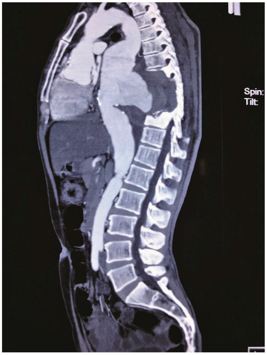
Figure 3.
CT chest lateral view demonstrating erosion of vertebra and presence of clot within the aneurysm.
Snowman appearance on X-ray is classically seen in total anomalous pulmonary venous connection (TAPVC). Although localised globular descending thoracic aneurysm can also simulate a snowman appearance on the X-ray, this cannot be considered as differential diagnosis for X-ray appearance of TAPVC. This is because such aneurysms do not occur in infants and uncorrected TAPVC rarely reaches adulthood. The other conditions that may rarely produce a snowman-like appearance in this age group include hiatus hernia, extramedullary haematopoiesis and tumours like neurofibromas and lymphomas. This image highlights the “Neo-snowman”-like appearance of descending aortic aneurysm.
© 2011 by the author. Attribution - Non-Commercial - NoDerivatives 4.0.