You are currently viewing a new version of our website. To view the old version click .
Nutraceuticals
  • Review
  • Open Access

10 November 2025

A Narrative Review on Breast Cancer Treatment Supported by Focused and Systemic Phytotherapy

,
,
,
,
,
,
,
,
and
1
CBSin—Center of BioSciences in Integrative Health, 4250-105 Porto, Portugal
2
ICBAS—School of Medicine and Biomedical Sciences, University of Porto, 4050-313 Porto, Portugal
3
Associate Laboratory i4HB—Institute for Health and Bioeconomy, University Institute of Health Sciences—CESPU, 4585-116 Gandra, Portugal
4
UCIBIO—Applied Molecular Biosciences Unit, Toxicologic Pathology Research Laboratory, University Institute of Health Sciences (1H-TOXRUN, IUCS-CESPU), 4585-116 Gandra, Portugal

Abstract

Cancer remains a persistent global health challenge, continuously driving the search for novel and effective therapeutic strategies. In the case of breast cancer, treatment decisions are primarily guided by factors such as the disease stage, histological grade, molecular receptor status, and the presence of genetic mutations. Understanding these parameters is crucial for tailoring interventions and improving clinical outcomes. To enhance prognostic and diagnostic accuracy, attention has increasingly turned to identifying molecular targets that play key roles in breast cancer development. Currently, standard treatments include surgery, chemotherapy, and radiotherapy. However, these approaches are often associated with significant side effects and a diminished quality of life. As a result, many breast cancer patients are turning to complementary therapies—including phytotherapy, nutritional interventions, and dietary supplements—to support conventional treatment, alleviate adverse effects, and improve overall well-being. Within the vast realm of medicinal flora, anticancer plants represent a compelling area of study, serving as a rich reservoir of bioactive compounds. These compounds have demonstrated significant promise in the ongoing battle against cancer. Often highlighted in traditional medicinal practices, these plants harbor a wide array of phytochemicals, such as alkaloids, flavonoids, polyphenols, and terpenoids. These phytochemicals manifest diverse biological activities, notably exhibiting pronounced anticancer properties. The exploration of these natural compounds has opened new avenues for developing innovative and targeted therapeutic strategies in cancer treatment. They achieve definitive chemotherapeutic and chemopreventive roles by integrating with specific molecular signals. Their multiple biological functions include antimutagenic, antiproliferative, antimetastatic, anti-angiogenesis, anti-inflammatory, antioxidant, and immunomodulatory properties, which collectively enable them to control cancer progression and intervene at various stages of cancer cell development. Moreover, these compounds are involved in regulating the cell cycle and microRNA, ultimately leading to cancer cell death by promoting apoptosis and autophagy, often mediated through ROS signaling. Thus, based on a large theoretical revision, we conclude that high-quality evidence is necessary in order to advise these products concerning their efficacy and safety. Also, clinical evidence should be supported by a comprehensive individual diagnosis and adequate research protocols in order to evaluate whether the benefits of these plant-produced interventions can outweigh their harms.

1. Introduction

According to the World Health Organization (WHO), cancer is the second leading cause of death in the world [,,,]. In the period between 2013 and 2021, the most common cancers in men were prostate (22.4%), colorectal (16.6%), lung (15.1%) and urinary bladder (11.7%) cancer; in women, breast (28.0%), colorectal (16.9%), uterus (6.2%) and lung (6.0%) cancer were the most common [,,].
The literature indicates that breast cancer commonly initiates within the mammary ducts and/or lobules [,,]. The initial stage, carcinoma in situ, is generally not considered life-threatening. However, malignant cells can extend into adjacent breast tissue (invasion), resulting in tumor formation detectable as lumps or areas of thickening. This invasive progression can lead to metastatic disease, where cancer cells disseminate to regional lymph nodes or other organs. Therapeutic strategies are tailored to the patient, cancer subtype, and disease dissemination, typically integrating surgery, radiation, and systemic treatments including hormonal therapies, immunotherapy, chemotherapy, and targeted biological agents [,,].

1.1. “Who Is at Risk”

The epidemiologic parameters (e.g., incidence, mortality, and survival rates) related to breast cancer diverge significantly between countries and regions []. It is well known that certain factors can increase the risk of breast cancer, such as gender, increasing age, family history of breast cancer, health habits, lifestyle (e.g., dietary, harmful alcohol abuse, tobacco use), obesity, history of radiation exposure, reproductive history (such as age at menarche and age at first pregnancy) and postmenopausal hormone therapy. Nevertheless, approximately half of breast cancers develop in women who have no identifiable breast cancer risk factor other than gender (female) and age (over 40 years) [,].
With respect to family history of breast cancer, most women diagnosed with the pathology do not have a known family history of the disease, with less than 5% of total breast cancers being hereditary cases [,]. Certain inherited high-penetrance gene mutations greatly increase breast cancer risk, accounting for up to 25% of hereditary cases due to a mutation in one of the identified, rare but highly penetrant, genes (BRCA1, BRCA2, TP53, CDH1, STK11, PTEN/MMAC1), which confer up to an 80% lifetime risk of breast cancer [,,]. An additional 2–3% of cases are due to a mutation in rare moderate-penetrance genes (e.g., CHEK2, BRIP1, ATM, and PALB2), each associated with a twofold increase in risk [,,]. According to the WHO [], the most dominant mutations are in BRCA1, BRCA2, and PALB-2 genes.

1.2. Global Impact

Globally, breast cancer is the most prevalent malignancy in females and a leading cause of cancer-related death among women [,]. It has overtaken lung cancer as the most commonly diagnosed cancer worldwide, now responsible for one in eight cancer diagnoses and a combined total of 2.3 million new cases in both sexes []. Constituting a quarter of all cancer cases in females, it was the predominant cancer diagnosed in women in 2020. The disease’s burden is expanding in numerous parts of the world, particularly within developing countries. According to recent data from the Portuguese League against Cancer, in Portugal, around 7000 new cases of breast cancer are diagnosed annually, and 1800 women die from the disease [].
Breast cancer death rates have been decreasing steadily since 1989, for an overall decline of 43% through 2020. This decrease in death rates is believed to be the result of earlier diagnosis through screening and increased awareness, as well as more efficient treatments [,]. The WHO’s Global Breast Cancer Initiative aims to reduce global breast cancer mortality by 2.5% annually, which would prevent 2.5 million deaths between 2020 and 2040. This objective is supported by three key pillars: health promotion for early detection through public education on breast cancer signs and symptoms, timely diagnosis, and comprehensive breast cancer management [,]. Consequently, modern medicine must prioritize targeted multidisciplinary research and improve the efficiency of health prevention. This involves focusing on primary prevention and modifying risk factors for early disease detection, promptly initiating treatment (secondary prevention), and ensuring diligent follow-up. The ultimate goal is to continuously reduce the morbidity, mortality, and economic burden associated with breast cancer [].
The WHO [] outlines several potential symptoms of breast cancer, which include a palpable breast lump or area of thickening, frequently without associated pain; alterations in breast size, shape, or overall appearance; skin changes such as dimpling, redness, or pitting; modifications in nipple morphology or the periareolar skin; and atypical or sanguineous nipple fluid. When breast cancer metastasizes to other bodily regions, it can elicit additional symptoms. The axillary lymph nodes commonly represent the initial site of detectable dissemination, though some cancerous lymph nodes may not be palpable. Subsequently, malignant cells can spread to distant organs, including the lungs, liver, brain, and bones. Upon reaching these secondary sites, novel cancer-related symptoms, such as bone pain or headaches, may manifest [,].

1.3. Molecular Factors with a Significant Role in Breast Cancer Development

Several molecular targets critical to breast cancer development and progression have been identified (Table 1) and are explored further in this section.
Table 1. Main molecular targets involved in breast cancer and their mechanisms of action.

1.3.1. Estrogen and Progesterone

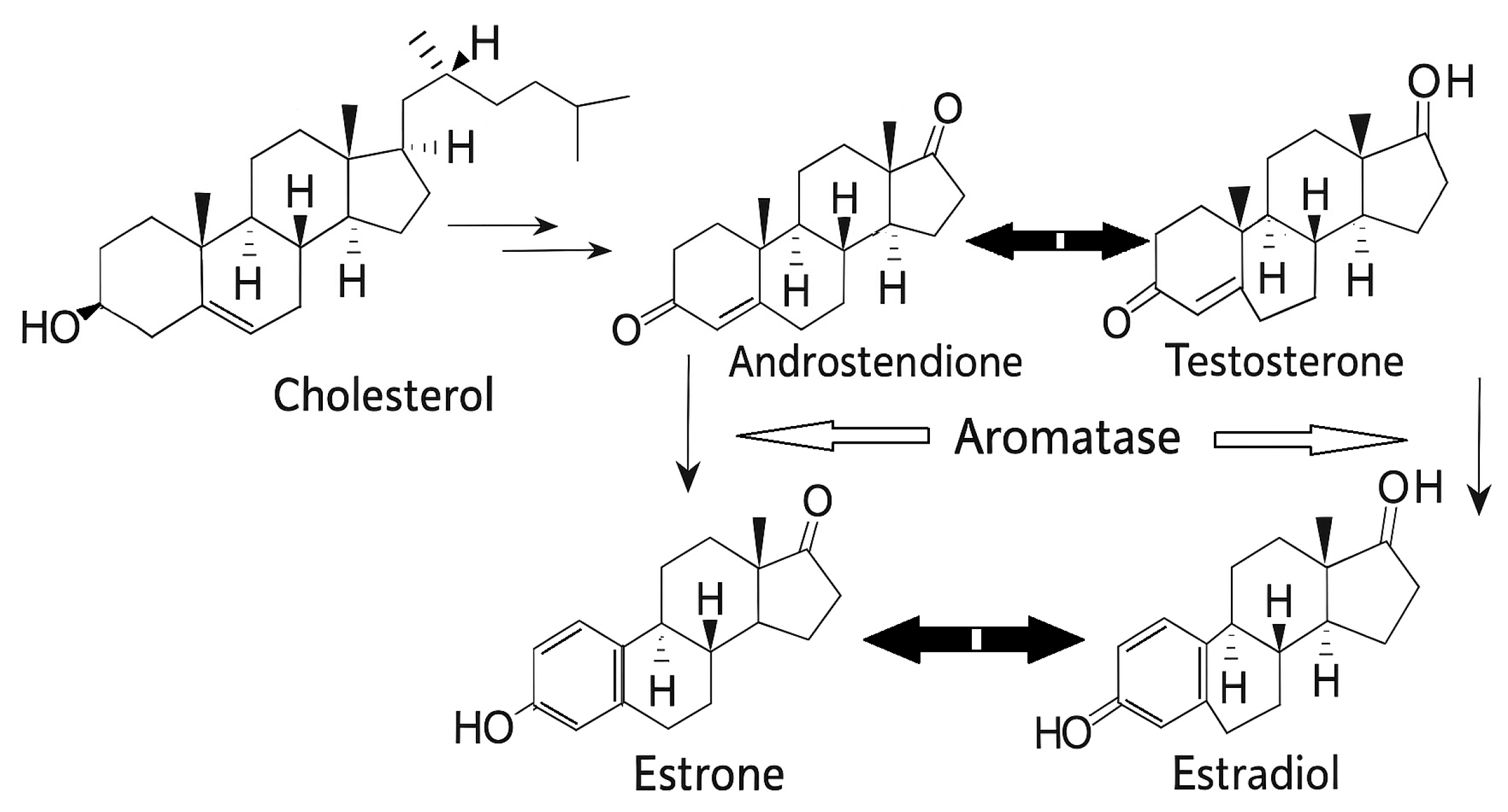
Estrogen is the major mitogenic hormone in pubertal mammary glands, mainly responsible for the typically “feminine” secondary characteristics, such as breasts, in the thickening of the endometrium and other features of regulating the menstrual cycle (egg releasing). Progesterone particularly supports the pregnancy process and stimulates adult mammary epithelial proliferation [,]. In premenopausal women, estrogen is synthesized primarily in theca cells, while progesterone is synthesized in theca and granulosa cells of the ovarian corpus luteum and additionally in the adrenal gland, placenta, and adipose tissue. The synthesis process of estrogens ends with the conversion of androgens in granulosa cells by the aromatase enzyme, as illustrated in Figure 1. There are three main endogenous forms of physiological estrogens in women: estrone (E1), estradiol (E2), and estriol (E3). After menopause, E1 has an important role as it is formed in adipose tissue from adrenal dehydroepiandrosterone. E2 is considered the major and most potent product of the estrogen biosynthesis process. E3 form is the least prevalent estrogen and is formed from the E1 or E2.
Figure 1. Schematic representation of the production of estrone (E1) and estradiol (E2), and progesterone in the ovarian corpus luteum. The conversion of androstenedione and testosterone to E1 and E2, respectively, occurs through a series of enzymatic reactions primarily in peripheral tissues, which is known as aromatization and is mediated by the enzyme aromatase. E1 is a less potent estrogen compared to E2, but still plays significant roles in various physiological processes, particularly in postmenopausal women, where it becomes the primary source of estrogen [,].
Attending that in female mammals, the development and regulation of the reproductive system and non-reproductive system are significantly influenced by estrogens and respective estrogen receptors (ERs), which present a critical involvement in the etiology and advancement of breast cancer. In fact, endogenous estrogens (estrone and estradiol) mediate a range of systemic effects, playing a regulatory role in a myriad of physiological processes from reproduction to lipid metabolism. While offering protective benefits for organs such as the brain, bone, heart, liver, and vagina, extended exposure to these hormones is associated with adverse outcomes, specifically an increased risk of developing breast and uterine cancers [,,]. Since its involvement in the breast cancer phenomenon is well established, it is naturally consequent to positioning them as essential molecular targets [,,,,,].
Estrogens can indirectly influence breast cancer risk through the upregulation of progesterone receptors, thereby enhancing progesterone-mediated signaling pathways. However, increasing clinical and experimental data suggest that progestogens, whether in the form of natural progesterone or synthetic analogs (progestins), may represent the predominant hormonal factor contributing to what has traditionally been viewed as estrogen-related breast cancer susceptibility [,]. Therapeutic strategies that block estrogen activity, such as selective estrogen receptor (ER) modulators or agents that inhibit estrogen biosynthesis, have demonstrated efficacy in decreasing recurrence and disease progression in patients with ER-positive breast tumors. Furthermore, elevated circulating estrogen concentrations have been consistently associated with higher breast cancer risk. Collectively, current findings highlight the broad oncogenic and tumor-promoting influence of estrogen in the initiation, recurrence, and progression of breast cancer [].
Estrogen and its mechanisms: Concerning specific molecular mechanisms, estrogens influence breast cancer development and progression through several pathways, including the stimulation of cellular proliferation via the ERα pathway, direct augmentation of genetic mutation rates, or modulation of DNA repair mechanisms [,,,].
Estrogens exert their biological effects through two main categories of receptors: the classical nuclear ERs (ERα and ERβ) and the more recently identified membrane-associated receptors (GPR30 and ER-X). Both receptor types are distributed throughout the brain and peripheral tissues, exhibiting cell- and tissue-specific expression patterns. The nuclear ERs belong to the superfamily of transcription factors and function primarily via a traditional estrogen-dependent signaling pathway. Within the cytoplasm, these receptors bind lipophilic estrogen molecules, forming complexes that translocate into the nucleus. Once there, receptor dimerization occurs, allowing interaction with specific estrogen response elements in target gene promoter regions. This process, facilitated by the recruitment of co-activator proteins, initiates transcriptional activation (Figure 2).
Figure 2. The estrogen signaling pathway. The estrogen signaling mainly includes activation of the intracellular ER that, upon ligand binding and dimerization, translocates to the nucleus, where it directly binds responsive elements of target genes involved in cell growth, inflammation, proliferation, survival, and protein synthesis. Differently, estrogens mediate non-genomic effects and activate intracellular signaling through the binding of the plasma membrane receptors, ER variants, and the G protein-coupled receptor (GPR30). This binding induces the rapid activation of several protein kinases, like mitogen-activated protein kinase (MAPK), which, in the end, regulate the gene expression of estrogen target genes.
Relative to the signaling pathway of ERs, Zimmerman et al. [] and Al-Shami et al. [] point out that estrogen can cross the plasma membrane, where it interacts with intracellular ER to affect DNA directly. Moreover, estradiol stimulates mitogen-activated protein kinases (MAPK/ERK) within two minutes in MCF-7 breast cancer cells []. Furthermore, conjugated estrogen that is membrane-impermeable rapidly activates the MAPK/ERK pathways despite its inability to reach intracellular ERα or Erβ []. So, considering that conjugated estrogen is membrane-impermeable, several studies proposed that an ER should be localized in the plasma membrane and initiate the nongenomic effects phase, perhaps via a novel receptor, renamed G protein-coupled estrogen receptor (GPER) []. However, GPER is not the only candidate for a membrane-bound ERs, as there is a truncated isoform of ERα that also induces acute signaling. Therefore, and most likely, multiple membrane-bound receptors mediate the acute actions of estrogen [,].
In summary, it has been proposed that GPER1, ERα, and ERβ can interact with estrogens to trigger a variety of intracellular signaling pathways. Estrogen-driven signaling can be broadly divided into genomic and non-genomic mechanisms, which differ in the cellular and molecular processes governing gene regulation. In these pathways, the estrogen–receptor complexes may associate with DNA either directly or indirectly. Once translocated into the nucleus, these complexes bind specific chromatin regions known as estrogen response elements (EREs), where they modulate gene transcription through two sequential genomic phases [,].
Tumors that express high levels of estrogen receptors are categorized as ER-positive, while those with few or no detectable receptors are classified as ER-negative, distinctions that significantly influence therapeutic decisions. ER-negative breast cancers tend to display greater aggressiveness and are associated with poorer survival outcomes. Conversely, patients with ER-positive tumors, although initially more responsive to hormone therapy, have a higher likelihood of disease recurrence more than a decade after the initial diagnosis [].
Taken together, the accumulated evidence indicates that estrogen and its metabolic derivatives play a crucial yet complex role in both the initiation and progression of breast cancer [,]. Large-scale clinical studies using selective estrogen receptor modulators have further reinforced the association between estrogen exposure and breast cancer risk [,]. Clinical parameters such as mammographic breast density, circulating estrogen concentrations, and bone mineral density serve as useful biomarkers of cumulative estrogen exposure, thereby contributing to risk assessment in women []. Structurally and functionally, the ERα and ERβ isoforms share substantial homology and interact similarly with endogenous estrogens, particularly E2, which are pivotal in both normal cellular development and malignant transformation in various cancers. The oncogenic potential of estrogen has been linked to the formation of reactive aromatic metabolites, specifically catechol estrogen quinones, which originate from naturally occurring catechol estrogens and can contribute to DNA damage and carcinogenesis [].
Progesterone and its Mechanisms: Steroid hormones such as progesterone exert their effects primarily through binding to intracellular receptor proteins located in the cytoplasm and nucleus. Progesterone specifically interacts with two main receptor isoforms, PR-A and PR-B, which are derived from the same gene (PGR) but transcribed via distinct promoters, resulting in receptors with differing regulatory and functional capacities. The relative expression and ratio of PR-A to PR-B in reproductive tissues fluctuate according to the developmental phase and hormonal milieu of the tissue []. Both PR-A and PR-B can bind progesterone, undergo dimerization, and associate with specific progesterone response elements (PREs) on DNA to influence transcriptional activity. Once activated by ligand binding, these receptors recruit transcriptional co-regulators—either coactivators or corepressors—that interact with the transcriptional machinery to modulate gene expression []. In breast tissue, ligand-bound PR regulates the expression of multiple genes, including those encoding estrogen receptors, insulin receptors, epidermal growth factor (EGF) and its receptor, as well as transforming growth factor (TGF)-α. Through this mechanism, progesterone is thought to influence cell proliferation indirectly by altering the abundance of growth factors and their corresponding receptors. Evidence from reproductive cancer cell lines further indicates that PR-regulated gene expression depends on MAPK signaling, paralleling the mechanism observed for estrogen receptor–mediated genes [].
Concerning progesterone signaling via the PR in breast cancer development, it is almost consensual that equal amounts of PR-A and PR-B are present in healthy adult breast tissue, and the same is true in benign breast lesions, whereas this ratio is altered in breast cancer [,]. It has been proposed that progesterone stimulates normal human breast epithelial cells via a paracrine signaling mechanism, a process considered a potential risk factor for breast cancer development. This occurs through progesterone-induced cyclic proliferation of mammary stem cell populations or latent tumor-initiating cells within mature breast epithelium, thereby facilitating preneoplastic transformation [,]. In breast cancer, the proliferative influence of progesterone is largely mediated by the PR-B isoform, which also plays a predominant role in extranuclear progesterone signaling. In contrast, PR-A exhibits limited capacity to rapidly activate protein kinase pathways, rendering it less effective in mediating extranuclear responses []. Studies using breast cancer cell lines indicate that PR-A is required for optimal progesterone responsiveness, while PR-B-driven transcriptional activity tends to enhance tumorigenic potential [].
Analogous to ERs, the classical nuclear PRs are the principal mediators of progestogen-dependent genomic effects. Nevertheless, progesterone can also exert biological actions through rapid, non-genomic mechanisms that involve activation of intracellular signaling cascades initiated at the plasma membrane. These “non-classical” progesterone actions occur more quickly than the transcriptionally mediated effects of nuclear PRs, as they do not rely on gene transcription or protein synthesis [,,].
Therefore, two distinct membrane-associated receptor systems have been identified as mediators of these rapid progesterone effects: the membrane progesterone receptors (mPRs) and the progesterone receptor membrane component 1 (PGRMC1) []. PGRMC1 forms part of a larger progesterone-binding multiprotein complex with diverse functions, including activation of cytochrome P450 enzymes involved in steroid metabolism. This receptor is expressed in breast and various other tissues and is believed to contribute to the antiapoptotic activity of progesterone []. Of note, PGRMC1 expression is decreased in postmenopausal women, as well as in those with premature ovarian failure or polycystic ovary syndrome, but is found to be upregulated in breast cancer tissues. Elevated estrogen levels are known to induce PR expression (a process referred to as estrogen priming), which is characteristic of many progesterone-responsive tissues [].
As well, recent research has highlighted that PR-A and PR-B may also modulate estrogen signaling and influence ER–chromatin interactions []. This suggests that the function of PR in breast cancer extends beyond serving merely as a biomarker of ER activity. In particular, PR appears capable of acting as both a co-regulatory partner and a modulator of ER-driven gene targeting, although findings in this area remain variable across studies.
According to Tian et al. [], exposure of MCF-7 breast cancer cells to either estrogen or progesterone individually enhances cell viability, with progesterone exerting a comparatively weaker effect. However, when both hormones are administered together, MCF-7 cells exhibit the greatest increase in viability. Moreover, the study identified cyclin G1 as a key regulator of cell cycle progression, which showed a similar upregulation pattern under combined hormone treatment.
Moreover, genetic mutations affecting DNA repair pathways, such as BRCA1, BRCA2, CHEK2, PALB2, ATM, and TP53, have been strongly associated with an elevated risk of developing breast cancer. While certain mutations, like BRCA1, are primarily linked with triple-negative breast cancer, others, including CHEK2, are more often associated with ER-positive subtypes []. A study examining BRCA1 mutation carriers reported that this mutation can promote abnormal expansion of luminal progenitor cells independently of progesterone signaling. Follow-up research by the same group revealed that BRCA1 mutation carriers exhibit dysregulated progesterone signaling, leading to increased cellular proliferation and genomic instability [].
In summary, they found that E2 plus progesterone exerted greater detrimental effects on the risk of breast cancer than either E2 or progesterone alone []. Collectively, animal and human studies indicate that progesterone is the principal hormonal factor driving the development and recurrence of adult mammary/breast epithelial proliferation, while estrogens may contribute to breast cancer risk by amplifying progesterone signaling [].
Therefore, both estrogen and progesterone play distinct yet interconnected roles in modulating breast cancer risk. Elevated estrogen levels are associated with an increased likelihood of developing breast cancer, as excessive estrogen activity can promote uncontrolled cellular proliferation, a key step in tumor initiation and progression [,]. Approximately 70% of breast cancer cases are classified as hormone receptor–positive, meaning tumor growth is stimulated by hormonal signals. Such cancers may express ER-positive, PR-positive, or both, indicating that the malignant cells depend on these hormones for proliferation and survival [,,].
While the link between estrogen dominance and heightened breast cancer risk is well established, the role of progesterone in breast carcinogenesis remains less clearly defined. Current evidence on progesterone’s contribution to breast cancer development is more limited and, in some cases, conflicting [,,,].

1.3.2. Aromatase and Breast Cancer

The cytochrome P450 enzyme, aromatase, plays a pivotal role in catalyzing the biosynthesis of estrogens (estrone and estradiol) from androgens (androstenedione and testosterone) (Figure 3) [,]. This enzyme is widely distributed, found in tissues such as the breast, skin, brain, adipose tissue, muscle, and bone [,]. A notable finding is the twenty-fold higher concentration of estrogens in breast cancer tissues compared to circulating plasma. This localized estrogen production, often occurring within or near malignant tissues, strongly suggests an elevated local expression of aromatase [,]. Given this, inhibiting aromatase represents a vital therapeutic approach to decrease estrogen levels, a strategy that has proven clinically valuable for postmenopausal women with breast cancer. The suppression of aromatase activity effectively reduces estrogen production to near-undetectable concentrations, profoundly impacting the development and progression of hormone-responsive breast cancers. As a result, aromatase inhibitors (AIs) are utilized for both anticancer therapy and chemoprevention, helping to prevent secondary breast cancer recurrence [,,]. Several natural compounds and plant extracts have been identified with aromatase inhibitory activities, which is relevant in the context of estrogen-dependent conditions such as breast cancer [].
Figure 3. Schematic representation of the estrogen biosynthesis pathway: Cholesterol is converted to androstenedione and testosterone, which are then aromatase-catalyzed into estrone and estradiol, respectively.
A wide range of natural compounds has demonstrated the ability to inhibit aromatase, a key enzyme in estrogen biosynthesis. Flavonoids such as apigenin, chrysin, and genistein, found in foods like parsley, chamomile, and soy, inhibit aromatase activity either through direct enzyme binding or by modulating gene expression. Similarly, plant extracts including white button mushroom, green tea polyphenols (e.g., EGCG), and red clover (rich in biochanin A) have shown aromatase inhibition in vitro and in vivo. In addition, other natural compounds like resveratrol (from grapes and red wine) and curcumin (from turmeric) suppress aromatase expression at the transcriptional level [,,,,,]. Collectively, these findings highlight the therapeutic potential of dietary phytochemicals in modulating estrogen production and possibly contributing to the prevention or treatment of hormone-dependent cancers.

1.3.3. Cathepsin D—Lysosomal Aspartic Protease

Found in all tissues, Cathepsin-D (CD) is an aspartyl lysosomal protease responsible for broad protein degradation in the highly acidic lysosomal environment []. A key distinction in metastatic breast cancer is the high secretion of pro-cathepsin-D (pCD). This pCD acts as a mitogen, stimulating the pro-invasive and pro-metastatic behavior of cancer and stromal cells. Given that pCD/CD levels have been identified as an independent prognostic factor in various cancers through numerous studies, CD is now considered a promising target for anti-cancer therapy [,,]. This abnormal secretion is due to two mechanisms, overexpression of the CD gene and altered processing of the precursor protein [,]. In ER-positive breast cancer cells, CD gene transcription is increased by estrogen and growth factors, but in estrogen-receptor-negative cells, the mechanism is unknown. Estrogen, through its ERα, can directly bind to DNA and activate the transcription of target genes, while growth factors can activate signaling pathways that ultimately lead to ER phosphorylation and enhanced transcriptional activity. This interplay between estrogen and growth factor signaling is crucial for tumor growth and survival in ER-positive breast cancer [,]. A large number of independent clinical studies associated high CD concentrations in the cytosol of primary breast cancers with increased risk of subsequent metastasis [,,]. This cytosol localization of CD suggests CD functions in apoptosis []. Rather than increasing the primary tumor cancer cell escape through basement membrane degradation, as proposed for neutral proteinases, CD appears to facilitate cell growth at distant sites [].
The major requirement for pCD to be autoactivated effectively seems to be its localization in an acidic microenvironment. This acidic milieu has been previously described in pre-lysosomal compartments such as late endosomes, often large intracellular acidic vesicles [,]. The lysosomal system is composed of several distinct and dynamically interacting vesicular compartments that are major sites for intracellular protein turnover and the limited proteolytic processing of certain proteins. After secretion, pCD could potentially be activated extracellularly in tumor tissue, where pH is generally more acidic, due to macrophage activity, than that in the corresponding normal tissue [,]. However, most studies point to a role of CD in facilitating distant metastasis via an indirect mitogenic activity rather than stimulation of local invasion and extravasation. Thus, the limiting step for metastasis was not intravasation of cancer cells, their survival in the circulation, or their extravasation, but more probably the ability of cancer cells to form colonies at distant sites in a foreign acidic environment. This could explain why cancer cells overexpressing CD have a higher capability of developing metastasis. On the other hand, this overexpression might also facilitate invasion following digestion of the basement membrane by extracellular proteases [,,,]. Expression of several proteases is generally increased during tumor progression, which has been proposed to also play a critical role in metastasis. Thus, high concentrations of CD have been found to be associated with increased risk of developing clinical metastasis in human breast cancer by degrading the extracellular matrix, liberating growth factors, and influencing cell proliferation and migration [,]. However, despite CD being a promising target in cancer cells, the effects and underlying mechanism of its inhibition remain unclear, and the proposed pathways are very controversial. Immunohistochemistry does not allow for determining the sites of mRNA CD synthesis, which is required to understand the mechanism by which this protease might facilitate metastasis [,].
Elevated CD concentrations detected in the peritumoral microenvironment of both human and animal subjects suggest their involvement in cancer cell invasiveness. While CD activity is typically optimal at pH values below that of normal tissue [], primarily within the lysosomal environment (where pH is generally thought to be less than 6), studies have demonstrated its capacity to cleave aggrecan within both interglobular and chondroitin sulfate domains over a broader pH range of 5.2–6.5 [,]. In some pathological states, excessive CD levels arise from an overburdened secretory pathway, leading to the release of the pCD rather than the mature form. Both CD and its proenzyme have been proposed to function as growth factors, stimulating the proliferation of malignant cells [,]. Upon secretion by tumor cells, CD mediates the cleavage of various structural and functional proteins and peptides, and is also known to inactivate chemokines, including macrophage inflammatory protein-1 alpha (CCL3), CCL4, and CCL21 []; it also cleaves prolactin [] and osteopontin [], thereby modulating their functions. The secretion at increased levels of CD has been linked to size, grade, aggressiveness, metastasis, and prognosis of endometrial, ovarian, and breast cancers [,]. In human breast carcinomas, CD is highly expressed and it is considered an independent marker of poor prognosis [,].
Importantly, the role of CD in cancer progression intersects with another critical cellular mechanism: autophagy. Autophagy is a highly normal and conserved process of cell degradation and cycling in all eukaryotes []. Autophagy disruption has the potential to sensitize cancer cells to programmed cell death and augment chemotherapy-induced apoptosis. Chloroquine and its analog, hydroxychloroquine, are currently the sole clinically utilized autophagy inhibitors []. Nevertheless, their association with ocular toxicity necessitates the development of novel autophagy inhibitors possessing a superior therapeutic index. The small molecule lucanthone, a prodrug that undergoes metabolism to hycanthone, also inhibits autophagy, induces lysosomal membrane permeabilization, and exhibits significantly greater potency in breast cancer models compared to chloroquine. Given that lucanthone effectively reduces cancer cell viability irrespective of p53 status, its mechanism of action appears to be more significantly driven by autophagy inhibition than by effects on DNA repair. Furthermore, lucanthone’s capacity to inhibit autophagy suggests its potential to enhance the efficacy of chemotherapeutic agents that modulate this pathway. Consistent with this, Carew et al. [] demonstrated that lucanthone acts as an autophagic inhibitor, inducing apoptosis through CD accumulation and enhancing vorinostat-mediated cell death in breast cancer models. On the other hand, more recent studies reported that it remains unknown whether the effect of CD on autophagy can change the cell death caused by ionizing radiation []. Constitutively high-expressed CD activates the fusion of autophagosome and lysosome, enhancing autophagic flux, reducing tumor radio-resistance, and thus decreasing radiation-induced apoptosis. In this sense, recent studies suggested CD inhibitors to be a potential drug to improve the radio-therapeutic efficacy of patients with glioblastoma cancer [].

1.3.4. Urokinase Plasminogen Activator

The intricate processes of cancer cell invasion and metastasis are driven by a concerted interplay between proteolytic enzymes and adhesive proteins, critical for cellular attachment and migration []. Compelling data from preclinical and clinical investigations underscore the pivotal role of the urokinase-type plasminogen activator (uPA) system in these degradative pathways [,,]. Furthermore, the antigen levels of uPA system components correlate with prognostic outcomes across numerous malignancies, including breast cancer. Specifically, the serine protease uPA binds to its cognate cell surface receptor, uPAR, thereby facilitating the conversion of plasminogen into the active serine protease plasmin. This expansive protease is capable of directly or indirectly degrading a wide array of extracellular matrix (ECM) constituents, additionally activating metalloproteinases that subsequently break down collagens and other matrix proteins. Supporting this, a previously developed enzyme-linked immunosorbent assay (ELISA) revealed uPAR levels in pellet extracts to be more than threefold higher than those measured in cytosols (p < 0.001). Consequently, elevated uPAR levels signify its profound involvement in plasmin-mediated ECM degradation, reflecting the substantial prognostic implications of uPA:uPAR complexes during cancer invasion and metastasis [,].
Beyond their proteolytic functions, most components of the uPA system involved in plasminogen activation have been associated with cell adhesion and migration via both proteolytic and non-proteolytic mechanisms [,,]. Cellular migration, in particular, necessitates the interaction of cell-surface adhesion receptors, such as integrins and uPAR, with their corresponding extracellular matrix-associated ligands, including vitronectin [,]. The activity of uPA can be modulated by its serpin inhibitors, PAI-1 and PAI-2 []. Given their multifaceted roles, several components of the uPA system represent compelling targets for anti-angiogenic, anti-invasive, and/or anti-metastatic therapeutic interventions.
Various strategies have been developed to modulate the expression and activity of uPA and uPAR at both gene and protein levels. These therapeutic approaches include the use of antisense oligonucleotides (ASOs), monoclonal antibodies, enzyme inhibitors, and both recombinant and synthetic analogs of uPA and uPAR [,,]. Among these, ASOs represent a particularly promising modality due to their capacity for sequence-specific inhibition of gene expression at the transcriptional and translational stages [,]. This ability to selectively suppress gene expression highlights the significant clinical potential of ASOs as therapeutic tools.
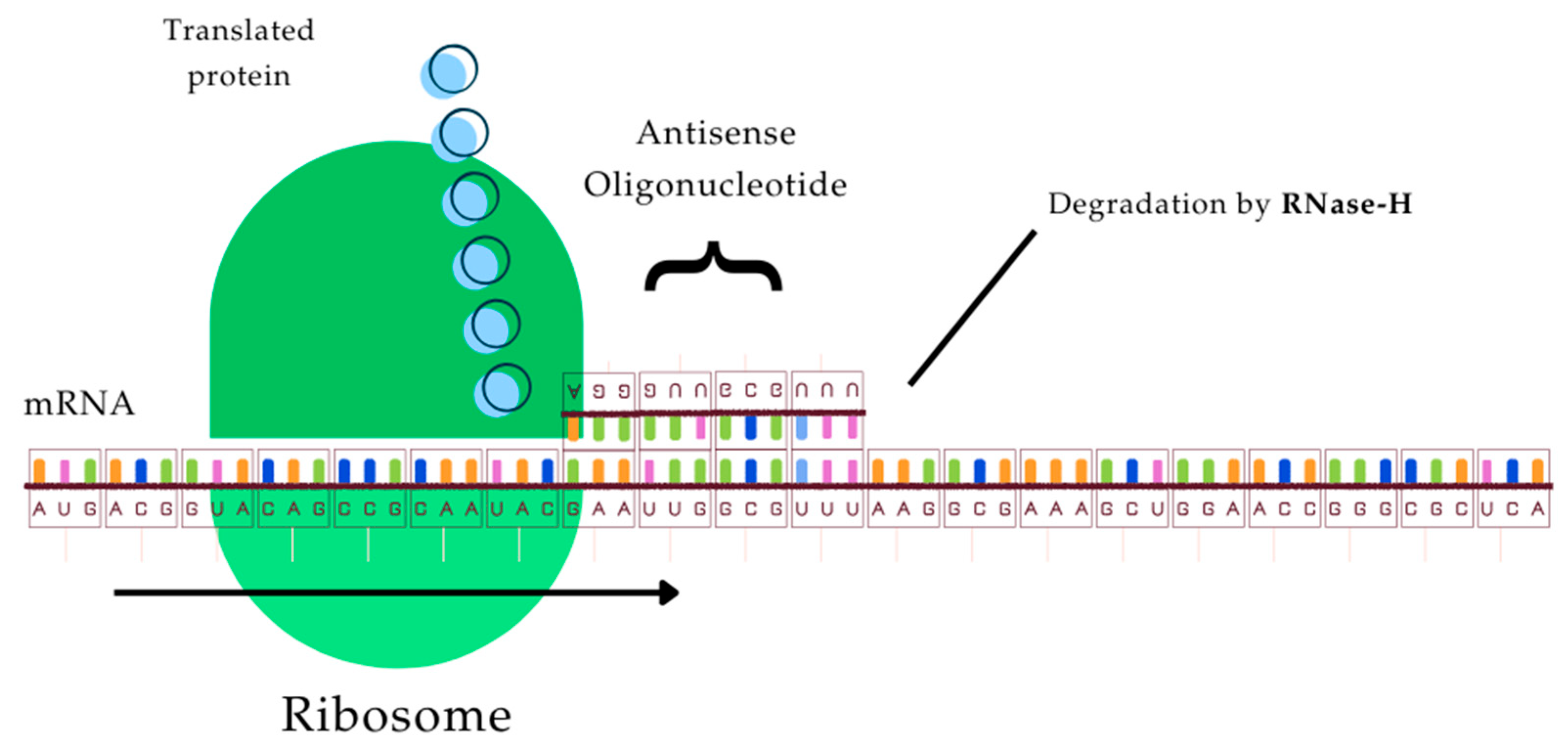
Although the precise mechanisms by which ASOs exert their inhibitory effects are not fully understood, several pathways have been proposed. One widely accepted mechanism involves steric hindrance of ribosome assembly or elongation during translation, thereby preventing protein synthesis (Figure 4). Another proposed mechanism of action involves the enzymatic cleavage of the RNA strand within DNA–RNA hybrids by RNase H, an endonuclease that selectively targets the RNA component of such duplexes (Figure 4). Although RNase H is widely expressed in various cell types, several studies have reported that ASOs can suppress specific gene expression without necessarily inducing mRNA degradation [,,,]. In addition, ASOs may interfere with the secondary and tertiary folding of mRNA molecules, thereby preventing the formation of structural motifs required for the binding of regulatory nonribosomal proteins. Given the pivotal role of the urokinase-type plasminogen activator (uPA) system in mediating tumor cell adhesion and migration, therapeutic approaches using targeted biomolecules such as antisense oligonucleotides have been explored to disrupt the non-proteolytic functions of this complex system.
Figure 4. Antisense oligonucleotides can interfere with the translation process after sequence-specific binding to the target mRNA by a (steric) block of translation and/or activation of RNase-H.
As knowledge about these mechanisms increases, the targeting of such protein-binding sites may become another highly efficient route for therapy supported by the antisense-mediated gene regulation.
Several waves of preclinical and clinical studies on ASOs have made it possible to optimize their pharmacological, pharmacodynamic, and pharmacokinetic properties, increasing their field of use and decreasing their overall toxicities [,,,]. The pathogenic mRNAs are now major therapeutic targets in diseases previously considered incurable [,,]. Therefore, numerous clinical trials evaluating the use of downregulating antisense oligonucleotides for the treatment of cancer and other diseases are now underway.

1.3.5. Fucosiltransferase 3 (FUT3)

Invasive ductal carcinoma is the primary and most relevant cause of increasing cancer-related mortality among women, where among the molecules used in the pathological characterization of tumors, glycoconjugates play a key role as potential biomarkers, due to relevant glycosylation cellular mechanisms. Dysregulation of glycosylation in cell surface and serum glycoproteins has been linked to carcinogenesis and cancer progression. Malignant tumors often exhibit abnormal fucosylated glycan expression with different localizations and functions []. This is due to the process’s complexity, which is highly modulated by different cellular conditions [,,].
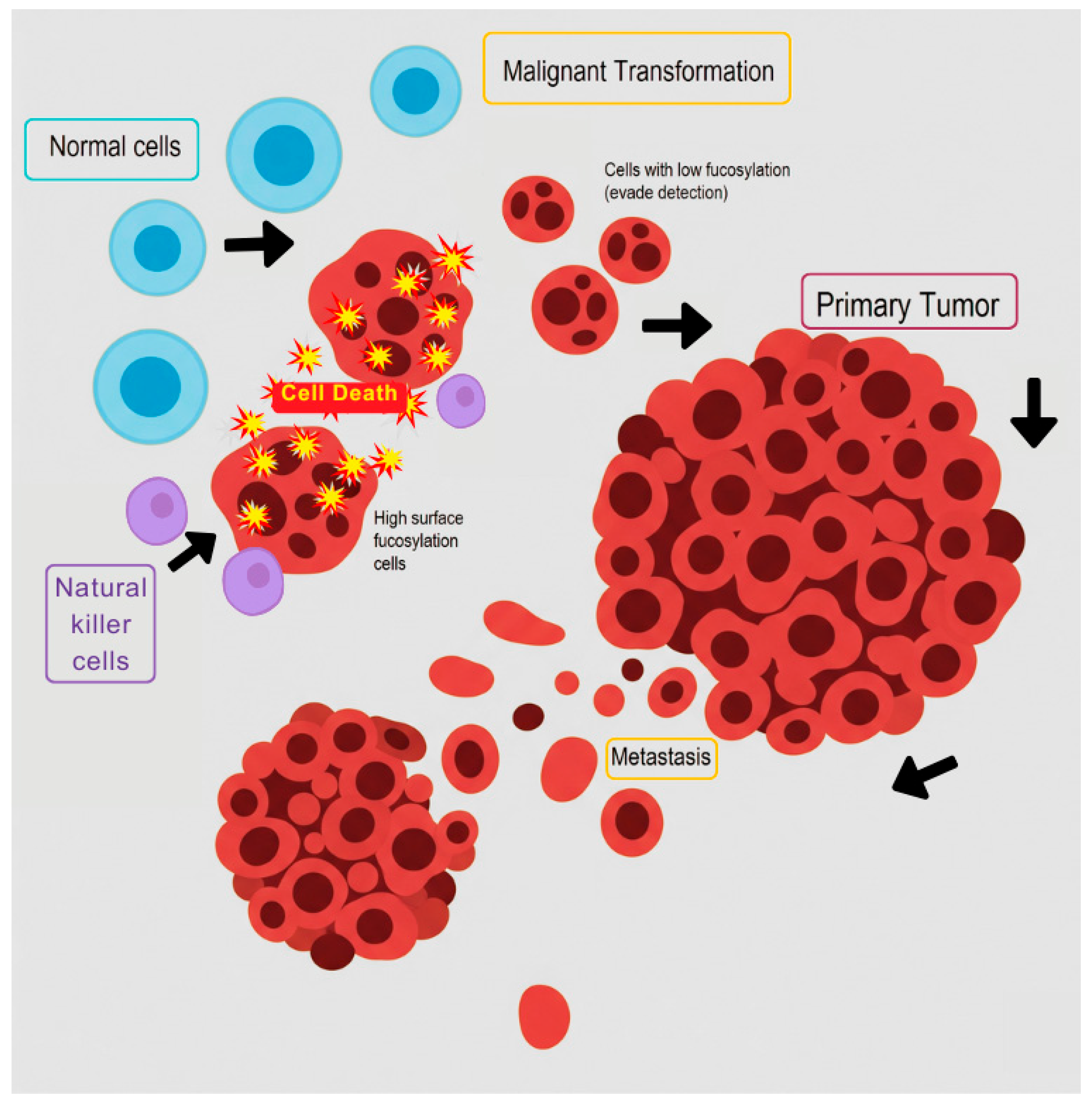
Among the prominent glycosylation reactions, fucosylation frequently undergoes alterations in cancer progression (Figure 5).
Figure 5. A schematic representation of how cell fucolysation governs breast cancer establishment and progression, particularly highlighting immune evasion. Normal mammary cells (round, blue) undergo malignant transformation following mutagenesis, generating two main cellular populations: low-fucosylated cells (round, small-nucleated) characterized by low FUT3 and high FUCA1 expression, and high-fucosylated cells (irregularly shaped). Effector immune cells, like Natural Killer (NK) cells (round purple), detect surface fucosylated antigens via lectin-like receptors, leading to the elimination of high-fucosylated cells. Critically, the low-fucosylated cells escape immune recognition, enabling them to establish the primary tumor. Figure by the authors, based on do Nascimento et al. [].
The biosynthesis of fucosylated glycans is catalyzed by fucosyltransferases (FUTs), enzymes responsible for transferring L-fucose residues from their activated donor, GDP-fucose, to a specific acceptor glycan. In in vitro models, α-linked L-fucose present in breast cancer cells is related to their adhesion to components of the extracellular matrix and endothelial cells in in vitro models []. Moreover, in vitro investigations utilizing diverse tumor cell lines have demonstrated a correlation between FUT activity and processes such as cell migration, invasion, epithelial-mesenchymal transition, and the induction of NK cell-mediated cytotoxicity [,,,]. The FUT3 gene is responsible for encoding a fucosyltransferase with α1-3 and α1-4 activity. Alpha-1,3/4-fucosyltransferase has been linked to pro-tumor and anti-tumor pathways depending on the cancer type, whose main products are the Lewis antigens []. The products of the action of the FUT3 enzyme, the sialyl Lewis X antigen (sLex) and its isomer sialyl Lewis A (sLea), are found in high levels in solid tumors, including breast cancer []. Both, sLea and sLex, are ligands recognized by selectin transmembrane glycoproteins involved in cell–cell interactions, necessary in inflammatory processes, hemostasis/thrombosis, wound healing, and tumor metastasis.
In recent years, several investigations have elucidated the crucial role of fucosylation in TRAIL-induced apoptosis within colon cancer [,]. These studies initially established a positive correlation between TRAIL sensitivity and the mRNA expression levels of the fucosyltransferase enzymes FUT3 and FUT6 in various colon adenocarcinoma cell lines. The divergence between in vitro and in vivo experimental outcomes could be attributed to the engagement of the immunological system against tumor cells in vivo, and the specific involvement of FUT3 in this process.
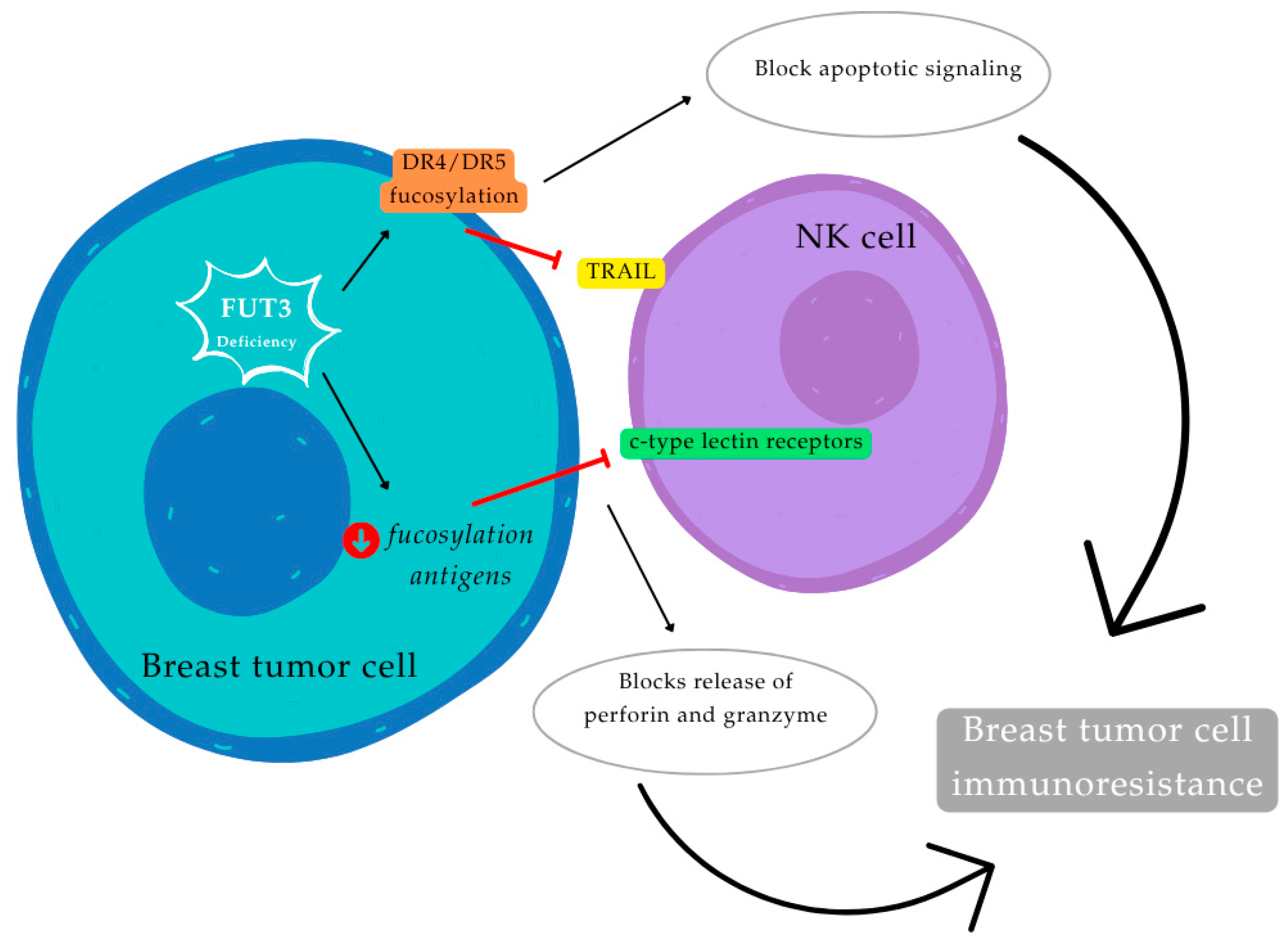
Notwithstanding the documented pro-tumorigenic activity of the FUT3 enzyme and its products, numerous studies have concurrently highlighted its importance in mediating NK cell cytotoxicity against tumor cells. This dual role is underpinned by the recognition of sLex antigen by C-type lectin receptors and the essential fucosylation of DR4 and DR5 receptors, a critical step for the extrinsic apoptotic pathway stimulated by Apo2L-TRAIL. The TRAIL pathway is demonstrably linked to NK cell-induced cytotoxicity toward tumor cells [,,,,]. Employing agonistic receptor-specific TRAIL variants to precisely determine the contribution of FUT3 and FUT6-mediated fucosylation to TRAIL-induced apoptosis through its two death receptors, DR4 and DR5 (Figure 6 and Figure 7) [,]. It is hypothesized that the absence of FUT3 in breast cancer cells may preclude the fucosylation of DR4 and DR5 receptors, consequently abrogating TRAIL apoptotic signaling. Moreover, a deficiency in FUT3 would diminish the presence of fucosylated antigens on the cell surface, preventing the recognition of Lewis antigens by C-type lectin receptors on NK cells and thereby hindering the release of perforin and granzyme. Both of these mechanisms are posited to contribute to the immunoresistance of breast tumor cells [,,,].
Figure 6. This schematic model illustrates the role of α1,3/4-fucosyltransferase (FUT3) in NK cell-induced cytotoxicity against breast tumor cells. The absence of FUT3 in breast cancer cells can prevent the fucosylation of Death Receptor 4 (DR4) and Death Receptor 5 (DR5), thereby blocking TNF-related apoptosis-inducing ligand (TRAIL) apoptotic signaling. Furthermore, FUT3 deficiency reduces fucosylated antigens on the cell surface, leading to non-recognition of Sialyl Lewis X (sLex) antigens by C-type lectin receptors on NK cells. This prevents the release of perforin and granzyme, and together, both mechanisms can promote breast tumor cell immunoresistance. (Figure by the authors, adapted from do Nascimento et al. [].
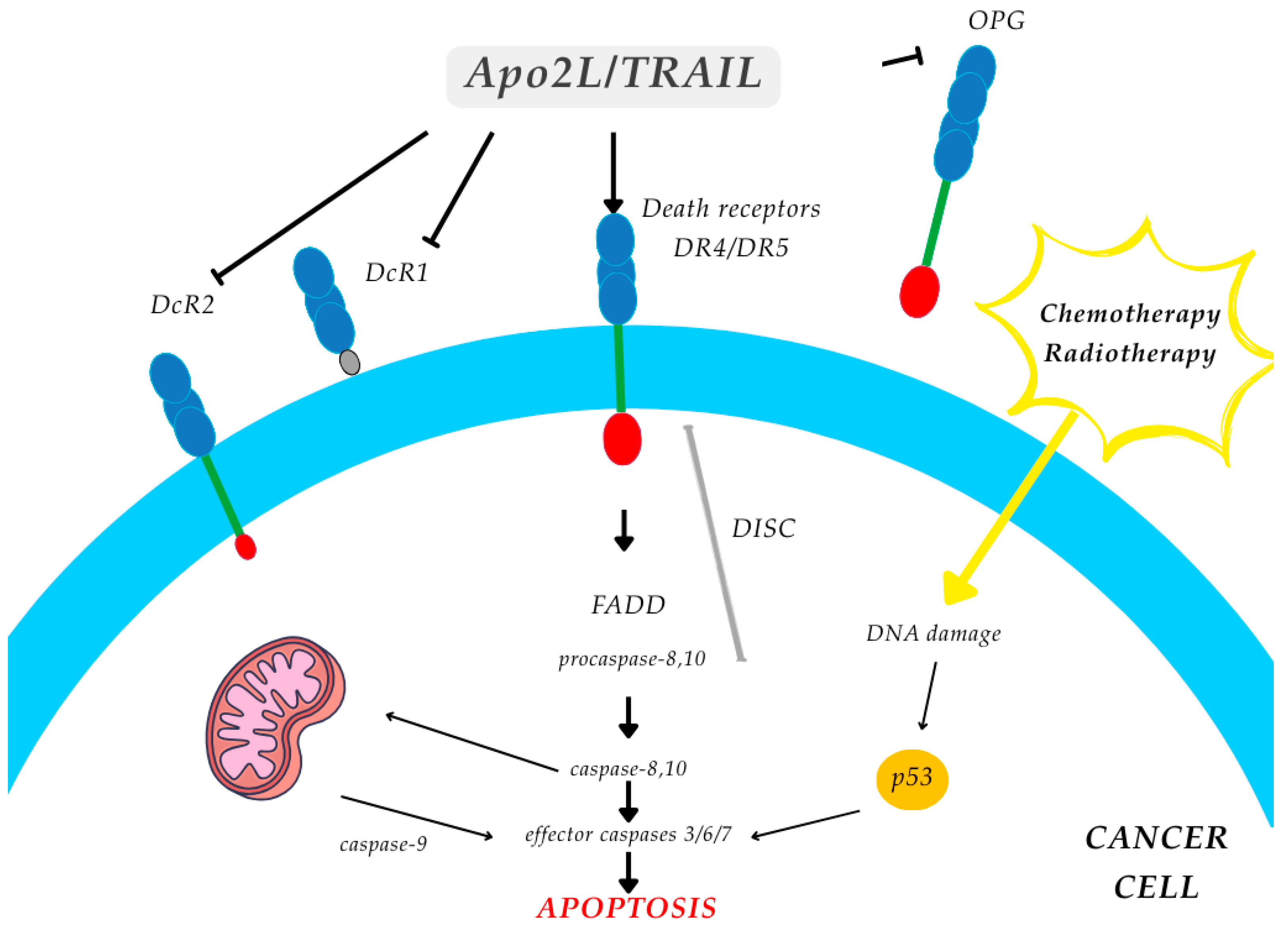
Figure 7. Apoptosis cascade through Apo2LTRAIL and its death receptors DR4 and DR5, which contain a cytoplasmic death domain and signal apoptosis. Apo2L/TRAIL is a homotrimeric ligand that interacts with four closely related members of the TNF receptor superfamily. Non-death receptors DcR1 linked to the plasma membrane by a glycophosphatidylinositol moiety, and without signaling activity, and DcR2 with a truncated non-functional death domain, are inihibited. Furthermore, it stimulates DR4 and DR5, which will induce signaling of the extrinsic pathway through the direct intervention of Caspase 8 towards cellular apoptosis, while the intrinsic pathway is signaled with indirect intervention of Caspase 8 acting first on the mitochondria in order to then trigger apoptosis by another additional mechanism.
Considering the substantial contribution of fucosyltransferases to cancer biology, changes in FUT (FUT1 to FUT-13) expression are significantly implicated in oncogenesis and malignancy. A review by Vasconcelos et al. [] underscores FUTs as promising biomarkers for cancer prognosis and diagnosis, alongside their utility as therapeutic targets. Moreover, the results from do Nascimento et al. [], coupled with prior understanding of FUT3’s involvement in both pro-tumorigenic and anti-tumorigenic mechanisms, prompted the proposal of a model for FU6T3 expression regulation during breast cancer establishment and progression. Breast cancer tissue frequently exhibits overexpression of fucosylated glycans, such as sialyl-Lewis X/A, and α1,3/4-FUTs, which correlates with enhanced disease progression and metastatic potential. The observed dual role of FUT3 in tumor cells may indicate that these cells modulate its expression to acquire beneficial attributes during their primary site establishment and subsequent metastatic dissemination. From a therapeutic perspective, developing strategies and/or protocols to inhibit the enzymatic activity of fucosyltransferases and their gene expression represents a critical advancement in cancer treatment [,].
Apoptosis is essential for the maintenance of cell populations. Perturbation of this normal process alters the balance between cell death and cell proliferation and may lead to the development of various types of disease, in which cancer is most prominent. The control of apoptosis and necrosis mechanisms involves several protein factors, receptors, and the activation of the extracellular and intracellular cell death pathway [,,]. In fact, the tumor necrosis factor family of cytokines, type II transmembrane protein, is an important regulator of homeostasis and mediators of disease, while respective ligand molecules-receptors are type I transmembrane proteins capable of mediating a wide range of responses in vitro and in vivo.
Although tumor necrosis factor (TNF) was first identified in 1975, its effects had been first noted in the late 19th century by W. B. Coley, a New York surgeon []. Only recently has it become apparent that TNF-alfa is a representative member of a large family of cytokines that exert a fundamental effect on cell proliferation and death, inflammation, and immunological and neuronal cell function. The cDNAs for TNF-alfa and a related factor called lymphotoxin-alfa (LT-alfa, also called TNF-beta) were cloned independently. TNF-alfa is produced primarily by monocytes/macrophages, whereas LT-alfa by activated lymphocytes []. TNF-alfa and LT-alfa are 28% identical at the amino acid level. The features of a unique new TNF-like ligand, TRAIL/APO-2L, and its receptors will be discussed here in the context of the TNF superfamily.
Tumor necrosis factor-associated apoptosis-inducing ligand (TRAIL), a cytokine also known as TNFSF10 because it is encoded by the gene TNFSF10, is a member of the tumor necrosis factor (TNF) superfamily [,]. Beyond the TRAIL, other molecules in this family include LT-alpha, LT-beta, CD40L, CD30L, and CD27L, which are all known for their roles in apoptosis, inflammation, and immune responses [,]. TRAIL is a protein that can exist in both membrane-bound and soluble forms, and like other members of the TNF family, it is a typical type II transmembrane protein capable of forming trimers or dimers in solution. TRAIL showed that it is closely related to other death ligands, with the highest sequence similarity to Fas ligand [], hence the name APO2L [,]. TRAIL plays a significant role in regulating cell death, specifically through the induction of apoptosis in tumor cells and transformed cells.
Supported by specific extra and intracellular mechanisms, TRAIL has emerged as a promising cancer therapeutic due to its ability to selectively induce apoptosis in tumor cells while leaving normal cells unharmed. TRAIL primarily signals via two transmembrane DR receptors, specifically DR4 (TRAIL-R1) and DR5 (TRAIL-R2). DR4 and DR5 contain a death domain to convey apoptotic signals, and therefore are denoted as death receptors, whereas DcR1 and DcR2 are unable to induce cell death [,,,] (Figure 6 and Figure 7). In addition, details of the extracellular and successive intracellular events induced by TRAIL-DR complexation have been reported as apoptotic exogenous (extrinsic) and endogenous (intrinsic) pathways. The extrinsic apoptosis pathway involves the binding of TRAIL to death receptors DR4 and DR5 on the cell membrane surface. Upon binding of TRAIL with DR, TRAIL-DR assembles a death-inducing signalling complex and recruits the intracellular adaptor protein via the death effector domain [,,]. This process of apoptosis is caspase-8-dependent. Caspase-8 activates downstream effector caspases, including procaspase-3, -6, and -7, leading to activation of specific kinases to execute cell death [,,]. On the other hand, the intrinsic pathway is initiated in the mitochondria. Caspase 8 mediates programmed apoptosis from mitochondria by activating Caspase-9, which is capable of amplifying Caspase-3 activation, via a Caspase pathway similar to Fas, contributing to apoptotic cell death too []. TRAIL is also capable of binding to the decoy receptors DcR1 and DcR2, which differ structurally from the death-inducing receptors. DcR1 lacks an intracellular signaling domain, whereas DcR2 possesses only a truncated death domain. Functionally, DcR1 acts as a neutralizing receptor that sequesters TRAIL, thereby preventing it from triggering apoptosis. In contrast, DcR2 retains a partial signaling capability through its cytoplasmic region, which can activate the NF-κB pathway. Consequently, in cells expressing DcR2, TRAIL binding results in NF-κB activation, promoting the transcription of genes that counteract apoptotic signaling and/or enhance proinflammatory responses. Like other members of the TNF superfamily, Apo2L/TRAIL can trigger various biological regulatory mechanisms in cancer and normal cells. For example, changes in protein expression can influence TRAIL resistance, and pro-inflammatory cytokines and lipopolysaccharides can induce up-regulation of TRAIL on the surface of monocytes, macrophages, dendritic cells, and NKs []. Although the complete biological role of Apo2L/TRAIL remains to be fully elucidated, increasing evidence indicates that this apoptosis-inducing ligand serves as a vital component of immune surveillance against oncogenically transformed and virally infected cells. Furthermore, Apo2L/TRAIL may function to mitigate autoimmune responses, promising a candidate for cancer therapy [,]. Especially when TRAIL is used in combination with low-dose chemotherapeutic agents, it shows obvious synergistic effects, and this specificity makes it a very promising anticancer agent [].
Distinguishing them from conventional cancer therapeutics, DR ligands induce tumor cell apoptosis irrespective of the p53 tumor suppressor gene, which is frequently inactivated in malignant cells. Consequently, Apo2L/TRAIL holds promise for treating tumors resistant to conventional therapies and may augment the effectiveness of existing treatments in a wide range of cancers [].

1.3.6. Tyrosine Kinase Receptor

Tyrosine kinases are part of the broader family of protein kinases—enzymes that catalyze the transfer of phosphate groups to amino acid residues such as serine, threonine, or tyrosine, thereby serving as molecular “switches” that regulate numerous cellular activities. By 2004, a total of 58 human receptor tyrosine kinases (RTKs) had been identified and classified into 20 subfamilies. Among these, eight membrane-associated proteins contain tyrosine kinase–like domains but lack catalytic function and are therefore considered pseudokinases.
RTKs are structurally composed of three primary regions: an extracellular domain responsible for ligand binding, a single transmembrane domain, and an intracellular catalytic domain that mediates phosphorylation of specific substrates. Ligand engagement with the extracellular region induces conformational changes that activate the kinase domain. This activation facilitates access of adenosine triphosphate (ATP) and target substrates to the active site, initiating phosphorylation events that propagate intracellular signaling. The ensuing phosphorylation cascade ultimately conveys extracellular cues to the nucleus, resulting in altered gene expression patterns. Aberrations in RTK signaling, whether caused by activating mutations, chromosomal rearrangements, or receptor overexpression, are strongly implicated in oncogenesis, often producing constitutively active kinases that drive uncontrolled, ligand-independent cell proliferation [,].
Functionally, tyrosine kinases catalyze the transfer of phosphate groups from ATP to tyrosine residues within target proteins, leading to their activation or functional modulation []. These enzymes play pivotal roles in coordinating physiological and biochemical processes, serving as essential mediators of intracellular signaling cascades that regulate cellular growth, differentiation, metabolism, and apoptosis in response to diverse stimuli [,]. Recent findings further underscore the central role of tyrosine kinases in the pathogenesis of cancer. By participating in pathways that control proliferation and apoptotic sensitivity, tyrosine kinases contribute both to normal tissue homeostasis and to the progression of malignant transformation [,]. Within these signaling cascades, tyrosine kinases act as intermediaries that relay extracellular signals across the plasma membrane into the cytoplasm and, in many cases, to the nucleus, where they influence transcriptional regulation. Mutations that result in constitutive activation of tyrosine kinases can drive continuous signaling, promoting tumor initiation and progression. While receptor tyrosine kinases primarily mediate transmembrane signaling, intracellular or non-receptor tyrosine kinases participate in cytoplasmic and nuclear signaling events that regulate cell cycle progression and transcription factor activity. Phosphorylation at tyrosine residues thereby modulates diverse protein characteristics, including enzymatic activity, intracellular localization, and molecular interactions, processes fundamental to mitogenic signaling and cell proliferation [,,,].
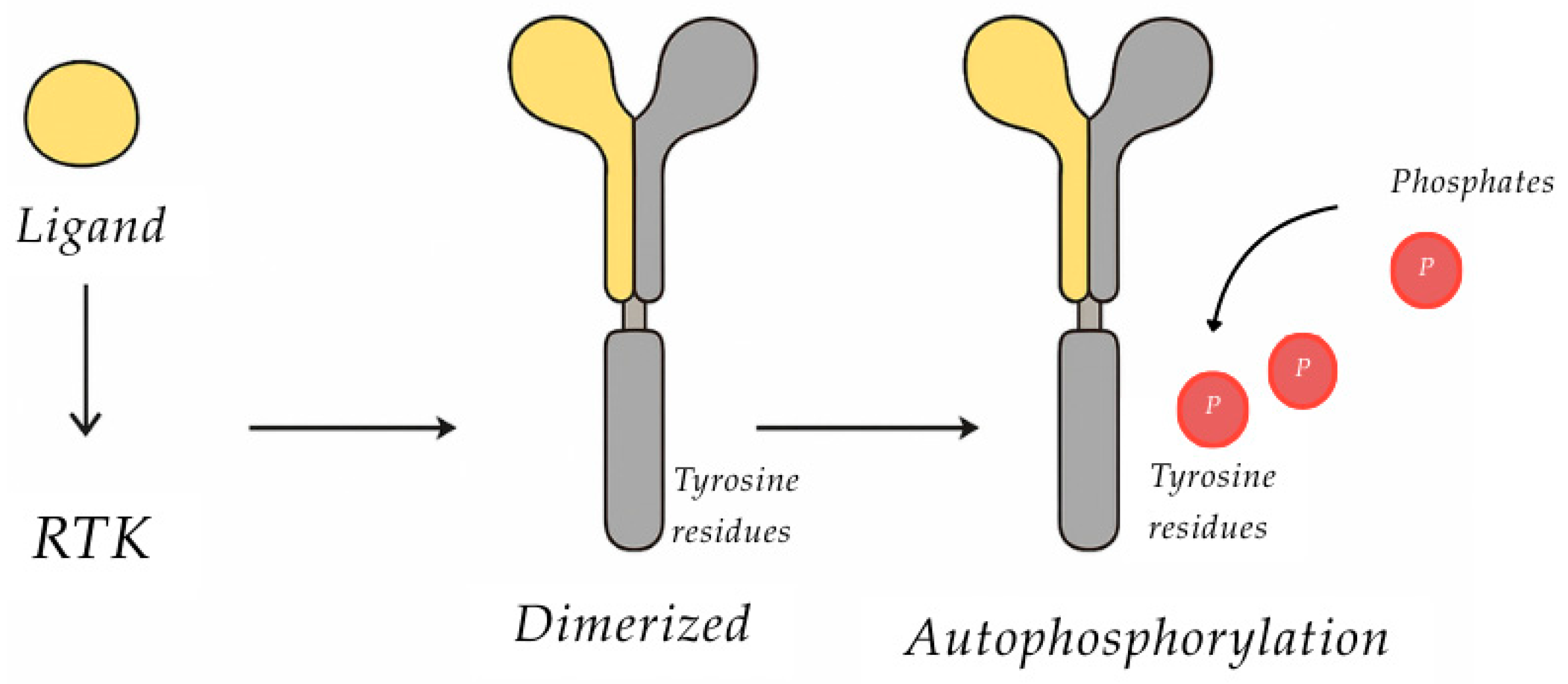
Biochemical mechanism of action of tyrosine kinase: tyrosine kinases specifically catalyze phosphorylation of tyrosine residues in various substrate proteins upon activation by ligand binding to their extracellular domains. This interaction induces a conformational shift within the activation loop, enabling ATP and substrate access to the catalytic site. The enzyme then transfers a phosphate group from Mg-ATP to tyrosine residues on the receptor itself and on downstream signaling proteins, initiating a cascade of intracellular events [,]. Tyrosine kinase receptors and their ligands are often degraded by lysosomal enzymes after being internalized into the cell. This process, known as receptor downregulation, is a crucial mechanism for regulating cell signaling and preventing overstimulation. Receptor-specific ligands, such as growth factors, result in RTK dimerization or oligomerization. This often results in the receptors’ activation and autophosphorylation (Figure 8), allowing them to recruit and activate downstream mediators via their SH2 domains [,]. Following receptor activation, a cascade of intracellular biochemical events is initiated, ultimately resulting in the activation or suppression of specific gene subsets that determine the cell’s biological response to the external signal. As part of this signaling process, receptors often undergo lateral movement within the plasma membrane and are subsequently internalized via clathrin-mediated endocytosis. This involves the invagination of the membrane to form clathrin-coated vesicles, which eventually detach to create endocytic compartments. These vesicles can fuse with lysosomes, where both the ligand and receptor may be degraded by lysosomal enzymes. In certain cases, however, receptors are recycled back to the plasma membrane for reuse. Throughout receptor internalization, the ligand–receptor complex typically dissociates, marking the termination of the signaling event and contributing to the regulation of distinct cellular and physiological outcomes [,].
Figure 8. Diagram concerning the activation process of the tyrosine phosphorylation.
Ligands are extracellular signal molecules (e.g., epidermal growth factor-EGF, platelet-derived growth factor-PDGF, etc.) that induce receptor dimerization (except the insulin receptor) by which different ligands employ different strategies, achieving the stable dimeric conformation [,]. One ligand may bind with two receptor molecules to form a 1:2 ligand receptor complex, while in other cases, two ligands bind simultaneously to two receptors 2:2 ligand receptor complex. The receptor dimerization is also stabilized by receptor–receptor interactions. Some ligand receptors are not sufficient for some complexes and are stabilized by accessory molecules. Ligand binding to the extracellular domain stabilizes the formation of active dimmers and consequently protein tyrosine kinase activation [,].
Among the RTKs, the epidermal growth factor receptor (EGFR), also known as ErbB1 or HER1, is particularly significant. The epidermal growth factor receptor is a transmembrane receptor tyrosine kinase that is part of the human epidermal growth factor receptor (HER or Erb) family, which also includes HER2 (ErbB2), HER3 (ErbB3), and HER4 (ErbB4), playing a fundamental role in cell signaling pathways [,,,,]. Its signaling network is extremely complex, involving 211 biochemical reactions and 322 signaling molecules []. The EGFR-dependent intracellular signaling pathway is initiated by the binding of a specific ligand (such as endothelial growth factor, transforming growth factor alpha (TGFα), among others) in the extracellular portion of EGFR, or one of the other EGFR-related receptors (HER2, HER3, or HER4) [,,,]. The formation of functionally active homodimers (EGFR-EGFR) or heterodimers (EGFR-HER2, EGFR-HER3, EGFR-HER4) causes ATP-dependent phosphorylation of specific tyrosine residues in the intracellular domain of EGFR [,]. This autophosphorylation in turn triggers a complex set of intracellular signaling, first in the cytoplasm and then in the nucleus, which can result in tumor cell proliferation, blockade of apoptosis, potential for invasion and metastasis, and stimulation of neovascularization [,].
Thus, the activity of these kinase receptors is dependent on the phosphorylation of signaling molecules and the downstream activation of transcription factors, which will mediate the expression of target genes [,]. The three major downstream signaling pathways that undergo EGFR-mediated activation are (i) the rat sarcoma (RAS)/rapidly accelerated fibrosarcoma (RAF)/mitogen-activated protein kinase (MAPK) pathway, which controls gene transcription, cell cycle progression from G1 to S phase, and cell proliferation; (ii) the phosphatidylinositol-3-kinase (PI3K)/protein kinase B (AKT) pathway, which activates the anti-apoptotic and pro-survival signaling cascade; and (iii) the janus kinase (JAK)/signal transducers and activators of transcription (STAT) pathway [,,,].
Because receptor tyrosine kinases such as HER1 and HER2 play central roles in regulating cell division and tumor growth, their inhibition effectively disrupts cell cycle progression and can suppress tumor development. In support of this, recent studies have shown that nitric oxide, nitric oxide synthase, and non-receptor tyrosine kinases-SRC are involved in estrogen (E2)-stimulated non-nuclear signaling pathways that promote proliferation in MCF-7 breast cancer cells. These pathways, while essential for normal cellular growth and function, can contribute to the development of various diseases, including cancer, when dysregulated or overactivated [,,,].

1.3.7. Deiodinase-Type 3 Enzyme—DIO3

Thyroid hormones are essential regulators of several cellular processes, and changes in their status interfere with tumor progression through virtually all hallmarks of cancer. The type 3 deiodinase enzyme (DIO3) is the main enzyme responsible for the inactivation of thyroid hormones, with a relevant role in cell proliferation and physiological growth. Additionally, its expression levels are associated with seasonal adaptations changing in response to photoperiod variations [,]. Changes in the DIO3 enzyme expression have been observed in cell pathological proliferation and differentiation, described in several human neoplasms []. Clinical studies have associated thyroid hormone levels with the risk of developing breast cancer, while in vitro studies have demonstrated that they influence the proliferation, apoptosis, and migration of mammary tumor cells [,]. According to Goemann et al. [], supported by immunohistochemistry studies and RNA sequencing data from the TCGA database, DIO3 expression was found in 35/39 (89.7%) of the invasive ductal carcinoma samples, with an average H-Score of 104.9 ± 55, and in only one sample of the three invasive lobular carcinomas analyzed (H-Score = 86) []. Thus, these results indicate that the DIO3 enzyme is expressed in normal breast tissue and in breast cancer, while low levels of DIO3 expression were associated with increased mortality in the breast cancer patients. Accordingly, low DIO3 mRNA levels were associated with an increased risk of death in a multivariate model in the validation cohort. The reduction in DIO3 expression in breast cancer can be explained at least in part by hypermethylation of the gene’s promoter region. These results point to DIO3 as a prognostic marker in breast cancer, with reduced expression associated with survival [,].
Thus, DIO3, a thyroid hormone inactivating enzyme, shows promise as a diagnostic and prognostic marker in breast cancer. Decreased DIO3 expression has been linked to poor overall survival in breast cancer patients, suggesting that DIO3 expression may be altered in breast cancer and could be used to identify patients with a higher risk of metastasis or poor prognosis [,]. Given its potential to modulate intracellular thyroid hormone levels and interact with estrogen metabolism in breast cancer, DIO3 expression may hold therapeutic implications. In conclusion, while DIO3 is expressed in both normal and tumoral breast tissue, decreased expression correlates with poor overall survival in breast cancer patients [,].
In one embodiment, it uses a proportional hazards model to determine the correlations between clinical outcomes of patients with breast cancer and changes in tumor gene expression, DIO3/DIO3 protein, and diagnosis of tumor tissue []. According research study, it is suggested that DLK1delta, such as non-canonical Notch ligand 1 versus DIO3 (DLK1-DIO3) genes, miR-379/656 cluster, and its target gene network, might be potential diagnostic and prognostic markers for cancer management and therapy.
Several reported studies suggest that DIO3 overexpression should be assessed before pharmacological or surgical interventions in gastrointestinal stromal tumors because it might have both prognostic and therapeutic roles [,]. Consequently, further elucidation of the developmental roles of deiodinases in mediating cell fate (death or survival) may present novel opportunities for leveraging these enzymes as therapeutic targets in a spectrum of pathological states.

1.4. Immune Response and Inflammation

In addition to genetic and epigenetic changes, the immune system plays a central role in the initiation, progression, and control of breast cancer. The concept of cancer immunoediting describes three key phases in the interaction between tumors and the immune system: (i) elimination of tumor cells, (ii) equilibrium between immune surveillance and tumor growth, and (iii) immune escape, where tumor cells evade detection and proliferate unchecked [,,,].
This immune surveillance involves both innate and adaptive immunity, particularly NK cells and cytotoxic T lymphocytes, which can detect and eliminate transformed cells. Therefore, therapeutic strategies aimed at stimulating immune mechanisms, especially after chemotherapy or radiotherapy, which often deplete immune cells, may be crucial to prevent relapse [,].
Furthermore, psychoneuroimmunology reveals that emotional and psychological stress can suppress immune function. Stress activates the hypothalamic–pituitary–adrenal axis and the autonomic nervous system, leading to reduced NK and T-cell activity and increased inflammation. This immunosuppressive state may contribute to tumor progression [,].
Breast tumors develop within a complex tumor microenvironment (TME), which includes not only cancer cells but also stromal cells such as fibroblasts, endothelial cells, and immune cells like neutrophils, lymphocytes, and dendritic cells []. Within this niche, mesenchymal stem cells and tumor-associated fibroblasts promote angiogenesis and metastasis through the secretion of cytokines like TNF-α and IL-1β [].
The inflammatory component of the TME is especially relevant. Chronic inflammation fosters a milieu that supports carcinogenesis. Activated macrophages and other immune cells in this environment release high levels of pro-inflammatory mediators, including cytokines (e.g., TNF-α, IL-6), chemokines (e.g., IL-8, CCL5), and enzymes such as inducible NO synthase and cyclooxygenase-2 (COX-2) [,,]. These mediators promote proliferation, inhibit tumor suppressor genes, induce DNA damage, and enhance angiogenesis.
Systemic inflammation is also linked to poor prognosis in breast cancer. A high neutrophil-to-lymphocyte ratio has been consistently associated with worse survival outcomes in multiple epithelial cancers, including breast cancer [,]. It is estimated that up to 25% of cancers may arise from chronic inflammatory or infectious conditions. Also, pathological angiogenesis and inflammation are mutually reinforcing processes. Inflammatory cells secrete vascular endothelial growth factor (VEGF) and other angiogenic cytokines, which promote blood vessel formation, which, in turn, supports tumor growth and metastasis [].
Nowadays, it is well established that platelets interact intimately with tumor cells. In this context, platelets play a dual role in cancer. On one hand, tumor cells induce platelet aggregation, which facilitates immune evasion and metastasis. On the other hand, platelets release growth and angiogenic factors in the TME, promoting tumor proliferation. Platelets can also suppress immune surveillance, undermining the efficacy of immunotherapies [,].
Cancer is associated with increased thrombotic risk, including myocardial infarction and stroke. This risk is partly due to imbalances in COX-1/COX-2-derived prostanoids and lipoxygenase (LOX) activity []. In this context, natural compounds from plants exhibiting dual inhibitory action on cyclooxygenase (COX-1 and COX-2) and lipoxygenase enzymes have emerged as promising agents in managing inflammation with a reduced risk of cardiovascular side effects, particularly due to their antiplatelet properties. Thus, flavonoids such as quercetin, kaempferol, and apigenin demonstrated non-selective COX and LOX inhibition, along with inhibition of platelet aggregation through downregulation of thromboxane A2 (TXA2) synthesis and modulation of intracellular calcium signaling []. Curcumin inhibits both COX-2 and 5-LOX, reducing the production of pro-inflammatory eicosanoids while exhibiting mild antiplatelet effects without significantly affecting hemostasis []. Similarly, resveratrol inhibits COX-1, COX-2, and 5-LOX while also attenuating platelet aggregation through NO-mediated and TXA2-suppressive mechanisms []. Thus, leveraging phytochemicals with COX/LOX inhibitory and antiplatelet activities, but lower adverse effects, represents a promising strategy to mitigate both inflammation and atherothrombotic risk in cancer patients.

1.5. Breast Cancer Biomarkers

Tumor markers can be defined as molecules that indicate the presence of cancer or provide information about its likely behavior in the future. That is, they are molecular products metabolized and secreted by neoplastic tissue and biochemically characterized in cells or body fluids [,]. They are also indicators of tumor stage and grade, being useful for diagnosing, monitoring responses to treatment, and predicting the recurrence of carcinomas.

1.5.1. Blood Biomarkers

Soluble molecules, termed serum tumor markers, are aberrantly released into the circulation by malignant cells or components of the tumor microenvironment [,]. The ideal serum tumor marker detects disease early, predicts response or resistance to specific therapy, and monitors the patient after the first therapy []. This information, taken together, reveals the importance of using these markers in clinical practice. In the case of breast cancer, different serum markers have been tested for these purposes: Carcinoembryonic Antigen (CEA), the soluble form of the MUC-1 protein (CA15-3), Tissue Polypeptide Antigen (TPA), Tissue-Specific Polypeptide Antigen (TPS), and Human Epithelial Growth Factor Receptor type 2 (HER2) [,].
Once breast cancer treatment depends on the stage of the disease at the time of diagnosis, histological grade, and presence or absence of certain receptors and genetic mutations, for the molecular characterization of these tumors, it is important to identify three biomarkers: estrogen and progesterone hormone receptors and HER2 [,]. Around 70% of breast cancers are ER-positive, which allows therapy with estrogen antagonist molecules. In turn, HER2 is present in around 25% of breast cancers and is considered a more aggressive phenotype with a worse prognosis. In addition to these biomarkers, CD, CA 15.3, and CA 27.29 are also widely used [,,,]. As it was mentioned before, the role of CD in carcinogenesis is associated with the stimulation of DNA and mitosis during tissue regeneration, and due to its proteolytic power, it facilitates tumor dissemination by digestion of proteoglycans from the interstitial matrix and the basement membrane. CA 15.3 is one of the most specific biomarkers for breast cancer. Its sensitivity varies according to the tumor mass and the patient’s clinical status [,,,]. However, it is understood that, at present, CA 15.3 is not safe for an accurate diagnosis. Tumor antigen 27.29 is found in the blood of breast cancer patients, but without sufficient sensitivity and specificity to be used as an isolated diagnostic test, being indicated for detecting recurrent breast cancer and monitoring advanced treatment [].
Prompt detection of breast cancer not only enhances therapeutic efficacy but also mitigates its associated psychological, economic, and social burdens [,]. This pressing demand for novel and effective detection modalities has propelled the pursuit of cost-effective, non-invasive diagnostic tools for breast cancer [,,]. Diagnosis via blood-based biomarker measurement is regarded as a non-invasive and economical method capable of conveying information regarding disease presence, absence, and evolution. In this vein, extensive research endeavors have been directed towards identifying and assessing the utility of liquid biomarkers [,]. In particular, the liquid biopsy paradigm, which focuses on identifying tumor-derived biomarkers (cellular, molecular, and genomic) within blood and other biological fluids such as saliva, has attracted substantial interest for the diagnostic and prognostic assessment of diverse malignancies, including breast cancer [,].
Within this framework, the significance of microRNAs (miRNAs) has gained widespread recognition. These 20–22 nucleotide-long molecules serve as epigenetic regulators, primarily functioning as repressors of gene expression [,,]. miRNAs are well-established for their critical regulatory involvement in disease pathogenesis, especially in oncology [,,]. Moreover, miRNAs exert significant influence over numerous cancer-related biological processes, including cell proliferation, cell cycle progression, apoptosis, differentiation, migration, metabolic regulation, and stress responses [,,]. Several miRNAs have been identified as key regulators across a variety of cancers, such as breast, colorectal, gastric, lung, prostate, and thyroid malignancies [,]. Their tissue-specific expression, remarkable stability, and detectability in bodily fluids (including blood, serum, and urine) make them promising candidates for the development of miRNA-based biomarkers for cancer diagnosis and therapeutic monitoring [].
Recent genome-wide analyses leveraging high-throughput datasets have highlighted the diagnostic and prognostic potential of a subset of 15 miRNAs from the MiR-379/656 cluster, particularly for breast cancer and its molecular subtypes, including Basal and Luminal B tumors [,].

1.5.2. Salivary Biomarkers

According to the studies of Wan et al. [] and Koopaie et al. [], salivary biomarkers offer a noninvasive and accessible means of identifying breast cancer. In these works, commercially available disposable-based strips, similar to those commonly used for glucose detection, were used and functionalized to detect breast cancer with biomarkers of HER2 and CA15-3.
The past years have seen diverse investigations propose a wide array of salivary biomarkers, from proteomic and metabolomic profiles to transcriptomic and reagent-free biophotonic markers [,,,,,,,]. This active research area has, in fact, given rise to the term “salivaomics,” specifically referring to saliva-based diagnostics [,]. The appeal of saliva for diagnostic purposes is multifaceted, offering advantages such as facile collection, minimal personnel training, rapid sampling, convenient storage, straightforward transportation, reduced susceptibility to clotting, and diminished health worker risks [,].
Nevertheless, a key challenge remains: the concentration and presence of biomarkers in saliva may not always align with those in other biological fluids [], making it crucial to determine which salivary biomarkers offer acceptable sensitivity and specificity for breast cancer diagnosis. Efforts have also focused on detecting exosome-like microvesicles from salivary gland cells, which encapsulate detectable proteins and mRNAs []. Additionally, the efficacy of salivary HER2 levels [], sialic acid, and sialo-glycomic levels (including the impact of disease stage and chemotherapeutics) [,] has been assessed in breast cancer patients. Multiple studies have also examined salivary levels of lectins, polyamines, and N-acetylated compounds in breast cancer patients [,,,]. It has been concluded that salivary biomarkers may be more readily detectable in advanced compared to early stages of breast cancer patients [,,,]. However, Wood and Streckfus [] suggested that combined biomarker approaches show better predictive ability than individual biomarkers. Among single biomarkers, proline demonstrated strong capacity for both early and late-stage breast cancer diagnosis, while taurine proved effective in identifying early breast cancer, and valine exhibited excellent diagnostic accuracy for advanced stages [].

1.5.3. Urinary Tumor Markers

Urinary tumor markers are substances—typically proteins, enzymes, DNA/RNA fragments, or metabolites—excreted in the urine and associated with the presence of cancer. They have gained attention as non-invasive tools for screening, early diagnosis, monitoring of recurrence, and evaluating treatment response in various types of cancer.
The urine has advantages as a diagnostic medium as it represents non-invasive, painless, and easy to collect, can be used for follow up and there is a lower risk of contamination compared with blood, being ideal for large-scale population screening.
Examples of urinary markers studied in breast cancer are (i) elevated urinary levels of matrix metalloproteinases that have been associated with more aggressive or advanced disease [], (ii) elevated urinary levels of 8-oxo-dG (8-hydroxy-2′-deoxyguanosine) that are found in breast cancer patients and may correlate with disease progression or oxidative stress burden [] and also (iii) patterns of urinary estrogen metabolites that have been explored as potential risk indicators, especially in hormone receptor-positive breast cancers [].
The main limitations for their use concern that not all markers are specific to one cancer type, false positives can occur due to infections, inflammation, or benign conditions, and most tests should be used in combination with clinical and imaging assessments.
Urinary tumor markers are not yet established as standard diagnostic or prognostic tools in breast cancer, but ongoing research is promising. The development of multi-marker panels, integration with liquid biopsy approaches, and advancements in proteomic and metabolomic profiling may enhance their clinical utility.

1.6. Breast Cancer Treatment

Advances in molecular research have allowed a greater understanding of the cellular pathways that govern the development of breast tumors, facilitating progress in the identification of diagnostic markers and the development of new therapeutic strategies. The complexity of the molecular mechanisms that regulate tumor initiation and progression determines the heterogeneity of breast cancer, which represents a challenge in the selection of treatment options and disease prognosis [,,]. Thus, the progressive accumulation of genetic and epigenetic changes results in a heterogeneous population of cells that have different clinical behaviors and that trigger different responses to treatment, even in tumors that, from a diagnostic point of view, have the same classification [,,].
A crucial element in managing any cancer is early detection and accurate diagnosis. Breast cancer treatment varies depending on the subtype and extent of spread—whether it’s confined to the breast, involves lymph nodes (stages II or III), or has metastasized to other organs (stage IV) [,,]. Treatments are more effective and better tolerated when initiated promptly and completed as prescribed. These therapies include surgery to remove the tumor, radiation therapy to reduce local recurrence risk, and systemic medical drugs designed to kill cancer cells and prevent dissemination. The latter category encompasses hormonal therapies, immunotherapy, chemotherapy, and targeted biological therapies [,,]. Surgery may involve either a lumpectomy (removal of cancerous tissue only) or a mastectomy (removal of the entire breast). Lymph nodes may also be removed to assess the cancer’s metastatic potential. Historically, a complete axillary dissection (removal of the entire lymph node bed under the arm) was considered necessary for invasive cancers to prevent spread. However, the less invasive sentinel node biopsy is now preferred due to fewer complications [,,,,]. Radiotherapy plays a vital role in breast cancer treatment. In early-stage cases, radiation can often spare a woman from undergoing a mastectomy. It also treats residual microscopic cancer cells in breast tissue and/or lymph nodes, minimizing the chances of chest wall recurrence [,,]. For later-stage cancers, radiotherapy can still reduce recurrence risk, even after a mastectomy [,,].
Medical treatments for breast cancer, administered either before (neoadjuvant) or after (adjuvant) surgery, are determined by the cancer’s biological subtype, as identified through tumor marker detection. The vast majority of drugs used for breast cancer are already listed on the WHO Essential Medicines List [].
Cancers expressing the ER and/or PR receptor are highly likely to respond to hormone therapies such as tamoxifen or aromatase inhibitors. These treatments can reduce the recurrence of “hormone-positive” cancers by nearly half [,,]. Although endocrine therapies can induce menopausal symptoms, they are generally well tolerated.
On the contrary, triple-negative breast cancer (TNBC), an aggressive subtype lacking estrogen, progesterone, and HER2 receptors, presents limited treatment options. In this context, phytochemicals have emerged as promising agents in TNBC due to their ability to target multiple cancer-related pathways. Compounds such as polyphenols have demonstrated anti-proliferative, pro-apoptotic, anti-metastatic, and epigenetic regulatory effects in TNBC models. These agents often exert their effects by modulating signaling pathways like PI3K/Akt/mTOR, NF-κB, Wnt/β-catenin, and MAPK, with reduced toxicity compared to conventional therapies [,]. Nevertheless, while preclinical data are encouraging, clinical validation is still needed to determine their efficacy and safety in TNBC patients.

1.6.1. Estrogen Modulation

Strategies involving the modulation of estrogen exposure for breast cancer therapy originated as early as the late nineteenth century, concurrent with observations of the favorable effects of complete ovariectomy on disease progression. While ovarian ablation (achieved through surgical, radiotherapeutic, or pharmacological means) continues to be clinically employed for certain pre-menopausal breast cancer patients [,], extensive research has been dedicated to the pharmacological modification of estrogen exposure. Modulation of estrogens and their receptors can be accomplished by interfering with ER binding, inducing ER downregulation, or diminishing estrogen biosynthesis [,,].
For the past quarter-century, tamoxifen (Nolvadex®), a selective estrogen receptor modulator (SERM) that acts by competitively blocking estrogen binding to the ER, has been considered the primary therapeutic choice for estrogen deprivation [,,]. Nevertheless, its use is accompanied by notable adverse effects, including an elevated risk of endometrial cancer and thromboembolism []. Subsequent SERMs, such as raloxifene (Evista®) (approved in the United States for osteoporosis) and toremifene (Fareston®) (approved in the United States for breast cancer treatment), were developed to mitigate these side effects while preserving therapeutic efficacy in breast cancer management [,,].
Several plant-derived compounds exhibit anti-estrogenic or estrogen-regulating effects, making them valuable in the prevention or management of estrogen-dependent breast cancer. Key phytochemicals include flavonoids, lignans, isoflavones, and polyphenols, which can act by inhibiting aromatase, blocking ERs, or modulating estrogen metabolism. Flavonoids like apigenin (parsley, chamomile), chrysin (passionflower, honey), and genistein (soy) have shown aromatase inhibition and SERM-like activity. Lignans, found in flaxseed and sesame, are converted by gut microbiota into enterolactone and enterodiol, which exhibit weak estrogenic or anti-estrogenic activity depending on endogenous estrogen levels. Some phytochemicals, phytoestrogens, which are synthesized from plants and vegetables, show low estrogenic activity or anti-estrogenic activity with potentially anti-proliferative effects that offer nutraceutical or pharmacological advantages. Resveratrol (from grapes/red wine) and curcumin (from turmeric) downregulate ER expression and inhibit aromatase transcription, with demonstrated anti-proliferative effects in breast cancer cells. Green tea catechins (especially epigallocatechin gallate-EGCG) and red clover extracts (rich in biochanin A) also inhibit aromatase and interfere with estrogen-mediated signaling [,].
These phytotherapeutics offer multi-targeted, low-toxicity options for modulating estrogen activity, though their clinical efficacy and safety require further validation in controlled human studies. They may complement conventional therapies in estrogen-sensitive conditions, particularly when used in integrative or preventive settings.

1.6.2. Aromatase Inhibitors

Beyond direct modulation of ERs, aromatase inhibitors (AIs) represent another crucial class of endocrine therapy in the treatment of hormone-dependent breast cancer. By inhibiting the conversion of androgens to estrogens, AIs help reduce estrogen levels, especially in postmenopausal women.
When considering current breast cancer therapies, AIs generally demonstrate improved efficacy with fewer side effects compared to other treatments [,]. Currently, two new synthetic AIs and one dietary supplement are undergoing clinical trials as single agents. Atamestane, with or without toremifene, has shown fewer side effects than letrozole, along with favorable effects on bone, serum, and uterine markers []. Testolactone, a first-generation AI, is approved in the United States for advanced breast cancer treatment []. Clinical trials are also being conducted on IH636 grape seed extract, a botanical dietary supplement, for breast cancer prevention in postmenopausal women at increased risk. The IH636 extract, rich in proanthocyanidins, has demonstrated aromatase inhibition in both in vitro and in vivo models [,].
While synthetic AIs typically have a better side effect profile than tamoxifen, serious adverse effects related to estrogen deprivation can still occur []. These include decreased bone mineral density, osteoporosis, increased musculoskeletal disorders [,,], and a higher incidence of cardiovascular events along with altered lipid profiles [,]. Additionally, AIs can affect cognition by reducing the protective effects of estrogens on memory loss with aging [,], and can be responsible for various other health problems that can diminish quality of life, such as diarrhea, vaginal dryness, diminished libido, and dyspareunia [].
Natural products, traditionally used for nutritional or medicinal purposes, such as botanical dietary supplements and traditional medicinal plants, present a promising source of AIs with potentially fewer side effects than synthetic alternatives. These compounds are derived from a wide range of natural sources, including terrestrial plants and animals, marine organisms, bacteria, fungi, and other microbes, offering a chemically diverse repertoire often unattainable through conventional synthetic chemistry techniques [,,,]. The reduced side effects associated with natural products might stem from their complex matrix, which includes compounds that inhibit aromatase alongside others that mitigate the side effects of estrogen deprivation (e.g., phytoestrogens). Consequently, AIs from natural products could be crucial for expanding the clinical utility of AIs in their current role as chemotherapeutic agents to future applications in breast cancer chemoprevention, particularly in preventing secondary recurrence []. In essence, these compounds could offer increased clinical efficacy with fewer adverse effects. Screening for novel natural product AIs could also yield improved leads for future drug development.
The groups most frequently tested for AIs include various classes of flavonoids, a prominent category of polyphenolic compounds characterized by a phenylchroman backbone. Flavonoids are classified into more than ten distinct structural groups, among which the most significant are flavones, flavanones, flavonols, flavanonols, isoflavones, flavanols (catechins), and anthocyanidins [,].
The aromatase-inhibitory activity of polyphenolic compounds, especially flavonoids, such as chrysin, apigenin, naringenin, epigallocatechin gallate (EGCG), quercetin, and resveratrol, among others, has been extensively studied [,,]. Table 2 shows some examples of polyphenolic compounds with aromatase inhibition activity.
Table 2. Examples of polyphenolic compounds with known aromatase inhibition activity.
The utility of flavonoids has been established in numerous cellular and animal studies, showcasing a range of beneficial properties including cytoprotective, antioxidant, free radical scavenging, antiviral, antibacterial, anti-tumor (relevant for cancer prevention and treatment), and anti-inflammatory effects [,]. Based on these properties, it has recently been considered for remarkable applications for the treatment of various diseases [,]. The protective properties of flavonoids are attributed to their ability to safeguard DNA, support immune and hematopoietic systems, scavenge free radicals, enhance immune function, and increase leukocyte counts, demonstrating protective effects in animal models [,,]. Flavonoids are particularly effective at neutralizing oxygen-derived free radicals, mitigating the indirect cellular damage caused by ionizing radiation [,]. At the cellular and molecular levels, flavonoids have been shown to induce apoptosis in cancer cells through multiple mechanisms []. Of note, since breast cancer tissues often overexpress aromatase and produce higher estrogen levels compared to non-cancerous tissues, the capacity of flavonoids to inhibit aromatase has attracted significant attention as a therapeutic strategy for hormone-dependent breast cancer [,].
Moreover, chalcones (commonly referred to as “open-chain flavonoids” or “bioprecursors of flavonoids” due to their role in flavonoid biosynthesis) exhibit broad pharmacological potential. They have demonstrated antidiabetic, anticancer, anti-inflammatory, antimicrobial, antioxidant, antiparasitic, psychoactive, and neuroprotective effects []. Through complex and multifaceted mechanisms, chalcones can modulate various cancer cell lines, inhibit diverse pathogenic microorganisms and parasites, and regulate multiple signaling pathways involved in disease processes [].
On the other hand, Alkaloids, the largest group of secondary metabolites, are a structurally diverse class of low-molecular-weight compounds containing a heterocyclic ring and at least one nitrogen atom. Approximately 17,000 alkaloids have been identified with notable biological and pharmacological activities, generally exhibiting low toxicity and good chemical stability []. Their pharmacological effects span antimalarial, antiasthmatic, vasodilatory, antiarrhythmic, analgesic, antibacterial, antihyperglycemic, and anticancer activities [,]. As anticancer agents, alkaloids have shown chemopreventive potential and the ability to sensitize tumor cells resistant to standard chemotherapies. The extensive structural diversity of these compounds has enabled their development as lead molecules for anticancer drug discovery and therapy [,].
Furthermore, ten alkaloids were tested for aromatase inhibition; five of them were isolated from Nicotiana tabacum L. [,] and the others from Hydrastis canadensis L. (goldenseal), and Piper L. sp. [], but none of them were found to inhibit aromatase. Currently, numerous alkaloids are being tested for their cytotoxicity or are undergoing clinical evaluation, and some have received FDA approval for cancer treatment. Their anti-tumor activity stems from their ability to induce DNA cleavage, which is effectively mediated by topoisomerase I and II inhibition, in addition to causing mitotic arrest, mitochondrial permeabilization, and inhibiting key enzymes involved in cell signaling and metabolism []. Several other compounds have been tested for aromatase inhibition, such as fatty acids, lignans, peptides, terpenoids, isoprenoids, sesquiterpene lactone, withanolides, xanthones, and anthraquinones, and showed diverse activity from weak to strong, depending on each property and targets.
To summarize, a substantial number of natural product extracts, sourced from diverse terrestrial and marine organisms, have undergone evaluation for their aromatase inhibitory potential through a range of noncellular, cell-based, and in vivo methodologies. The most potent compounds generally belonged to the flavonoid or xanthone structural classes, with the exception of certain active sesquiterpene lactones and anthraquinones. The ability of flavonoids to inhibit aromatase has been thoroughly substantiated [,]. A subset of these flavonoids has advanced to in vivo investigations, with varied outcomes [,].

1.6.3. HER2-Positive Cancers

Hormone receptor-negative cancers, defined by the absence of ER or PR expression, generally mandate chemotherapy unless the malignancy is minute. Contemporary chemotherapy regimens are highly efficacious in mitigating the likelihood of cancer dissemination or recurrence and are typically delivered in an outpatient capacity. Hospitalization for breast cancer chemotherapy is generally not required in the absence of adverse events [,,,].
Independent overexpression of the HER2/neu oncogene can occur in breast cancers. Such HER2-positive (HER2+) malignancies are amenable to targeted biological therapies. About 1 in 5 women with breast cancer have overexpression of HER2 on the surface of their tumor cells. These cancers, known as HER2+breast cancers, tend to grow and spread more aggressively. Different types of drugs have been developed to target the HER2 protein [,]. HER2 status is determined by analyzing tumor tissue, and when positive, it can be blocked with targeted drugs, reducing the growth and proliferation of tumors. The so-called anti-HER2 therapies consist of monoclonal antibodies (MABs) that are designed to bind to the HER2 protein on cancer cells, which prevents the cells from growing [,]. These biological agents are very effective antibodies and should only be used in HER2+ breast cancer. Targeted biological therapies are combined with chemotherapy to make them more effective [,,,]:
-
Trastuzumab is an antibody that targets HER2+ cancer cells. When bound to the HER2 protein, it slows or stops the growth of these cells. For advanced breast cancer, treatment is usually given with the goal of reducing the size of the tumors or slowing their development [].
-
Pertuzumab also targets HER2+ cancer cells but in a different way than trastuzumab. When combined with trastuzumab and chemotherapy, it can slow disease progression and increase survival compared to the isolated administration of trastuzumab and chemotherapy [,].
-
Ado-trastuzumab emtansine (T-DM1) is also a targeted therapy for HER2+ advanced breast cancer. It consists of trastuzumab, along with chemotherapy. T-DM1 is approved for the treatment of HER2+ advanced breast cancer that continues to progress after treatment with trastuzumab and taxane chemotherapy. A recent study showed that T-DM1 increases overall survival more than lapatinib in addition to capecitabine in women with HER2+ advanced breast cancer [,].
-
Trastuzumab deruxtecan is also an antibody for the treatment of HER2+ advanced breast cancer. It consists of trastuzumab along with the chemotherapy drug deruxtecan. Its use is approved for the treatment of advanced HER2+ breast cancer that continues to progress after two or more treatments with drugs that target HER2+ cells [,].
The evolution of immunotherapy, particularly through advancements in antibody-enabled killing mechanisms, has demonstrated substantial promise in suppressing or preventing cancer progression [,]. Indeed, contemporary investigations into antibody-mediated cytotoxic effects on tumor cells, coupled with the proliferation of anti-cancer antibody development, have culminated in the generation of MABs designed to selectively recognize specific antigens on the cancer cell surface. Nevertheless, considerable impediments restrict their universal application as anticancer therapeutics; for example, MABs derived from murine cells often co-purify with host proteins and adventitious pathogens. Consequently, plants present an innovative paradigm for monoclonal antibody production against cancer, furnishing an essentially inexhaustible reservoir of MABs, commonly referred to as “plantibodies”. Plants inherently have high expression capability, efficacy, are cheaper, and, on large-scale production, are safer and ethically adequate [,]. Conventional MABs, including trastuzumab, face inherent shortcomings in the therapeutic management of HER2+ breast cancer, especially in patients exhibiting acquired drug resistance. A study by Park et al. [] detailed the successful expression of plant-derived anti-HER2 variable fragments of a camelid heavy-chain domain (VHH) antibody. This VHH antibody demonstrated robust specific affinity and suggested significant promise as an effective immunotherapeutic agent for HER2+ breast malignancies, even those that have developed trastuzumab resistance [,]. Moreover, Bulaon et al. [] reported on the antitumor efficacy of a plant-derived anti-CTLA-4 2C8 MAB. This MAB, expressed in N. benthamiana plants, offers therapeutic potential comparable to that of clinically effective Ipilimumab, while providing more advantageous features for rapid expression, straightforward scale-up, and cost-effective production of such recombinant therapeutics [,].
Other antibodies commonly used as immunotherapeutic agents, such as Nivolumab, Pembrolizumab, and Atezolizumab (Tecentriq), function as immune checkpoint inhibitors (ICIs). These ICIs reduce the immunosuppressive effect exerted by neoplastic cells. Specifically, they target the PD-1/PD-L1 checkpoint by interacting with PD-1 on T-lymphocytes and blocking its binding with PD-L1 expressed on tumorigenic cells. In contrast, the Varlilumab monoclonal antibody targets CD27 [,,]. These antibodies can be extracted from the leaves of N. benthamiana with the great advantage of greatly decreasing the capital required for the manufacture of these ICIs [,,,]. These findings corroborate the utility of plants as effective production systems for immunotherapeutic antibodies, which could significantly lower the cost of current anticancer treatments [].

1.6.4. Other Enzyme Inhibitors for Breast Cancer Reduction

Other pathways of metabolic mechanisms involved in the progression of breast cancer may be explored in order to understand the adequate methodological process for preventing or blocking its development. Enzyme inhibitors that have been shown to be useful in inhibiting breast cancer promotion include the following:
-
Tyrosine kinase receptors: Since tyrosine kinases are enzymes that control cell division and tumor growth by acting at the level of HER1 and HER2 receptors, their inhibition leads to the interruption of the cell cycle, preventing tumor growth. Tyrosine kinase inhibitors approved for the treatment of advanced breast cancer are tucatinib, neratinib, and lapatinib. The addition of tucatinib to treatment with trastuzumab and chemotherapy showed increased overall survival in women with HER2+ advanced breast cancer [,,].
-
Poly (ADP-ribose) polymerase (PARP) Enzyme Inhibitors: DNA damage and the mechanisms of its repair represent pivotal factors in the emergence of mutations that instigate and drive tumorigenesis []. Genetic instability, secondary to changes in the DNA molecule and the number and/or structure of chromosomes, is present in the majority of solid tumors []. Poly (ADP-ribose) polymerases are a group of enzymes that play a key role in signaling and repairing DNA errors. So, the inhibition of its activity is a therapeutic strategy that takes advantage of the mechanism of synthetic lethality and can be used in the treatment of tumors with specific defects in DNA repair pathways, namely in tumors with mutations in BRCA1 and BRCA2 tumor suppressor genes [,,,]. As tumor cells with a mutated BRCA gene already have trouble repairing damaged DNA, blocking PARP proteins often leads to the death of these cells []. Consequently, PARP deficiency impairs homologous recombination (HR), thereby promoting the dominance of non-conservative DNA repair pathways []. The HR process involves proteins including BRCA1, BRCA2, PALB2, ATM, CHEK1, CHEK2, and RAD51. In contrast, the enzymes PARP1 and PARP2 are foundational to the base-excision repair (BER) pathway []. The discovery of the PARP family of enzymes and the knowledge of their role in DNA repair pathways made it possible to develop a new class of anti-neoplastic drugs—PARP inhibitors (iPARP) []. iPARPs, which target the PARP enzyme, were the inaugural clinically approved agents to leverage the principle of synthetic lethality [,]. Synthetic lethality describes a genetic interaction in which the simultaneous functional abrogation of two genes leads to cellular demise, while the isolated functional loss of either gene maintains cell viability [,]. Consequently, iPARPs constitute a novel therapeutic approach for the treatment of tumors harboring BRCA1/2 mutations or those displaying a “BRCAness” phenotype, given their inherent deficiencies in HR []. There are several iPARPs already approved by the USA Food and Drug Administration (FDA) and the European Medicines Agency used in the treatment of breast, ovarian, pancreatic, and prostate cancer []. However, as with other target therapies, despite being well tolerated and widely used in the clinical practice, iPARPs resistance is common and can be developed through various molecular mechanisms []. iPARPs may be employed as a monotherapy or in conjunction with other agents, particularly alongside chemotherapy, immunotherapy, and targeted therapies that compromise DNA repair mechanisms [,].
Presently, at least four iPARPs have secured approval from the FDA for the management of certain malignancies:
-
Olaparib: Initially approved by the FDA only for the treatment of patients with advanced ovarian cancer with BRCA1/2 germline mutations (gBRCAm) undergoing three or more chemotherapy regimens. It was the first iPARP to enter clinical practice [,].
-
Rucaparib: The efficacy of rucaparib was evaluated in the ARIEL3 study, in which progression-free survival was estimated in patients with recurrent ovarian, fallopian tube, or primary peritoneal epithelial tumors being treated with this drug. This intervention was observed to enhance patient prognosis, even in the absence of BRCA1/2 mutations [].
-
Niraparib: The NOVA study allowed the approval of niraparib in the maintenance treatment of epithelial tumors of the ovary, fallopian tubes, or primary peritoneal, recurrent and sensitive to platinum [,]. In this study, it was found that the benefit of niraparib is transversal to tumors of the ovary, regardless of HR status and BRCA mutations [].
-
Talazoparib: It is a potent iPARP. In addition to having a high capacity to inhibit the catalytic activity of enzymes, it has greater potential to trap PARP1 in DNA errors []. According to the results of the EMBRACA study, talazoparib was approved for the treatment of breast tumors associated with gBRCAm, HER2-negative, locally advanced or metastatic [].
-
Cyclin-dependent kinase inhibitor: Cyclin-dependent kinases (CDKs) play an essential role in regulating cell cycle progression, allowing the transition between different phases. Its activation depends on cyclins, molecules that are synthesized and degraded during the cell cycle []. As cell cycle regulators, their inhibition ensures that tumor cells do not enter cell division, thus preventing them from proliferating and dying, breaking the tumor growth cycle. A targeted therapy known as a CDK inhibitor stops the activity of CDK4/6 []. Among this type of inhibitors, three were approved by ANVISA: Palbociclib, Abemaciclib, and Ribociclib []. On the basis of their impressive efficacy, all three CDK4/6 inhibitors now play an important role in the treatment of patients with HR+, HER2- breast cancer; however, their optimal use still needs to be established [].
-
Oncolytic viruses are gaining significant clinical value due to their effectiveness against cancer. Plant viruses, specifically, cannot infect mammalian cells, eliminating the infection-related drawbacks seen with other viral therapies. This makes them a valuable tool for manipulating tumors and inducing anti-tumor immunity [,]. Rather than directly replicating in or destroying cancer cells, plant virus nanoparticles (PVNPs) represent a novel class of immunostimulatory agents []. There are two main types of PVNPs: viral nanoparticles (VNPs), which are whole viruses with both a coat protein and internal nucleic acid, and virus-like particles (VLPs), which consist solely of the coat protein [,,]. VLPs are genome-free versions of VNPs; they cannot replicate in plants and closely resemble the native structure of plant viruses. Both VLPs and VNPs can act as immune adjuvants and delivery systems for tumor-specific antigens that the human immune system can recognize.
VNPs and VLPs are efficiently taken up by antigen-presenting cells, allowing them to elicit robust immune responses. They have been utilized in cancer immunotherapy through direct tumor injection to induce anti-tumor immunity. This works by disrupting local immunosuppression, which then supports the development of systemic immunity against the tumor, a process known as “in situ vaccination” [,,,].
PVNPs can be modified to carry immunoadjuvants, further boosting their antitumor efficacy. An excellent example of this is the use of the cowpea chlorotic mottle virus (CCMV), loaded with CpG oligonucleotides, to stimulate the activation of tumor-associated macrophages in both in vitro and in vivo settings [,,]. This particular study demonstrated improved knockdown efficacy and reduced cancer cell invasiveness, emphasizing the potential of plant-derived VLPs as effective nucleic acid carriers [,].
In general, although all conventional therapies target neoplastic cells, they are associated with multiple adverse effects such as risk of post-surgical complications, lymphoedema, fatigue, anemia, nausea and diarrhea, hair loss, immunosuppression, and increased risk of infectious diseases. These adverse effects, in addition to causing a decrease in patients’ quality of life, limit the use of these treatments in therapeutic doses and compromise the success of the treatment [,,]. Biological therapies might lead the next front in the battle against cancer, as many hold promise of selectively targeting tumors while minimizing toxic side effects, as well as circumventing or overcoming acquired tumor resistance against conventional treatments.

1.7. Integrative and Complementary Medicine: Phytomedicine as the Most Expeditious Therapy

Integrative medicine is an approach to medical care that combines conventional medicine with complementary and alternative medicine (CAM) practices that have been shown through science to be safe and effective [,]. This holistic approach often stresses the patient’s preferences, and it attempts to address the physical, mental, and spiritual aspects of health [,]. Terms such as “natural,” “holistic,” “home remedy,” or “Eastern Medicine” are also used to refer to CAM. This medicine comprises a set of knowledge, practices, and skills based on indigenous experiences, beliefs, and theories from different cultures—whether or not they can be explained—used both for health maintenance and for the prevention, diagnosis, improvement, or treatment of physical or mental illnesses [,,]. The success of patient survival rates explains the acceptance of the CAM approach to cancer care by medical professionals and by a great proportion of cancer patients [,,]. Among these patients, women with breast cancer remain the most likely users of some form of CAM modalities [,,,,] with an estimated rate as high as 75% [,]. Overall, breast cancer patients’ reasons for widespread use of CAM diverge. These reasons may include their capacity to boost the immune system, ease side effects from conventional treatments, improve quality of life, give the feeling of more control over the disease, and prevent relapse to prolong survival [,,,,]. Essentially, patients want to feel more active and autonomous in their treatment journey [].
Recently, innovative approaches involve using nanocarriers as nutraceutical delivery systems. These platforms are excellent for simultaneously enhancing all factors that influence a nutraceutical’s bioavailability [,]. The choice of carrier materials and their structures, small size, surface electrical charge, and large surface area significantly protect nutraceuticals from external factors. This also improves their aqueous solubility, performance, and residence time in the gastrointestinal tract []. Among these, nanostructured lipid carriers are a particularly attractive and versatile option. Their chemical composition and architecture ensure high protection of nutraceuticals from both external and physiological factors, optimizing their bioavailability [,,].
Conventional medicine (allopathic medicine, biomedicine, Western, mainstream, or orthodox medicine) is a health system that uses drugs, radiation, or surgery to treat cancer. Complementary and alternative medicine is used along with standard medical treatment, but is not considered by itself to be a standard treatment. Nevertheless, some conventional medical care practitioners are also practitioners of CAM []. This approach is increasing due to the accessibility and affordability of CAM therapy [,], as well as its strong relationship with traditional medicine. It is considered integrative therapy when these treatments are used in combination with conventional medicine. In the context of cancer treatment, CAM, like phytotherapy and acupuncture, is usually used to help lessen some side effects of cancer treatment, such as nausea, pain, and fatigue [,,]. However, the research involving most types of complementary medicine is scarce [,].
Based on scientific studies, the three following groups of CAM were proposed: first, the group of natural products as phytopharmaceuticals and medicinal plants used as dietary supplements; second, the mind and body practices group including yoga, acupuncture (electroacupuncture, auriculotherapy, laser moxibustion), relaxation techniques, therapeutic touch, movement therapies, Taichi, Qigong (biofeedback exercises) and hypnotherapy; lastly, the final group incorporates other complementary methods like traditional Chinese medicine, Ayurveda, naturopathy, and homeopathy [,,].
In spite of its increasing use, the traditional or complementary medicine is not usually included in the public health systems of developed countries, although in Canada, the United Kingdom, Germany, and Switzerland it is integrated in their national health system [,]. In this regard, the population who has used complementary therapies at least once amounts to 70% in Canada, 42% in the United States, 81% in Spain, 49% in France, and 31% in Belgium. The prevalence of their use is 80% in African countries and 40% in China, where there is an integration between traditional Chinese medicine and acupuncture within the public health system [,].
According to Rodrigues et al. [] and Kong et al. [], based on systematic reviews, meta-analyses, and in vivo and cell line studies, it is possible to say that phytotherapy is one of the CAM modalities more traditionally accepted and used in cancer treatment all around the world. Among CAMs used by cancer patients, herbal preparations derived from all or parts of plants (or phytotherapy) are the most commonly used and the oldest used group of treatments []. From the patient’s point of view, the reasons for using phytotherapeutic products include lessening symptoms of disease and preventing disease [] (e.g., garlic contains high levels of organosulfur compounds that have been experimentally proven to prevent cancer in animals []). Most of the time, patients use plant products for self-medication. Using dietary phytochemicals for cancer prevention is a practical and cost-effective strategy in clinical settings. These naturally occurring compounds are especially promising because they can act on multiple levels to prevent or reverse the early genetic changes that lead to cancer, all while sparing healthy cells and reducing the likelihood of tumor development [,,,,]. Found mostly in whole plant foods, phytochemicals offer strong antioxidant effects and play a key role in preventing cancer during its initial stages [,,,,]. Their anti-cancer actions include neutralizing free radicals, boosting the body’s production of antioxidant enzymes, enhancing DNA repair, detoxifying carcinogens, and inhibiting enzymes that promote oxidative damage [,,,,]. Additionally, as previously mentioned, plants serve as an ideal source for producing biopharmaceuticals—such as monoclonal antibodies, viral nanoparticles, and phytochemicals—thanks to their low cost, fewer processing requirements, and high levels of safety and efficacy. However, herbal preparations may have a superior risk of adverse effects and therapy interactions than other complementary therapies due to the potential bioactive compounds of various plants. Usually, phytotherapeutic products are not tested with the scientific rigor required for conventional drugs, nor are usually controlled by any purity and potency certificate [].
Presently, the relentless pursuit of new and more effective cancer therapies has driven research on multiple fronts, and natural products are emerging as a field of crucial importance. The recognition of medicinal plants as effective and inexpensive sources of novel synthetic chemotherapeutic compounds has increased in recent decades, and many researchers have focused their research on this promising area []. Historically, many of the most successful anti-cancer drugs originated from nature, from vinca alkaloids (such as vincristine and vinblastine, derived from Catharanthus roseus) to paclitaxel (extracted from Taxus brevifolia). This rich heritage underscores the untapped potential of biodiversity for discovering compounds with anti-tumor activity. Currently, research is not limited to identifying new molecules but also explores the use of complex plant extracts as complementary and integrative therapies, aiming to improve patients’ quality of life and mitigate the side effects of conventional treatments.
In the realm of cancer, herbal medicinal products exhibit diverse biological effects (Figure 9). These include the defense against malignancy by enhancing detoxification, cleansing processes, and modulating the action of certain hormones and enzymes; the reduction in side effects and complications associated with chemotherapy and radiotherapy; and the improvement of immune cell function, such as stimulating the production of cytokines like interleukin, interferon, TNF-α, and colony-stimulating factor []. Many plant-derived compounds, therefore, demonstrate multiple mechanisms for combating cancer: (i) induction of apoptosis; (ii) inhibition of angiogenesis; (iii) inhibition of cell proliferation; (iv) immunomodulation by enhancing the activity of immune cells (e.g., natural killer cells or T-cells) or reducing immunosuppression caused by the tumor; (v) antioxidant action by neutralizing harmful free radicals, potentially playing a role in cancer prevention and reducing treatment side effects; (vi) chemosensitization, making cancer cells more susceptible to conventional chemotherapy drugs.
Figure 9. Plant-derived anti-cancer therapeutics and biopharmaceuticals [].
Thus, a wide array of plant species and their derived compounds are under active investigation for their potential anti-cancer properties. The antiproliferative activity and cancer cell selectivity of a curcumin derivative, curcumin nicotinate, or the induction of selective death of cancer cells through reactive oxygen species (ROS) generation of Epigallocatechin-3-gallate (EGCG), a major polyphenol component in green tea, are good examples [,,,,].
Curcuma longa (Turmeric): The active compound, curcumin, a polyphenol extracted from the rhizomes of Curcuma longa, has been extensively studied in various cancer types, including colon, breast, and prostate cancer, exploring their potential as a chemopreventive agent and as a chemosensitizer. Its anti-inflammatory and antioxidant properties are also key areas of interest. As reported in recent reviews, curcumin seems to exhibit anticancer ability by targeting different cell signaling pathways, including growth factors, cytokines, transcription factors, and genes modulating cellular proliferation and apoptosis [].
Several clinical trials have to date studied curcumin in colorectal cancer and have shown varying degrees of efficacy. The data from a clinical study involving curcumin showed that the number of peripheral Tregs significantly decreased, while Th1 cells increased. Furthermore, curcumin treatment seems to promote conversion of Tregs to Th1 cells, as well as enhancing their IFN-γ production. Also, in another clinical study, curcumin was shown to induce apoptosis in the three types of urological cancers, limiting their proliferative potential. Notably, curcumin also decreased the expression of MMPs, therefore interfering with urological cancer metastasis. When used in combination with chemotherapy agents, curcumin displays synergistic effects in suppressing cancer progression. Although it showed poor bioavailability due to the fact of low absorption, rapid metabolism, and systemic elimination, several new curcumin formulations have been developed, which heighten its potential to garner further attention as a safe and inexpensive option available, particularly for the prevention and treatment of colorectal cancer [,].
Camellia sinensis (Green Tea): Epigallocatechin gallate (EGCG), a prominent catechin in green tea, is also the focus of research in the cancer area, investigating its potential in prostate, breast, and lung cancer, looking at its ability to inhibit cancer cell proliferation and induce apoptosis. Catechins can chelate metal ions, particularly copper. Notably, when lymphocytes with excess copper were treated with EGCG, DNA damage significantly increased []. In addition, catechins and other active compounds from green tea can also repair DNA damage caused by UV-B radiation []. Several studies indicate that EGCG can induce apoptosis and halt the cell cycle. Its ability to suppress metalloproteinase activity is also widely recognized [].
The pro-apoptotic and anti-proliferative effects of green tea catechins are mediated by various molecular signaling pathways. These include, but are not limited to, the suppression of nuclear factor kappa-B (NF-κB) [,]. NF-κB is a crucial transcription factor sensitive to oxidative stress, regulating diverse biological responses such as inflammation, cell proliferation, and cancer cell death.
Furthermore, EGCG stimulates endothelial nitric oxide synthase (eNOS). It’s also well-established that EGCG exposure inhibits the action of tumor necrosis factor (TNF-α), which subsequently triggers apoptosis in cancer cells [].
Panax ginseng (Ginseng): The active components of Ginseng, ginsenosides, have been extensively researched for their effects on cancer-related fatigue, their immunomodulatory properties, and their direct antitumor potential. Panax ginseng root is widely used for its therapeutic anti-oxidative and immunomodulatory activities. It has a good safety profile and a minor incidence of adverse effects. Ginseng has been extensively used to treat chronic fatigue, and several meta-analyses and systematic reviews focused on the efficacy of ginseng on fatigue, such as cancer-related fatigue, chronic fatigue syndrome, idiopathic chronic fatigue, and physical fatigue [].
In addition, a recent in vitro study showed the effects of 20(S)-protopanaxadiol (PPD), a metabolite of Panax ginseng, against human gastric cancer in vitro. PPD exhibited significantly suppressed cell viability, migration, colony formation, and invasion. PPD seems to enhance apoptosis and autophagy in a dose- and time-dependent manner by inhibiting non-receptor tyrosine kinases-SRC [].
Artemisia annua (Sweet Wormwood): Artemisinin, a compound traditionally used for malaria, has garnered attention for its potential in different types of cancer. Research suggests it may selectively target cancer cells, particularly those with high iron levels, through mechanisms involving oxidative stress. Terpenes (mainly mono and sesqui), flavonoids, and polyphenolic acids are the main chemical constituents of this herb. In the anti-cancer activity of this plant, several factors are involved: (i) presence of endoperoxide group, (ii) Interaction with iron complexes in the blood, (iii) influence of flavonoids as flavones and flavonols. Specifically, flavonoids were shown to inhibit cancer cell growth as well as cell proliferation and cell apoptosis [,].
A recent study also suggested that casticin and chrysosplenol D, compounds found in Artemisia annua, may reduce topo IIα activity by binding to topo IIα-DNA, affecting the replication of DNA, triggering DNA damage, and inducing apoptosis in lung cancer cells [].
Brown algae: One of the three main macroalgae is brown algae, which is a famous and relevant marine plant with excellent edible and pharmaceutical value []. Fucoxanthin (Fx), a dietary carotenoid primarily found in edible brown algae like Undaria pinnatifida (wakame), is responsible for many of the anticancer effects observed from the lipophilic bioactive compounds in these algae []. Both Fx and its deacetylated form, fucoxanthinol (FxOH), have demonstrated potential anticancer effects in preclinical models. They achieve this by suppressing numerous cancer-related signaling pathways, modulating the tumor microenvironment, or altering the gut microbiota [].
For instance, an ethanol extract of U. pinnatifida was shown to induce apoptosis in human colorectal carcinoma HCT116 cells through the activation of caspase-3 []. Other studies have revealed that fucoxanthin can prevent tumor cell proliferation by targeting classical pathways involved in metastasis and the cell cycle, including the PI3K/Akt and NF-κB pathways [].
Other Promising Plants: The list of plants with potential anti-cancer activities extends to many other botanicals. Gynostemma pentaphyllum (Jiaogulan) has been studied for its adaptogenic and potential anti-cancer effects []. Ganoderma lucidum (Reishi mushroom) is known for its immunomodulatory properties and is being investigated as a supportive therapy in cancer []. Silybum marianum (Milk Thistle), with its active compound silymarin, is explored for its hepatoprotective and potential anti-cancer properties []. While Catharanthus roseus (Madagascar Periwinkle) is the source of established anti-cancer drugs like vinblastine and vincristine (known as vinca alkaloids) []. Ginger (Zingiber officinale) is known for reducing nausea during chemotherapy and potentially having anti-inflammatory and anticancer effects []. Garlic (Allium sativum) contains sulfur compounds like allicin, which may help prevent cancer and support immune function []. Mistletoe (Viscum album), widely used in Europe, especially in integrative oncology, is believed to stimulate the immune system and improve quality of life in cancer patients [].
Moreover, plants containing chalcones, for instance, Glycyrrhiza, Piper, Angelica, and the Ruscus genus, have long been utilized as therapeutic remedies. Licochalcones segregated from Licorice have been stated to have a range of biological activities, like antispasmodic, chemopreventive, antimalarial, antitumor, anti-inflammatory, antifungal, antioxidant, and antibacterial activities. At the same time, both apples and sour fruits are rich in nutritional sources of dihydrochalcones and chalcones []
Therefore, new research continues to explore other compounds from many plants, emphasizing nature’s enduring role as a source of novel therapeutics.
In response to the individual behavior of each tumor, conventional oncology therapy seeks to adapt treatment to each patient, providing the right therapy at the right quantities. This personalized approach has always been a therapeutic strategy of CAMs. For instance, in traditional Chinese medicine (TCM), after individualized diagnosis, cancer treatment and prevention are based on four pillars: (i) removing blocks, (ii) regulating the flow of Qi, (iii) eliminating pathogens, and (iv) treating risk factors involved in the development of cancer. Thus, several studies indicated that TCM can play an important role in all phases of cancer treatment, including the postoperative period and chemotherapy and radiotherapy cycles. Suppression of tumor progression, improvement of surgical complications, reduction in adverse effects, and increased sensitivity to radio and chemotherapy, and stimulation of the immune system are some of the advantages described for plants used in Chinese herbal medicine for cancer treatment [,]. It is important to note that in CAM approaches, a substance can be combined with others in formulas that act synergistically to enhance therapeutic efficacy or minimize and reduce its toxicity or possible adverse reactions, and modify or harmonize its action.
Nevertheless, evidence on the effect of herbal medicinal products on cancer is not consensual. For instance, in a prospective study using an exploratory analysis, it was not found that phytotherapeutic products used among long-term breast cancer survivors (for at least 10 years) were associated with better survival rates []. The most frequent phytotherapeutic products used by the participants of the study were Echinacea, herbal teas, and Ginkgo biloba. However, authors reported limitations in the study, such as a lack of information on when phytotherapeutics use was initiated, duration, or application.
Another important aspect is the interactions of herbals with other therapies. A study [] reported that 38% of treated breast cancer patients (in a total of 360) used herbal preparations (echinacea, pomegranate, peppermint, chamomile, grapefruit, garlic, and ginseng). These herbal products have the potential to interact with adjuvant endocrine therapies (e.g., tamoxifen, anastrozole, letrozole, and exemestane).
Moreover, despite their undeniable potential, high-quality evidence is necessary in order to advise these products concerning efficacy and safety. Despite encouraging findings, significant limitations in current research need to be addressed for plant-based therapies to gain broader acceptance and integration into mainstream oncology. Thus, research into natural products for cancer faces significant challenges that need to be overcome for their therapeutic value to be fully realized:
Standardization and quality to ensure the consistency of chemical composition between different batches and suppliers;
Pharmacokinetics and pharmacodynamics to know how multiple compounds within an extract are absorbed, distributed, metabolized, and eliminated by the body, and how they interact with biological targets;
Safety and toxicity at high doses or negative interactions with other medications, compromising patient safety, particularly in poly-medicated oncology patients;
Clinical trial design, attending to the complexity of extracts, makes blinding and randomization in trials difficult, and determining the ideal dosage can be a challenge;
The classification and regulation of plant-derived products vary widely, which can delay the approval process and their integration into clinical practice.
Overcoming these limitations through well-designed, large-scale, and rigorously standardized clinical trials is crucial to unlock the full therapeutic potential of plants in the fight against cancer. At the same time, clinical evidence should be supported by a theoretical framework with a comprehensive individual diagnosis, adequate research protocols determining relevant outcomes, correct and amplified analyses, and respective results interpretation in order to evaluate whether the benefits of this type of intervention outweigh its harms.
In this sense, recent innovative cancer evaluation methods have been developed that contribute in a great manner to unlock the full therapeutic potential of plants against cancer. In this sense, novel in vitro approaches based on 3D tumor models can be an excellent opportunity for improving the experimental and clinical studies with better facilities [,,,,]. To more closely mimic the tumor microenvironment, researchers are increasingly utilizing 3D models. Approaches for creating more biomimetic 3D cancer models include, but are not limited to, the following:
  • Providing appropriate matrix components in a 3D configuration that mirrors in vivo conditions.
  • Co-culturing cancer cells, endothelial cells, and other associated cells in a spatially relevant manner.
  • Monitoring and controlling hypoxia levels to mimic those found in native tumors.
  • Monitoring the release of angiogenic factors by cancer cells in response to hypoxia.
On the other hand, nowadays, throughout medical artificial intelligence equipment, based on bioresonance waves, it is possible to accomplish the characterization of several physiological human parameters and to personalize therapy for a specific cancer patient, and also monitor the disease status during the cancer treatment period. This novel in silico approach, which we intend to address in future works of our group, has the advantage of allowing a functional and systemic diagnosis before and during treatment.

2. Discussion

Different therapies are currently available for breast cancer treatment, targeting neoplastic cells, but they are often associated with adverse effects and postsurgical complications. On the other hand, phytochemicals show strong potential as anticancer agents due to their relative safety and selectivity for cancer cells. These compounds can offer multiple benefits, including the prevention and repair of DNA damage, regulation of hormonal activity, inhibition of cancer cell proliferation, and induction of apoptosis. They also seem to exert antioxidant, anti-inflammatory, antimutagenic, antiproliferative, antimetastatic, and immunomodulatory effects. Importantly, the efficacy of phytochemicals can vary depending on genetic background, and combinations of plant-derived compounds often produce synergistic effects superior to single agents.
Despite these encouraging findings, significant limitations in current research need to be addressed to gain broader acceptance and integration into mainstream oncology: (i) sample size, especially in Phase I and II studies; (ii) study design issues, such as a lack of proper randomization, blinding or suitable control groups, that can introduce bias and weaken the reliability of the results; (iii) standardization, as without rigorous standardiza-tion of the active compounds, it is difficult to compare results across different studies and establish clear dose–response relationships. Given these limitations and the contradictory findings in the literature, it is necessary that the efficacy and safety of complementary and alternative medicine be confirmed through well-designed clinical trials.
It is urgent to investigate the potential of phytotherapeutic formulations, with or without nutritional supplements or specific diets, as neoadjuvant or adjuvant strategies in breast cancer care. In this context, preclinical studies using innovative 3D tumor models could provide important insights into the physicochemical properties and biological effects of these formulations, including their ability to inhibit tumor growth and modulate inflammatory responses. Such investigations should also be integrated with personalized diagnostic and therapeutic approaches, potentially supported by artificial intelligence-based bioresonance technologies. Together, these efforts would contribute to building robust evidence on the efficacy, safety, and clinical applicability of phytoproducts. Thus, our review highlights the need for comprehensive, well-designed, large-scale, and rigorously standardized research programs, ranging from preclinical studies to personalized clinical trials, in which treatment strategies could be continuously adapted according to patient response. This includes adjusting the concentration or combination of phytoproducts throughout the therapeutic process.
Interest in complementary medicine is steadily increasing among both researchers and patients, particularly in the context of breast cancer. Many health professionals and associations now recognize its potential benefits. Evidence suggests that complementary approaches may help alleviate side effects of conventional therapies, such as chemotherapy and radiotherapy, by supporting immune function, improving metabolism, and modulating hormonal and enzymatic activity. These mechanisms could ultimately enhance patients’ overall health and quality of life [,]. However, high-quality clinical evidence remains essential before such approaches can be integrated into standard neoadjuvant or adjuvant treatment protocols. Strong evidence would also justify new cost-effectiveness analyses, paving the way for broader clinical application.

3. Conclusions

Conventional breast cancer therapies primarily target neoplastic cells but are often limited by adverse effects that compromise treatment success and reduce patients’ quality of life. This scenario highlights the urgent need for alternative therapeutic strategies.
A major advantage of phytochemicals lies in their ability to modulate multiple signaling pathways simultaneously, thereby reducing tumor cell survival and preventing disease progression. Nonetheless, significant challenges remain, particularly the lack of robust evidence on their mechanisms of action and clinical applicability in breast cancer treatment or in mitigating therapy-related side effects.
Future research should address these gaps through high-quality preclinical and clinical studies. Innovative approaches that integrate complementary medicine procedures with artificial intelligence-based diagnostic and therapeutic tools may enable highly personalized treatments, helping to relieve symptoms, minimize side effects, and improve overall quality of life. Accordingly, new research strategies are needed to strengthen immune responses, support tissue repair, and enhance global health in breast cancer patients. These should combine mechanistic studies of phytotherapeutic compounds with personalized therapeutic strategies tailored to individual patient conditions, allowing dynamic adjustment of treatments over time. In this context, our group has already initiated efforts to develop an artificial intelligence-driven bioresonance methodology for clinical diagnosis and personalized therapeutic planning within the context of complementary health sciences.
In conclusion, phytochemicals hold strong promise as complementary strategies in breast cancer care, offering multitargeted anticancer effects with fewer adverse outcomes than conventional therapies. However, robust mechanistic and clinical evidence is urgently needed to translate this potential into practice. Integrating phytotherapy with personalized, artificial intelligence-driven approaches may ultimately improve efficacy, reduce side effects, and enhance breast cancer patients’ quality of life.

Author Contributions

Conceptualization, H.M., J.M. and M.B.C.; methodology, H.M., J.M., M.-d.-C.M., C.P., A.C., C.G., M.B.C. and C.A.; validation, J.M., M.B.C., and J.M.R.; investigation, H.M., J.M., M.-d.-C.M., A.C., C.P., C.G., M.B.C. and J.M.R.; resources, H.M., J.M. and J.M.R.; writing—original draft preparation, H.M., J.M. and M.B.C.; writing—review and editing, J.M., M.B.C., M.-d.-C.M., C.S. and J.M.R.; visualization, H.M., J.M., M.-d.-C.M., A.C., C.P., C.G., M.B.C. and J.M.R.; supervision, J.M., A.C., C.P., M.B.C. and J.M.R. All authors have read and agreed to the published version of the manuscript.

Funding

This research received no external funding.

Institutional Review Board Statement

Not applicable.

Data Availability Statement

The original contributions presented in this study are included in the article. Further inquiries can be directed to the corresponding author.

Conflicts of Interest

The authors declare that there are no conflicts of interest.

References

  1. Bray, F.; Ferlay, J.; Soerjomataram, I.; Siegel, R.L.; Torre, L.A.; Jemal, A. Global cancer statistics 2018: GLOBOCAN estimates of incidence and mortality worldwide for 36 cancers in 185 countries. CA Cancer J. Clin. 2018, 68, 394–424. [Google Scholar] [CrossRef]
  2. Ferlay, J.; Colombet, M.; Soerjomataram, I.; Parkin, D.M.; Pineros, M.; Znaor, A.; Bray, F. Cancer statistics for the year 2020: An overview. Int. J. Cancer 2021, 149, 778–789. [Google Scholar] [CrossRef]
  3. Siegel, R.L.; Miller, K.D.; Wagle, N.S.; Jemal, A. Cancer statistics, 2023. CA Cancer J. Clin. 2023, 73, 17–48. [Google Scholar] [CrossRef]
  4. Brierley, J.D.; Sullivan, R. Union for international cancer control (UICC). Cancer Syst. Control Health Prof. 2025, 126, 277. [Google Scholar]
  5. Gradishar, W.J.; Moran, M.S.; Abraham, J.; Abramson, V.; Aft, R.; Agnese, D.; Allison, K.H.; Anderson, B.; Burstein, H.J.; Chew, H. NCCN Guidelines® insights: Breast cancer, version 4.2023: Featured updates to the NCCN guidelines. J. Natl. Compr. Cancer Netw. 2023, 21, 594–608. [Google Scholar] [CrossRef] [PubMed]
  6. Shiovitz, S.; Korde, L.A. Genetics of breast cancer: A topic in evolution. Ann. Oncol. 2015, 26, 1291–1299. [Google Scholar] [CrossRef] [PubMed]
  7. World Health Organization. Global Strategies and Plans of Action That Are Scheduled to Expire Within One Year: WHO Traditional Medicine Strategy: 2014–2023 (EB152/37). 2023. Available online: https://pesquisa.bvsalud.org/portal/resource/pt/biblio-1567633 (accessed on 1 February 2025).
  8. Rossi, R.E.; Pericleous, M.; Mandair, D.; Whyand, T.; Caplin, M.E. The role of dietary factors in prevention and progression of breast cancer. Anticancer Res. 2014, 34, 6861–6875. [Google Scholar]
  9. Mills, G.B.; Rieger, P.T. The M. D. Anderson Cancer Care Series: Breast Cancer; Hunt, K.K., Robb, G.L., Strom, E.A., Ueno, N.T., Eds.; Springer: New York, NY, USA, 2001; pp. 55–92. [Google Scholar]
  10. Parkin, D.M. Global cancer statistics in the year 2000. Lancet Oncol. 2001, 2, 533–543. [Google Scholar] [CrossRef]
  11. Peto, J. Cancer epidemiology in the last century and the next decade. Nature 2001, 411, 390–395. [Google Scholar] [CrossRef]
  12. Liede, A.; Karlan, B.Y.; Narod, S.A. Cancer risks for male carriers of germline mutations in BRCA1 or BRCA2: A review of the literature. J. Clin. Oncol. 2004, 22, 735–742. [Google Scholar] [CrossRef]
  13. Adank, M.A.; van Mil, S.E.; Gille, J.J.; Waisfisz, Q.; Meijers-Heijboer, H. PALB2 analysis in BRCA2-like families. Breast Cancer Res. Treat. 2011, 127, 357–362. [Google Scholar] [CrossRef] [PubMed]
  14. World Health Organization. Global Breast Cancer Initiative Implementation Framework: Assessing, Strengthening and Scaling-Up of Services for the Early Detection and Management of Breast Cancer; World Health Organization: Geneva, Switzerland, 2023. [Google Scholar]
  15. Sung, H.; Ferlay, J.; Siegel, R.L.; Laversanne, M.; Soerjomataram, I.; Jemal, A.; Bray, F. Global Cancer Statistics 2020: GLOBOCAN Estimates of Incidence and Mortality Worldwide for 36 Cancers in 185 Countries. CA Cancer J. Clin. 2021, 71, 209–249. [Google Scholar] [CrossRef] [PubMed]
  16. Sharma, R. Breast cancer incidence, mortality and mortality-to-incidence ratio (MIR) are associated with human development, 1990–2016: Evidence from Global Burden of Disease Study 2016. Breast Cancer 2019, 26, 428–445. [Google Scholar] [CrossRef] [PubMed]
  17. Arnold, M.; Morgan, E.; Rumgay, H.; Mafra, A.; Singh, D.; Laversanne, M.; Vignat, J.; Gralow, J.R.; Cardoso, F.; Siesling, S.; et al. Current and future burden of breast cancer: Global statistics for 2020 and 2040. Breast 2022, 66, 15–23. [Google Scholar] [CrossRef]
  18. Islami, F.; Baeker Bispo, J.; Lee, H.; Wiese, D.; Yabroff, K.R.; Bandi, P.; Sloan, K.; Patel, A.V.; Daniels, E.C.; Kamal, A.H. American Cancer Society’s report on the status of cancer disparities in the United States, 2023. CA A Cancer J. Clin. 2024, 74, 136–166. [Google Scholar] [CrossRef]
  19. Kolak, A.; Kaminska, M.; Sygit, K.; Budny, A.; Surdyka, D.; Kukielka-Budny, B.; Burdan, F. Primary and secondary prevention of breast cancer. Ann. Agric. Environ. Med. 2017, 24, 549–553. [Google Scholar] [CrossRef]
  20. Lopes, C.M.; Dourado, A.; Oliveira, R. Phytotherapy and Nutritional Supplements on Breast Cancer. Biomed. Res. Int. 2017, 2017, 7207983. [Google Scholar] [CrossRef]
  21. Bernstein, C.; Bernstein, H.; Payne, C.M.; Garewal, H. DNA repair/pro-apoptotic dual-role proteins in five major DNA repair pathways: Fail-safe protection against carcinogenesis. Mutat. Res. 2002, 511, 145–178. [Google Scholar] [CrossRef]
  22. Bhat, K.P.; Pezzuto, J.M. Natural modulators of estrogen biosynthesis and function as chemopreventive agents. Arch. Pharmacal Res. 2001, 24, 473–484. [Google Scholar] [CrossRef]
  23. Jordan, V.C.; Osipo, C.; Schafer, J.M.; Fox, J.E.; Cheng, D.; Liu, H. Changing role of the oestrogen receptor in the life and death of breast cancer cells. Breast 2003, 12, 432–441. [Google Scholar] [CrossRef]
  24. Osborne, C.K.; Schiff, R.; Fuqua, S.A.; Shou, J. Estrogen receptor: Current understanding of its activation and modulation. Clin. Cancer Res. 2001, 7, 4338s–4342s; discussion 4411s–4412s. [Google Scholar]
  25. Simpson, E.R. Sources of estrogen and their importance. J. Steroid Biochem. Mol. Biol. 2003, 86, 225–230. [Google Scholar] [CrossRef]
  26. Simpson, E.R.; Zhao, Y.; Agarwal, V.R.; Michael, M.D.; Bulun, S.E.; Hinshelwood, M.M.; Graham-Lorence, S.; Sun, T.; Fisher, C.R.; Qin, K.; et al. Aromatase expression in health and disease. Recent. Prog. Horm. Res. 1997, 52, 185–213; discussion 184–213. [Google Scholar]
  27. Benes, P.; Vetvicka, V.; Fusek, M. Cathepsin D—Many functions of one aspartic protease. Crit. Rev. Oncol./Hematol. 2008, 68, 12–28. [Google Scholar] [CrossRef]
  28. Ohri, S.S.; Vashishta, A.; Proctor, M.; Fusek, M.; Vetvicka, V. The propeptide of cathepsin D increases proliferation, invasion and metastasis of breast cancer cells. Int. J. Oncol. 2008, 32, 491–498. [Google Scholar] [CrossRef]
  29. Vashishta, A.; Ohri, S.S.; Proctor, M.; Fusek, M.; Vetvicka, V. Role of activation peptide of procathepsin D in proliferation and invasion of lung cancer cells. Anticancer Res. 2006, 26, 4163–4170. [Google Scholar]
  30. Garcia, M.; Platet, N.; Liaudet, E.; Laurent, V.; Derocq, D.; Brouillet, J.P.; Rochefort, H. Biological and clinical significance of cathepsin D in breast cancer metastasis. Stem Cells 1996, 14, 642–650. [Google Scholar] [CrossRef]
  31. Christensen, B.; Schack, L.; Kläning, E.; Sørensen, E.S. Osteopontin is cleaved at multiple sites close to its integrin-binding motifs in milk and is a novel substrate for plasmin and cathepsin D. J. Biol. Chem. 2010, 285, 7929–7937. [Google Scholar] [CrossRef] [PubMed]
  32. Piwnica, D.; Touraine, P.; Struman, I.; Tabruyn, S.b.; Bolbach, G.r.; Clapp, C.; Martial, J.A.; Kelly, P.A.; Goffin, V. Cathepsin D processes human prolactin into multiple 16K-like N-terminal fragments: Study of their antiangiogenic properties and physiological relevance. Mol. Endocrinol. 2004, 18, 2522–2542. [Google Scholar] [CrossRef] [PubMed]
  33. Wolf, M.; Clark-Lewis, I.; Buri, C.; Langen, H.; Lis, M.; Mazzucchelli, L. Cathepsin D specifically cleaves the chemokines macrophage inflammatory protein-1α, macrophage inflammatory protein-1β, and SLC that are expressed in human breast cancer. Am. J. Pathol. 2003, 162, 1183–1190. [Google Scholar] [CrossRef]
  34. Andreasen, P.A.; Kjoller, L.; Christensen, L.; Duffy, M.J. The urokinase-type plasminogen activator system in cancer metastasis: A review. Int. J. Cancer 1997, 72, 1–22. [Google Scholar] [CrossRef]
  35. Chapman, H.A. Plasminogen activators, integrins, and the coordinated regulation of cell adhesion and migration. Curr. Opin. Cell Biol. 1997, 9, 714–724. [Google Scholar] [CrossRef] [PubMed]
  36. Conese, M.; Blasi, F. The urokinase/urokinase-receptor system and cancer invasion. Baillieres Clin. Haematol. 1995, 8, 365–389. [Google Scholar] [CrossRef] [PubMed]
  37. Hirakawa, M.; Takimoto, R.; Tamura, F.; Yoshida, M.; Ono, M.; Murase, K.; Sato, Y.; Osuga, T.; Sato, T.; Iyama, S.; et al. Fucosylated TGF-beta receptors transduces a signal for epithelial-mesenchymal transition in colorectal cancer cells. Br. J. Cancer 2014, 110, 156–163. [Google Scholar] [CrossRef] [PubMed]
  38. Wagner, K.W.; Punnoose, E.A.; Januario, T.; Lawrence, D.A.; Pitti, R.M.; Lancaster, K.; Lee, D.; von Goetz, M.; Yee, S.F.; Totpal, K.; et al. Death-receptor O-glycosylation controls tumor-cell sensitivity to the proapoptotic ligand Apo2L/TRAIL. Nat. Med. 2007, 13, 1070–1077. [Google Scholar] [CrossRef]
  39. Du, Z.; Lovly, C.M. Mechanisms of receptor tyrosine kinase activation in cancer. Mol. Cancer 2018, 17, 58. [Google Scholar] [CrossRef]
  40. Goemann, I.M.; Marczyk, V.R.; Recamonde-Mendoza, M.; Wajner, S.M.; Graudenz, M.S.; Maia, A.L. Decreased expression of the thyroid hormone-inactivating enzyme type 3 deiodinase is associated with lower survival rates in breast cancer. Sci. Rep. 2020, 10, 13914. [Google Scholar] [CrossRef]
  41. Visser, T.J. Mechanism of action of iodothyronine-5′-deiodinase. Biochim. Biophys. Acta 1979, 569, 302–308. [Google Scholar] [CrossRef]
  42. Al-Shami, K.; Awadi, S.; Khamees, A.; Alsheikh, A.M.; Al-Sharif, S.; Ala’ Bereshy, R.; Al-Eitan, S.F.; Banikhaled, S.H.; Al-Qudimat, A.R.; Al-Zoubi, R.M.; et al. Estrogens and the risk of breast cancer: A narrative review of literature. Heliyon 2023, 9, e20224. [Google Scholar] [CrossRef]
  43. Kim, J.; Munster, P.N. Estrogens and breast cancer. Ann. Oncol. 2025, 36, 134–148. [Google Scholar] [CrossRef]
  44. Wang, X.; Feng, S.; Deng, Q.; Wu, C.; Duan, R.; Yang, L. The role of estrogen in Alzheimer’s disease pathogenesis and therapeutic potential in women. Mol. Cell. Biochem. 2025, 480, 1983–1998. [Google Scholar] [CrossRef] [PubMed]
  45. Kuller, L.H.; Matthews, K.A.; Meilahn, E.N. Estrogens and women’s health: Interrelation of coronary heart disease, breast cancer and osteoporosis. J. Steroid Biochem. Mol. Biol. 2000, 74, 297–309. [Google Scholar] [CrossRef] [PubMed]
  46. Stevenson, J.C. Cardiovascular effects of oestrogens. J. Steroid Biochem. Mol. Biol. 2000, 74, 387–393. [Google Scholar] [CrossRef] [PubMed]
  47. Pfaff, D.W.; Vasudevan, N.; Kia, H.K.; Zhu, Y.S.; Chan, J.; Garey, J.; Morgan, M.; Ogawa, S. Estrogens, brain and behavior: Studies in fundamental neurobiology and observations related to women’s health. J. Steroid Biochem. Mol. Biol. 2000, 74, 365–373. [Google Scholar] [CrossRef]
  48. Zimmerman, M.A.; Budish, R.A.; Kashyap, S.; Lindsey, S.H. GPER-novel membrane oestrogen receptor. Clin. Sci. 2016, 130, 1005–1016. [Google Scholar] [CrossRef]
  49. Zhu, B.T.; Han, G.-Z.; Shim, J.-Y.; Wen, Y.; Jiang, X.-R. Quantitative Structure-Activity Relationship of Various Endogenous Estrogen Metabolites for Human Estrogen Receptor α and β Subtypes: Insights into the Structural Determinants Favoring a Differential Subtype Binding. Endocrinology 2006, 147, 4132–4150. [Google Scholar] [CrossRef]
  50. Trabert, B.; Sherman, M.E.; Kannan, N.; Stanczyk, F.Z. Progesterone and Breast Cancer. Endocr. Rev. 2020, 41, 320–344. [Google Scholar] [CrossRef]
  51. Boonyaratanakornkit, V.; McGowan, E.; Sherman, L.; Mancini, M.A.; Cheskis, B.J.; Edwards, D.P. The role of extranuclear signaling actions of progesterone receptor in mediating progesterone regulation of gene expression and the cell cycle. Mol. Endocrinol. 2007, 21, 359–375. [Google Scholar] [CrossRef]
  52. Bellance, C.; Khan, J.A.; Meduri, G.; Guiochon-Mantel, A.; Lombes, M.; Loosfelt, H. Progesterone receptor isoforms PRA and PRB differentially contribute to breast cancer cell migration through interaction with focal adhesion kinase complexes. Mol. Biol. Cell 2013, 24, 1363–1374. [Google Scholar] [CrossRef]
  53. Richer, J.K.; Jacobsen, B.M.; Manning, N.G.; Abel, M.G.; Wolf, D.M.; Horwitz, K.B. Differential gene regulation by the two progesterone receptor isoforms in human breast cancer cells. J. Biol. Chem. 2002, 277, 5209–5218. [Google Scholar] [CrossRef]
  54. Thomas, P.; Pang, Y.; Dong, J. Enhancement of cell surface expression and receptor functions of membrane progestin receptor alpha (mPRalpha) by progesterone receptor membrane component 1 (PGRMC1): Evidence for a role of PGRMC1 as an adaptor protein for steroid receptors. Endocrinology 2014, 155, 1107–1119. [Google Scholar] [CrossRef]
  55. Revelli, A.; Massobrio, M.; Tesarik, J. Nongenomic actions of steroid hormones in reproductive tissues. Endocr. Rev. 1998, 19, 3–17. [Google Scholar] [CrossRef] [PubMed]
  56. Thomas, P. Rapid steroid hormone actions initiated at the cell surface and the receptors that mediate them with an emphasis on recent progress in fish models. Gen. Comp. Endocrinol. 2012, 175, 367–383. [Google Scholar] [CrossRef] [PubMed]
  57. Li, Z.; Wei, H.; Li, S.; Wu, P.; Mao, X. The Role of Progesterone Receptors in Breast Cancer. Drug Des. Devel Ther. 2022, 16, 305–314. [Google Scholar] [CrossRef]
  58. Swinstead, E.; Need, E.; Grover, P.; Smith, E.; Buchanan, G. Estrogen priming is critical for effective progesterone receptor engagement with DNA in breast cancer cells: ChIP sequencing and microarray studies. In Proceedings of the Endocrine Abstracts, Florence, Italy, 5–9 May 2012. [Google Scholar]
  59. Tian, J.M.; Ran, B.; Zhang, C.L.; Yan, D.M.; Li, X.H. Estrogen and progesterone promote breast cancer cell proliferation by inducing cyclin G1 expression. Braz. J. Med. Biol. Res. 2018, 51, 1–7. [Google Scholar] [CrossRef]
  60. Smith, I.E.; Dowsett, M. Aromatase inhibitors in breast cancer. N. Engl. J. Med. 2003, 348, 2431–2442. [Google Scholar] [CrossRef]
  61. Simpson, E.; Rubin, G.; Clyne, C.; Robertson, K.; O’Donnell, L.; Jones, M.; Davis, S. The role of local estrogen biosynthesis in males and females. Trends Endocrinol. Metab. 2000, 11, 184–188. [Google Scholar] [CrossRef]
  62. Pasqualini, J.R.; Chetrite, G.; Blacker, C.; Feinstein, M.C.; Delalonde, L.; Talbi, M.; Maloche, C. Concentrations of estrone, estradiol, and estrone sulfate and evaluation of sulfatase and aromatase activities in pre- and postmenopausal breast cancer patients. J. Clin. Endocrinol. Metab. 1996, 81, 1460–1464. [Google Scholar] [CrossRef]
  63. Buzdar, A.; Chlebowski, R.; Cuzick, J.; Duffy, S.; Forbes, J.; Jonat, W.; Ravdin, P. Defining the role of aromatase inhibitors in the adjuvant endocrine treatment of early breast cancer. Curr. Med. Res. Opin. 2006, 22, 1575–1585. [Google Scholar] [CrossRef]
  64. Brueggemeier, R.W. Update on the use of aromatase inhibitors in breast cancer. Expert. Opin. Pharmacother. 2006, 7, 1919–1930. [Google Scholar] [CrossRef]
  65. Kendall, A.; Dowsett, M. Novel concepts for the chemoprevention of breast cancer through aromatase inhibition. Endocr.-Relat. Cancer 2006, 13, 827–837. [Google Scholar] [CrossRef]
  66. Balunas, M.J.; Su, B.; Brueggemeier, R.W.; Kinghorn, A.D. Natural products as aromatase inhibitors. Anti-Cancer Agents Med. Chem. 2008, 8, 646–682. [Google Scholar] [CrossRef]
  67. Grube, B.J.; Eng, E.T.; Kao, Y.C.; Kwon, A.; Chen, S. White button mushroom phytochemicals inhibit aromatase activity and breast cancer cell proliferation. J. Nutr. 2001, 131, 3288–3293. [Google Scholar] [CrossRef]
  68. Satoh, K.; Sakamoto, Y.; Ogata, A.; Nagai, F.; Mikuriya, H.; Numazawa, M.; Yamada, K.; Aoki, N. Inhibition of aromatase activity by green tea extract catechins and their endocrinological effects of oral administration in rats. Food Chem. Toxicol. 2002, 40, 925–933. [Google Scholar] [CrossRef]
  69. Wu, A.H.; Butler, L.M. Green tea and breast cancer. Mol. Nutr. Food Res. 2011, 55, 921–930. [Google Scholar] [CrossRef] [PubMed]
  70. Han, Y.; Jo, H.; Cho, J.H.; Dhanasekaran, D.N.; Song, Y.S. Resveratrol as a Tumor-Suppressive Nutraceutical Modulating Tumor Microenvironment and Malignant Behaviors of Cancer. Int. J. Mol. Sci. 2019, 20, 925. [Google Scholar] [CrossRef] [PubMed]
  71. Lu, H.; Zheng, J.; Hu, C.; He, J.; Wang, S.; Chen, Z.; Wang, Y.; Li, H.; Ge, R.-S.; Tang, Y.; et al. Cyclocurcumin potently inhibits human aromatase as a potential therapeutic agent. J. Steroid Biochem. Mol. Biol. 2025, 247, 106672. [Google Scholar] [CrossRef] [PubMed]
  72. Cataldo, A.M.; Barnett, J.L.; Berman, S.A.; Li, J.; Quarless, S.; Bursztajn, S.; Lippa, C.; Nixon, R.A. Gene expression and cellular content of cathepsin D in Alzheimer’s disease brain: Evidence for early up-regulation of the endosomal-lysosomal system. Neuron 1995, 14, 671–680. [Google Scholar] [CrossRef]
  73. Di, Y.Q.; Han, X.L.; Kang, X.L.; Wang, D.; Chen, C.H.; Wang, J.X.; Zhao, X.F. Autophagy triggers CTSD (cathepsin D) maturation and localization inside cells to promote apoptosis. Autophagy 2021, 17, 1170–1192. [Google Scholar] [CrossRef]
  74. Escot, C.; Zhao, Y.; Puech, C.; Rochefort, H. Cellular localisation by in situ hybridisation of cathepsin D, stromelysin 3, and urokinase plasminogen activator RNAs in breast cancer. Breast Cancer Res. Treat. 1996, 38, 217–226. [Google Scholar] [CrossRef]
  75. Seo, S.U.; Woo, S.M.; Im, S.S.; Jang, Y.; Han, E.; Kim, S.H.; Lee, H.; Lee, H.S.; Nam, J.O.; Gabrielson, E.; et al. Cathepsin D as a potential therapeutic target to enhance anticancer drug-induced apoptosis via RNF183-mediated destabilization of Bcl-xL in cancer cells. Cell Death Dis. 2022, 13, 115. [Google Scholar] [CrossRef] [PubMed]
  76. Handley, C.J.; Mok, M.T.; Ilic, M.Z.; Adcocks, C.; Buttle, D.J.; Robinson, H.C. Cathepsin D cleaves aggrecan at unique sites within the interglobular domain and chondroitin sulfate attachment regions that are also cleaved when cartilage is maintained at acid pH. Matrix Biol. 2001, 20, 543–553. [Google Scholar] [CrossRef] [PubMed]
  77. Clark, I.M.; Murphy, G. Matrix Proteinases. In Dynamics of Bone and Cartilage Metabolism; Seibel, M.J., Robins, S.P., Bilezikian, J.P., Eds.; Academic Press: Burlington, OA, Canada, 2006; pp. 181–198. [Google Scholar]
  78. Ji, K.; Sloane, B.F. Cancer—Proteases in the Progression and Metastasis. In Encyclopedia of Cell Biology; Bradshaw, R.A., Stahl, P.D., Eds.; Academic Press: Waltham, MA, USA, 2016; pp. 753–762. [Google Scholar]
  79. Guerra-Martin, M.D.; Tejedor-Bueno, M.S.; Correa-Casado, M. Effectiveness of Complementary Therapies in Cancer Patients: A Systematic Review. Int. J. Environ. Res. Public Health 2021, 18, 1017. [Google Scholar] [CrossRef] [PubMed]
  80. Glondu, M.; Coopman, P.; Laurent-Matha, V.; Garcia, M.; Rochefort, H.; Liaudet-Coopman, E. A mutated cathepsin-D devoid of its catalytic activity stimulates the growth of cancer cells. Oncogene 2001, 20, 6920–6929. [Google Scholar] [CrossRef]
  81. Fitzgibbons, P.L.; Page, D.L.; Weaver, D.; Thor, A.D.; Allred, D.C.; Clark, G.M.; Ruby, S.G.; O’Malley, F.; Simpson, J.F.; Connolly, J.L.; et al. Prognostic factors in breast cancer. College of American Pathologists Consensus Statement 1999. Arch. Pathol. Lab. Med. 2000, 124, 966–978. [Google Scholar] [CrossRef]
  82. Rochefort, H.; Garcia, M.; Glondu, M.; Laurent, V.; Liaudet, E.; Rey, J.M.; Roger, P. Cathepsin D in breast cancer: Mechanisms and clinical applications, a 1999 overview. Clin. Chim. Acta 2000, 291, 157–170. [Google Scholar] [CrossRef]
  83. Parzych, K.R.; Klionsky, D.J. An overview of autophagy: Morphology, mechanism, and regulation. Antioxid. Redox Signal 2014, 20, 460–473. [Google Scholar] [CrossRef]
  84. Carew, J.S.; Espitia, C.M.; Esquivel, J.A., 2nd; Mahalingam, D.; Kelly, K.R.; Reddy, G.; Giles, F.J.; Nawrocki, S.T. Lucanthone is a novel inhibitor of autophagy that induces cathepsin D-mediated apoptosis. J. Biol. Chem. 2011, 286, 6602–6613. [Google Scholar] [CrossRef]
  85. Zheng, W.; Chen, Q.; Wang, C.; Yao, D.; Zhu, L.; Pan, Y.; Zhang, J.; Bai, Y.; Shao, C. Inhibition of Cathepsin D (CTSD) enhances radiosensitivity of glioblastoma cells by attenuating autophagy. Mol. Carcinog. 2020, 59, 651–660. [Google Scholar] [CrossRef]
  86. Foekens, J.A.; Peters, H.A.; Look, M.P.; Portengen, H.; Schmitt, M.; Kramer, M.D.; Brunner, N.; Janicke, F.; Meijer-van Gelder, M.E.; Henzen-Logmans, S.C.; et al. The urokinase system of plasminogen activation and prognosis in 2780 breast cancer patients. Cancer Res. 2000, 60, 636–643. [Google Scholar]
  87. De Witte, H.; Sweep, F.; Brunner, N.; Heuvel, J.; Beex, L.; Grebenschikov, N.; Benraad, T. Complexes between urokinase-type plasminogen activator and its receptor in blood as determined by enzyme-linked immunosorbent assay. Int. J. Cancer 1998, 77, 236–242. [Google Scholar] [CrossRef]
  88. Deng, G.; Curriden, S.A.; Wang, S.; Rosenberg, S.; Loskutoff, D.J. Is plasminogen activator inhibitor-1 the molecular switch that governs urokinase receptor-mediated cell adhesion and release? J. Cell Biol. 1996, 134, 1563–1571. [Google Scholar] [CrossRef] [PubMed]
  89. Kanse, S.M.; Kost, C.; Wilhelm, O.G.; Andreasen, P.A.; Preissner, K.T. The urokinase receptor is a major vitronectin-binding protein on endothelial cells. Exp. Cell Res. 1996, 224, 344–353. [Google Scholar] [CrossRef]
  90. Andreasen, P.A.; Georg, B.; Lund, L.R.; Riccio, A.; Stacey, S.N. Plasminogen activator inhibitors: Hormonally regulated serpins. Mol. Cell. Endocrinol. 1990, 68, 1–19. [Google Scholar] [CrossRef] [PubMed]
  91. Brysch, W.; Schlingensiepen, K.H. Design and application of antisense oligonucleotides in cell culture, in vivo, and as therapeutic agents. Cell. Mol. Neurobiol. 1994, 14, 557–568. [Google Scholar] [CrossRef]
  92. Brysch, W.; Magal, E.; Louis, J.C.; Kunst, M.; Klinger, I.; Schlingensiepen, R.; Schlingensiepen, K.H. Inhibition of p185c-erbB-2 proto-oncogene expression by antisense oligodeoxynucleotides down-regulates p185-associated tyrosine-kinase activity and strongly inhibits mammary tumor-cell proliferation. Cancer Gene Ther. 1994, 1, 99–105. [Google Scholar]
  93. Chiang, M.Y.; Chan, H.; Zounes, M.A.; Freier, S.M.; Lima, W.F.; Bennett, C.F. Antisense oligonucleotides inhibit intercellular adhesion molecule 1 expression by two distinct mechanisms. J. Biol. Chem. 1991, 266, 18162–18171. [Google Scholar] [CrossRef]
  94. Wahlestedt, C.; Golanov, E.; Yamamoto, S.; Yee, F.; Ericson, H.; Yoo, H.; Inturrisi, C.E.; Reis, D.J. Antisense oligodeoxynucleotides to NMDA-R1 receptor channel protect cortical neurons from excitotoxicity and reduce focal ischaemic infarctions. Nature 1993, 363, 260–263. [Google Scholar] [CrossRef]
  95. Agrawal, S. Antisense oligonucleotides: Towards clinical trials. Trends Biotechnol. 1996, 14, 376–387. [Google Scholar] [CrossRef]
  96. Frazier, K.S. Antisense oligonucleotide therapies: The promise and the challenges from a toxicologic pathologist’s perspective. Toxicol. Pathol. 2015, 43, 78–89. [Google Scholar] [CrossRef]
  97. Dhuri, K.; Bechtold, C.; Quijano, E.; Pham, H.; Gupta, A.; Vikram, A.; Bahal, R. Antisense Oligonucleotides: An Emerging Area in Drug Discovery and Development. J. Clin. Med. 2020, 9, 2004. [Google Scholar] [CrossRef] [PubMed]
  98. Khuu, A.; Verreault, M.; Colin, P.; Tran, H.; Idbaih, A. Clinical Applications of Antisense Oligonucleotides in Cancer: A Focus on Glioblastoma. Cells 2024, 13, 1869. [Google Scholar] [CrossRef] [PubMed]
  99. Crooke, S.T.; Witztum, J.L.; Bennett, C.F.; Baker, B.F. RNA-Targeted Therapeutics. Cell Metab. 2018, 27, 714–739. [Google Scholar] [CrossRef] [PubMed]
  100. Sazani, P.; Kole, R. Therapeutic potential of antisense oligonucleotides as modulators of alternative splicing. J. Clin. Investig. 2003, 112, 481–486. [Google Scholar] [CrossRef]
  101. Vasconcelos, A.O.; Vieira, L.M.; Rocha, C.R.C.; Beltrao, E.I.C. The contribution of fucosyltransferases to cancer biology. Braz. J. Biol. 2024, 84, e278681. [Google Scholar] [CrossRef]
  102. do Nascimento, J.C.; Ferreira Sde, A.; Vasconcelos, J.L.; da Silva-Filho, J.L.; Barbosa, B.T.; Bezerra, M.F.; Rocha, C.R.; Beltrao, E.I. Fut3 role in breast invasive ductal carcinoma: Investigating its gene promoter and protein expression. Exp. Mol. Pathol. 2015, 99, 409–415. [Google Scholar] [CrossRef]
  103. do Nascimento, J.C.F.; Beltrao, E.I.C.; Rocha, C.R.C. High FUT3 expression is a marker of lower overall survival of breast cancer patients. Glycoconj. J. 2020, 37, 263–275. [Google Scholar] [CrossRef]
  104. Yuan, K.; Kucik, D.; Singh, R.K.; Listinsky, C.M.; Listinsky, J.J.; Siegal, G.P. Alterations in human breast cancer adhesion-motility in response to changes in cell surface glycoproteins displaying alpha-L-fucose moieties. Int. J. Oncol. 2008, 32, 797–807. [Google Scholar]
  105. Higai, K.; Ichikawa, A.; Matsumoto, K. Binding of sialyl Lewis X antigen to lectin-like receptors on NK cells induces cytotoxicity and tyrosine phosphorylation of a 17-kDa protein. Biochim. Biophys. Acta 2006, 1760, 1355–1363. [Google Scholar] [CrossRef]
  106. Silsirivanit, A.; Araki, N.; Wongkham, C.; Vaeteewoottacharn, K.; Pairojkul, C.; Kuwahara, K.; Narimatsu, Y.; Sawaki, H.; Narimatsu, H.; Okada, S.; et al. CA-S27: A novel Lewis a associated carbohydrate epitope is diagnostic and prognostic for cholangiocarcinoma. Cancer Sci. 2013, 104, 1278–1284. [Google Scholar] [CrossRef]
  107. Ohyama, C.; Kanto, S.; Kato, K.; Nakano, O.; Arai, Y.; Kato, T.; Chen, S.; Fukuda, M.N.; Fukuda, M. Natural killer cells attack tumor cells expressing high levels of sialyl Lewis x oligosaccharides. Proc. Natl. Acad. Sci. USA 2002, 99, 13789–13794. [Google Scholar] [CrossRef] [PubMed]
  108. Javaud, C.; Dupuy, F.; Maftah, A.; Julien, R.; Petit, J.M. The fucosyltransferase gene family: An amazing summary of the underlying mechanisms of gene evolution. Genetica 2003, 118, 157–170. [Google Scholar] [CrossRef] [PubMed]
  109. Jeschke, U.; Mylonas, I.; Shabani, N.; Kunert-Keil, C.; Schindlbeck, C.; Gerber, B.; Friese, K. Expression of sialyl lewis X, sialyl Lewis A, E-cadherin and cathepsin-D in human breast cancer: Immunohistochemical analysis in mammary carcinoma in situ, invasive carcinomas and their lymph node metastasis. Anticancer Res. 2005, 25, 1615–1622. [Google Scholar] [PubMed]
  110. Zhang, B.; van Roosmalen, I.A.M.; Reis, C.R.; Setroikromo, R.; Quax, W.J. Death receptor 5 is activated by fucosylation in colon cancer cells. FEBS J. 2019, 286, 555–571. [Google Scholar] [CrossRef]
  111. Kelley, S.K.; Ashkenazi, A. Targeting death receptors in cancer with Apo2L/TRAIL. Curr. Opin. Pharmacol. 2004, 4, 333–339. [Google Scholar] [CrossRef]
  112. Orlinick, J.R.; Chao, M.V. TNF-related ligands and their receptors. Cell. Signal 1998, 10, 543–551. [Google Scholar] [CrossRef]
  113. Masum, A.A.; Yokoi, K.; Hisamatsu, Y.; Naito, K.; Shashni, B.; Aoki, S. Design and synthesis of a luminescent iridium complex-peptide hybrid (IPH) that detects cancer cells and induces their apoptosis. Bioorg Med. Chem. 2018, 26, 4804–4816. [Google Scholar] [CrossRef]
  114. Deng, Q.; Chen, L.; Zhang, G.; Liu, L.; Luo, S.M.; Gao, X. TRIAL-based combination therapies in cancers. Int. Immunopharmacol. 2024, 138, 112570. [Google Scholar] [CrossRef]
  115. Paul, M.K.; Mukhopadhyay, A.K. Tyrosine kinase—Role and significance in Cancer. Int. J. Med. Sci. 2004, 1, 101–115. [Google Scholar] [CrossRef]
  116. Huang, L.; Fu, L. Mechanisms of resistance to EGFR tyrosine kinase inhibitors. Acta Pharm. Sin. B 2015, 5, 390–401. [Google Scholar] [CrossRef]
  117. Tomasello, C.; Baldessari, C.; Napolitano, M.; Orsi, G.; Grizzi, G.; Bertolini, F.; Barbieri, F.; Cascinu, S. Resistance to EGFR inhibitors in non-small cell lung cancer: Clinical management and future perspectives. Crit. Rev. Oncol. Hematol. 2018, 123, 149–161. [Google Scholar] [CrossRef]
  118. Campos Bastos, S. Mecanismos De Resistência Aos Inibidores Da Tirosina Cinase Do RECETOR Do Fator De Crescimento Epidérmico No Cancro De Pulmão De Não Pequenas Células. Master’s Thesis, ICBAS—School of Medicine and Biomedical Sciences of the University of Porto, Porto, Portugal, 2022. [Google Scholar]
  119. Hashim, G.M.; Shahgolzari, M.; Hefferon, K.; Yavari, A.; Venkataraman, S. Plant-Derived Anti-Cancer Therapeutics and Biopharmaceuticals. Bioengineering 2024, 12, 7. [Google Scholar] [CrossRef]
  120. Pottier, C.; Fresnais, M.; Gilon, M.; Jerusalem, G.; Longuespee, R.; Sounni, N.E. Tyrosine Kinase Inhibitors in Cancer: Breakthrough and Challenges of Targeted Therapy. Cancers 2020, 12, 731. [Google Scholar] [CrossRef]
  121. Ciardiello, F.; Tortora, G. EGFR antagonists in cancer treatment. N. Engl. J. Med. 2008, 358, 1160–1174. [Google Scholar] [CrossRef]
  122. Liu, Q.; Yu, S.; Zhao, W.; Qin, S.; Chu, Q.; Wu, K. EGFR-TKIs resistance via EGFR-independent signaling pathways. Mol. Cancer 2018, 17, 53. [Google Scholar] [CrossRef] [PubMed]
  123. Murphrey, M.B.; Quaim, L.; Rahimi, N.; Varacallo, M.A. Biochemistry, Epidermal Growth Factor Receptor. In StatPearls; StatPearls Publishing: Treasure Island, FA, USA, 2023. [Google Scholar]
  124. Wee, P.; Wang, Z. Epidermal Growth Factor Receptor Cell Proliferation Signaling Pathways. Cancers 2017, 9, 52. [Google Scholar] [CrossRef] [PubMed]
  125. Saif, M.W.; Shah, M. K-ras mutations in colorectal cancer: A practice changing discovery. Clin. Adv. Hematol. Oncol. 2009, 7, 45–53, 64. [Google Scholar]
  126. Zaryouh, H.; Van Loenhout, J.; Peeters, M.; Vermorken, J.B.; Lardon, F.; Wouters, A. Co-Targeting the EGFR and PI3K/Akt Pathway to Overcome Therapeutic Resistance in Head and Neck Squamous Cell Carcinoma: What about Autophagy? Cancers 2022, 14, 6128. [Google Scholar] [CrossRef]
  127. Andl, C.D.; Mizushima, T.; Oyama, K.; Bowser, M.; Nakagawa, H.; Rustgi, A.K. EGFR-induced cell migration is mediated predominantly by the JAK-STAT pathway in primary esophageal keratinocytes. Am. J. Physiol. Gastrointest. Liver Physiol. 2004, 287, G1227–G1237. [Google Scholar] [CrossRef]
  128. Benedetti, V.; Lavecchia, A.M.; Locatelli, M.; Brizi, V.; Corna, D.; Todeschini, M.; Novelli, R.; Benigni, A.; Zoja, C.; Remuzzi, G.; et al. Alteration of thyroid hormone signaling triggers the diabetes-induced pathological growth, remodeling, and dedifferentiation of podocytes. JCI Insight 2019, 4, e130249. [Google Scholar] [CrossRef]
  129. Kolcsar, M.; Zeces, I.A.; Kovecsi, A.; Kovacs, Z.; Gall, Z. Iodothyronine Deiodinase 3 Gene Expression in Gastrointestinal Stromal Tumors: A Pilot Study to Contribute to Risk Assessment. Cureus 2024, 16, e67426. [Google Scholar] [CrossRef] [PubMed]
  130. Karunakara, S.H.; Mehtani, R.; Kabekkodu, S.P.; Kumar, D.P.; Santhekadur, P.K. Genes of DLK1-DIO3 Locus and miR-379/656 Cluster is a Potential Diagnostic and Prognostic Marker in Patients with Hepatocellular Carcinoma: A Systems Biology Study. J. Clin. Exp. Hepatol. 2025, 15, 102450. [Google Scholar] [CrossRef] [PubMed]
  131. Jiang, X.; Shapiro, D.J. The immune system and inflammation in breast cancer. Mol. Cell. Endocrinol. 2014, 382, 673–682. [Google Scholar] [CrossRef] [PubMed]
  132. de Martel, C.; Georges, D.; Bray, F.; Ferlay, J.; Clifford, G.M. Global burden of cancer attributable to infections in 2018: A worldwide incidence analysis. Lancet Glob. Health 2020, 8, e180–e190. [Google Scholar] [CrossRef]
  133. Goff, S.L.; Danforth, D.N. The Role of Immune Cells in Breast Tissue and Immunotherapy for the Treatment of Breast Cancer. Clin. Breast Cancer 2021, 21, e63–e73. [Google Scholar] [CrossRef]
  134. Bailey, C.; Black, J.R.M.; Reading, J.L.; Litchfield, K.; Turajlic, S.; McGranahan, N.; Jamal-Hanjani, M.; Swanton, C. Tracking Cancer Evolution through the Disease Course. Cancer Discov. 2021, 11, 916–932. [Google Scholar] [CrossRef]
  135. Hong, J.T.; Son, D.J.; Lee, C.K.; Yoon, D.Y.; Lee, D.H.; Park, M.H. Interleukin 32, inflammation and cancer. Pharmacol. Ther. 2017, 174, 127–137. [Google Scholar] [CrossRef]
  136. Emens, L.A. Breast Cancer Immunotherapy: Facts and Hopes. Clin. Cancer Res. 2018, 24, 511–520. [Google Scholar] [CrossRef]
  137. Albini, A.; Sporn, M.B. The tumour microenvironment as a target for chemoprevention. Nat. Rev. Cancer 2007, 7, 139–147. [Google Scholar] [CrossRef]
  138. Augimeri, G.; Gonzalez, M.E.; Paoli, A.; Eido, A.; Choi, Y.; Burman, B.; Djomehri, S.; Karthikeyan, S.K.; Varambally, S.; Buschhaus, J.M.; et al. A hybrid breast cancer/mesenchymal stem cell population enhances chemoresistance and metastasis. JCI Insight 2023, 8, e164216. [Google Scholar] [CrossRef]
  139. Riera Romo, M.; Perez-Martinez, D.; Castillo Ferrer, C. Innate immunity in vertebrates: An overview. Immunology 2016, 148, 125–139. [Google Scholar] [CrossRef]
  140. Baek, S.H.; Park, T.; Kang, M.G.; Park, D. Anti-Inflammatory Activity and ROS Regulation Effect of Sinapaldehyde in LPS-Stimulated RAW 264.7 Macrophages. Molecules 2020, 25, 4089. [Google Scholar] [CrossRef] [PubMed]
  141. Abdulkhaleq, L.A.; Assi, M.A.; Abdullah, R.; Zamri-Saad, M.; Taufiq-Yap, Y.H.; Hezmee, M.N.M. The crucial roles of inflammatory mediators in inflammation: A review. Vet. World 2018, 11, 627–635. [Google Scholar] [CrossRef] [PubMed]
  142. Wellenstein, M.D.; Coffelt, S.B.; Duits, D.E.M.; van Miltenburg, M.H.; Slagter, M.; de Rink, I.; Henneman, L.; Kas, S.M.; Prekovic, S.; Hau, C.S.; et al. Loss of p53 triggers WNT-dependent systemic inflammation to drive breast cancer metastasis. Nature 2019, 572, 538–542. [Google Scholar] [CrossRef] [PubMed]
  143. Liubomirski, Y.; Lerrer, S.; Meshel, T.; Rubinstein-Achiasaf, L.; Morein, D.; Wiemann, S.; Korner, C.; Ben-Baruch, A. Tumor-Stroma-Inflammation Networks Promote Pro-metastatic Chemokines and Aggressiveness Characteristics in Triple-Negative Breast Cancer. Front. Immunol. 2019, 10, 757. [Google Scholar] [CrossRef]
  144. Koupenova, M.; Clancy, L.; Corkrey, H.A.; Freedman, J.E. Circulating Platelets as Mediators of Immunity, Inflammation, and Thrombosis. Circ. Res. 2018, 122, 337–351. [Google Scholar] [CrossRef]
  145. Coy-Barrera, E.D.; Cuca-Suarez, L.E. In vitro anti-inflammatory effects of naturally-occurring compounds from two Lauraceae plants. An. Acad. Bras. Cienc. 2011, 83, 1397–1402. [Google Scholar] [CrossRef]
  146. Jaeschke, R.; Gajewski, P.; Brozek, J. Cardiovascular thrombotic events in controlled, clinical trials of rofecoxib. Circulation 2002, 106, e18. [Google Scholar] [CrossRef]
  147. Garcia-Lafuente, A.; Guillamon, E.; Villares, A.; Rostagno, M.A.; Martinez, J.A. Flavonoids as anti-inflammatory agents: Implications in cancer and cardiovascular disease. Inflamm. Res. 2009, 58, 537–552. [Google Scholar] [CrossRef]
  148. Jurenka, J.S. Anti-inflammatory properties of curcumin, a major constituent of Curcuma longa: A review of preclinical and clinical research. Altern. Med. Rev. 2009, 14, 141–153. [Google Scholar]
  149. Wallerath, T.; Deckert, G.; Ternes, T.; Anderson, H.; Li, H.; Witte, K.; Forstermann, U. Resveratrol, a polyphenolic phytoalexin present in red wine, enhances expression and activity of endothelial nitric oxide synthase. Circulation 2002, 106, 1652–1658. [Google Scholar] [CrossRef]
  150. Duffy, M.J. Clinical uses of tumor markers: A critical review. Crit. Rev. Clin. Lab. Sci. 2001, 38, 225–262. [Google Scholar] [CrossRef]
  151. Duffy, M.J. Role of tumor markers in patients with solid cancers: A critical review. Eur. J. Intern. Med. 2007, 18, 175–184. [Google Scholar] [CrossRef] [PubMed]
  152. Maric, P.; Ozretic, P.; Levanat, S.; Oreskovic, S.; Antunac, K.; Beketic-Oreskovic, L. Tumor markers in breast cancer--evaluation of their clinical usefulness. Coll. Antropol. 2011, 35, 241–247. [Google Scholar] [PubMed]
  153. Briguenti Ramalho, R.; De Luca Castro, G.; Santana Sakamoto, G.; Romana Correia Sales, A.K.; Nicole Lino do Nascimento, W.; Gomes Lourencini, Y.; Oliveira de Lima, L.; Weiler Prado, L.; Oliveira Freire do Nascimento, L.; De Almeida Andrade Pereira, L.; et al. Diagnóstico e tratamento do câncer de mama: Uma revisão de literatura. Braz. J. Implantol. Health Sci. 2024, 6, 1040–1050. [Google Scholar] [CrossRef]
  154. Plebani, M. Biochemical and imaging biomarkers: The search for the Holy Grail. Clin. Chem. Lab. Med. 2010, 48, 1055–1056. [Google Scholar] [CrossRef] [PubMed]
  155. Cunat, S.; Hoffmann, P.; Pujol, P. Estrogens and epithelial ovarian cancer. Gynecol. Oncol. 2004, 94, 25–32. [Google Scholar] [CrossRef]
  156. Dunn, B.M. Aspartic Proteases. In Encyclopedia of Biological Chemistry; Lennarz, W.J., Lane, M.D., Eds.; Academic Press: Waltham, MA, USA, 2013; pp. 137–140. [Google Scholar]
  157. Fusek, M.; Mares, M.; Vetvicka, V.; Cathepsin, D. Handbook of Proteolytic Enzymes; Rawlings, N.D., Salvesen, G., Eds.; Academic Press: Waltham, MA, USA, 2013; pp. 54–63. [Google Scholar]
  158. Pashayan, N.; Antoniou, A.C.; Ivanus, U.; Esserman, L.J.; Easton, D.F.; French, D.; Sroczynski, G.; Hall, P.; Cuzick, J.; Evans, D.G.; et al. Personalized early detection and prevention of breast cancer: ENVISION consensus statement. Nat. Rev. Clin. Oncol. 2020, 17, 687–705. [Google Scholar] [CrossRef]
  159. Barba, D.; Leon-Sosa, A.; Lugo, P.; Suquillo, D.; Torres, F.; Surre, F.; Trojman, L.; Caicedo, A. Breast cancer, screening and diagnostic tools: All you need to know. Crit. Rev. Oncol. Hematol. 2021, 157, 103174. [Google Scholar] [CrossRef]
  160. Gibson, A.F.; Broom, A.; Kirby, E.; Wyld, D.K.; Lwin, Z. The Social Reception of Women with Cancer. Qual. Health Res. 2017, 27, 983–993. [Google Scholar] [CrossRef]
  161. Tejeda, S.; Gallardo, R.I.; Ferrans, C.E.; Rauscher, G.H. Breast cancer delay in Latinas: The role of cultural beliefs and acculturation. J. Behav. Med. 2017, 40, 343–351. [Google Scholar] [CrossRef]
  162. Wan, H.H.; Zhu, H.; Chiang, C.C.; Li, J.S.; Ren, F.; Tsai, C.T.; Liao, Y.T.; Neal, D.; Esquivel-Upshaw, J.F.; Pearton, S.J. High sensitivity saliva-based biosensor in detection of breast cancer biomarkers: HER2 and CA15-3. J. Vac. Sci. Technol. B Nanotechnol. Microelectron. 2024, 42, 023202. [Google Scholar] [CrossRef] [PubMed]
  163. Agide, F.D.; Sadeghi, R.; Garmaroudi, G.; Tigabu, B.M. A systematic review of health promotion interventions to increase breast cancer screening uptake: From the last 12 years. Eur. J. Public Health 2018, 28, 1149–1155. [Google Scholar] [CrossRef] [PubMed]
  164. Jin, L.; Zhao, W.; Zhang, J.; Chen, W.; Xie, T.; Wang, L.; Fan, W.; Xie, S.; Shen, J.; Zheng, H.; et al. Evaluation of the diagnostic value of circulating tumor cells with CytoSorter ® CTC capture system in patients with breast cancer. Cancer Med. 2020, 9, 1638–1647. [Google Scholar] [CrossRef]
  165. Feng, J.; Li, B.; Ying, J.; Pan, W.; Liu, C.; Luo, T.; Lin, H.; Zheng, L. Liquid Biopsy: Application in Early Diagnosis and Monitoring of Cancer. Small Struct. 2020, 1, 2000063. [Google Scholar] [CrossRef]
  166. Wang, X.; Kaczor-Urbanowicz, K.E.; Wong, D.T. Salivary biomarkers in cancer detection. Med. Oncol. 2017, 34, 7. [Google Scholar] [CrossRef]
  167. Lal, M.; Ansari, A.H.; Agrawal, A.; Mukhopadhyay, A. Diagnostic and Prognostic Potential of MiR-379/656 MicroRNA Cluster in Molecular Subtypes of Breast Cancer. J. Clin. Med. 2021, 10, 4071. [Google Scholar] [CrossRef]
  168. Lee, R.C.; Feinbaum, R.L.; Ambros, V. The C. elegans heterochronic gene lin-4 encodes small RNAs with antisense complementarity to lin-14. Cell 1993, 75, 843–854. [Google Scholar] [CrossRef]
  169. Sowa, P.M.; Downes, M.J.; Gordon, L.G. Cost-effectiveness of dual-energy X-ray absorptiometry plus antiresorptive treatment in Australian women with breast cancer who receive aromatase inhibitors. J. Bone Miner. Metab. 2017, 35, 199–208. [Google Scholar] [CrossRef]
  170. Smykla, A.; Walewicz, K.; Trybulski, R.; Halski, T.; Kucharzewski, M.; Kucio, C.; Mikusek, W.; Klakla, K.; Taradaj, J. Effect of Kinesiology Taping on breast cancer-related lymphedema: A randomized single-blind controlled pilot study. Biomed. Res. Int. 2013, 2013, 767106. [Google Scholar] [CrossRef]
  171. McCann, S.E.; Edge, S.B.; Hicks, D.G.; Thompson, L.U.; Morrison, C.D.; Fetterly, G.; Andrews, C.; Clark, K.; Wilton, J.; Kulkarni, S. A pilot study comparing the effect of flaxseed, aromatase inhibitor, and the combination on breast tumor biomarkers. Nutr. Cancer 2014, 66, 566–575. [Google Scholar] [CrossRef]
  172. Mao, J.J.; Farrar, J.T.; Bruner, D.; Zee, J.; Bowman, M.; Seluzicki, C.; DeMichele, A.; Xie, S.X. Electroacupuncture for fatigue, sleep, and psychological distress in breast cancer patients with aromatase inhibitor-related arthralgia: A randomized trial. Cancer 2014, 120, 3744–3751. [Google Scholar] [CrossRef] [PubMed]
  173. Koopaie, M.; Kolahdooz, S.; Fatahzadeh, M.; Manifar, S. Salivary biomarkers in breast cancer diagnosis: A systematic review and diagnostic meta-analysis. Cancer Med. 2022, 11, 2644–2661. [Google Scholar] [CrossRef] [PubMed]
  174. Kaczor-Urbanowicz, K.E.; Wei, F.; Rao, S.L.; Kim, J.; Shin, H.; Cheng, J.; Tu, M.; Wong, D.T.W.; Kim, Y. Clinical validity of saliva and novel technology for cancer detection. Biochim. Biophys. Acta Rev. Cancer 2019, 1872, 49–59. [Google Scholar] [CrossRef] [PubMed]
  175. Zanotti, L.; Paderno, A.; Piazza, C.; Pagan, E.; Bignotti, E.; Romani, C.; Bandiera, E.; Calza, S.; Del Bon, F.; Nicolai, P.; et al. Epidermal growth factor receptor detection in serum and saliva as a diagnostic and prognostic tool in oral cancer. Laryngoscope 2017, 127, E408–E414. [Google Scholar] [CrossRef]
  176. Koopaie, M.; Abedinejad, F.; Manifar, S.; Mousavi, R.; Kolahdooz, S.; Shamshiri, A. Salivary miRNA-21 expression as a potential non-invasive diagnostic biomarker in breast cancer. Gene Rep. 2021, 25, 101317. [Google Scholar] [CrossRef]
  177. Hernandez-Arteaga, A.C.; de Jesus Zermeno-Nava, J.; Martinez-Martinez, M.U.; Hernandez-Cedillo, A.; Ojeda-Galvan, H.J.; Jose-Yacaman, M.; Navarro-Contreras, H.R. Determination of Salivary Sialic Acid Through Nanotechnology: A Useful Biomarker for the Screening of Breast Cancer. Arch. Med. Res. 2019, 50, 105–110. [Google Scholar] [CrossRef]
  178. Farahani, H.; Amri, J.; Alaee, M.; Mohaghegh, F.; Rafiee, M. Serum and Saliva Levels of Cancer Antigen 15-3, Carcinoembryonic Antigen, Estradiol, Vaspin, and Obestatin as Biomarkers for the Diagnosis of Breast Cancer in Postmenopausal Women. Lab. Med. 2020, 51, 620–627. [Google Scholar] [CrossRef]
  179. Rapado-Gonzalez, O.; Majem, B.; Muinelo-Romay, L.; Lopez-Lopez, R.; Suarez-Cunqueiro, M.M. Cancer Salivary Biomarkers for Tumours Distant to the Oral Cavity. Int. J. Mol. Sci. 2016, 17, 1531. [Google Scholar] [CrossRef]
  180. Kaczor-Urbanowicz, K.E.; Martin Carreras-Presas, C.; Kaczor, T.; Tu, M.; Wei, F.; Garcia-Godoy, F.; Wong, D.T. Emerging technologies for salivaomics in cancer detection. J. Cell. Mol. Med. 2017, 21, 640–647. [Google Scholar] [CrossRef]
  181. Ghizoni, J.S.; Nichele, R.; de Oliveira, M.T.; Pamato, S.; Pereira, J.R. The utilization of saliva as an early diagnostic tool for oral cancer: microRNA as a biomarker. Clin. Transl. Oncol. 2020, 22, 804–812. [Google Scholar] [CrossRef]
  182. Al-Muhtaseb, S.I. Serum and saliva protein levels in females with breast cancer. Oncol. Lett. 2014, 8, 2752–2756. [Google Scholar] [CrossRef]
  183. Myllymaki, S.M.; Mikkola, M.L. Inductive signals in branching morphogenesis—Lessons from mammary and salivary glands. Curr. Opin. Cell Biol. 2019, 61, 72–78. [Google Scholar] [CrossRef] [PubMed]
  184. Laidi, F.; Bouziane, A.; Lakhdar, A.; Khabouze, S.; Rhrab, B.; Zaoui, F. Salivary expression of soluble HER2 in breast cancer patients with positive and negative HER2 status. Onco Targets Ther. 2014, 7, 1285–1289. [Google Scholar] [CrossRef] [PubMed]
  185. Hernández-Arteaga, A.; de Jesús Zermeño Nava, J.; Kolosovas-Machuca, E.S.; Velázquez-Salazar, J.J.; Vinogradova, E.; José-Yacamán, M.; Navarro-Contreras, H.R. Diagnosis of breast cancer by analysis of sialic acid concentrations in human saliva by surface-enhanced Raman spectroscopy of silver nanoparticles. Nano Res. 2017, 10, 3662–3670. [Google Scholar] [CrossRef]
  186. Liu, X.; Yu, H.; Qiao, Y.; Yang, J.; Shu, J.; Zhang, J.; Zhang, Z.; He, J.; Li, Z. Salivary Glycopatterns as Potential Biomarkers for Screening of Early-Stage Breast Cancer. EBioMedicine 2018, 28, 70–79. [Google Scholar] [CrossRef]
  187. Yang, J.; Liu, X.; Shu, J.; Hou, Y.; Chen, M.; Yu, H.; Ma, T.; Du, H.; Zhang, J.; Qiao, Y.; et al. Abnormal Galactosylated-Glycans recognized by Bandeiraea Simplicifolia Lectin I in saliva of patients with breast Cancer. Glycoconj. J. 2020, 37, 373–394. [Google Scholar] [CrossRef]
  188. Murata, T.; Yanagisawa, T.; Kurihara, T.; Kaneko, M.; Ota, S.; Enomoto, A.; Tomita, M.; Sugimoto, M.; Sunamura, M.; Hayashida, T.; et al. Salivary metabolomics with alternative decision tree-based machine learning methods for breast cancer discrimination. Breast Cancer Res. Treat. 2019, 177, 591–601. [Google Scholar] [CrossRef]
  189. Porto-Mascarenhas, E.C.; Assad, D.X.; Chardin, H.; Gozal, D.; De Luca Canto, G.; Acevedo, A.C.; Guerra, E.N. Salivary biomarkers in the diagnosis of breast cancer: A review. Crit. Rev. Oncol. Hematol. 2017, 110, 62–73. [Google Scholar] [CrossRef]
  190. Wood, N.; Streckfus, C.F. The Expression of Lung Resistance Protein in Saliva: A Novel Prognostic Indicator Protein for Carcinoma of the Breast. Cancer Investig. 2015, 33, 510–515. [Google Scholar] [CrossRef]
  191. Ando, W.; Kikuchi, K.; Uematsu, T.; Yokomori, H.; Takaki, T.; Sogabe, M.; Kohgo, Y.; Otori, K.; Ishikawa, S.; Okazaki, I. Novel breast cancer screening: Combined expression of miR-21 and MMP-1 in urinary exosomes detects 95% of breast cancer without metastasis. Sci. Rep. 2019, 9, 13595. [Google Scholar] [CrossRef]
  192. Kuo, H.W.; Chou, S.Y.; Hu, T.W.; Wu, F.Y.; Chen, D.J. Urinary 8-hydroxy-2′-deoxyguanosine (8-OHdG) and genetic polymorphisms in breast cancer patients. Mutat. Res. 2007, 631, 62–68. [Google Scholar] [CrossRef]
  193. Wang, T.; Nichols, H.B.; Nyante, S.J.; Bradshaw, P.T.; Moorman, P.G.; Kabat, G.C.; Parada, H., Jr.; Khankari, N.K.; Teitelbaum, S.L.; Terry, M.B.; et al. Urinary Estrogen Metabolites and Long-Term Mortality Following Breast Cancer. JNCI Cancer Spectr. 2020, 4, pkaa014. [Google Scholar] [CrossRef] [PubMed]
  194. Tewari, D.; Patni, P.; Bishayee, A.; Sah, A.N.; Bishayee, A. Natural products targeting the PI3K-Akt-mTOR signaling pathway in cancer: A novel therapeutic strategy. Semin. Cancer Biol. 2022, 80, 1–17. [Google Scholar] [CrossRef] [PubMed]
  195. Bashar, M.A.; Hossain, M.A.; Kavey, M.R.H.; Shazib, R.; Islam, M.S.; Ansari, S.A.; Rahman, M.H. Network Pharmacology and In silico Elucidation of Phytochemicals Extracted from Ajwa Dates (Phoenix dactylifera L.) to Inhibit Akt and PI3K Causing Triple Negative Breast Cancer (TNBC). Curr. Pharm. Des. 2025, 31, 774–796. [Google Scholar] [CrossRef] [PubMed]
  196. Strom, E.A.; Buzdar, A.U.; Hunt, K.K. Multidisciplinary Care of the Breast Cancer Patient: Overview and Implementation. In Breast Cancer; Hunt, K.K., Robb, G.L., Strom, E.A., Ueno, N.T., Eds.; Springer: New York, NY, USA, 2001; pp. 1–19. [Google Scholar]
  197. Buzdar, A.U. Breast cancer in men. Oncology 2003, 17, 1361–1364; discussion 1364, 1369–1372. [Google Scholar]
  198. Mouridsen, H.T.; Rose, C.; Brodie, A.H.; Smith, I.E. Challenges in the endocrine management of breast cancer. Breast 2003, 12 (Suppl. S2), S2–S19. [Google Scholar] [CrossRef]
  199. Forbes, J.F.; Gradishar, W.J.; Ravdin, P.M. Choosing between endocrine therapy and chemotherapy—or is there a role for combination therapy? Breast Cancer Res. Treat. 2002, 75 (Suppl. S1), S37–S44; discussion S39–S57. [Google Scholar] [CrossRef]
  200. Gabrielson, M.; Hammarstrom, M.; Bergqvist, J.; Lang, K.; Rosendahl, A.H.; Borgquist, S.; Hellgren, R.; Czene, K.; Hall, P. Baseline breast tissue characteristics determine the effect of tamoxifen on mammographic density change. Int. J. Cancer 2024, 155, 339–351. [Google Scholar] [CrossRef]
  201. Jordan, V.C.; Gapstur, S.; Morrow, M. Selective estrogen receptor modulation and reduction in risk of breast cancer, osteoporosis, and coronary heart disease. J. Natl. Cancer Inst. 2001, 93, 1449–1457. [Google Scholar] [CrossRef]
  202. Dunn, B.K.; Ford, L.G. From adjuvant therapy to breast cancer prevention: BCPT and STAR. Breast J. 2001, 7, 144–157. [Google Scholar] [CrossRef]
  203. Ellmen, J.; Hakulinen, P.; Partanen, A.; Hayes, D.F. Estrogenic effects of toremifene and tamoxifen in postmenopausal breast cancer patients. Breast Cancer Res. Treat. 2003, 82, 103–111. [Google Scholar] [CrossRef]
  204. Lecomte, S.; Demay, F.; Ferriere, F.; Pakdel, F. Phytochemicals Targeting Estrogen Receptors: Beneficial Rather Than Adverse Effects? Int. J. Mol. Sci. 2017, 18, 1381. [Google Scholar] [CrossRef] [PubMed]
  205. Cella, D.; Fallowfield, L.; Barker, P.; Cuzick, J.; Locker, G.; Howell, A.; Group, A.T. Quality of life of postmenopausal women in the ATAC (“Arimidex”, tamoxifen, alone or in combination) trial after completion of 5 years’ adjuvant treatment for early breast cancer. Breast Cancer Res. Treat. 2006, 100, 273–284. [Google Scholar] [CrossRef] [PubMed]
  206. Goss, P.E.; Qi, S.; Hu, H.; Cheung, A.M. The effects of atamestane and toremifene alone and in combination compared with letrozole on bone, serum lipids and the uterus in an ovariectomized rat model. Breast Cancer Res. Treat. 2007, 103, 293–302. [Google Scholar] [CrossRef] [PubMed]
  207. Cocconi, G. First generation aromatase inhibitors—aminoglutethimide and testololactone. Breast Cancer Res. Treat. 1994, 30, 57–80. [Google Scholar] [CrossRef]
  208. Kijima, I.; Phung, S.; Hur, G.; Kwok, S.L.; Chen, S. Grape seed extract is an aromatase inhibitor and a suppressor of aromatase expression. Cancer Res. 2006, 66, 5960–5967. [Google Scholar] [CrossRef]
  209. Eng, E.T.; Ye, J.; Williams, D.; Phung, S.; Moore, R.E.; Young, M.K.; Gruntmanis, U.; Braunstein, G.; Chen, S. Suppression of estrogen biosynthesis by procyanidin dimers in red wine and grape seeds. Cancer Res. 2003, 63, 8516–8522. [Google Scholar]
  210. Santen, R.J.; Lobenhofer, E.K.; Afshari, C.A.; Bao, Y.; Song, R.X. Adaptation of estrogen-regulated genes in long-term estradiol deprived MCF-7 breast cancer cells. Breast Cancer Res. Treat. 2005, 94, 213–223. [Google Scholar] [CrossRef]
  211. Gnant, M. Management of bone loss induced by aromatase inhibitors. Cancer Investig. 2006, 24, 328–330. [Google Scholar] [CrossRef]
  212. Chowdhury, S.; Pickering, L.M.; Ellis, P.A. Adjuvant aromatase inhibitors and bone health. J. Br. Menopause Soc. 2006, 12, 97–103. [Google Scholar] [CrossRef] [PubMed]
  213. Eastell, R.; Hannon, R.A.; Cuzick, J.; Dowsett, M.; Clack, G.; Adams, J.E. Effect of an aromatase inhibitor on bmd and bone turnover markers: 2-year results of the Anastrozole, Tamoxifen, Alone or in Combination (ATAC) trial (18233230). J. Bone Miner. Res. 2006, 21, 1215–1223. [Google Scholar] [CrossRef] [PubMed]
  214. Geisler, J.; Lonning, P.E.; Krag, L.E.; Lokkevik, E.; Risberg, T.; Hagen, A.I.; Schlichting, E.; Lien, E.A.; Ofjord, E.S.; Eide, G.E.; et al. Changes in bone and lipid metabolism in postmenopausal women with early breast cancer after terminating 2-year treatment with exemestane: A randomised, placebo-controlled study. Eur. J. Cancer 2006, 42, 2968–2975. [Google Scholar] [CrossRef] [PubMed]
  215. Chlebowski, R.T.; Anderson, G.L.; Geller, M.; Col, N. Coronary heart disease and stroke with aromatase inhibitor, tamoxifen, and menopausal hormone therapy use. Clin. Breast Cancer 2006, 6 (Suppl. S2), S58–S64. [Google Scholar] [CrossRef]
  216. Yue, X.; Lu, M.; Lancaster, T.; Cao, P.; Honda, S.; Staufenbiel, M.; Harada, N.; Zhong, Z.; Shen, Y.; Li, R. Brain estrogen deficiency accelerates Abeta plaque formation in an Alzheimer’s disease animal model. Proc. Natl. Acad. Sci. USA 2005, 102, 19198–19203. [Google Scholar] [CrossRef]
  217. Jones, W.P.; Chin, Y.W.; Kinghorn, A.D. The role of pharmacognosy in modern medicine and pharmacy. Curr. Drug Targets 2006, 7, 247–264. [Google Scholar] [CrossRef]
  218. Butler, M.S. The role of natural product chemistry in drug discovery. J. Nat. Prod. 2004, 67, 2141–2153. [Google Scholar] [CrossRef]
  219. Clardy, J.; Walsh, C. Lessons from natural molecules. Nature 2004, 432, 829–837. [Google Scholar] [CrossRef]
  220. Daly, J.W.; Spande, T.F.; Garraffo, H.M. Alkaloids from amphibian skin: A tabulation of over eight-hundred compounds. J. Nat. Prod. 2005, 68, 1556–1575. [Google Scholar] [CrossRef]
  221. Brodowska, K.M. Natural flavonoids: Classification, potential role, and application of flavonoid analogues. Eur. J. Biol. Res. 2017, 7, 108–123. [Google Scholar]
  222. Szaefer, H.; Licznerska, B.; Sobierajska, H.; Baer-Dubowska, W. Breast Cancer Cytochromes P450: Chemopreventive and/or Therapeutic Targets for Naturally Occurring Phytochemicals. Molecules 2025, 30, 3079. [Google Scholar] [CrossRef]
  223. Balam, F.H.; Ahmadi, Z.S.; Ghorbani, A. Inhibitory effect of chrysin on estrogen biosynthesis by suppression of enzyme aromatase (CYP19): A systematic review. Heliyon 2020, 6, e03557. [Google Scholar] [CrossRef]
  224. Alhadrami, H.A.; Sayed, A.M.; Melebari, S.A.; Khogeer, A.A.; Abdulaal, W.H.; Al-Fageeh, M.B.; Algahtani, M.; Rateb, M.E. Targeting allosteric sites of human aromatase: A comprehensive in-silico and in-vitro workflow to find potential plant-based anti-breast cancer therapeutics. J. Enzym. Inhib. Med. Chem. 2021, 36, 1334–1345. [Google Scholar] [CrossRef] [PubMed]
  225. Hoensch, H.P.; Oertel, R. The value of flavonoids for the human nutrition: Short review and perspectives. Clin. Nutr. Exp. 2015, 3, 8–14. [Google Scholar] [CrossRef]
  226. Sarbu, L.G.; Bahrin, L.G.; Babii, C.; Stefan, M.; Birsa, M.L. Synthetic flavonoids with antimicrobial activity: A review. J. Appl. Microbiol. 2019, 127, 1282–1290. [Google Scholar] [CrossRef] [PubMed]
  227. Mashhadi Akbar Boojar, M. An Overview of the Cellular Mechanisms of Flavonoids Radioprotective Effects. Adv. Pharm. Bull. 2020, 10, 13–19. [Google Scholar] [CrossRef]
  228. Yahyapour, R.; Shabeeb, D.; Cheki, M.; Musa, A.E.; Farhood, B.; Rezaeyan, A.; Amini, P.; Fallah, H.; Najafi, M. Radiation Protection and Mitigation by Natural Antioxidants and Flavonoids: Implications to Radiotherapy and Radiation Disasters. Curr. Mol. Pharmacol. 2018, 11, 285–304. [Google Scholar] [CrossRef]
  229. Wang, Q.; Xie, C.; Xi, S.; Qian, F.; Peng, X.; Huang, J.; Tang, F. Radioprotective Effect of Flavonoids on Ionizing Radiation-Induced Brain Damage. Molecules 2020, 25, 5719. [Google Scholar] [CrossRef]
  230. Moon, H.I.; Jeong, M.H.; Jo, W.S. Protective activity of C-geranylflavonoid analogs from Paulownia tomentosa against DNA damage in 137Cs irradiated AHH-1 cells. Nat. Prod. Commun. 2014, 9, 1295–1298. [Google Scholar]
  231. Landauer, M.R.; Harvey, A.J.; Kaytor, M.D.; Day, R.M. Mechanism and therapeutic window of a genistein nanosuspension to protect against hematopoietic-acute radiation syndrome. J. Radiat. Res. 2019, 60, 308–317. [Google Scholar] [CrossRef]
  232. Gvilava, I.; Ormotsadze, G.; Chkhikvishvili, I.; Giorgobiani, M.; Kipiani, N.V.; Sanikidze, T. Radioprotective Activity of Polymetoxy-Lated Flavonoids of Citrus Extract. Georgian Med. News 2018, 285, 119–124. [Google Scholar]
  233. Ghasemi, S.; Lorigooini, Z. A review of significant molecular mechanisms of flavonoids in prevention of prostate cancer. J. Chem. Pharm. Sci. 2016, 9, 3388–3394. [Google Scholar]
  234. Hamadeh, I.S.; Patel, J.N.; Rusin, S.; Tan, A.R. Personalizing aromatase inhibitor therapy in patients with breast cancer. Cancer Treat. Rev. 2018, 70, 47–55. [Google Scholar] [CrossRef] [PubMed]
  235. Seo, J.I.; Yu, J.S.; Zhang, Y.; Yoo, H.H. Evaluating flavonoids as potential aromatase inhibitors for breast cancer treatment: In vitro studies and in silico predictions. Chem. Biol. Interact. 2024, 392, 110927. [Google Scholar] [CrossRef]
  236. Salehi, B.; Quispe, C.; Chamkhi, I.; El Omari, N.; Balahbib, A.; Sharifi-Rad, J.; Bouyahya, A.; Akram, M.; Iqbal, M.; Docea, A.O.; et al. Pharmacological Properties of Chalcones: A Review of Preclinical Including Molecular Mechanisms and Clinical Evidence. Front. Pharmacol. 2020, 11, 592654. [Google Scholar] [CrossRef]
  237. Habli, Z.; Toumieh, G.; Fatfat, M.; Rahal, O.N.; Gali-Muhtasib, H. Emerging Cytotoxic Alkaloids in the Battle against Cancer: Overview of Molecular Mechanisms. Molecules 2017, 22, 250. [Google Scholar] [CrossRef]
  238. Lu, J.J.; Bao, J.L.; Chen, X.P.; Huang, M.; Wang, Y.T. Alkaloids isolated from natural herbs as the anticancer agents. Evid.-Based Complement. Altern. Med. Ecam 2012, 2012, 485042. [Google Scholar] [CrossRef]
  239. Ijaz, S.; Akhtar, N.; Khan, M.S.; Hameed, A.; Irfan, M.; Arshad, M.A.; Ali, S.; Asrar, M. Plant derived anticancer agents: A green approach towards skin cancers. Biomed. Pharmacother. 2018, 103, 1643–1651. [Google Scholar] [CrossRef]
  240. Osawa, Y.; Tochigi, B.; Tochigi, M.; Ohnishi, S.; Watanabe, Y.; Bullion, K.; Osawa, G.; Nakabayashi, Y.; Yarborough, C. Aromatase inhibitors in cigarette smoke, tobacco leaves and other plants. J. Enzym. Inhib. 1990, 4, 187–200. [Google Scholar] [CrossRef]
  241. Kadohama, N.; Shintani, K.; Osawa, Y. Tobacco alkaloid derivatives as inhibitors of breast cancer aromatase. Cancer Lett. 1993, 75, 175–182. [Google Scholar] [CrossRef]
  242. Demain, A.L.; Vaishnav, P. Natural products for cancer chemotherapy. Microb. Biotechnol. 2011, 4, 687–699. [Google Scholar] [CrossRef]
  243. Basly, J.P.; Lavier, M.C. Dietary phytoestrogens: Potential selective estrogen enzyme modulators? Planta Med. 2005, 71, 287–294. [Google Scholar] [CrossRef]
  244. Seralini, G.; Moslemi, S. Aromatase inhibitors: Past, present and future. Mol. Cell. Endocrinol. 2001, 178, 117–131. [Google Scholar] [CrossRef] [PubMed]
  245. Saarinen, N.; Joshi, S.C.; Ahotupa, M.; Li, X.; Ammala, J.; Makela, S.; Santti, R. No evidence for the in vivo activity of aromatase-inhibiting flavonoids. J. Steroid Biochem. Mol. Biol. 2001, 78, 231–239. [Google Scholar] [CrossRef] [PubMed]
  246. Goodin, M.G.; Rosengren, R.J. Epigallocatechin gallate modulates CYP450 isoforms in the female Swiss-Webster mouse. Toxicol. Sci. 2003, 76, 262–270. [Google Scholar] [CrossRef] [PubMed]
  247. Cheng, X. A Comprehensive Review of HER2 in Cancer Biology and Therapeutics. Genes 2024, 15, 903. [Google Scholar] [CrossRef]
  248. Loibl, S.; Gianni, L. HER2-positive breast cancer. Lancet 2017, 389, 2415–2429. [Google Scholar] [CrossRef]
  249. Mercogliano, M.F.; Bruni, S.; Mauro, F.L.; Schillaci, R. Emerging Targeted Therapies for HER2-Positive Breast Cancer. Cancers 2023, 15, 1987. [Google Scholar] [CrossRef]
  250. Stanowicka-Grada, M.; Senkus, E. Anti-HER2 Drugs for the Treatment of Advanced HER2 Positive Breast Cancer. Curr. Treat. Options Oncol. 2023, 24, 1633–1650. [Google Scholar] [CrossRef]
  251. Zimmerman, B.S.; Esteva, F.J. Next-Generation HER2-Targeted Antibody–Drug Conjugates in Breast Cancer. Cancers 2024, 16, 800. [Google Scholar] [CrossRef]
  252. Zhu, K.; Yang, X.; Tai, H.; Zhong, X.; Luo, T.; Zheng, H. HER2-targeted therapies in cancer: A systematic review. Biomark. Res. 2024, 12, 16. [Google Scholar] [CrossRef] [PubMed]
  253. Kamta, J.; Chaar, M.; Ande, A.; Altomare, D.A.; Ait-Oudhia, S. Advancing Cancer Therapy with Present and Emerging Immuno-Oncology Approaches. Front. Oncol. 2017, 7, 64. [Google Scholar] [CrossRef] [PubMed]
  254. Moussavou, G.; Ko, K.; Lee, J.H.; Choo, Y.K. Production of monoclonal antibodies in plants for cancer immunotherapy. Biomed. Res. Int. 2015, 2015, 306164. [Google Scholar] [CrossRef] [PubMed]
  255. Hefferon, K. Reconceptualizing cancer immunotherapy based on plant production systems. Future Sci. OA 2017, 3, FSO217. [Google Scholar] [CrossRef]
  256. Park, C.; Kim, K.; Kim, Y.; Zhu, R.; Hain, L.; Seferovic, H.; Kim, M.H.; Woo, H.J.; Hwang, H.; Lee, S.H.; et al. Plant-Derived Anti-Human Epidermal Growth Factor Receptor 2 Antibody Suppresses Trastuzumab-Resistant Breast Cancer with Enhanced Nanoscale Binding. ACS Nano 2024, 18, 16126–16140. [Google Scholar] [CrossRef]
  257. Bulaon, C.J.I.; Khorattanakulchai, N.; Rattanapisit, K.; Sun, H.; Pisuttinusart, N.; Strasser, R.; Tanaka, S.; Soon-Shiong, P.; Phoolcharoen, W. Antitumor effect of plant-produced anti-CTLA-4 monoclonal antibody in a murine model of colon cancer. Front. Plant Sci. 2023, 14, 1149455. [Google Scholar] [CrossRef]
  258. Lu, R.M.; Hwang, Y.C.; Liu, I.J.; Lee, C.C.; Tsai, H.Z.; Li, H.J.; Wu, H.C. Development of therapeutic antibodies for the treatment of diseases. J. Biomed. Sci. 2020, 27, 1. [Google Scholar] [CrossRef]
  259. Phakham, T.; Bulaon, C.J.I.; Khorattanakulchai, N.; Shanmugaraj, B.; Buranapraditkun, S.; Boonkrai, C.; Sooksai, S.; Hirankarn, N.; Abe, Y.; Strasser, R.; et al. Functional Characterization of Pembrolizumab Produced in Nicotiana benthamiana Using a Rapid Transient Expression System. Front. Plant Sci. 2021, 12, 736299. [Google Scholar] [CrossRef]
  260. Rattanapisit, K.; Phakham, T.; Buranapraditkun, S.; Siriwattananon, K.; Boonkrai, C.; Pisitkun, T.; Hirankarn, N.; Strasser, R.; Abe, Y.; Phoolcharoen, W. Structural and In Vitro Functional Analyses of Novel Plant-Produced Anti-Human PD1 Antibody. Sci. Rep. 2019, 9, 15205. [Google Scholar] [CrossRef]
  261. Rattanapisit, K.; Bulaon, C.J.I.; Strasser, R.; Sun, H.; Phoolcharoen, W. In vitro and in vivo studies of plant-produced Atezolizumab as a potential immunotherapeutic antibody. Sci. Rep. 2023, 13, 14146. [Google Scholar] [CrossRef]
  262. Lord, C.J.; Ashworth, A. PARP inhibitors: Synthetic lethality in the clinic. Science 2017, 355, 1152–1158. [Google Scholar] [CrossRef]
  263. Lord, C.J.; Ashworth, A. The DNA damage response and cancer therapy. Nature 2012, 481, 287–294. [Google Scholar] [CrossRef]
  264. Lord, C.J.; Ashworth, A. Mechanisms of resistance to therapies targeting BRCA-mutant cancers. Nat. Med. 2013, 19, 1381–1388. [Google Scholar] [CrossRef]
  265. Farmer, H.; McCabe, N.; Lord, C.J.; Tutt, A.N.; Johnson, D.A.; Richardson, T.B.; Santarosa, M.; Dillon, K.J.; Hickson, I.; Knights, C.; et al. Targeting the DNA repair defect in BRCA mutant cells as a therapeutic strategy. Nature 2005, 434, 917–921. [Google Scholar] [CrossRef] [PubMed]
  266. McLornan, D.P.; List, A.; Mufti, G.J. Applying synthetic lethality for the selective targeting of cancer. N. Engl. J. Med. 2014, 371, 1725–1735. [Google Scholar] [CrossRef] [PubMed]
  267. Branco, C.; Paredes, J. PARP Inhibitors: From the Mechanism of Action to Clinical Practice. Acta Med. Port. 2022, 35, 135–143. [Google Scholar] [CrossRef] [PubMed]
  268. Matulonis, U.A.; Harter, P.; Gourley, C.; Friedlander, M.; Vergote, I.; Rustin, G.; Scott, C.; Meier, W.; Shapira-Frommer, R.; Safra, T.; et al. Olaparib maintenance therapy in patients with platinum-sensitive, relapsed serous ovarian cancer and a BRCA mutation: Overall survival adjusted for postprogression poly(adenosine diphosphate ribose) polymerase inhibitor therapy. Cancer 2016, 122, 1844–1852. [Google Scholar] [CrossRef]
  269. Mirza, M.R.; Pignata, S.; Ledermann, J.A. Latest clinical evidence and further development of PARP inhibitors in ovarian cancer. Ann. Oncol. 2018, 29, 1366–1376. [Google Scholar] [CrossRef]
  270. Livraghi, L.; Garber, J.E. PARP inhibitors in the management of breast cancer: Current data and future prospects. BMC Med. 2015, 13, 188. [Google Scholar] [CrossRef]
  271. Fong, P.C.; Boss, D.S.; Yap, T.A.; Tutt, A.; Wu, P.; Mergui-Roelvink, M.; Mortimer, P.; Swaisland, H.; Lau, A.; O’Connor, M.J.; et al. Inhibition of poly(ADP-ribose) polymerase in tumors from BRCA mutation carriers. N. Engl. J. Med. 2009, 361, 123–134. [Google Scholar] [CrossRef]
  272. Konecny, G.E.; Kristeleit, R.S. PARP inhibitors for BRCA1/2-mutated and sporadic ovarian cancer: Current practice and future directions. Br. J. Cancer 2016, 115, 1157–1173. [Google Scholar] [CrossRef]
  273. Yi, M.; Dong, B.; Qin, S.; Chu, Q.; Wu, K.; Luo, S. Advances and perspectives of PARP inhibitors. Exp. Hematol. Oncol. 2019, 8, 29. [Google Scholar] [CrossRef]
  274. Moore, K.; Colombo, N.; Scambia, G.; Kim, B.G.; Oaknin, A.; Friedlander, M.; Lisyanskaya, A.; Floquet, A.; Leary, A.; Sonke, G.S.; et al. Maintenance Olaparib in Patients with Newly Diagnosed Advanced Ovarian Cancer. N. Engl. J. Med. 2018, 379, 2495–2505. [Google Scholar] [CrossRef]
  275. Coleman, R.L.; Oza, A.M.; Lorusso, D.; Aghajanian, C.; Oaknin, A.; Dean, A.; Colombo, N.; Weberpals, J.I.; Clamp, A.; Scambia, G.; et al. Rucaparib maintenance treatment for recurrent ovarian carcinoma after response to platinum therapy (ARIEL3): A randomised, double-blind, placebo-controlled, phase 3 trial. Lancet 2017, 390, 1949–1961. [Google Scholar] [CrossRef]
  276. Mirza, M.R.; Monk, B.J.; Herrstedt, J.; Oza, A.M.; Mahner, S.; Redondo, A.; Fabbro, M.; Ledermann, J.A.; Lorusso, D.; Vergote, I.; et al. Niraparib Maintenance Therapy in Platinum-Sensitive, Recurrent Ovarian Cancer. N. Engl. J. Med. 2016, 375, 2154–2164. [Google Scholar] [CrossRef]
  277. Litton, J.K.; Rugo, H.S.; Ettl, J.; Hurvitz, S.A.; Goncalves, A.; Lee, K.H.; Fehrenbacher, L.; Yerushalmi, R.; Mina, L.A.; Martin, M.; et al. Talazoparib in Patients with Advanced Breast Cancer and a Germline BRCA Mutation. N. Engl. J. Med. 2018, 379, 753–763. [Google Scholar] [CrossRef] [PubMed]
  278. McCann, K.E. Advances in the use of PARP inhibitors for BRCA1/2-associated breast cancer: Talazoparib. Future Oncol. 2019, 15, 1707–1715. [Google Scholar] [CrossRef] [PubMed]
  279. Braal, C.L.; Jongbloed, E.M.; Wilting, S.M.; Mathijssen, R.H.J.; Koolen, S.L.W.; Jager, A. Inhibiting CDK4/6 in Breast Cancer with Palbociclib, Ribociclib, and Abemaciclib: Similarities and Differences. Drugs 2021, 81, 317–331. [Google Scholar] [CrossRef] [PubMed]
  280. Buijs, P.R.; Verhagen, J.H.; van Eijck, C.H.; van den Hoogen, B.G. Oncolytic viruses: From bench to bedside with a focus on safety. Hum. Vaccin. Immunother. 2015, 11, 1573–1584. [Google Scholar] [CrossRef]
  281. Shukla, S.; Hu, H.; Cai, H.; Chan, S.K.; Boone, C.E.; Beiss, V.; Chariou, P.L.; Steinmetz, N.F. Plant Viruses and Bacteriophage-Based Reagents for Diagnosis and Therapy. Annu. Rev. Virol. 2020, 7, 559–587. [Google Scholar] [CrossRef]
  282. Steele, J.F.C.; Peyret, H.; Saunders, K.; Castells-Graells, R.; Marsian, J.; Meshcheriakova, Y.; Lomonossoff, G.P. Synthetic plant virology for nanobiotechnology and nanomedicine. Wiley Interdiscip. Rev. Nanomed. Nanobiotechnol. 2017, 9, e1447. [Google Scholar] [CrossRef]
  283. Shahgolzari, M.; Pazhouhandeh, M.; Milani, M.; Yari Khosroushahi, A.; Fiering, S. Plant viral nanoparticles for packaging and in vivo delivery of bioactive cargos. Wiley Interdiscip. Rev. Nanomed. Nanobiotechnol. 2020, 12, e1629. [Google Scholar] [CrossRef] [PubMed]
  284. Masarapu, H.; Patel, B.K.; Chariou, P.L.; Hu, H.; Gulati, N.M.; Carpenter, B.L.; Ghiladi, R.A.; Shukla, S.; Steinmetz, N.F. Physalis Mottle Virus-Like Particles as Nanocarriers for Imaging Reagents and Drugs. Biomacromolecules 2017, 18, 4141–4153. [Google Scholar] [CrossRef] [PubMed]
  285. Kim, K.R.; Lee, A.S.; Kim, S.M.; Heo, H.R.; Kim, C.S. Virus-like nanoparticles as a theranostic platform for cancer. Front. Bioeng. Biotechnol. 2022, 10, 1106767. [Google Scholar] [CrossRef]
  286. Cai, H.; Shukla, S.; Steinmetz, N.F. The Antitumor Efficacy of CpG Oligonucleotides is Improved by Encapsulation in Plant Virus-Like Particles. Adv. Funct. Mater. 2020, 30, 1908743. [Google Scholar] [CrossRef]
  287. Iravani, S.; Varma, R.S. Vault, viral, and virus-like nanoparticles for targeted cancer therapy. Mater. Adv. 2023, 4, 2909–2917. [Google Scholar] [CrossRef]
  288. Chan, S.K.; Steinmetz, N.F. microRNA-181a silencing by antisense oligonucleotides delivered by virus-like particles. J. Mater. Chem. B 2023, 11, 816–825. [Google Scholar] [CrossRef]
  289. Rodrigues, J.M.; Santos, C.; Ribeiro, V.; Silva, A.; Lopes, L.; Machado, J.P. Mental health benefits of traditional Chinese medicine—An umbrella review of meta-analyses. Brain Behav. Immun. Integr. 2023, 2, 100013. [Google Scholar] [CrossRef]
  290. Health, U.D.o.; Services, H. Thinking About Complementary and Alternative Medicine: A Guide for People with Cancer. NIH Publication: Bethesda, MD, USA, 2005; pp. 1–20. [Google Scholar]
  291. Complementary Therapy. Available online: https://www.breastcancer.org/treatment/complementary-therapy (accessed on 15 December 2024).
  292. Che, C.-T.; George, V.; Ijinu, T.; Pushpangadan, P.; Andrae-Marobela, K. Traditional medicine. In Pharmacognosy; Elsevier: Amsterdam, The Netherlands, 2024; pp. 11–28. [Google Scholar]
  293. Micke, O.; Bruns, F.; Glatzel, M.; Schönekaes, K.; Micke, P.; Mücke, R.; Büntzel, J. Predictive factors for the use of complementary and alternative medicine (CAM) in radiation oncology. Eur. J. Integr. Med. 2009, 1, 19–25. [Google Scholar] [CrossRef]
  294. Horneber, M.; Bueschel, G.; Dennert, G.; Less, D.; Ritter, E.; Zwahlen, M. How many cancer patients use complementary and alternative medicine: A systematic review and metaanalysis. Integr. Cancer Ther. 2012, 11, 187–203. [Google Scholar] [CrossRef]
  295. Eschiti, V.S. Lesson from comparison of CAM use by women with female-specific cancers to others: It’s time to focus on interaction risks with CAM therapies. Integr. Cancer Ther. 2007, 6, 313–344. [Google Scholar] [CrossRef]
  296. Gansler, T.; Kaw, C.; Crammer, C.; Smith, T. A population-based study of prevalence of complementary methods use by cancer survivors: A report from the American Cancer Society’s studies of cancer survivors. Cancer 2008, 113, 1048–1057. [Google Scholar] [CrossRef] [PubMed]
  297. Wanchai, A.; Armer, J.M.; Stewart, B.R. Complementary and alternative medicine use among women with breast cancer: A systematic review. Clin. J. Oncol. Nurs. 2010, 14, E45–E55. [Google Scholar] [CrossRef] [PubMed]
  298. Vidal, M.; Carvalho, C.; Bispo, R. Use of complementary and alternative medicine in a sample of women with breast cancer. Sage Open 2013, 3, 2158244013502497. [Google Scholar] [CrossRef]
  299. Astin, J.A.; Reilly, C.; Perkins, C.; Child, W.L.; Susan, G.K.B.C.F. Breast cancer patients’ perspectives on and use of complementary and alternative medicine: A study by the Susan, G. Komen Breast Cancer Foundation. J. Soc. Integr. Oncol. 2006, 4, 157–169. [Google Scholar] [CrossRef]
  300. Dobos, G.J.; Voiss, P.; Schwidde, I.; Choi, K.E.; Paul, A.; Kirschbaum, B.; Saha, F.J.; Kuemmel, S. Integrative oncology for breast cancer patients: Introduction of an expert-based model. BMC Cancer 2012, 12, 539. [Google Scholar] [CrossRef]
  301. Dobos, G.; Tao, I. The model of Western integrative medicine: The role of Chinese medicine. Chin. J. Integr. Med. 2011, 17, 11–20. [Google Scholar] [CrossRef]
  302. Tung, N. What is the optimal endocrine therapy for postmenopausal women with hormone receptor-positive early breast cancer? J. Clin. Oncol. 2013, 31, 1391–1397. [Google Scholar] [CrossRef]
  303. Bilia, A.R.; Piazzini, V.; Guccione, C.; Risaliti, L.; Asprea, M.; Capecchi, G.; Bergonzi, M.C. Improving on Nature: The Role of Nanomedicine in the Development of Clinical Natural Drugs. Planta Med. 2017, 83, 366–381. [Google Scholar] [CrossRef]
  304. Grifoni, L.; Vanti, G.; Donato, R.; Sacco, C.; Bilia, A.R. Promising Nanocarriers to Enhance Solubility and Bioavailability of Cannabidiol for a Plethora of Therapeutic Opportunities. Molecules 2022, 27, 6070. [Google Scholar] [CrossRef]
  305. Blaskovich, M.A.T.; Kavanagh, A.M.; Elliott, A.G.; Zhang, B.; Ramu, S.; Amado, M.; Lowe, G.J.; Hinton, A.O.; Pham, D.M.T.; Zuegg, J.; et al. The antimicrobial potential of cannabidiol. Commun. Biol. 2021, 4, 7. [Google Scholar] [CrossRef]
  306. Paulus, V.; Billieux, J.; Benyamina, A.; Karila, L. Cannabidiol in the context of substance use disorder treatment: A systematic review. Addict. Behav. 2022, 132, 107360. [Google Scholar] [CrossRef] [PubMed]
  307. Sivesind, T.E.; Maghfour, J.; Rietcheck, H.; Kamel, K.; Malik, A.S.; Dellavalle, R.P. Cannabinoids for the Treatment of Dermatologic Conditions. JID Innov. 2022, 2, 100095. [Google Scholar] [CrossRef] [PubMed]
  308. Borm, K.J.; Schiller, K.; Asadpour, R.; Combs, S.E. Complementary and Alternative Medicine in Radiotherapy: A Comprehensive Review. Top. Magn. Reson. Imaging 2020, 29, 149–156. [Google Scholar] [CrossRef] [PubMed]
  309. Harris, P.E.; Cooper, K.L.; Relton, C.; Thomas, K.J. Prevalence of complementary and alternative medicine (CAM) use by the general population: A systematic review and update. Int. J. Clin. Pract. 2012, 66, 924–939. [Google Scholar] [CrossRef]
  310. Johnson, S.B.; Park, H.S.; Gross, C.P.; Yu, J.B. Complementary Medicine, Refusal of Conventional Cancer Therapy, and Survival Among Patients wsith Curable Cancers. JAMA Oncol. 2018, 4, 1375–1381. [Google Scholar] [CrossRef]
  311. Scaria, B.; Sood, S.; Raad, C.; Khanafer, J.; Jayachandiran, R.; Pupulin, A.; Grewal, S.; Okoko, M.; Arora, M.; Miles, L.; et al. Natural Health Products (NHP’s) and Natural Compounds as Therapeutic Agents for the Treatment of Cancer; Mechanisms of Anti-Cancer Activity of Natural Compounds and Overall Trends. Int. J. Mol. Sci. 2020, 21, 8480. [Google Scholar] [CrossRef]
  312. World Health Organization. WHO International Standard Terminologies on Traditional Chinese Medicine, Geneva. World Health Organization: Geneva, Switzerland, 2022. [Google Scholar]
  313. Kong, Y.; Zhu, X.; Zhang, X.; Li, Z.; Yin, Y.; Wang, J.; Jia, H. Traditional Chinese medicine combined with radiofrequency ablation improves primary liver cancer outcomes: A systematic review with meta-analysis. Heliyon 2023, 9, e18591. [Google Scholar] [CrossRef]
  314. Ma, H.; Carpenter, C.L.; Sullivan-Halley, J.; Bernstein, L. The roles of herbal remedies in survival and quality of life among long-term breast cancer survivors--results of a prospective study. BMC Cancer 2011, 11, 222. [Google Scholar] [CrossRef]
  315. Wargovich, M.J.; Woods, C.; Hollis, D.M.; Zander, M.E. Herbals, cancer prevention and health. J. Nutr. 2001, 131, 3034S–3036S. [Google Scholar] [CrossRef]
  316. He, Y.C.; He, L.; Khoshaba, R.; Lu, F.G.; Cai, C.; Zhou, F.L.; Liao, D.F.; Cao, D. Curcumin Nicotinate Selectively Induces Cancer Cell Apoptosis and Cycle Arrest through a P53-Mediated Mechanism. Molecules 2019, 24, 4179. [Google Scholar] [CrossRef]
  317. Pukhov, S.A.; Semakov, A.V.; Globa, A.A.; Anikina, L.V.; Afanasyeva, S.V.; Yandulova, E.Y.; Aleksandrova, Y.R.; Neganova, M.E.; Klochkov, S.G. New Conjugates of Daunorubicin with Sesquiterpene Lactones and Their Biological Activity. ChemistrySelect 2021, 6, 8446–8451. [Google Scholar] [CrossRef]
  318. Lee, H.W.; Choi, J.H.; Seo, D.; Gavaachimed, L.; Choi, J.; Park, S.; Min, N.Y.; Lee, D.H.; Bang, H.W.; Ham, S.W.; et al. EGCG-induced selective death of cancer cells through autophagy-dependent regulation of the p62-mediated antioxidant survival pathway. Biochim. Biophys. Acta Mol. Cell Res. 2024, 1871, 119659. [Google Scholar] [CrossRef] [PubMed]
  319. Neganova, M.E.; Aleksandrova, Y.R.; Sharova, E.V.; Smirnova, E.V.; Artyushin, O.I.; Nikolaeva, N.S.; Semakov, A.V.; Schagina, I.A.; Akylbekov, N.; Kurmanbayev, R.; et al. Conjugates of 3,5-Bis(arylidene)-4-piperidone and Sesquiterpene Lactones Have an Antitumor Effect via Resetting the Metabolic Phenotype of Cancer Cells. Molecules 2024, 29, 2765. [Google Scholar] [CrossRef] [PubMed]
  320. Omogbadegun, Z.O. Medicinal plants-based foods for breast cancer treatment: An ethnobotanical survey and digitization. Int. J. Med. Plants Altern. Med. 2013, 1, 137–163. [Google Scholar]
  321. Nagykalnai, T.; Landherr, L.; Nagy, A.C. Vitamin D and breast cancer. Orv. Hetil. 2014, 155, 1091–1096. [Google Scholar] [CrossRef]
  322. Giordano, A.; Tommonaro, G. Curcumin and Cancer. Nutrients 2019, 11, 2376. [Google Scholar] [CrossRef]
  323. Hashemi, M.; Mirzaei, S.; Barati, M.; Hejazi, E.S.; Kakavand, A.; Entezari, M.; Salimimoghadam, S.; Kalbasi, A.; Rashidi, M.; Taheriazam, A.; et al. Curcumin in the treatment of urological cancers: Therapeutic targets, challenges and prospects. Life Sci. 2022, 309, 120984. [Google Scholar] [CrossRef]
  324. Weng, W.; Goel, A. Curcumin and colorectal cancer: An update and current perspective on this natural medicine. Semin. Cancer Biol. 2022, 80, 73–86. [Google Scholar] [CrossRef]
  325. Farhan, M. Green Tea Catechins: Nature’s Way of Preventing and Treating Cancer. Int. J. Mol. Sci. 2022, 23, 10713. [Google Scholar] [CrossRef]
  326. Ye, Y.; Yan, J.; Cui, J.; Mao, S.; Li, M.; Liao, X.; Tong, H. Dynamic changes in amino acids, catechins, caffeine and gallic acid in green tea during withering. J. Food Compos. Anal. 2018, 66, 98–108. [Google Scholar] [CrossRef]
  327. Toden, S.; Tran, H.M.; Tovar-Camargo, O.A.; Okugawa, Y.; Goel, A. Epigallocatechin-3-gallate targets cancer stem-like cells and enhances 5-fluorouracil chemosensitivity in colorectal cancer. Oncotarget 2016, 7, 16158–16171. [Google Scholar] [CrossRef]
  328. Palatty, P.L.; Azmidah, A.; Rao, S.; Jayachander, D.; Thilakchand, K.R.; Rai, M.P.; Haniadka, R.; Simon, P.; Ravi, R.; Jimmy, R.; et al. Topical application of a sandal wood oil and turmeric based cream prevents radiodermatitis in head and neck cancer patients undergoing external beam radiotherapy: A pilot study. Br. J. Radiol. 2014, 87, 20130490. [Google Scholar] [CrossRef]
  329. Han, Y.; Pei, D.; Li, W.; Luo, B.; Jiang, Q. Epigallocatechin gallate attenuates tumor necrosis factor (TNF)-alpha-induced inhibition of osteoblastic differentiation by up-regulating lncRNA TUG1 in osteoporosis. Bioengineered 2022, 13, 8950–8961. [Google Scholar] [CrossRef] [PubMed]
  330. Sadeghian, M.; Rahmani, S.; Zendehdel, M.; Hosseini, S.A.; Zare Javid, A. Ginseng and Cancer-Related Fatigue: A Systematic Review of Clinical Trials. Nutr. Cancer 2021, 73, 1270–1281. [Google Scholar] [CrossRef] [PubMed]
  331. Song, C.; Shen, T.; Kim, H.G.; Hu, W.; Cho, J.Y. 20(S)-Protopanaxadiol from Panax ginseng Induces Apoptosis and Autophagy in Gastric Cancer Cells by Inhibiting Src. Am. J. Chin. Med. 2023, 51, 205–221. [Google Scholar] [CrossRef] [PubMed]
  332. Alesaeidi, S.; Miraj, S. A Systematic Review of Anti-malarial Properties, Immunosuppressive Properties, Anti-inflammatory Properties, and Anti-cancer Properties of Artemisia Annua. Electron. Physician 2016, 8, 3150–3155. [Google Scholar] [CrossRef]
  333. Albaqami, J.J.; Benny, T.P.; Hamdi, H.; Altemimi, A.B.; Kuttithodi, A.M.; Job, J.T.; Sasidharan, A.; Narayanankutty, A. Phytochemical Composition and In Vitro Antioxidant, Anti-Inflammatory, Anticancer, and Enzyme-Inhibitory Activities of Artemisia nilagirica (C.B. Clarke) Pamp. Molecules 2022, 27, 7119. [Google Scholar] [CrossRef]
  334. Fu, C.; Zhang, K.; Wang, M.; Qiu, F. Casticin and chrysosplenol D from Artemisia annua L. induce apoptosis by inhibiting topoisomerase IIalpha in human non-small-cell lung cancer cells. Phytomedicine 2022, 100, 154095. [Google Scholar] [CrossRef]
  335. Terasaki, M.; Kubota, A.; Kojima, H.; Maeda, H.; Miyashita, K.; Kawagoe, C.; Mutoh, M.; Tanaka, T. Fucoxanthin and Colorectal Cancer Prevention. Cancers 2021, 13, 2379. [Google Scholar] [CrossRef]
  336. Wang, J.; Ma, Y.; Yang, J.; Jin, L.; Gao, Z.; Xue, L.; Hou, L.; Sui, L.; Liu, J.; Zou, X. Fucoxanthin inhibits tumour-related lymphangiogenesis and growth of breast cancer. J. Cell. Mol. Med. 2019, 23, 2219–2229. [Google Scholar] [CrossRef] [PubMed]
  337. Li, Y.; Lin, W.; Huang, J.; Xie, Y.; Ma, W. Anti-cancer effects of Gynostemma pentaphyllum (Thunb.) Makino (Jiaogulan). Chin. Med. 2016, 11, 43. [Google Scholar] [CrossRef] [PubMed]
  338. Sohretoglu, D.; Huang, S. Ganoderma lucidum Polysaccharides as An Anti-cancer Agent. Anticancer. Agents Med. Chem. 2018, 18, 667–674. [Google Scholar] [CrossRef] [PubMed]
  339. Emadi, S.A.; Ghasemzadeh Rahbardar, M.; Mehri, S.; Hosseinzadeh, H. A review of therapeutic potentials of milk thistle (Silybum marianum L.) and its main constituent, silymarin, on cancer, and their related patents. Iran. J. Basic. Med. Sci. 2022, 25, 1166–1176. [Google Scholar] [CrossRef]
  340. Bangroo, A.; Malhotra, A.; Sharma, U.; Jain, A.; Kaur, A. Biosynthesis of Zinc Oxide Nanoparticles Using Catharanthus Roseus Leaves and Their Therapeutic Response in Breast Cancer (MDA-MB-231) Cells. Nutr. Cancer 2022, 74, 1489–1496. [Google Scholar] [CrossRef]
  341. Hafuth, S.; Randhawa, S. Investigating the Anti-Cancer Properties of 6-Shogaol in Zingiber officinale. Crit. Rev. Oncog. 2022, 27, 15–22. [Google Scholar] [CrossRef]
  342. Rauf, A.; Abu-Izneid, T.; Thiruvengadam, M.; Imran, M.; Olatunde, A.; Shariati, M.A.; Bawazeer, S.; Naz, S.; Shirooie, S.; Sanches-Silva, A.; et al. Garlic (Allium sativum L.): Its Chemistry, Nutritional Composition, Toxicity, and Anticancer Properties. Curr. Top. Med. Chem. 2022, 22, 957–972. [Google Scholar] [CrossRef]
  343. Yosri, N.; Kamal, N.; Mediani, A.; AbouZid, S.; Swillam, A.; Swilam, M.; Ayyat, A.M.; Jantan, I. Immunomodulatory Activity and Inhibitory Effects of Viscum album on Cancer Cells, Its Safety Profiles and Recent Nanotechnology Development. Planta Med. 2024, 90, 1059–1079. [Google Scholar] [CrossRef]
  344. Zou, K.; Li, Z.; Zhang, Y.; Zhang, H.Y.; Li, B.; Zhu, W.L.; Shi, J.Y.; Jia, Q.; Li, Y.M. Advances in the study of berberine and its derivatives: A focus on anti-inflammatory and anti-tumor effects in the digestive system. Acta Pharmacol. Sin. 2017, 38, 157–167. [Google Scholar] [CrossRef]
  345. Konkimalla, V.B.; Efferth, T. Evidence-based Chinese medicine for cancer therapy. J. Ethnopharmacol. 2008, 116, 207–210. [Google Scholar] [CrossRef]
  346. McLay, J.S.; Stewart, D.; George, J.; Rore, C.; Heys, S.D. Complementary and alternative medicines use by Scottish women with breast cancer. What, why and the potential for drug interactions? Eur. J. Clin. Pharmacol. 2012, 68, 811–819. [Google Scholar] [CrossRef]
  347. Nyga, A.; Cheema, U.; Loizidou, M. 3D tumour models: Novel in vitro approaches to cancer studies. J. Cell Commun. Signal 2011, 5, 239–248. [Google Scholar] [CrossRef]
  348. Lin, R.Z.; Chang, H.Y. Recent advances in three-dimensional multicellular spheroid culture for biomedical research. Biotechnol. J. 2008, 3, 1172–1184. [Google Scholar] [CrossRef]
  349. Hehlgans, S.; Eke, I.; Storch, K.; Haase, M.; Baretton, G.B.; Cordes, N. Caveolin-1 mediated radioresistance of 3D grown pancreatic cancer cells. Radiother. Oncol. 2009, 92, 362–370. [Google Scholar] [CrossRef]
  350. do Amaral, J.B. Células MCF-7 Como Modelo 3D no Estudo de Câncer de Mama Humano. Ph.D. Thesis, Universidade de São Paulo, São Paulo, Brazil, 2010. [Google Scholar]
  351. Storch, K.; Eke, I.; Borgmann, K.; Krause, M.; Richter, C.; Becker, K.; Schrock, E.; Cordes, N. Three-dimensional cell growth confers radioresistance by chromatin density modification. Cancer Res. 2010, 70, 3925–3934. [Google Scholar] [CrossRef]
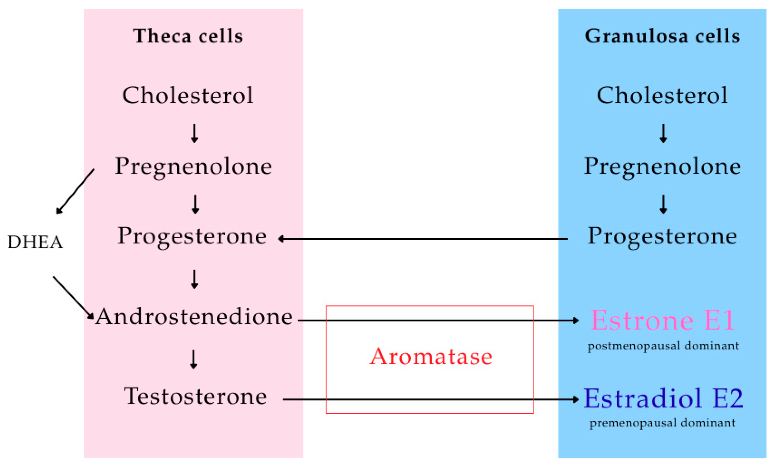
Disclaimer/Publisher’s Note: The statements, opinions and data contained in all publications are solely those of the individual author(s) and contributor(s) and not of MDPI and/or the editor(s). MDPI and/or the editor(s) disclaim responsibility for any injury to people or property resulting from any ideas, methods, instructions or products referred to in the content.

Article Metrics

Citations

Article Access Statistics

Multiple requests from the same IP address are counted as one view.