

Exercise-Induced Muscle Damage after a High-Intensity Interval Exercise Session: Systematic Review

 , , and
Abstract
:1. Introduction
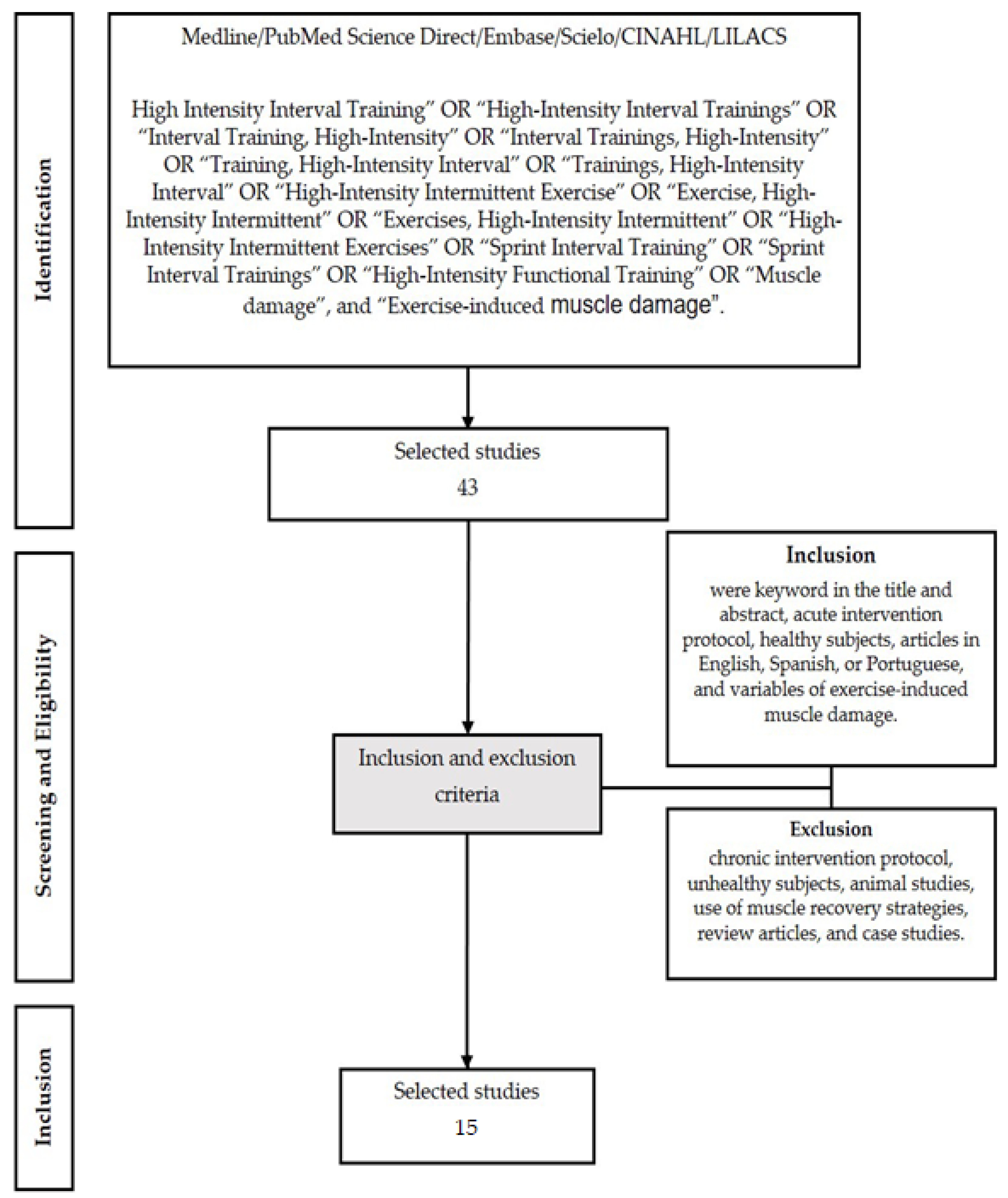
2. Materials and Methods
3. Results
4. Discussion
5. Conclusions
Author Contributions
Funding
Institutional Review Board Statement
Informed Consent Statement
Data Availability Statement
Acknowledgments
Conflicts of Interest
References
- Gibala, M.J.; McGee, S.L. Metabolic Adaptations to short-term high-intensity interval training: A little pain for a lot of gain. Exerc. Sport Sci. Rev. 2008, 36, 58–63. [Google Scholar] [CrossRef] [PubMed]
- Gibala, M.J.; Little, J.P. Just HIT it! A time-efficient exercise strategy to improve muscle insulin sensitivity. J. Physiol. 2010, 588, 3341–3342. [Google Scholar] [CrossRef] [PubMed]
- Buchheit, M.; Laursen, P.B. High-intensity interval training, solutions to the programming puzzle: Part I: Cardiopulmonary emphasis. Sports Med. 2013, 43, 313–338. [Google Scholar] [CrossRef]
- Gibala, M.J.; Gillen, J.B.; Percival, M.E. Physiological and health-related adaptations to low-volume interval training: Influences of nutrition and sex. Sports Med. 2014, 44, 127–137. [Google Scholar] [CrossRef]
- Gillen, J.B.; Martin, B.J.; MacInnis, M.J.; Skelly, L.E.; Tarnopolsky, M.A.; Gibala, M.J. Twelve Weeks of Sprint Interval Training Improves Indices of Cardiometabolic Health Similar to Traditional Endurance Training despite a Five-Fold Lower Exercise Volume and Time Commitment. PLoS ONE. 2016, 11, e0154075. [Google Scholar] [CrossRef] [PubMed]
- Rozenek, R.; Salassi, J.W.; Pinto, N.M.; Fleming, J.D. Acute Cardiopulmonary and Metabolic Responses to High-Intensity Interval Training Protocols Using 60 s of Work and 60 s Recovery. J. Strength Cond. Res. 2016, 30, 3014–3023. [Google Scholar] [CrossRef]
- Dun, Y.; Smith, J.R.; Liu, S.; Olson, T.P. High-Intensity Interval Training in Cardiac Rehabilitation. Clin. Geriatr. Med. 2019, 35, 469–487. [Google Scholar] [CrossRef]
- Osawa, Y.; Azuma, K.; Tabata, S.; Katsukawa, F.; Ishida, H.; Oguma, Y.; Kawai, T.; Itoh, H.; Okuda, S.; Matsumoto, H. Effects of 16-week high-intensity interval training using upper and lower body ergometers on aerobic fitness and morphological changes in healthy men: A preliminary study. Open Access J. Sports Med. 2014, 5, 257–265. [Google Scholar] [CrossRef]
- Wiewelhove, T.; Fernandez-Fernandez, J.; Raeder, C.; Kappenstein, J.; Meyer, T.; Kellmann, M.; Pfeiffer, M.; Ferrauti, A. Acute responses and muscle damage in different high-intensity interval running protocols. J. Sports Med. Physi. Fit. 2015, 56, 606–615. [Google Scholar]
- Thompson, W.R. Worldwide Survey of Fitness Trends for 2021. ACSM’s Health Fit. J. 2021, 25, 10–19. [Google Scholar] [CrossRef]
- Peake, J.M.; Tan, S.J.; Markworth, J.F.; Broadbent, J.A.; Skinner, T.L.; Cameron-Smith, D. Metabolic and hormonal responses to isoenergetic high-intensity interval exercise and continuous moderate-intensity exercise. Ame J. Physiol. Endocrinol. Metab. 2014, 307, E539–E552. [Google Scholar] [CrossRef] [PubMed]
- Weston, K.S.; Wisløff, U.; Coombes, J.S. High-intensity interval training in patients with lifestyle-induced cardiometabolic disease: A systematic review and meta-analysis. Br. J. Sports Med. 2014, 48, 1227–1234. [Google Scholar] [CrossRef] [PubMed]
- Billat, L.V. Interval Training for Performance: A Scientific and Empirical Practice. Special recommendations for middle- and long-distance running. Part I: Aerobic interval training. Sports Med. 2001, 31, 13–31. [Google Scholar] [CrossRef] [PubMed]
- Fountaine, C.J.; Schmidt, B.J. Metabolic Cost of Rope Training. J. Strength Cond. Res. 2015, 29, 889–893. [Google Scholar] [CrossRef] [PubMed]
- Evangelista, A.L.; Teixeira, C.L.S.; Machado, A.F.; Pereira, P.E.; Machado, R.L.; Bocalini, D.S. Effects of a short-term of whole-body, high-intensity, intermittent training program on morphofunctional parameters. J. Bodyw. Mov. Ther. 2019, 23, 456–460. [Google Scholar] [CrossRef] [PubMed]
- Machado, A.F.; Evangelista, A.L.; Miranda, J.M.Q.; Teixeira, C.V.L.S.; Rica, R.L.; Lopes, C.R.; Figueira-Júnior, A.; Baker, J.S.; Bocalini, D.S. Description of training loads using whole-body exercise during high-intensity interval training. Clinics 2018, 73, e516. [Google Scholar] [CrossRef]
- Cipryan, L.; Tschakert, G.; Hofmann, P. Acute and post-exercise physiological responses to high-intensity interval training in endurance and sprint athletes. J. Sports Scien. Med. 2017, 16, 219–229. [Google Scholar]
- Chen, T.C.; Nosaka, K.; Sacco, P. Intensity of eccentric exercise, shift of optimum angle, and the magnitude of repeated-bout effect. J. Appl. Physiol. 2007, 102, 992–999. [Google Scholar] [CrossRef]
- Clarkson, P.M.; Hubal, M.J. Exercise-Induced Muscle Damage in Humans. Am. J. Phys. Med. Rehabil. 2002, 81, S52–S69. [Google Scholar] [CrossRef]
- Gibala, M.J.; MacDougall, J.D.; Tarnopolsky, M.A.; Stauber, W.T.; Elorriaga, A.; Peake, J.M.; Neubauer, O.; Della Gatta, P.A.; Nosaka, K.; Kanzaki, K.; et al. Changes in human skeletal muscle ultrastructure and force production after acute resistance exercise. J. Appl. Physiol. 1995, 78, 702–708. [Google Scholar] [CrossRef]
- Cleak, M.; Eston, R. Delayed onset muscle soreness: Mechanisms and management. J. Sports Sci. 1992, 10, 325–341. [Google Scholar] [CrossRef] [PubMed]
- Beaton, L.J.; Tarnopolsky, M.A.; Phillips, S.M. Contraction-induced muscle damage in humans following calcium channel blocker administration. Clin. Trial J. Physiol. 2002, 544, 849–859. [Google Scholar] [CrossRef] [PubMed]
- Hyldahl, R.D.; Nelson, B.; Xin, L.; Welling, T.; Groscost, L.; Hubal, M.J.; Chipkin, S.; Clarkson, P.M.; Parcell, A.C. Extracellular matrix remodeling and its contribution to protective adaptation following lengthening contractions in human muscle. FASEB J. 2015, 29, 2894–2904. [Google Scholar] [CrossRef] [PubMed]
- Warren, G.L.; Ingalls, C.P.; Lowe, D.A.; Armstrong, R.B. What Mechanisms Contribute to the Strength Loss That Occurs During and in the Recovery from Skeletal Muscle Injury? J. Orthop. Sports Phys. Ther. 2002, 32, 58–64. [Google Scholar] [CrossRef]
- Jamurtas, A.Z.; Theocharis, V.; Tofas, T.; Tsiokanos, A.; Yfanti, C.; Paschalis, V.; Koutedakis, Y.; Nosaka, K. Comparison between leg and arm eccentric exercises of the same relative intensity on indices of muscle damage. Eur. J. Appl. Physiol. 2005, 95, 179–185. [Google Scholar] [CrossRef]
- Nosaka, K.; Newton, M.; Sacco, P. Delayed-onset muscle soreness does not reflect the magnitude of eccentric exercise-induced muscle damage. Scand. J. Med. Sci. Sports. 2002, 12, 337–346. [Google Scholar] [CrossRef]
- Joo, C.H. Development of a non-damaging high-intensity intermittent running protocol. J. Exerc. Rehabil. 2015, 11, 112–118. [Google Scholar] [CrossRef]
- Nosaka, K.; Newton, M.; Sacco, P.; Chapman, D.; Lavender, A. Partial Protection against Muscle Damage by Eccentric Actions at Short Muscle Lengths. Med. Sci. Sports Exerc. 2005, 37, 746–753. [Google Scholar] [CrossRef]
- Yu, J.-Y.; Jeong, J.-G.; Lee, B.-H. Evaluation of muscle damage using ultrasound imaging. J. Phys. Ther. Sci. 2015, 27, 531–534. [Google Scholar] [CrossRef]
- Cui, S.; Sun, B.; Yin, X.; Guo, X.; Chao, D.; Zhang, C.; Zhang, C.-Y.; Chen, X.; Ma, J. Time-course responses of circulating microRNAs to three resistance training protocols in healthy young men. Sci. Rep. 2017, 7, 2203. [Google Scholar] [CrossRef]
- Damas, F.; Libardi, C.A.; Ugrinowitsch, C. The development of skeletal muscle hypertrophy through resistance training: The role of muscle damage and muscle protein synthesis. Eur. J. Appl. Physiol. 2018, 118, 485–500. [Google Scholar] [CrossRef] [PubMed]
- Jadad, A.R.; Moore, R.A.; Carroll, D.; Jenkinson, C.; Reynolds, D.J.M.; Gavaghan, D.J.; McQuay, H.J. Assessing the quality of reports of randomized clinical trials: Is blinding necessary? Control. Clin. Trials. 1996, 17, 1–12. [Google Scholar] [CrossRef] [PubMed]
- Whiting, P.; Savović, J.; Higgins, J.P.T.; Caldwell, D.M.; Reeves, B.C.; Shea, B.; Davies, P.; Kleijnen, J.; Churchill, R.; ROBIS group. ROBIS: A new tool to assess risk of bias in systematic reviews was developed. J. Clin. Epidemiol. 2016, 69, 225–234. [Google Scholar] [CrossRef] [PubMed]
- Kjaergard, L.L.; Villumsen, J.; Gluud, C. Reported Methodologic quality and discrepancies between large and small randomized trials in meta-analyses. Ann. Intern. Med. 2001, 135, 982–989. [Google Scholar] [CrossRef]
- Deminice, R.; Trindade, C.S.; Degiovanni, G.C.; Garlip, M.R.; Portari, G.V.; Teixeira, M.; Jordao, A.A. Oxidative stress biomarkers response to high intensity interval training and relation to performance in competitive swimmers. J. Sport Med. Physi. Fit. 2010, 50, 356–362. [Google Scholar]
- Cipryan, L. The effect of fitness level on cardiac autonomic regulation, IL-6, total antioxidant capacity, and muscle damage responses to a single bout of high-intensity interval training. J. Sport Health Sci. 2016, 7, 363–371. [Google Scholar] [CrossRef]
- Franchini, E.; Julio, U.F.; Panissa, V.L.G.; Lira, F.S.; Gerosa-Neto, J.; Branco, B.H.M. High-intensity intermittent training positively affects aerobic and anaerobic performance in judo athletes independently of exercise mode. Front. Physiol. 2016, 7, 268. [Google Scholar] [CrossRef]
- Cipryan, L. IL-6, Antioxidant capacity and muscle damage markers following high-intensity interval training protocols. J. Hum. Kinet. 2017, 56, 139–148. [Google Scholar] [CrossRef]
- Spada, T.C.; Silva, J.M.R.D.; Francisco, L.S.; Marçal, L.J.; Antonangelo, L.; Zanetta, D.M.T.; Yu, L.; Burdmann, E.A. High intensity resistance training causes muscle damage and increases biomarkers of acute kidney injury in healthy individuals. PLoS ONE. 2018, 13, e0205791. [Google Scholar] [CrossRef]
- Farias-Júnior, L.F.; Browne, R.A.V.; Frazão, D.T.; Dantas, T.C.B.; Silva, P.H.M.; Freitas, R.P.A.; Aoki, M.S.; Costa, E.C. Effect of Low-Volume High-Intensity Interval Exercise and Continuous Exercise on Delayed-Onset Muscle Soreness in Untrained Healthy Males. J Strength Cond Res. 2019, 33, 774–782. [Google Scholar] [CrossRef]
- Farias-Junior, L.F.; Browne, R.A.V.; Freire, Y.A.; Oliveira-Dantas, F.F.; Lemos, T.M.A.M.; Galvão-Coelho, N.L.; Hardcastle, S.J.; Okano, A.H.; Aoki, M.S.; Costa, E.C. Psychological responses, muscle damage, inflammation, and delayed onset muscle soreness to high-intensity interval and moderate-intensity continuous exercise in overweight men. Physiol. Behav. 2019, 199, 200–209. [Google Scholar] [CrossRef] [PubMed]
- Timón, R.; Olcina, G.; Camacho-Cardeñosa, M.; Camacho-Cardenosa, A.; Martinez-Guardado, I.; Marcos-Serrano, M. 48-h recovery of biochemical parameters and physical performance after two modalities of CrossFit workouts. Biol. Sport. 2019, 36, 283–289. [Google Scholar] [CrossRef] [PubMed]
- Gomes, J.H.; Mendes, R.R.; Franca, C.S.; Da Silva-Grigoletto, M.E.; da Silva, D.R.P.; Antoniolli, A.R.; Silva, A.M.d.O.e.; Quintans-Júnior, L.J. Acute leucocyte, muscle damage, and stress marker responses to high-intensity functional training. PLoS ONE. 2020, 15, e0243276. [Google Scholar] [CrossRef]
- Boullosa, D.; Dragutinovic, B.; Deutsch, J.-P.; Held, S.; Donath, L.; Bloch, W.; Schumann, M. Acute and Delayed Effects of Time-Matched Very Short “All Out” Efforts in Concentric vs. Eccentric Cycling. Int. J. Environ. Res. Public Health 2021, 18, 7968. [Google Scholar] [CrossRef] [PubMed]
- Alves, J.W.; Farias-Junior, L.F.; Alves, C.P.d.L.; Mortatti, A.L.; Costa, E.C. Low-Volume High-Intensity Interval Training Sessions With Different Work–Recovery Durations and Muscle Damage in Trained Men. Res. Q. Exerc. Sport. 2022, 94, 73–81. [Google Scholar] [CrossRef] [PubMed]
- Rohnejad, B.; Monazzami, A. Effects of high-intensity intermittent training on some inflammatory and muscle damage indices in overweight middle-aged men. Apunt. Sports Med. 2023, 58, 1–9. [Google Scholar] [CrossRef]
- Newton, M.J.; Morgan, G.T.; Sacco, P.; Chapman, D.W.; Nosaka, K. Comparison of Responses to Strenuous Eccentric Exercise of the Elbow Flexors Between Resistance-Trained and Untrained Men. J. Strength Cond. Res. 2008, 22, 597–607. [Google Scholar] [CrossRef]
- Clarkson, P.M.; Nosaka, K.; Braun, B. Muscle function after exercise-induced muscle damage and rapid adaptation. Med. Sci. Sports Exerc. 1992, 24, 512–520. [Google Scholar] [CrossRef]
- Tee, J.C.; Bosch, A.N.; Lambert, M.I. Metabolic consequences of exercise-induced muscle damage. Sports Med. 2007, 37, 827–836. [Google Scholar] [CrossRef]
- De Freitas, M.C.; Gerosa-Neto, J.; Zanchi, N.E.; Lira, F.S.; Rossi, F.E. Role of metabolic stress for enhancing muscle adaptations: Practical applications. World J. Methodol. 2017, 7, 46–54. [Google Scholar] [CrossRef]
- Tabata, I.; Nishimura, K.; Kouzaki, M.; Hirai, Y.; Ogita, F.; Miyachi, M.; Yamamoto, K. Effects of moderate-intensity endurance and high-intensity intermittent training on anaerobic capacity and VO2max. Med. Sci. Sports Exerc. 1996, 28, 1327–1330. [Google Scholar] [CrossRef]
- Hicks, K.M.; Onambélé, G.L.; Winwood, K.; Morse, C.I. Muscle Damage following Maximal Eccentric Knee Extensions in Males and Females. PLoS ONE. 2016, 11, e0150848. [Google Scholar] [CrossRef] [PubMed]
- Damas, F.; Phillips, S.M.; Libardi, C.A.; Vechin, F.C.; Lixandrão, M.E.; Jannig, P.R.; Costa, L.A.R.; Bacurau, A.V.; Snijders, T.; Parise, G.; et al. Resistance training-induced changes in integrated myofibrillar protein synthesis are related to hypertrophy only after attenuation of muscle damage. J. Physiol. 2016, 594, 5209–5222. [Google Scholar] [CrossRef] [PubMed]
- Wiig, H.; Cumming, K.T.; Handegaard, V.; Stabell, J.; Spencer, M.; Raastad, T. Muscular heat shock protein response and muscle damage after semi-professional football match. Scand. J. Med. Sci. Sports. 2022, 32, 984–996. [Google Scholar] [CrossRef] [PubMed]
- Ebbeling, C.B.; Clarkson, P.M. Exercise-Induced Muscle Damage and Adaptation. Sports Med. 1989, 7, 207–234. [Google Scholar] [CrossRef] [PubMed]

| Authors | Language | Journal | IF |
|---|---|---|---|
| Deminice et al., 2010 [35] | English | The Journal of Sports Medicine and Physical Fitness | 1.432 |
| Joo, 2015 [27] | English | Journal of Exercise Rehabilitation | 1.170 |
| Wiewelhove et al., 2016 [9] | English | The Journal of Sports Medicine and Physical Fitness | 1.432 |
| Cipryan, 2016 [36] | English | Journal of Sport and Health Science | 5.200 |
| Franchini et al., 2016 [37] | English | Frontiers in Physiology | 3.367 |
| Cyprian, 2017 [38] | English | Journal of Human Kinetics | 1.664 |
| Cipryan et al., 2017 [17] | English | Journal of Sports Science and Medicine | 1.806 |
| Spada et al., 2018 [39] | English | Plos One | 2.740 |
| Farias-Junior et al., 2019 [40] | English | The Journal of Strength and Conditioning Research | 3.200 |
| Farias-Junior et al., 2019a [41] | English | Physiology & Behavior | 3.742 |
| Timón et al., 2019 [42] | English | Biology of Sport | 2.000 |
| Gomes et al., 2020 [43] | English | Plos One | 2.740 |
| Boullosa et al., 2021 [44] | English | International Journal of Environmental Research and Public Health | 4.614 |
| Alves et al., 2023 [45] | English | Research Quarterly for Exercise and Sport | 2.098 |
| Rohnejad and Monazzami, 2023 [46] | English | Apunts Sports Medicine | 1.150 |
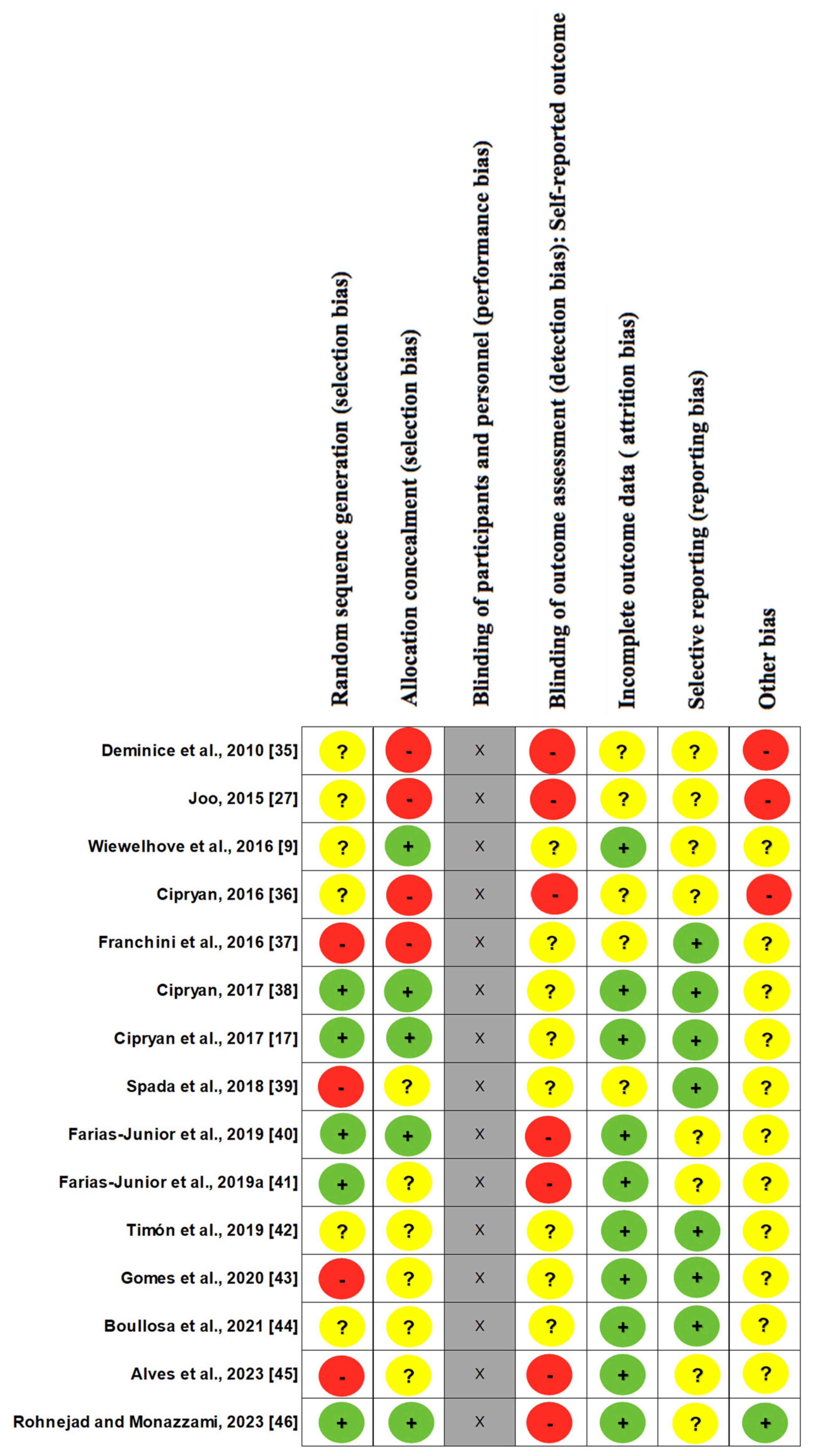
| Study | Was the Study Described as Randomized? | Was There a Description of Randomization? Was it Adequate? | Were There Comparisons and Results? | Was There a Description of Comparisons and Results? Were They Adequate? | Was There a Description of Withdrawals and Dropouts? | Total |
|---|---|---|---|---|---|---|
| Deminice et al., 2010 [35] | 0 | 0 | 1 | 1 | 1 | 3 |
| Joo, 2015 [27] | 0 | 0 | 1 | 1 | 1 | 3 |
| Wiewelhove et al., 2016 [9] | 0 | 1 | 1 | 1 | 1 | 4 |
| Cipryan, 2016 [36] | 0 | 0 | 1 | 1 | 1 | 3 |
| Franchini et al., 2016 [37] | 0 | 1 | 1 | 1 | 1 | 4 |
| Cipryan, 2017 [38] | 0 | 0 | 1 | 1 | 1 | 3 |
| Cipryan et al., 2017 [17] | 0 | 0 | 1 | 1 | 1 | 3 |
| Spada et al., 2018 [39] | 0 | 0 | 1 | 1 | 1 | 3 |
| Farias-Junior et al., 2019 [40] | 1 | 1 | 1 | 1 | 1 | 5 |
| Farias-Junior et al., 2019a [41] | 1 | 0 | 1 | 1 | 1 | 4 |
| Timón et al., 2019 [42] | 0 | 1 | 1 | 1 | 1 | 4 |
| Gomes et al., 2020 [43] | 0 | 0 | 1 | 1 | 1 | 3 |
| Boullosa et al., 2021 [44] | 0 | 0 | 1 | 1 | 1 | 3 |
| Alves et al., 2023 [45] | 0 | 0 | 1 | 1 | 1 | 3 |
| Rohnejad and Monazzami, 2023 [46] | 1 | 1 | 1 | 1 | 1 | 5 |
| Reference | Subjects/Sample | Age (Years) | VO2 | HIIT Protocol | Intensity |
|---|---|---|---|---|---|
| Deminice et al., 2010 [35] | A total of 10 well-trained swimmers among the top 10 best Brazilian swimmers in their styles (men n = 8; women n = 2), familiar with HIIT series in their training routine | 20 ± 2 | N/A | A set of 8 maximal swims over 100 m per their style specialty, with 10 min rest. | Maximum effort |
| Joo, 2015 [27] | A total of 10 healthy, moderately trained men used to frequent high-intensity exercise | 31 ± 7.1 | VO2max 58 ± 7.1 mL kg min | A total of 8 sets of 3 min jogging sessions on a treadmill, interspersed with 3 min active intervals (1.5 min at 25% VO2max and 1.5 min at 50% VO2max). | 90% VO2max |
| Wiewelhove et al., 2016 [9] | A total of 16 well-trained male athletes from intermittent sports (tennis, handball, and soccer) | 24.6 ± 2.7 | VO2max 58.3 ± 5.9 mL kg min | A total of five different HIIT protocols, separated by six days each, were performed. HIIT-P240: 4 sets of 4 min with 3 min passive interval (2/1 work/rest ratio). HIIT-P120: 7 sets of 2 min with 2 min passive interval (1/1 work/rest ratio), 40 m come-and-go run test. HIIT-P30: 2 blocks with 10 sets of 30 s with 45 s interval and 3 min passive recovery between the blocks (1/2 work/rest ratio). HIIT-P15: 3 blocks with 9 sets of 15 s with 30 s interval and 3 min of passive recovery between blocks (1/4 work/rest ratio), sprint straight. HIIT-P5: 4 blocks of 6 sets of 5 s with 25 s interval and 5 min of passive recovery between blocks (1/12 work/rest ratio). | %V 30–15 intermittent fitness test HIIT-P240—80% HIIT-P120—85% HIIT-P30—90% HIIT-P15—95% HIIT-P5—all out |
| Cipryan, 2016 [36] | The sample consisted of 30 healthy young subjects distributed in well trained (WT; n = 11; h/week 12.00 ± 5.89), moderately trained (MT; n = 10; h/week 6.05 ± 2.22), untrained (UT; n = 9; no intentional sports activities) | WT: 24.18 ± 1.80 MT: 24.18 ± 1.80 UT: 24.44 ± 2.54 | VO2 max WT: 61.39 ± 3.63 mL kg min MT: 53.46 ± 2.80 mL kg min UT: 47.21 ± 3.98 mL kg min | All participants performed a 30 min HIIT composed of 6 × 2 min interval exercise with work-to-rest ratio = 1. | 100% vVO2max |
| Franchini et al., 2016 [37] | The sample consisted of 35 male judo athletes divided into HIIT in stationary bicycle for lower limbs (HIIT-L; n = 9), HIIT in stationary bicycle for upper limbs (HIIT-U; n = 9), Uchi-Komi judo technique (HIIT-UK; n = 9), and control (C; n = 8) | HIIT-L: 22.3 ± 5.2 HIIT-U: 23.6 ± 6.7 HIIT-UK: 23.4 ± 4.2 control: 26.4 ± 7.0 | VO2 peak Gradual maximal upper limb stationary bicycle test for each group (PRE values) HIIT-L: 2.78 ± 0.41 L.min HIIT-U: 3.10 ± 0.70 L.min HIIT-UK: 3.16 ± 0.30 L.min control: 2.86 ± 0.37 L.min Gradual maximal lower limb stationary bicycle test for each group (PRE values) HIIT-L: 3.62 ± 0.50 L.min HIIT-U: 3.82 ± 0.59 L.min HIIT-UK: 3.87 ± 0.44 L.min control: 3.56 ± 0.49 L.min | Tests on the stationary bicycle with 70 rpm fixed cadence for lower limbs and 90 rpm for upper limbs, totaling 22 min/session. Session divided into 2 HIIT blocks, each block lasting 4 min (10 times/20 s effort and 10 s break), and 5 min rest between each block. | All out |
| Cyprian, 2017 [38] | A total of 12 moderately trained men participated in three HIIT trials | 22.8 ± 1.7 | VO2max 57.2 ± 6.3 mL kg min | The three different HIIT protocols were performed on a treadmill with work/rest ratio = 1 (HIIT 15 s/15 s, HIIT 30 s/30 s, and HIIT 60 s/60 s), the total duration was 12 min with identical external work with active recovery of 6 min at 60% vVO2max. | 100% vVO2max |
| Cipryan et al., 2017 [17] | In total, 16 highly trained men were divided into endurance athletes (E = n = 8; h/week 13.9 ± 4.0) and sprint athletes (S = n = 8; h/week 9.9 ± 1.9), both groups performed 3 HIIT protocols | E: 22.1 ± 2.5 S: 22.9 ± 3.5 | VO2max E: 66.2 ± 5.0 mL kg min S: 56.8 ± 5.0 mL kg min | A total of two HIIT protocols were performed on a treadmill. The 3 min HIIT consisted of 4 sets of 3 min of work with 3 min of passive recovery interval. The 30 s HIIT consisted of 21 sets of 30 s of work with 30 s of passive recovery interval. The control group ran for 21 min. | 3 min HIIT: 100% vVO2max 30 s HIIT: 100% vVO2max C: 50% vVO2max |
| Spada et al., 2018 [39] | A total of 58 healthy volunteers (29 men and 29 women), able to correctly perform the prescribed exercise, and with serum and urinary laboratory parameters within normal ranges, performed a high-intensity interval resistance training (HIIRT) session. | 24 (21–28) | N/A | The HIIRT session consisted of 8 sets of squats with the fastest speed and the highest number of repetitions achievable for 20 s with 10 s of rest between sets. | Maximum effort |
| Farias-júnior et al., 2019 [40] | The sample consisted of 15 untrained healthy males | 25.1 ± 4.4 | N/A | The low-volume HIIE consisted of 10 × 60 s work bouts interspersed with 60 s of active recovery at 30% of MV. | 90% of Maximal velocity (MV) |
| Farias-júnior et al., 2019a [41] | The sample consisted of 20 overweight inactive men | 28.9 ± 5.0 | VO2pico 39.0 ± 4.1 | The HIIE consisted of 10 × 1 min intervals interspersed with 1 min of passive recovery. | 100% of Vmax |
| Timón et al., 2019 [42] | A total of 12 trained men and CrossFit practitioners completed two modalities of WODs on separate days: WOD1 (as many rounds as possible) and WOD2 (rounds for time) | 30.4 ± 5.37 | VO2max 47.8 ± 3.63 mL kg min | They practiced two modalities of workout of the day (WODs) on separate days. WOD1: as many rounds as possible of burpees and toes to bar with increasing repetitions (1-1, 2-2, 3-3,...) in five minutes. WOD2: 3 blocks of 20 wall ball (9 kg) repetitions and then 20 power clean repetitions (load of 40% of 1 RM) in the shortest time possible. | N.I |
| Gomes et al., 2020 [43] | A total of 23 subjects, 12 men and 11 women, were divided into experienced (EXP: ≥18 months of experience; n = 13) and beginners (BEG: 3–8 months experience; n = 10) and were submitted to a specific protocol of the modality | EXP: 31.1 ± 4.9 BEG: 30.9 ± 4.8 ALL: 31.0 ± 4.8 | VO2max EXP: 40.7 ± 1.8 mL kg min BEG: 39.2 ± 1.4 mL kg min ALL: 40.0 ± 1.7 mL kg min | The high-intensity functional training session (HIFT) WOD developed was called “Cindy”. This WOD consisted of as many rounds as possible of 5 pull-ups, 10 push-ups, and 15 air squats in 20 min. | All out |
| Boullosa et al., 2021 [44] | The sample consisted of 12 physically active men involved in recreational endurance sports | 23.4 ± 2.8 | VO2max: ≥90% of the maximum predicted heart rate for age (HRmax) | A total of 8 maximal efforts for 5 s, with 55 s of active recovery interval at 80 rpm, in concentric vs. eccentric cycling. | All out |
| Alves et al., 2023 [45] | The sample consisted of 24 trained adult males | 22.3 ± 2.9 | N.I | Two LV-HIIT sessions: The 60/60 s LV-HIIT protocol consisted of 10 × 60 s of maximal aerobic speed on treadmill interspersed by 60 s of passive recovery. The 30/30 s LV-HIIT protocol with 20 × 30 s of maximal aerobic speed on treadmill interspersed by 30 s of passive recovery. | 100% Vmax |
| Rohnejad and Monazzami, 2023 [46] | The sample consisted of 22 overweight middle-aged active men | Control: 47.80 ± 7.50 HIIT group: 45.90 ± 6.17 | VO2max Control: 28.5 1± 1.55 mL kg min HIIT group: 28.14 ± 1.30 mL kg min | The HIIT training program consisted of intermittent running for 30 s, 30 s of active recovery at 50% aerobic speed (4 sets, 4 rounds, and 5 min of passive recovery between each round). | 100% Maximal aerobic velocity (MAV) |
| Reference | Damage Markers | POS | 30 min | 1 h | 2 h | 3 h | 4 h | 24 h | 48 h | 72 h | 7 Days | Conclusion |
|---|---|---|---|---|---|---|---|---|---|---|---|---|
| Deminice et al., 2010 [35] | 1. CK | ↑ CK | N/A | N/A | N/A | N/A | N/A | N/A | N/A | N/A | N/A | Proposed session-specific HIIT induces increased creatine kinase in competitive swimmers. |
| Joo, 2015 [27] | 1. CK * 2. Mb 3. Pain-VAS 4. Muscle Pain Sensitivity (distal myotendinous junction and middle belly of rectus femoris) * 5. MVC * | ↑ Mb | N/A | N/A | N/A | N/A | N/A | ↑ Mb | ↔ Mb ↑ Pain-VAS | ↔ Mb ↔ Pain-VAS | ↔ Mb ↑ Pain-VAS | The results show that, in moderately trained subjects used to high-intensity exercise, the exercise protocol used in this study was able to increase post-exercise myoglobin levels as well as muscle pain perception 48 h after the protocol. No other marker changed. |
| Wiewelhove et al., 2016 [9] | 1. CK 2. Pain-VAS * 3. CMJ | N/A | HIIT-P240 * CK HIIT-5 ↓ CMJ | N/A | N/A | N/A | N/A | HIIT-P240 ↑ CK HIIT-5 ↑ CK ↓ CMJ | N/A | N/A | N/A | The HIIT-P240 straight running and the HIIT-P5 sprint showed an increase in CK 24 h after exercise. HIIT-P5 showed a CMJ reduction 30 min and 24 h after exercise, which suggests that short intervals of high-intensity training possibly cause greater muscle damage compared to long intervals of submaximal-intensity training. |
| Cipryan, 2016 [36] | 1. CK 2. Mb | WT ↑ CK ↑ Mb MT ↑ CK ↑ Mb UT ↑ CK ↑ Mb | N/A | N/A | WT ↔ CK ↑ Mb MT ↑ CK ↑ Mb UT ↑ CK ↑ Mb | N/A | WT ↔ CK ↑ Mb MT ↑ CK ↑ Mb UT ↑ CK ↑ Mb | N/A | N/A | N/A | N/A | Although the HIIT protocol increased markers of exercise-induced muscle damage, CK and Mb increases were less pronounced in well-trained athletes compared to moderately trained or untrained individuals. |
| Fanchini et al., 2016 [37] | 1. CK 2. LDH 3. AST 4. ALT | Wingate test values in the stationary bicycle performed before the upper and lower limbs training period. HIIT-L ↑ CK ↑ LDH ↑ AST ↑ ALT All vs. PRE for both tests HIIT-U ↑ CK ↑ LDH ↑ AST ↑ ALT All vs. PRE for both tests HIIT-UK ↑ CK ↑ LDH ↑ AST ↑ ALT All vs. PRE for both tests | N/A | N/A | N/A | N/A | N/A | N/A | N/A | N/A | N/A | Both Wingate tests in stationary bicycle (lower and upper segment) increased muscle damage markers (CK, LDH, AST, and ALT) compared to pre-test in HIIT-I, HIIT-S, and HIIT-UK groups. |
| Cyprian, 2017 [38] | 1. CK 2. Mb 3. LDH | HIIT 15 s/15 s ↑ CK ↑ Mb ↑ LDH HIIT 30 s/30 s ↑ CK ↑ Mb ↑ LDH HIIT 60 s/60 s ↑ CK ↑ Mb ↑ LDH | N/A | N/A | N/A | HIIT 15 s/15 s ↑ CK ↑ Mb ↑ LDH HIIT 30 s/30 s ↔ CK ↑ Mb ↑ LDH HIIT 60 s/60 s ↔ CK ↑ Mb ↑ LDH | N/A | HIIT 15 s/15 s ↑ CK ↔ Mb ↑ LDH HIIT 30 s/30 s ↑ CK ↔ Mb ↑ LDH HIIT 60 s/60 s ↑ CK ↑ Mb ↑ LDH | N/A | N/A | N/A | All three HIIT protocols with short intervals and fixed external work caused an immediate elevation in muscle damage markers in circulation. However, these changes differed, prejudicing to assess the magnitude of exercise-induced muscle damage. The HIIT 30 s/30 s protocol showed a lower response in Mb. |
| Cipryan et al., 2017 [17] | 1. CK * 2. Mb | For both ET and ST athletes: 3 min HIIT ↑ Mb HIIT-30 s ↑ Mb | N/A | For both ET and ST athletes: 3 min HIIT ↑ Mb HIIT-30 s ↑ Mb | N/A | For both ET and ST athletes: 3 min HIIT ↑ Mb HIIT-30 s ↑ Mb | N/A | N/A | N/A | N/A | N/A | Markers of muscle damage monitored during the initial recovery failed to show any differences between individuals trained in endurance and sprint. Despite this, Mb values showed a moderate response 1 h and 3 h after the 30 min and 30 s HIIT session. The control group showed no change in markers. |
| Spada et al., 2018 [39] | 1. CK 2. Mb 3. Pain-Borg CR10 | N/A | N/A | N/A | ↑ CK ↑ Mb ↑ Pain | N/A | N/A | ↑ CK ↑ Mb ↑ Pain | N/A | N/A | N/A | A single session of HIIT in healthy and young individuals caused increases in CK, Mb, and pain, indicating the occurrence of muscle damage. |
| Farias-Junior et al., 2019 [40] | 1. PPT 2. PPTol 3. PPI Muscles analyzed: rectus femoris, biceps femoris, and gastrocnemius | N/A | N/A | N/A | N/A | N/A | N/A | HIIE RF ↑ PPI BF ↑ PPI G ↓ PPTol | N/A | N/A | N/A | Low-volume HIIE session elicited mild DOMS 24 h post exercise in untrained healthy males, which was similar to the traditional CE session. |
| Farias-Junior et al., 2019a [41] | 1. CK 2. LDH 3. PPT 4. PPTol 5. EVA-PPI Muscles analyzed: rectus femoris, biceps femoris, and gastrocnemius | N/A | N/A | N/A | N/A | N/A | N/A | ↑ CK G ↓ PPTol | ↑ CK BF ↑ PPI G ↔ PPTol | N/A | N/A | The subjects showed modest exercise-induced muscle damage for all individuals. |
| Timón et al., 2019 [42] | 1. CK 2. LDH * 3. AST 4. ALT 5. CMJ * 6. PT | WOD1 ↑ CK ↑ AST ↑ ALT ↓ TP WOD2 ↑ CK ↑ AST ↑ ALT ↓ TP | N/A | N/A | N/A | N/A | N/A | WOD1 ↑ CK ↔ AST ↔ ALT ↓ PT WOD2 ↑ CK ↑ AST ↑ ALT ↓ TP | WOD1 ↔ CK ↔ AST ↔ ALT ↔ PT WOD2 ↔ CK ↔ AST ↔ ALT ↔ PT | N/A | N/A | The effort intensity during WOD2 was higher than during WOD1. The performance of both CrossFit sessions (WOD1 and 2) caused significant changes in transaminases, markers of muscle damage, and reduction in physical performance. All values returned to baseline values in 48 h. |
| Gomes et al., 2020 [43] | 1. CK | EXP ↑ CK BEG ↑ CK ALL ↑ CK | EXP ↑ CK BEG ↑ CK ALL ↑ CK | N/A | N/A | N/A | N/A | EXP ↑ CK BEG ↑ CK ALL ↑ CK | N/A | N/A | N/A | A single HIFT session significantly increased CK levels in both EXPs and BEGs. |
| Boullosa et al., 2021 [44] | 1. CK 2. Pain–VAS 3. MC | Concentric protocol ↑ CK Eccentric protocol * CK | N/A | N/A | N/A | N/A | N/A | Concentric protocol ↔ CK * Pain–VAS * TC Eccentric protocol ↑ CK ↑ Pain–VAS ↑ MC | N/A | N/A | N/A | Single-session HIIT protocols are able to change damage markers mainly within 24 h. |
| Alves et al., 2023 [45] | 1. Countermovement vertical jump height (CVJH) * 2. PPT * 3. PPTol * 4. EVA–PPI * Muscles analyzed: rectus femoris (RF), biceps femoris (BF), and gastrocnemius (G). | N/A | N/A | N/A | N/A | N/A | N/A | For both groups (60/60 LV-HIIT and 30/30 LV-HIIT) No change | For both groups (60/60 LV-HIIT and 30/30 LV-HIIT) No change | N/A | N/A | The LV-HIIT sessions with different work–recovery durations (i.e., 10 × 60 s or 20 × 30 s at 100% of Vmax), matched by work–recovery ratio and total work performed (i.e., 1:1 and 10 min, respectively), elicit nonsignificant changes in exercise-induced muscle damage markers (i.e., DOMS and CVJH) following 24 and 48 h in recreationally trained men. |
| Rohnejad and Monazzami, 2023 [46] | 1. CK 2. LDH 3. AST 4. ALT | N/A | N/A | ↑ CK ↑ LDH ↑ ALT ↑ AST | N/A | N/A | N/A | ↑ CK ↔ LDH ↔ ALT ↔ AST | ↑ CK ↔ LDH ↔ ALT ↔ AST | N/A | N/A | The findings revealed that HIIT training led to a significant change in muscle damage variables in the training group in one hour after the training compared to the pre-test. Furthermore, the results showed that at 24 h and 48 h after training, no difference was observed between the training and control groups in the variables of LDH, ALT, and AST. |
Disclaimer/Publisher’s Note: The statements, opinions and data contained in all publications are solely those of the individual author(s) and contributor(s) and not of MDPI and/or the editor(s). MDPI and/or the editor(s) disclaim responsibility for any injury to people or property resulting from any ideas, methods, instructions or products referred to in the content. |
© 2023 by the authors. Licensee MDPI, Basel, Switzerland. This article is an open access article distributed under the terms and conditions of the Creative Commons Attribution (CC BY) license (https://creativecommons.org/licenses/by/4.0/).
Share and Cite
Leite, C.D.F.C.; Zovico, P.V.C.; Rica, R.L.; Barros, B.M.; Machado, A.F.; Evangelista, A.L.; Leite, R.D.; Barauna, V.G.; Maia, A.F.; Bocalini, D.S. Exercise-Induced Muscle Damage after a High-Intensity Interval Exercise Session: Systematic Review. Int. J. Environ. Res. Public Health 2023, 20, 7082. https://doi.org/10.3390/ijerph20227082
Leite CDFC, Zovico PVC, Rica RL, Barros BM, Machado AF, Evangelista AL, Leite RD, Barauna VG, Maia AF, Bocalini DS. Exercise-Induced Muscle Damage after a High-Intensity Interval Exercise Session: Systematic Review. International Journal of Environmental Research and Public Health. 2023; 20(22):7082. https://doi.org/10.3390/ijerph20227082
Chicago/Turabian StyleLeite, Carine D. F. C., Paulo V. C. Zovico, Roberta L. Rica, Bruna M. Barros, Alexandre F. Machado, Alexandre L. Evangelista, Richard D. Leite, Valerio G. Barauna, Adriano F. Maia, and Danilo S. Bocalini. 2023. "Exercise-Induced Muscle Damage after a High-Intensity Interval Exercise Session: Systematic Review" International Journal of Environmental Research and Public Health 20, no. 22: 7082. https://doi.org/10.3390/ijerph20227082
APA StyleLeite, C. D. F. C., Zovico, P. V. C., Rica, R. L., Barros, B. M., Machado, A. F., Evangelista, A. L., Leite, R. D., Barauna, V. G., Maia, A. F., & Bocalini, D. S. (2023). Exercise-Induced Muscle Damage after a High-Intensity Interval Exercise Session: Systematic Review. International Journal of Environmental Research and Public Health, 20(22), 7082. https://doi.org/10.3390/ijerph20227082