Abstract
This study aimed to evaluate the effects of short-term supplementation, with 2% Chlorella vulgaris (C. vulgaris) biomass and two 0.1% C. vulgaris extracts, on the health status (experiment one) and on the inflammatory response (experiment two) of gilthead seabream (Sparus aurata). The trial comprised four isoproteic (50% crude protein) and isolipidic (17% crude fat) diets. A fishmeal-based (FM), practical diet was used as a control (CTR), whereas three experimental diets based on CTR were further supplemented with a 2% inclusion of C. vulgaris biomass (Diet D1); 0.1% inclusion of C. vulgaris peptide-enriched extract (Diet D2) and finally a 0.1% inclusion of C. vulgaris insoluble fraction (Diet D3). Diets were randomly assigned to quadruplicate groups of 97 fish/tank (IBW: 33.4 ± 4.1 g), fed to satiation three times a day in a recirculation seawater system. In experiment one, seabream juveniles were fed for 2 weeks and sampled for tissues at 1 week and at the end of the feeding period. Afterwards, randomly selected fish from each group were subjected to an inflammatory insult (experiment two) by intraperitoneal injection of inactivated gram-negative bacteria, following 24 and 48 h fish were sampled for tissues. Blood was withdrawn for haematological procedures, whereas plasma and gut tissue were sampled for immune and oxidative stress parameters. The anterior gut was also collected for gene expression measurements. After 1 and 2 weeks of feeding, fish fed D2 showed higher circulating neutrophils than seabream fed CTR. In contrast, dietary treatments induced mild effects on the innate immune and antioxidant functions of gilthead seabream juveniles fed for 2 weeks. In the inflammatory response following the inflammatory insult, mild effects could be attributed to C. vulgaris supplementation either in biomass form or extract. However, the C. vulgaris soluble peptide-enriched extract seems to confer a protective, anti-stress effect in the gut at the molecular level, which should be further explored in future studies.
1. Introduction
In intensive farming facilities, fish are reared at high densities, which may increase stress and susceptibility to diseases, resulting in lower production yields. Consequently, there is an increasing pressure for disease management strategies, beyond the use of antibiotics or vaccination. In this sense, health promoting feeds designed not only to fulfil the nutrient requirements but also to strengthen the immune system are viewed as a way to reduce aquaculture dependency on chemotherapeutics and to mitigate its negative environmental effects [1,2]. Novel applications based on algal products are a fast emerging and a developing area, expected to reach 56.5 billion US$ by 2027 with a compound annual growth rate of 6% in the period from 2019 to 2027 [3]. The ability to grow in different environments and conditions as well as to produce large numbers of secondary metabolites makes microalgae a suitable raw material for different applications. These organisms are regarded as sustainable alternative sources of bioactive compounds, mostly sought out for the development of functional feeds, foods and health products [4,5,6].
Chlorella vulgaris is a green microalga with a wide distribution in freshwater, marine and terrestrial environments that is capable of rapid growth under autotrophic, mixotrophic and heterotrophic conditions [7]. These characteristics made C. vulgaris a successful candidate for large-scale cultivation and commercial production [8]. As with other microalgae species, C. vulgaris produces a different array of health-promoting biomolecules [9,10]. Notably, natural pigments such as lutein and astaxanthin extracted from Chlorella sp. show immunostimulatory and antioxidant protective effects [4,11,12]. Furthermore, these microalgae are characterised by a very high crude protein content (>50%) and a balanced amino acid (AA) profile, synthesising all essential AA in a considerable amount [4]. Already, C. vulgaris biomass has been successfully used in aquafeeds as a source of protein, improving growth performance, oxidative status and immune response in several fish species [13,14,15,16,17]. For instance, dietary supplementation of Chlorella sp. at 0.4 to 1.2%, stimulated the innate immunity of gibel carp (Carassius auratus gibelio), namely by increasing IgM, IgD, Interleukin-22 and chemokine levels [18]. Also, Zahran and Risha [16] reported that feed supplementation with powdered C. vulgaris protected Nile tilapia against arsenic-induced immunosuppression and oxidative stress.
Nonetheless, as with other algal biomasses, at high fishmeal replacement levels, studies start to report impaired growth performances [19,20]. Microalgae generally show thick cell walls that hinder the access of fish gut enzymes to intracellular nutrients. Hence, algae nutritional value increases if access is provided to macro and micronutrients [21,22,23]. Hydrolyses improve digestibility through the application of chemical or enzymatic methods to disrupt the cell wall and hydrolyse intact proteins [24]. The enzymatic method is sometimes advantageous because of milder processing conditions and peptide bond specificity, giving rise to digestible peptides believed to be more effective than the whole protein or the free AA [24,25]. Peptide bioactivity is influenced by molecular weight and peptide chain size [26]. In fact, low molecular weight peptides (<3 kDa) are described as having immune-stimulating or anti-inflammatory properties [26,27,28].
Several studies, have evaluated marine protein hydrolysates (MPH) as a dietary ingredient and their effects on growth performance, immune response and disease resistance in fish [26]. Results are promising, as the dietary inclusion of MPH has been shown to induce growth, antioxidant activity and fish immunity [28,29,30,31,32] as well as improve fish immune response and disease resistance to specific bacterial infections [27,33,34,35]. Moreover, regarding microalgae, different C. vulgaris protein hydrolysates and extracts have already been studied concerning its different bioactivities, namely, anticancer and antibacterial effects [36], as well as antioxidant and immune modulatory properties [37]. Results mentioned above suggest that C. vulgaris has the potential to act as a dietary supplement with nutraceutical properties and to stimulate the immune system. Therefore, the present study aimed to evaluate the effects of short-term dietary supplementation, with a 2% C. vulgaris biomass and a 0.1% supplementation with C. vulgaris soluble peptide-enriched extract, on the immune and the oxidative stress defences (health status; experiment one) and on the inflammatory response after an inflammatory insult (experiment two) of gilthead seabream (Sparus aurata).
2. Results
2.1. Haematology/Peripheral Leucocyte Responses
In experiment one, total WBC and RBC as well as MCH did not change significantly among different dietary treatments at both 1 and 2 weeks of feeding (Table 1). However, fish fed D2 presented a higher haemoglobin (Hb) concentration than D1 and D3 fed fish (Table 1). Differential leucocyte counts showed different modulation patterns between dietary treatments regardless of the sampling point (Table 2). For instance, the D1 fed group showed lower lymphocyte numbers at both 1 and 2 weeks, when compared to the other dietary treatments (Table 2). Whereas peripheral neutrophils increased in D2 fed fish compared to those fed CTR (Table 2). Circulating monocytes were not significantly modulated by dietary treatments at either 1 or 2 weeks of feeding.

Table 1.
Haemoglobin, mean corpuscular haemoglobin (MCH), red blood cells (RBC) and white blood cells (WBC) in gilthead seabream juveniles after 1 and 2 weeks of feeding (experiment one). Data are the mean ± SEM (n = 12).

Table 2.
Absolute values of peripheral blood leucocytes (thrombocytes, Lymphocytes, monocytes and neutrophils) in gilthead seabream juveniles after 1 and 2 weeks of feeding (experiment one). Data are the mean ± SEM (n = 12).
After the inflammatory insult (experiment two), Hb increased at 24 h following inoculation with the inactivated bacteria, while MCH, total WBC and RBC remained unchanged (Table 3). Peripheral lymphocyte numbers decreased at 24 h compared to 0 h, returning to resting values at 48 h (Table 4). Circulating neutrophil levels increased at 24 h and 48 h following pathogen inoculation compared to time 0 h (Table 4). Total thrombocyte and monocyte concentrations were unaffected (Table 4).

Table 3.
Haemoglobin, mean corpuscular haemoglobin (MCH), red blood cells (RBC) and white blood cells (WBC) in gilthead seabream juveniles following an inflammatory insult after 2 weeks of feeding (experiment two). Data are the mean ± SEM (n = 9).

Table 4.
Absolute values of peripheral blood leucocytes (thrombocytes, Lymphocytes, monocytes and neutrophils) in gilthead seabream juveniles following an inflammatory insult after 2 weeks of feeding (experiment two). Data are the mean ± SEM (n = 9).
2.2. Plasma Humoral Parameters
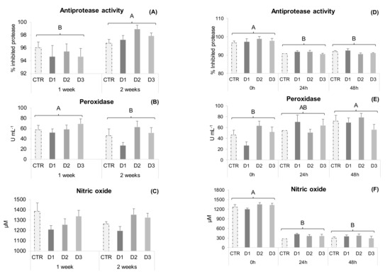
In experiment one, plasma humoral parameters (NO production, antiprotease and peroxidase activities) remained unaffected by the different dietary treatments at both sampling points (Figure 1A–C). However, antiprotease activity increased from 1 to 2 weeks of feeding, while peroxidase followed an opposite trend.

Figure 1.
Plasma immune parameters of gilthead seabream juveniles. Experiment one: (A) Antiprotease activity; (B) Peroxidase activity; (C) Nitric oxide. Data are the mean ± SEM (n = 12). Experiment two: (D) Antiprotease activity; (E) Peroxidase activity; (F) Nitric oxide. Data are the mean ± SEM (n = 9). Different capital letters represent significant differences in time regardless diet (p < 0.05).
2.3. Gut Innate Immune and Oxidative Stress Biomarkers
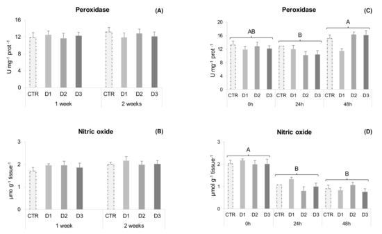
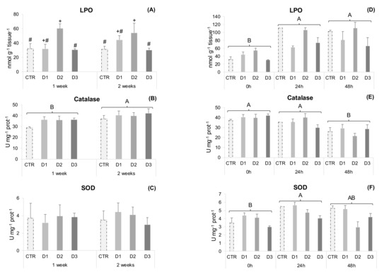
Peroxidase, NO production and SOD activity remained unchanged during the health status experiment in gut samples (Figure 2A–C). Nonetheless, D2 fed fish showed increased gut lipid peroxidation compared to D3 and CTR (Figure 3A), and catalase activity increased from 1 to 2 weeks of feeding.

Figure 2.
Gut immune parameters of gilthead seabream juveniles. Experiment one: (A) Peroxidase activity; (B) Nitric oxide (NO). Data are the mean ± SEM (n = 12). Experiment two: (C) Peroxidase activity; (D) Nitric oxide (NO). Data are the mean ± SEM (n = 9) Different capital letters represent significant differences in time regardless of diet (p < 0.05).

Figure 3.
Gut oxidative stress parameters of gilthead seabream juveniles. Experiment one: (A) Lipid peroxidation (LPO); (B) Catalase activity; (C) Superoxide dismutase activity (SOD). Data are the mean ± SEM (n = 12). Experiment two: (D) Lipid peroxidation (LPO); (E) Catalase activity; (F) Superoxide dismutase activity (SOD). Data are the mean ± SEM (n = 9). Different symbols represent significant differences between diets regardless of time (p < 0.05). Different capital letters represent significant differences in time regardless of diet (p < 0.05).
In experiment two, all measured parameters changed over time. Peroxidase activity increased from 24 to 48 h and NO production decreased after 24 and 48 h (Figure 2C,D). Antioxidant defences, such as catalase activity decreased 48 h after inoculation (Figure 3E), while lipid peroxidation increased at 24 and 48 h (Figure 3D). Superoxide dismutase activity increased at 24 h post-stimulus and D1 fed fish had higher activity than D3, irrespective of the sampling point (Figure 3F).
2.4. Gut Gene Expression Analysis
To evaluate the expression of gut health, immunity and oxidative stress related genes (Table 5 and Table 6), total RNA was isolated from fish anterior intestine. In experiment one, target genes transcriptomic analysis was not able to ascertain differences attributable to the dietary treatments, which could be related to the high intraspecific variability for some target genes (Table 5). However, cd8α, hsp70 and muc2 genes expression increased from 1 to 2 weeks.

Table 5.
Relative gene expression profiling of anterior intestine in gilthead seabream juveniles after 1 and 2 weeks of feeding (experiment one). Data are the mean ± SEM (n = 12). All data values for each gene were in reference to the expression level of CTR.

Table 6.
Relative gene expression profiling of anterior intestine in gilthead seabream juveniles following an inflammatory insult after 2 weeks of feeding (experiment two). Data are the mean ± SEM (n = 9). All data values for each gene were in reference to the expression level of 0 h CTR fish.
Following the inflammatory insult, changes attributed to dietary treatments were also not found in the majority of analysed genes, except for hsp70, which was down-regulated at 24 h in D2 fed fish (Table 6). Furthermore, tlr1 gene expression was up-regulated and gpx was down-regulated at 24 h in all dietary treatments.
3. Discussion
A main feature of C. vulgaris is its protein content and its balanced AA profile, making it a potential source of bioactive peptides. However, the presence of rigid cell walls limits the fish’s ability to access and to utilise the different nutrients inside microalgae cells. In the present study, cell wall disruption was obtained through a combination of chemical and enzymatic processes and the protein fraction was hydrolysed using a serine protease. Protein hydrolysates seem more effective than either intact protein or free AA in different applications for nutrition [25,38]. The current study was devised using two different approaches. First, there was a 2-week feeding trial to evaluate the health status of the fish, aiming to develop future prophylactic strategies (experiment one). After 2 weeks of feeding, fish were subjected to an inflammatory insult to evaluate the inflammatory response (experiment two) and to better discriminate any immunomodulatory effect from the different dietary treatments.
The overall haematological profile from the health status experiment showed some changes, mainly exerted by C. vulgaris biomass and peptide-enriched extract supplemented diets (D1 and D2 diets). Fish fed diet D1 showed lower lymphocyte numbers (Table 2). Accordingly, in a previous experiment with poultry, where different preparations of C. vulgaris were used, animals fed a supplemented diet with 1% chlorella powder showed decreased lymphocyte numbers [39]. Nonetheless, fish fed D2 diet not only had comparable lymphocyte numbers to CTR, but also showed a higher neutrophil concentration (Table 2). These higher circulating myeloid cell numbers in the D2 group might be of relevance during early responses to infection. Bøgwald et al. [40] have shown that medium-size peptides (500–3000 Da) from cod muscle protein hydrolysate, stimulated in vivo respiratory burst activity in Atlantic salmon (Salmo salar) head-kidney leucocytes. In the present study, the peptide-enriched extract protein/peptide profile (Figure S1) is mainly composed of small to medium size particles (<1200 Da) [41]. Size and molecular weight (MW) seem to be particularly important for peptide immunomodulatory activities, with small- to medium-sized particles showing the highest activity [26,28,40,42]. However, an increased leucocyte response in fish fed the D2 diet did not translate into an improved plasma humoral parameters response (NO concentration, antiprotease and peroxidase activities) at 1 or 2 weeks (Figure 1A–C), although those values tended to increase in seabream fed D2 and D3. Accordingly, former studies conducted on Coho salmon (Oncorhynchus kisutch) and turbot (Scophthalmus maximus) did not show any significant impacts on several innate immune defence mechanisms, when fish were fed MPH supplemented diets [43,44]. Nonetheless, beneficial effects have been reported in different fish species [26]. Khosravi et al. [33] supplemented red seabream (Pagrus major) and olive flounder (Paralichthys olivaceus) feeds with 2% krill and tilapia protein hydrolysates and supplementation improved lysozyme activity and respiratory burst in both species. Protein hydrolysates were mainly composed of small- (<500 Da) to medium-sized peptides (500–5000 Da). Furthermore, diet D2 shows a higher Hb concentration than D1 and D3 fed fish. The extraction method employed in a C. vulgaris biomass to obtain the soluble extract (diet D2) might increase iron availability, since most of the intracellular iron is associated with soluble proteins and iron is an essential element for Hb production [45].
In the present study, when fish were subjected to an inflammatory insult (experiment two), an immune response after the stimulus was observed through the time-dependent response pattern of peripheral leucocytes, plasma and gut immune parameters. Peripheral cell dynamics were significantly changed at 24 h post-stimulus, translating into a sharp increase in circulating neutrophils and a significant decrease in lymphocytes (Table 4), indicating that cells were differentiating and being recruited to the site of inflammation. Also, Hb concentration increased (Table 3) in line with a higher metabolic expenditure due to the inflammatory response, and peroxidase activity showed a clear augmentation following inflammation (Figure 1E). Even though circulating neutrophil numbers tended to increase in D1, 2 and 3 dietary treatments at 48 h following inflammation (Table 4), it was not possible to ascertain a clear Chlorella whole-biomass or extracts effect, a fact that could be related to high intraspecific variability in response to the stimulus and that reinforces the need for further studies to unravel the potential of these extracts.
Hydrogen peroxide and oxygen radicals are physiologically generated within cellular compartments and their build-up leads to tissue oxidative stress and damage [46]. Free radical effects are controlled endogenously by antioxidant enzymes and non-enzymatic antioxidants and also by exogenous dietary antioxidants that prevent oxidative damage. Chlorella sp. contain several phytochemicals, namely carotenoids, chlorophyll, flavonoids and polyphenols, which exhibit antioxidant activities [47,48]. Earlier studies showed a significant increase in serum SOD activity in gibel carp fed diets containing 0.8–2.0% dry Chlorella powder [20]. Rahimnejad et al. [14] reported increased plasma CAT activity and total antioxidant capacity (TAC) in olive flounder fed diets with 5% and 10% defatted C. vulgaris meal. As with other microalgae species, the antioxidant potential of C. vulgaris has been mainly assessed on serum and liver, though information is still scarce at the intestinal level. The intestinal epithelium, a highly selective barrier between the animal and the external environment, is constantly exposed to dietary and environmental oxidants. Consequently, it is more prone to oxidative stress and damage, which can impact gut functionality and health [49,50]. The dietary effects of microalgae biomass inclusion have been previously assessed on the intestine of gilthead seabream. Fish were fed diets supplemented with 0.5, 0.75 and 1.5% Nannochloropsis gaditana biomass and no signs of nutritional modulation were found for intestinal SOD and CAT transcription [51]. In the present study, D2 fed fish showed higher gut LPO than CTR and D3 at the end of experiment one (Figure 3A), which could be related to the extraction method employed, since most of the pigments present in the C. vulgaris biomass are not present in the peptide-enriched extract, diminishing the availability of exogenous dietary antioxidants. As pigments are mostly hydrophobic, they are extracted alongside the lipid fraction present in the insoluble extract (Diet D3). Regarding the activities of key enzymes involved in intestinal redox homeostasis (CAT and SOD), these remained unchanged among experimental groups. Castro et al. [17] replaced 100% FM by C. vulgaris biomass in plant protein rich diets for seabass (Dicentrarchus labrax) and found no differences in intestinal LPO, tGSH and GSH levels between dietary treatments. However, they reported lower SOD activity and higher GSSG levels in microalgae-enriched diets, suggesting an increased risk for oxidative stress when fish are subjected to pro-oxidative conditions. Such conditions might arise during an inflammatory insult. However, in experiment two of the present study, lipid peroxidation increased at 24 and 48 h (Figure 3D) post-stimulus but to the same extent for all the dietary treatments. It could be hypothesised that fish fed the D2 diet were able to cope with acute inflammation in a similar manner as the other experimental groups, despite their higher intestinal oxidative state. In other studies, C. vulgaris powdered biomass has been found to counteract the pro-oxidative effects of arsenic induced toxicity in both the gills and the liver of tilapia [16]. Furthermore, Grammes et al. [51] reported that substituting FM by C. vulgaris in aquafeeds containing 20% soybean meal (SBM) is an effective strategy to counteract soybean meal-induced enteropathy (SBMIE) in Atlantic salmon. Likely, this was by maintaining the integrity of the intestinal epithelial barrier and therefore preventing innate immune response activation and ROS generation [52,53].
In the present study, anterior gut transcriptional changes were also evaluated to determine the effect of dietary treatments on the expression patterns of different structural (muc2 and muc13), antioxidant (hsp70; gpx and sod(mn)) and immune related genes (il1β; il34; tlr1; cd8α; igm and hepc). The transcriptomic approach employed was not able to ascertain a clear dietary modulation, at least for the great majority of genes under evaluation in both experiments one and two. However, after the inflammatory insult, the hsp70 gene was down regulated in the D2 fed group after 24 h compared to those fed CTRL (Table 6). Heat shock protein 70 (HSP70) maintains cell integrity and function, and it promotes cell survival under stressful conditions [54]. Leduc et al. [28] reported that genes involved in cellular damage response and repair were also under-expressed in seabass fed a mix of tilapia (TH) and shrimp (SH) protein hydrolysates (5% dry matter diet), mainly composed of low molecular weight peptides. In the same study, fish that were fed the SH alone showed up-regulation of intestinal immune-related genes. Although composed of small-sized peptides, TH did not show the same pattern of stimulation, following what was observed in the current work. According to the authors, the immune-stimulatory effect of the SH was due to low molecular weight peptides, but also to its origin and its degree of hydrolysis [28]. Bioactive peptides are inactive when they are part of the native protein sequence; and, after hydrolysis, bioactivity can be gained depending on specific AA sequences and the size of the newly formed peptides [25]. Nevertheless, in the present study, the observed down-regulation of hsp70 gene expression in the gut of seabream fed D2 suggests a certain degree of anti-stress and/or antioxidant properties from the C. vulgaris peptide-enriched extract, in line with that hypothesized above.
In summary, the C. vulgaris peptide-enriched extract tested in the present study seems to confer a dual modulatory effect at both peripheral (blood) and local (gut) levels. In particular, it drives the proliferation of circulating neutrophils in resting seabream, which could be of assistance to fight against opportunistic pathogens. Following an inflammatory insult, this peptide-enriched extract may protect the gut against stress, and it should be considered for further studies.
4. Materials and Methods
4.1. C. vulgaris Hydrolysates Production
C. vulgaris was supplied, as powder, by AllMicroalgae—Natural Products, SA (Pataias, Portugal). The C. vulgaris hydrolysates were produced by an acid pre-treatment followed by an enzymatic hydrolysis, using a previously optimised method [41]. Briefly, C. vulgaris (Table 7) was mixed with an acetic acid solution (2% in deionised water) in a ratio of microalgae:water of 1:3 (w/v). The mixture was incubated for 1 h at 50 °C and 125 rpm in an orbital shaker (ThermoFisher Scientific, Waltham, MA, USA, MaxQ™ 6000). Then, deionised water was added until microalgae:water ratio reached 1:10 and the pH was adjusted to 7.5. For the enzymatic hydrolysis, first, 5% cellulase was added and the mixture was incubated for 2 h at 50 °C and 125 rpm. Secondly, 3.9% subtilisin was added and the mixture was incubated for 2 h at 40 °C at 125 rpm. During the enzymatic hydrolysis, pH was constantly verified and adjusted to 7.5, mainly in the subtilisin hydrolysis step. To stop the hydrolysis reaction, the mixture was incubated at 90 °C for 10 min to inactivate the enzymes. The resulting solution was centrifuged at 5000× g for 20 min, and both the water-soluble peptide-enriched supernatant (Table 8) and the pellet were collected and freeze-dried for further analysis.

Table 7.
Microalgae Chlorella vulgaris biomass composition (prior to extraction).

Table 8.
Chlorella vulgaris soluble extract protein concentration and in vitro bioactivities.
4.2. Diet Composition
The trial comprised 4 isoproteic (50% protein in dry matter (DM)), isolipidic (17% fat in DM) and isoenergetic (23 kJ/g) dietary treatments. A fishmeal-based (FM), practical diet was used as a control (CTR), whereas three experimental diets based on CTR were further supplemented with a 2% inclusion of C. vulgaris powdered biomass (Diet D1); 0.1% inclusion of C. vulgaris peptide-enriched extract (Diet D2) and finally 0.1% inclusion of C. vulgaris insoluble residue (Diet D3) (Table 9). Diets were manufactured by SPAROS (Olhão, Portugal). All powder ingredients were initially mixed and ground (<200 micron) in a micropulverizer hammer mill (SH1, Hosokawa-Alpine, Germany). Subsequently, the oils were added to the powder mixtures, which were humidified with 25% water and agglomerated by a low-shear and a low-temperature extrusion process (ITALPLAST, Parma, Italy). The resulting pellets of 2.0 mm were dried in a convection oven for 4 h at 55 °C (OP 750-UF, LTE Scientifics, Oldham, UK). Diets were packed in sealed plastic buckets and shipped to the research site (CIIMAR, Matosinhos, Portugal), where they were stored in a temperature-controlled room.

Table 9.
Ingredients and proximate composition of experimental diets.
4.3. Bacterial Growth and Inoculum Preparation
Photobacterium damselae subsp. piscicida (Phdp), strain PP3, was used for the inflammatory insult. Bacteria were routinely cultured at 22 °C in tryptic soy broth (TSB) or tryptic soy agar (TSA) (both from BD Difco™, Franklin Lakes, NJ, USA) supplemented with NaCl to a final concentration of 1% (w/v) (TSB-1 and TSA-1, respectively) and stored at −80 °C in TSB-1 supplemented with 15% (v/v) glycerol. To prepare the inoculum for injection into the fish peritoneal cavities, stocked bacteria were cultured for 48 h at 22 °C on TSA-1. Afterwards, exponentially growing bacteria were collected and resuspended in sterile HBSS and adjusted against its growth curve to 1 × 10⁷ colony forming units (cfu) mL−1. Plating serial dilutions of the suspensions onto TSA-1 plates and counting the number of cfu following incubation at 22 °C confirmed bacterial concentration of the inocula. Bacteria were then killed by heat at 70 °C for 10 min. Loss of bacterial viability following heat exposure was confirmed by plating resulting cultures on TSA-1 plates and failing to see any bacterial growth.
4.4. Fish Rearing Conditions and Feeding Scheme
The experiment was carried out in compliance with the Guidelines of the European Union Council (Directive 2010/63/EU) and Portuguese legislation for the use of laboratory animals at CIIMAR aquaculture and animal experimentation facilities in Matosinhos, Portugal. The protocol was approved by the CIIMAR Animal Welfare Committee in 29/04/2020 with the reference 0421/000/000/2020 from Direção Geral de Alimentação e Veterinária (DGAV). Seawater flow was kept at 4 L min−1 (mean temperature 22.4 ± 1 °C; mean salinity 35.2 ± 0.7 ‰) in a recirculation system with aeration (mean dissolved oxygen above 6 mg L−1). Water quality parameters were monitored daily and adjusted when necessary. Mortality was monitored daily. Diets were randomly assigned to triplicate groups of 97 fish/tank (IBW: 33.4 ± 4.1 g) that were fed to satiation three times a day for 2 weeks starting at a 1.5% biomass.
4.5. Experimental Procedures
To examine the influence that C. vulgaris biomass and protein-rich extract supplementation may have on the health status (trial 1) and the inflammatory response against bacteria (inactivated Phdp i.p. injection; trial 2), samples of blood and gut were collected at 1 and 2 weeks (Trial 1) and after 2 weeks of feeding at 0 h, 24 h and 48 h post-injection (Trial 2).
4.5.1. Health Status (Experiment One)
After 1 and 2 weeks, 12 fish/treatment were weighed and sampled for tissues (blood, head-kidney, liver and gut), after being sacrificed with a 2-phenoxyethanol lethal dose (0.5 mL L−1) [55]. Blood was collected from the caudal vein using heparinised syringes and centrifuged at 10,000× g for 10 min at 4 °C to obtain plasma samples. Plasma and tissue samples were immediately frozen in liquid nitrogen and stored at −80 °C until further analysis.
4.5.2. Inflammatory Response (Experiment Two)
At 2 weeks, 24 fish/treatment were subjected to an inflammatory insult by intraperitoneal (i.p.) injection of heat-inactivated Phdp (see Section 2.2) and immediately transferred to a similar recirculation system in triplicates. After 24 and 48 h post-injection (time-course), 9 fish/treatment were sampled as described above.
4.6. Haematological Procedures
The haematological profile consisted of total white (WBC) and red (RBC) blood cells counts. To determine WBC and RBC concentration, whole blood was diluted 1/20 and 1/200, respectively, in HBSS with heparin (30 U mL−1) and cell counts were done in a Neubauer chamber. Blood smears were prepared from peripheral blood, air-dried and stained with Wright’s stain (Haemacolor; Merck, Darmstadt, Germany), after fixation for 1 min with formol–ethanol (10% formaldehyde in ethanol). Neutrophils were labelled by detecting peroxidase activity revealed by Antonow’s technique described in Afonso et al. [56]. The slides were examined under oil immersion (1000×), and at least 200 leucocytes were counted and classified as thrombocytes, lymphocytes, monocytes and neutrophils. The relative percentage and absolute value (×104 mL−1) of each cell type was calculated.
4.7. Innate Humoral Parameters
4.7.1. Antiprotease Activity
The antiprotease activity was determined as described by Ellis et al. [57], with some modifications. Briefly, 10 µL of plasma were incubated with the same volume of trypsin solution (5 mg mL−1 in NaHCO3, 5 mg mL−1, pH 8.3) for 10 min at 22 °C. After incubation, 100 µL of phosphate buffer (NaH2PO4, 13.9 mg mL−1, pH 7.0) and 125 μL of azocasein solution (20 mg mL−1 in NaHCO3, 5 mg mL−1, pH 8.3) were added and incubated for 1 h at 22 °C. Finally, 250 μL of trichloroacetic acid were added to the reaction mixture and incubated for 30 min at 22 °C. The mixture was centrifuged at 10,000× g for 5 min at room temperature. Afterwards, 100 μL of the supernatant was transferred to a 96 well-plate and mixed with 100 μL of NaOH (40 mg mL−1). The OD was read at 450 nm in a Synergy HT microplate reader (Biotek, Winooski, VT, USA). Phosphate buffer instead of plasma and trypsin served as blank, whereas the reference sample was phosphate buffer instead of plasma. The sample inhibition percentage of trypsin activity was calculated as follows: 100 – ((sample absorbance/Reference absorbance) × 100). All analyses were conducted in duplicates.
4.7.2. Peroxidase Activity
Total peroxidase activity in plasma and intestine was measured, following the procedure described by Quade and Roth [58]. Briefly, 10 μL of plasma and 5 μL of intestine homogenate were diluted with 140 and 145 μL, respectively, of HBSS without Ca2+ and Mg2+ in 96-well plates. Then, 50 μL of 20 mM 3,3′,5,5′-tetramethylbenzidine hydrochloride (TMB; Sigma-Aldrich®, Merck, Darmstadt, Germany) and 50 μL of 5 mM H2O2 were added to the wells. The reaction was stopped after 2 min by adding 50 μL of H2SO4 (2 M) and the optical density (OD) was read at 450 nm in a Synergy HT microplate reader (Biotek, Winooski, VT, USA). Wells without plasma or mucus were used as blanks. The peroxidase activity (U mL−1 tissue) was determined, defining that one unit of peroxidase produces an absorbance change of 1 OD.
4.7.3. Nitric Oxide (NO) Production
NO production was measured in plasma (1:10 sample dilution) and intestine (1:5 sample dilution) samples. Total nitrite and nitrate concentrations in the sample were assessed using the Nitrite/Nitrate colorimetric method kit (Roche, Basel, Switzerland) adapted to microplates. Nitrite concentration was calculated by comparison with a sodium nitrite standard curve. Since nitrite and nitrate are endogenously produced as oxidative metabolites of the NO molecule, these compounds are considered as indicative of NO production.
4.8. Analysis of Oxidative Stress Biomarkers
Intestine samples were homogenised (1:10) in phosphate buffer 0.1 M (pH 7.4), using Precellys evolution tissue lyser homogenizer (Bertin Instruments, Montigny-le-Bretonneux, France).
4.8.1. Lipid Peroxidation (LPO)
One aliquot of tissue homogenate was used to determine the extent of endogenous LPO by measuring thiobarbituric acid-reactive species (TBARS) as suggested by Bird and Draper [59]. To prevent artifactual lipid peroxidation, butylhydroxytoluene (BHT 0.2 mM) was added to the aliquot. Briefly, 1 mL of 100% trichloroacetic acid and 1 mL of 0.73% thiobarbituric acid solution (in Tris–HCl 60 mM pH 7.4 with DTPA 0.1 mM) were added to 0.2 mL of intestine homogenate. After incubation at 100 °C for 60 min, the solution was centrifuged at 12,000× g for 5 min and LPO levels were determined at 535 nm.
4.8.2. Total Protein Quantification
The remaining tissue homogenate was centrifuged for 20 min at 12,000 rpm (4 °C) to obtain the post-mitochondrial supernatant fraction (PMS). Total proteins in homogenates were measured by using Pierce™ BCA Protein Assay Kit, as described by the manufacturer (ThermoFisher Scientific, Waltham, MA, USA).
4.8.3. Catalase (CAT)
CAT activity was determined in PMS by measuring substrate (H2O2) consumption at 240 nm according to Claiborne [60] adapted to microplate. Briefly, in a microplate well, 0.140 mL of phosphate buffer (0.05 M pH 7.0) and a 0.150 mL H2O2 solution (30 mM in phosphate buffer 0.05 M pH 7.0) were added to 0.01 mL of intestine PMS (0.7 mg mL−1 total protein). Enzymatic activity was determined in a microplate reader (BioTek Synergy HT, Winooski, VT, USA), reading the optical density at 240 nm for 2 min every 15 s interval.
4.8.4. Superoxide Dismutase (SOD)
SOD activity was measured according to Flohé and Otting [61], adapted to microplate by Lima, et al. [62]. Briefly, in a microplate well, 0.2 mL of the reaction solution [1 part xanthine solution 0.7 mM (in NaOH 1 mM) and 10 parts cytochrome c solution 0.03 mM (in phosphate buffer 50 mM pH 7.8 with 1 mM Na-EDTA)] was added to 0.05 mL of intestine PMS (0.25 mg mL−1 total protein). Optical density was measured at 550 nm in a microplate reader (BioTek Synergy HT, Winooski, VT, USA) every 20 sec interval for 3 min at 25 °C.
4.9. Gene Expression
RNA isolation from target tissue (anterior gut) and cDNA synthesis was conducted with NZY Total RNA Isolation kit and NZY first-strand cDNA synthesis kit (NZYTech, Lisbon, Portugal), following manufacturer’s specifications. Real-time quantitative PCR was carried out on a CFX384 Touch Real-Time PCR system (Bio-Rad Laboratories, Hercules, CA, USA). Genes comprised in the assay were selected for their involvement in gut integrity, health and immunity (Table 10). Specific primer pair sequences are listed in Table S1. Controls of general PCR performance were included on each array. Briefly, RT reactions were diluted to obtain the equivalent concentration of 20 ng of total input RNA which were used in a 10 μL volume for each PCR reaction. PCR wells contained a 2× SYBR Green Master Mix (Bio-Rad Laboratories, Hercules, CA, USA) and specific primers were used to obtain amplicons 50–250 bp in length. The program used for PCR amplification included an initial denaturation step at 95 °C for 10 min, followed by 40 cycles of 95 °C denaturation for 15 s, with primer annealing and extension temperature (Table S1) for 1 min. The efficiency of PCR reactions was always higher than 90%, and negative controls without sample templates were routinely performed for each primer set. The specificity of reactions was verified by analysis of melting curves (ramping rates of 0.5 °C/10 s over a temperature range of 55–95 °C). Fluorescence data acquired during the PCR extension phase were normalised using the Pfaffl [63] method. The geometric mean of two carefully selected housekeeping genes (elongation factor 1-α (ef1α) and ribosomal protein S18 (rps18)) was used as the normalisation factor to normalise the expression of target genes. For comparing the mRNA expression level of each gene in a given dietary treatment, all data values were in reference to the expression level of CTR fish.

Table 10.
PCR-array layout for gene expression profiling of anterior gut in sea bream.
4.10. Data Analysis
All results are expressed as mean ± standard error (mean ± SE). Residuals were tested for normality (Shapiro–Wilk’s test) and homogeneity of variance (Levene’s test). When residuals did not meet the assumptions, data was transformed by a Log10 or square root transformation. For gene expression data, a log2 transformation was applied to all expression values. Two-way ANOVAs were performed in data arising from both trials one and two, with “dietary treatment and time” as the fixed effects. Analysis of variance was followed by Tukey post-hoc tests. All statistical analyses were performed using the computer package SPSS 26 for WINDOWS. The level of significance used was p ≤ 0.05 for all statistical tests.
Supplementary Materials
The following supporting information can be downloaded at: https://www.mdpi.com/article/10.3390/md20070407/s1, Table S1. Relative gene expression profiling of anterior intestine in gilthead seabream juveniles fed experimental diets.; Figure S1. Protein/peptide profile of C. vulgaris hydrolysate, showing the main molecular weight ranges, the area of the main peak, and the localization of all the 42 identified peaks.
Author Contributions
Conceptualization, B.R., J.D., L.C., E.M. and B.C.; data curation, B.R.; formal analysis, B.R. and L.R.-P.; funding acquisition, J.D., L.C. and B.C.; investigation, B.R., L.R.-P. and S.A.C.; project administration, J.D. and B.C.; resources, M.P., J.L.d.S., J.D., E.M. and B.C.; supervision, J.D., L.C. and B.C.; writing—original draft, B.R.; writing—review and editing, L.R.-P., S.A.C., M.P., J.L.d.S., J.D., L.C., E.M. and B.C. All authors have read and agreed to the published version of the manuscript.
Funding
This work was funded by Compete 2020, Lisboa 2020, Algarve 2020, Portugal 2020, and the European Union through FEDER in the framework of VALORMAR project (POCI-01-0247-FEDER-024517) and by national funds through the Foundation for Science and Technology (FCT) within the scope of UIDB/50016/2020, UIDB/04423/2020, and UIDP/04423/2020. The views expressed in this work are the sole responsibility of the authors. B. Reis was supported by FCT, Soja de Portugal, SA, and Sparos Lda., through the grant PD/BDE/129262/2017. S.A. Cunha and B. Costas were supported by FCT, through grants SFRH/BD/144155/2019 and 2020.00290.CEECIND, respectively.
Institutional Review Board Statement
The experiment was carried out in compliance with the Guidelines of the European Union Council (Directive 2010/63/EU) and Portuguese legislation for the use of laboratory animals. CIIMAR facilities and their staff are certified to house and to conduct experiments with live animals (Group-C licences by the Direção Geral de Alimentação e Veterinária (DGAV), Ministério da Agricultura, Florestas e Desenvolvimento Rural, Portugal). The protocol was approved by the CIIMAR Animal Welfare Committee in 29/04/2020 with the reference 0421/000/000/2020 from DGAV.
Informed Consent Statement
Not applicable.
Data Availability Statement
Not applicable.
Conflicts of Interest
The authors declare that they have no known competing financial interests or personal relationships that could have appeared to influence the work reported in this paper.
References
- Meena, D.K.; Das, P.; Kumar, S.; Mandal, S.C.; Prusty, A.K.; Singh, S.K.; Akhtar, M.S.; Behera, B.K.; Kumar, K.; Pal, A.K.; et al. Beta-glucan: An ideal immunostimulant in aquaculture (a review). Fish Physiol. Biochem. 2013, 39, 431–457. [Google Scholar] [CrossRef] [PubMed]
- World Health Organization; Food and Agriculture Organization of the United Nations; International Office of Epizootics. Report of a Joint FAO/OIE/WHO Expert Consultation on Antimicrobial Use in Aquaculture and Antimicrobial Resistance, Seoul, Republic of Korea, 13–16 June 2006; World Health Organization: Geneva, Switzerland, 2006. [Google Scholar]
- Credence Research. Algae Products Market by Type (Spirulina, Chlorella, Astaxanthin, Beta Carotene, Hydrocolloids), By Source (Brown, Blue—Green, Green, Red, Others), By Application (Nutraceuticals, Food & Feed Supplements, Pharmaceuticals, Paints & Colorants, Chemicals, Fuels, Others)—Growth, Share, Opportunities & Competitive Analysis, 2019–2027. Available online: https://www.credenceresearch.com/report/algae-products-market (accessed on 20 February 2021).
- Ahmad, M.T.; Shariff, M.; Yusoff, F.M.; Goh, Y.M.; Banerjee, S. Applications of microalga Chlorella vulgaris in aquaculture. Rev. Aquac. 2020, 12, 328–346. [Google Scholar] [CrossRef]
- Ru, I.T.K.; Sung, Y.Y.; Jusoh, M.; Wahid, M.E.A.; Nagappan, T. Chlorella vulgaris: A perspective on its potential for combining high biomass with high value bioproducts. Appl. Phycol. 2020, 1, 2–11. [Google Scholar] [CrossRef] [Green Version]
- Cunha, S.A.; Pintado, M.E. Bioactive peptides derived from marine sources: Biological and functional properties. Trends Food Sci. Technol. 2022, 119, 348–370. [Google Scholar] [CrossRef]
- Tomaselli, L. The Microalgal Cell. In Handbook of Microalgal Culture: Biotechnology and Applied Phycology; Richmond, A., Ed.; Blackwell Science Ltd.: Oxford, UK, 2004. [Google Scholar]
- Borowitzka, M.A. Biology of Microalgae. In Microalgae in Health and Disease Prevention; Levine, I., Fleurence, J., Eds.; Academic Press: London, UK, 2018; pp. 23–72. [Google Scholar]
- Plaza, M.; Herrero, M.; Cifuentes, A.; Ibáñez, E. Innovative Natural Functional Ingredients from Microalgae. J. Agric. Food Chem. 2009, 57, 7159–7170. [Google Scholar] [CrossRef]
- Cuellar-Bermudez, S.P.; Aguilar-Hernandez, I.; Cardenas-Chavez, D.L.; Ornelas-Soto, N.; Romero-Ogawa, M.A.; Parra-Saldivar, R. Extraction and purification of high-value metabolites from microalgae: Essential lipids, astaxanthin and phycobiliproteins. Microb. Biotechnol. 2015, 8, 190–209. [Google Scholar] [CrossRef]
- Rahman, M.M.; Khosravi, S.; Chang, K.H.; Lee, S.-M. Effects of Dietary Inclusion of Astaxanthin on Growth, Muscle Pigmentation and Antioxidant Capacity of Juvenile Rainbow Trout (Oncorhynchus mykiss). Prev. Nutr. Food Sci. 2016, 21, 281–288. [Google Scholar] [CrossRef] [Green Version]
- Chew, B.P.; Park, J.S. Carotenoid action on the immune response. J. Nutr. 2004, 134, 257s–261s. [Google Scholar] [CrossRef]
- Bai, S.; Koo, J.-W.; Kim, K.; Kim, S.-G. Effects of Chlorella powder as a feed additive on growth performance in juvenile Korean rockfish, Sebastes schlegeli (Hilgendorf). Aquac. Res. 2001, 32, 92–98. [Google Scholar] [CrossRef]
- Rahimnejad, S.; Lee, S.-M.; Park, H.-G.; Choi, J. Effects of Dietary Inclusion of Chlorella vulgaris on Growth, Blood Biochemical Parameters, and Antioxidant Enzyme Activity in Olive Flounder, Paralichthys olivaceus. J. World Aquac. Soc. 2017, 48, 103–112. [Google Scholar] [CrossRef]
- Pakravan, S.; Akbarzadeh, A.; Sajjadi, M.M.; Hajimoradloo, A.; Noori, F. Chlorella vulgaris meal improved growth performance, digestive enzyme activities, fatty acid composition and tolerance of hypoxia and ammonia stress in juvenile Pacific white shrimp Litopenaeus vannamei. Aquac. Nutr. 2018, 24, 594–604. [Google Scholar] [CrossRef]
- Zahran, E.; Risha, E. Modulatory role of dietary Chlorella vulgaris powder against arsenic-induced immunotoxicity and oxidative stress in Nile tilapia (Oreochromis niloticus). Fish Shellfish Immunol. 2014, 41, 654–662. [Google Scholar] [CrossRef]
- Castro, C.; Coutinho, F.; Iglesias, P.; Oliva-Teles, A.; Couto, A. Chlorella sp. and Nannochloropsis sp. Inclusion in Plant-Based Diets Modulate the Intestine and Liver Antioxidant Mechanisms of European Sea Bass Juveniles. Front. Vet. Sci. 2020, 7, 607575. [Google Scholar] [CrossRef]
- Zhang, Q.; Qiu, M.; Xu, W.; Gao, Z.; Shao, R.; Qi, Z. Effects of Dietary Administration of Chlorella on the Immune Status of Gibel Carp, Carassius Auratus Gibelio. Ital. J. Anim. Sci. 2014, 13, 3168. [Google Scholar] [CrossRef]
- Lupatsch, I.; Blake, C. Algae alternative: Chlorella studied as protein source in tilapia feeds. Glob. Aquac. Advocate 2013, 16, 78–79. [Google Scholar]
- Xu, W.; Gao, Z.; Qi, Z.; Qiu, M.; Peng, J.; Shao, R. Effect of Dietary Chlorella on the Growth Performance and Physiological Parameters of Gibel carp, Carassius auratus gibelio. Turk. J. Fish. Aquat. Sci. 2014, 14, 53–57. [Google Scholar] [CrossRef]
- Tibbetts, S.M.; Mann, J.; Dumas, A. Apparent digestibility of nutrients, energy, essential amino acids and fatty acids of juvenile Atlantic salmon (Salmo salar L.) diets containing whole-cell or cell-ruptured Chlorella vulgaris meals at five dietary inclusion levels. Aquaculture 2017, 481, 25–39. [Google Scholar] [CrossRef] [Green Version]
- Teuling, E.; Wierenga, P.A.; Agboola, J.O.; Gruppen, H.; Schrama, J.W. Cell wall disruption increases bioavailability of Nannochloropsis gaditana nutrients for juvenile Nile tilapia (Oreochromis niloticus). Aquaculture 2019, 499, 269–282. [Google Scholar] [CrossRef]
- Valente, L.M.P.; Batista, S.; Ribeiro, C.; Pereira, R.; Oliveira, B.; Garrido, I.; Baião, L.F.; Tulli, F.; Messina, M.; Pierre, R.; et al. Physical processing or supplementation of feeds with phytogenic compounds, alginate oligosaccharide or nucleotides as methods to improve the utilization of Gracilaria gracilis by juvenile European seabass (Dicentrarchus labrax). Aquaculture 2021, 530, 735914. [Google Scholar] [CrossRef]
- Wang, X.; Zhang, X. Optimal extraction and hydrolysis of Chlorella pyrenoidosa proteins. Bioresour. Technol. 2012, 126, 307–313. [Google Scholar] [CrossRef]
- Kose, A.; Oncel, S.S. Properties of microalgal enzymatic protein hydrolysates: Biochemical composition, protein distribution and FTIR characteristics. Biotechnol. Rep. 2015, 6, 137–143. [Google Scholar] [CrossRef] [PubMed] [Green Version]
- Siddik, M.A.B.; Howieson, J.; Fotedar, R.; Partridge, G.J. Enzymatic fish protein hydrolysates in finfish aquaculture: A review. Rev. Aquac. 2021, 13, 406–430. [Google Scholar] [CrossRef]
- Kotzamanis, Y.P.; Gisbert, E.; Gatesoupe, F.J.; Zambonino Infante, J.; Cahu, C. Effects of different dietary levels of fish protein hydrolysates on growth, digestive enzymes, gut microbiota, and resistance to Vibrio anguillarum in European sea bass (Dicentrarchus labrax) larvae. Comp. Biochem. Physiol. A Mol. Integr. Physiol. 2007, 147, 205–214. [Google Scholar] [CrossRef] [PubMed] [Green Version]
- Leduc, A.; Zatylny-Gaudin, C.; Robert, M.; Corre, E.; Corguille, G.L.; Castel, H.; Lefevre-Scelles, A.; Fournier, V.; Gisbert, E.; Andree, K.B.; et al. Dietary aquaculture by-product hydrolysates: Impact on the transcriptomic response of the intestinal mucosa of European seabass (Dicentrarchus labrax) fed low fish meal diets. BMC Genomics 2018, 19, 396. [Google Scholar] [CrossRef] [Green Version]
- Siddik, M.A.B.; Howieson, J.; Fotedar, R. Beneficial effects of tuna hydrolysate in poultry by-product meal diets on growth, immune response, intestinal health and disease resistance to Vibrio harveyi in juvenile barramundi, Lates calcarifer. Fish Shellfish Immunol. 2019, 89, 61–70. [Google Scholar] [CrossRef]
- Zheng, K.; Xu, T.; Qian, C.; Liang, M.; Wang, X. Effect of low molecular weight fish protein hydrolysate on growth performance and IGF-I expression in Japanese flounder (Paralichthys olivaceus) fed high plant protein diets. Aquac. Nutr. 2014, 20, 372–380. [Google Scholar] [CrossRef]
- Ospina-Salazar, G.H.; Ríos-Durán, M.G.; Toledo-Cuevas, E.M.; Martínez-Palacios, C.A. The effects of fish hydrolysate and soy protein isolate on the growth performance, body composition and digestibility of juvenile pike silverside, Chirostoma estor. Anim. Feed Sci. Technol. 2016, 220, 168–179. [Google Scholar] [CrossRef]
- Xu, H.; Mu, Y.; Zhang, Y.; Li, J.; Liang, M.; Zheng, K.; Wei, Y. Graded levels of fish protein hydrolysate in high plant diets for turbot (Scophthalmus maximus): Effects on growth performance and lipid accumulation. Aquaculture 2016, 454, 140–147. [Google Scholar] [CrossRef]
- Khosravi, S.; Bui, H.T.D.; Rahimnejad, S.; Herault, M.; Fournier, V.; Kim, S.-S.; Jeong, J.-B.; Lee, K.-J. Dietary supplementation of marine protein hydrolysates in fish-meal based diets for red sea bream (Pagrus major) and olive flounder (Paralichthys olivaceus). Aquaculture 2015, 435, 371–376. [Google Scholar] [CrossRef]
- Bui, H.T.D.; Khosravi, S.; Fournier, V.; Herault, M.; Lee, K.-J. Growth performance, feed utilization, innate immunity, digestibility and disease resistance of juvenile red seabream (Pagrus major) fed diets supplemented with protein hydrolysates. Aquaculture 2014, 418-419, 11–16. [Google Scholar] [CrossRef]
- Chaklader, M.R.; Fotedar, R.; Howieson, J.; Siddik, M.A.B.; Foysal, M.J. The ameliorative effects of various fish protein hydrolysates in poultry by-product meal based diets on muscle quality, serum biochemistry and immunity in juvenile barramundi, Lates calcarifer. Fish Shellfish Immunol. 2020, 104, 567–578. [Google Scholar] [CrossRef]
- Sedighi, M.; Jalili, H.; Ranaei-Siadat, S.-O.; Amrane, A. Potential Health Effects of Enzymatic Protein Hydrolysates from Chlorella vulgaris. Appl. Food Biotechnol. 2016, 3, 160–169. [Google Scholar] [CrossRef]
- Sheih, I.C.; Wu, T.-K.; Fang, T.J. Antioxidant properties of a new antioxidative peptide from algae protein waste hydrolysate in different oxidation systems. Bioresour. Technol. 2009, 100, 3419–3425. [Google Scholar] [CrossRef]
- Clemente, A.; Vioque, J.; Sánchez-Vioque, R.; Pedroche, J.; Bautista, J.; Millán, F. Protein quality of chickpea (Cicer arietinum L.) protein hydrolysates. Food Chem. 1999, 67, 269–274. [Google Scholar] [CrossRef]
- Kang, H.K.; Salim, H.M.; Akter, N.; Kim, D.W.; Kim, J.H.; Bang, H.T.; Kim, M.J.; Na, J.C.; Hwangbo, J.; Choi, H.C.; et al. Effect of various forms of dietary Chlorella supplementation on growth performance, immune characteristics, and intestinal microflora population of broiler chickens. J. Appl. Poult. Res. 2013, 22, 100–108. [Google Scholar] [CrossRef]
- Bøgwald, J.; Dalmo, R.O.Y.; McQueen Leifson, R.; Stenberg, E.; Gildberg, A. The stimulatory effect of a muscle protein hydrolysate from Atlantic cod, Gadus morhua L., on Atlantic salmon, Salmo salar L.; head kidney leucocytes. Fish Shellfish Immunol. 1996, 6, 3–16. [Google Scholar] [CrossRef]
- Cunha, S.A.; Coscueta, E.R.; Nova, P.; Silva, J.L.; Pintado, M.M. Bioactive Hydrolysates from Chlorella vulgaris: Optimal Process and Bioactive Properties. Molecules 2022, 27, 2505. [Google Scholar] [CrossRef]
- Gildberg, A.; Bøgwald, J.; Johansen, A.; Stenberg, E. Isolation of acid peptide fractions from a fish protein hydrolysate with strong stimulatory effect on atlantic salmon (Salmo salar) head kidney leucocytes. Comp. Biochem. Physiol. B Biochem. Mol. Biol. 1996, 114, 97–101. [Google Scholar] [CrossRef]
- Murray, A.L.; Pascho, R.J.; Alcorn, S.W.; Fairgrieve, W.T.; Shearer, K.D.; Roley, D. Effects of various feed supplements containing fish protein hydrolysate or fish processing by-products on the innate immune functions of juvenile coho salmon (Oncorhynchus kisutch). Aquaculture 2003, 220, 643–653. [Google Scholar] [CrossRef]
- Zheng, K.; Liang, M.; Yao, H.; Wang, J.; Chang, Q. Effect of size-fractionated fish protein hydrolysate on growth and feed utilization of turbot (Scophthalmus maximus L.). Aquac. Res. 2013, 44, 895–902. [Google Scholar] [CrossRef]
- Botebol, H.; Lesuisse, E.; Šuták, R.; Six, C.; Lozano, J.-C.; Schatt, P.; Vergé, V.; Kirilovsky, A.; Morrissey, J.; Léger, T.; et al. Central role for ferritin in the day/night regulation of iron homeostasis in marine phytoplankton. Proc. Natl. Acad. Sci. USA 2015, 112, 14652. [Google Scholar] [CrossRef] [PubMed] [Green Version]
- Dirks, R.C.; Faiman, M.D.; Huyser, E.S. The role of lipid, free radical initiator, and oxygen on the kinetics of lipid peroxidation. Toxicol. Appl. Pharmacol. 1982, 63, 21–28. [Google Scholar] [CrossRef]
- Shibata, S.; Natori, Y.; Nishihara, T.; Tomisaka, K.; Matsumoto, K.; Sansawa, H.; Nguyen, V.C. Antioxidant and anti-cataract effects of Chlorella on rats with streptozotocin-induced diabetes. J. Nutr. Sci. Vitaminol. 2003, 49, 334–339. [Google Scholar] [CrossRef] [PubMed]
- Wang, H.-M.; Pan, J.-L.; Chen, C.-Y.; Chiu, C.-C.; Yang, M.-H.; Chang, H.-W.; Chang, J.-S. Identification of anti-lung cancer extract from Chlorella vulgaris C-C by antioxidant property using supercritical carbon dioxide extraction. Process Biochem. 2010, 45, 1865–1872. [Google Scholar] [CrossRef]
- Circu, M.L.; Aw, T.Y. Intestinal redox biology and oxidative stress. Semin. Cell Dev. Biol. 2012, 23, 729–737. [Google Scholar] [CrossRef] [Green Version]
- Yu, G.; Liu, Y.; Ou, W.; Dai, J.; Ai, Q.; Zhang, W.; Mai, K.; Zhang, Y. The protective role of daidzein in intestinal health of turbot (Scophthalmus maximus L.) fed soybean meal-based diets. Sci. Rep. 2021, 11, 3352. [Google Scholar] [CrossRef]
- Jorge, S.S.; Enes, P.; Serra, C.R.; Castro, C.; Iglesias, P.; Oliva Teles, A.; Couto, A. Short-term supplementation of gilthead seabream (Sparus aurata) diets with Nannochloropsis gaditana modulates intestinal microbiota without affecting intestinal morphology and function. Aquac. Nutr. 2019, 25, 1388–1398. [Google Scholar] [CrossRef]
- Grammes, F.; Reveco, F.E.; Romarheim, O.H.; Landsverk, T.; Mydland, L.T.; Øverland, M. Candida utilis and Chlorella vulgaris Counteract Intestinal Inflammation in Atlantic Salmon (Salmo salar L.). PLoS ONE 2013, 8, e83213. [Google Scholar] [CrossRef] [Green Version]
- Bravo-Tello, K.; Ehrenfeld, N.; Solís, C.J.; Ulloa, P.E.; Hedrera, M.; Pizarro-Guajardo, M.; Paredes-Sabja, D.; Feijóo, C.G. Effect of microalgae on intestinal inflammation triggered by soybean meal and bacterial infection in zebrafish. PLoS ONE 2017, 12, e0187696. [Google Scholar] [CrossRef]
- Silver, J.T.; Noble, E.G. Regulation of survival gene hsp70. Cell Stress Chaperones 2012, 17, 1–9. [Google Scholar] [CrossRef] [Green Version]
- Mylonas, C.C.; Cardinaletti, G.; Sigelaki, I.; Polzonetti-Magni, A. Comparative efficacy of clove oil and 2-phenoxyethanol as anesthetics in the aquaculture of European sea bass (Dicentrarchus labrax) and gilthead sea bream (Sparus aurata) at different temperatures. Aquaculture 2005, 246, 467–481. [Google Scholar] [CrossRef]
- Afonso, A.; Ellis, A.E.; Silva, M.T. The leucocyte population of the unstimulated peritoneal cavity of rainbow trout (Oncorhynchus mykiss). Fish Shellfish Immunol. 1997, 7, 335–348. [Google Scholar] [CrossRef]
- Ellis, A.E.; Cavaco, A.; Petrie, A.; Lockhart, K.; Snow, M.; Collet, B. Histology, immunocytochemistry and qRT-PCR analysis of Atlantic salmon, Salmo salar L.; post-smolts following infection with infectious pancreatic necrosis virus (IPNV). J. Fish Dis. 2010, 33, 803–818. [Google Scholar] [CrossRef]
- Quade, M.J.; Roth, J.A. A rapid, direct assay to measure degranulation of bovine neutrophil primary granules. Vet Immunol. Immunopathol. 1997, 58, 239–248. [Google Scholar] [CrossRef]
- Bird, R.P.; Draper, H.H. Comparative studies on different methods of malonaldehyde determination. Methods Enzymol. 1984, 105, 299–305. [Google Scholar] [CrossRef]
- Claiborne, A. Catalase activity. In CRC Handbook of Methods for Oxygen Radical Research; Greenwald, R.A., Ed.; CRC Press: Boca Raton, FL, USA, 1985; pp. 283–284. [Google Scholar]
- Flohé, L.; Otting, F. Superoxide dismutase assays. Methods Enzymol. 1984, 105, 93–104. [Google Scholar] [CrossRef]
- Lima, I.; Moreira, S.M.; Osten, J.R.-V.; Soares, A.M.V.M.; Guilhermino, L. Biochemical responses of the marine mussel Mytilus galloprovincialis to petrochemical environmental contamination along the North-western coast of Portugal. Chemosphere 2007, 66, 1230–1242. [Google Scholar] [CrossRef]
- Pfaffl, M.W. A new mathematical model for relative quantification in real-time RT-PCR. Nucleic Acids Res. 2001, 29, e45. [Google Scholar] [CrossRef]
Publisher’s Note: MDPI stays neutral with regard to jurisdictional claims in published maps and institutional affiliations. |
© 2022 by the authors. Licensee MDPI, Basel, Switzerland. This article is an open access article distributed under the terms and conditions of the Creative Commons Attribution (CC BY) license (https://creativecommons.org/licenses/by/4.0/).