Abstract
Origanum vulgare L., a medicinal herb rich in bioactive phenols and terpenes, is recognized for its anti-inflammatory and antimicrobial properties. Heavy-ion beam mutagenesis, a sophisticated breeding technique, can induce significant variations in plants, thereby affecting their secondary metabolite production. This study utilized metabolomic and transcriptomic approaches to investigate the effects of 12C6+ heavy-ion irradiation on oregano. Our results indicated substantial changes in mutant lines, including marked alterations in plant height, leaf morphology, and biomass accumulation. Metabolomic analysis indicated that the differentially accumulated volatile compounds were primarily terpenoids. Furthermore, transcriptomic analysis indicated a predominant enrichment of differentially expressed genes in terpenoid biosynthesis. Integrated analyses identified key transcriptional changes in genes encoding terpenoid backbone enzymes, such as GPPS, GGPPS, DXS, and HMGR, and pinpointed candidate genes, including TPS3, TPS6A, TPS6C, CYP71D178, CYP71D181, and CYP71D10B, whose expression patterns were closely associated with the differential accumulation of carvacrol and thymol. This comprehensive study elucidates the molecular mechanisms underlying metabolic reprogramming induced by heavy-ion irradiation in oregano and offers valuable genetic resources for future metabolic engineering and precision breeding initiatives aimed at enhancing the production of valuable bioactive compounds.
1. Introduction
Origanum vulgare L., commonly known as oregano, is a perennial herb of the lamiaceae family with a long history of culinary and medicinal use across diverse cultures [1,2]. Modern pharmacological studies have substantiated its traditional applications, revealing that oregano extracts possess significant antioxidant, antibacterial, anti-inflammatory, antitumor, and insecticidal properties [3,4,5]. These broad-spectrum bioactivities are primarily attributed to its rich profile of secondary metabolites, particularly terpenoids, which are the most prominent bioactive constituents [6]. Numerous terpenoids exhibit substantial pharmacological potential, including anticancer, antimicrobial, anti-inflammatory, and antioxidant effects, as well as protective properties against neural damage [7,8]. The clinical relevance of terpenoids is exemplified by drugs such as artemisinin (a sesquiterpene) for malaria and paclitaxel (a diterpene) for cancers [9,10]. Within oregano, the predominant active terpenoids are monoterpenes and sesquiterpenes. In oregano, the principal active terpenoids are monoterpenes and sesquiterpenes. The phenolic monoterpenes thymol and carvacrol have been identified as the key bioactive and commercially valuable signature compounds, conferring the distinctive pungent aroma and forming the basis of the plant’s potent antimicrobial activity [11,12]. Consequently, the quality and commercial value of oregano are directly determined by the content and ratio of thymol and carvacrol. However, the molecular mechanisms governing their biosynthesis in oregano remain largely elusive. Therefore, elucidating the regulatory pathways of thymol and carvacrol synthesis is imperative to meet the growing demand for high-quality oregano.
In plants, the biosynthesis of terpenoids primarily relies on two key pathways: the mevalonate (MVA) pathway in the cytoplasm and the methylerythritol phosphate (MEP) pathway in plastids [13]. For monoterpenes, the precursors isopentenyl pyrophosphate (IPP) and its isomer dimethylallyl pyrophosphate (DMAPP) are primarily supplied by the MEP pathway. These two five-carbon units are condensed by geranyl pyrophosphate synthase (GPPS) to form geranyl pyrophosphate (GPP), the universal carbon skeleton for monoterpenes. GPP is subsequently cyclized by specific terpene synthases (TPSs) to generate diverse monoterpene skeletons [14]. In Lamiaceae plants, thymol and carvacrol are biosynthesized from γ-terpinene through sequential oxidation by cytochrome P450 monooxygenases (P450s) of the CYP71D subfamily and dehydrogenation by a short-chain dehydrogenase/reductase (SDR), culminating in aromatic phenols via keto-enol tautomerism [15]. The entire biosynthetic pathway is subject to multi-layered regulation. Beyond the expression of structural genes (e.g., GPPS, TPS, and CYP71D), the pathway is finely tuned by various transcription factors (TFs). In other plant species, transcription factor (TF) families, including bZIP, TCP, MYB, and bHLH, have been shown to regulate terpenoid biosynthesis [16,17,18,19]. Given that alterations in the expression or activity of any these regulatory or structural genes can critically influence metabolic flux and end product accumulation, a comprehensive understanding of this regulatory network is essential.
Genetic diversity provides the essential foundation for variety improvement and germplasm innovation, directly influencing a population’s adaptive and evolutionary capacity [20,21]. Among methods to generate such diversity, heavy-ion beam irradiation is a promising physical mutagenesis technique. It boasts unique advantages such as low physiological damage, a high mutation rate, a broad mutation spectrum, and rapid stabilization of mutant traits [22]. At the molecular level, this mutagenesis primarily generates large-fragment deletions, insertions, and chromosomal structural variations [23]. These mutations are particularly impactful, as they often cause complete loss of gene function or critical amino acid changes, effectively disrupting rate-limiting steps and reprogramming the regulatory networks of secondary metabolic pathways to induce significant phenotypic alterations [24,25]. The application of heavy-ion beam irradiation has been demonstrated to be an effective method of inducing functional mutations while concurrently maintaining high survival rates. This combination renders it a potent instrument for germplasm innovation and gene discovery in the domain of plant breeding, including that of medicinal species [26,27,28]. However, the application of this potent technique to oregano breeding has not been documented.
In this study, headspace solid-phase microextraction (HS-SPME) coupled with gas chromatography–tandem mass spectrometry (GC-MS) and transcriptome sequencing (RNA-Seq) were employed to analyze DAVs and DEGs dynamic changes to 12C6+ heavy-ion beam irradiation for Origanum vulgare L. and to elucidate the molecular mechanisms underlying the biosynthesis of terpenoids, including thymol and carvacrol. These findings provide new insights into the molecular basis of heavy-ion mutagenesis and offer practical genetic resources for the targeted improvement of oregano.
2. Materials and Methods
2.1. Plant Materials
Seeds of oregano (Origanum vulgare L.) were chosen as the experimental material. A total of 1500 seeds were utilized for a dose-finding study. These seeds were exposed to a gradient of 12C6+ heavy-ion irradiation doses(0, 30, 60, 90,120 and 140 Gray (Gy)), with the non-irradiated seeds (0 Gy) serving as the control group. The germination rate, seedling emergence rate, and seedling survival rate were calculated for each dose. The semi-lethal radiation dose (LD50) was determined from this data (60 Gy) and was selected as the optimal dose for large-scale mutagenesis. Subsequently, a separate batch of 500 seeds was irradiated in a single session at this predetermined dose to establish the mutant population. 500 mutagenized seeds were cultivated as T0 plants in xian County, where plants were self-pollinated to advance the generations. This process of self-pollination continued to yield homozygous mutants. Subsequently, twenty seeds from each T2 line were selected and planted at the Henan Academy of Agricultural Sciences experimental station in Xinxiang City for cultivation. From this T2 population, homozygous lines exhibiting typical mutant traits were identified, screened, and selected for use in subsequent experiments.
2.2. Assessment of Morphological Traits
The growth status of oregano plants from the three groups was documented in the field through photographic records. Morphological evaluations were performed during the growth to seedling stage. Plant height, measured from apex to base, was determined using a standard ruler. The dimensions of the largest leaf, including maximum length and width, were assessed through image analysis utilizing ImageJ software (version: 1.54g). Additionally, the average fresh weights of the leaves and stems were recorded using an analytical balance.
2.3. Measurement of Soil and Plant Analyzer Development (SPAD)
After reaching the seedling stage, the chlorophyll content of the leaves in the three plant groups was measured using a handheld portable chlorophyll meter (Spad-502 plus, Konica Minolta, Inc., Osaka, Japan). Two measurements were obtained from each of the two largest pairs of opposite leaves per plant. Each sample was evaluated with a minimum of three biological replicates.
2.4. GC-MS/MS Analysis of Volatile Compounds in Oregano Leaves
Extract analysis and metabolic identification were conducted by MetWare (Wuhan, China) according to standard procedures. The total ion currents (TICs) of QC specimens are presented in Figure S2. The variable importance of projection (VIP) score of the application (O) partial least squares model was applied to fit the differentiated metabolites in comparison groups. Metabolites with FC ≥ 1 and VIP ≥ 1 are considered to be differentially accumulated metabolites. Metabolite data analysis was conducted using the MetaboAnalyst (version 6.0). Furthermore, the PCA method was used to calculate the variability in various groups, and functional annotation of DAVs was conducted via the KEGG pathway.
2.5. Transcriptome Sequencing and Analysis
Total RNA was extracted from 0.1 g of oregano leaves using a Plant Total RNA Extraction Kit (Omega Bio-Tek, Norcross, GA, USA). For each group, sequencing libraries were constructed from three independent biological replicates. Subsequently, a total of 4 μg of RNA per sample was used for library preparation, and the cDNA libraries were sequenced on the Illumina NovaSeq X Plus platform (version 1.3.1, San Diego, CA, USA). Raw reads were processed with fastp (v0.20.0) to obtain clean reads by removing adapter sequences, reads with >10% unknown bases (N), poly-A reads, and low-quality reads (where over 50% of the bases had a Phred score ≤ 20). De novo transcriptome assembly was performed using Trinity (v2.8.5) with default parameters. The assembly quality was assessed using the N50 statistic and BUSCO. The assembled unigenes were functionally annotated by aligning them against the NR (https://ftp.ncbi.nih.gov/blast/db, accessed on 12 December 2025), Swiss-Prot (https://www.expasy.org/resources/uniprotkb-swiss-prot, accessed on 12 December 2025), KEGG (https://www.genome.jp/kegg), and COG/KOG (https://www.ncbi.nlm.nih.gov/research/COG, accessed on 12 December 2025) databases using BLASTx (version 2.17.0+) with an E-value cutoff of 1 × 10⁻⁵. Protein domains were annotated using Pfam_Scan (version 1.6) against the Pfam database. For plant samples, transcription factors were identified using hmmscan against PlantTFDB (version 5.0) and BLASTp (version 2.17.0+) against PRGdb (version 4.0), respectively.
Sample relationships were assessed by Principal Component Analysis (PCA) and Pearson correlation heatmaps based on the gene expression matrix. We standardized the raw transcriptomic expression matrix (counts) using DESeq2 (v1.26.0) and perform differential expression analysis. Genes with a false discovery rate (FDR) < 0.05 and an absolute log2FC (fold change) > 1 were considered significantly DEGs. Use RSEM (v1.3.0) to convert raw count data to FPKM values.
Gene Ontology (GO) and Kyoto Encyclopedia of Genes and Genomes (KEGG) pathway enrichment analyses of the differentially expressed genes (DEGs) were performed using the clusterProfiler package (version 4.12). GO terms or KEGG pathways with a corrected p-value (Bonferroni for GO, FDR for KEGG) ≤ 0.05 were considered significantly enriched.
2.6. Real-Time Quantitative PCR (RT-qPCR) Analysis for Gene Expression
Total RNA was extracted from plant tissues utilizing the Plant RNA Kit (Omega Bio-Tek, USA). The cDNA template was synthesized with HiScript III All-in-one RT SuperMix Perfect for qPCR (+gDNA wiper) (Vazyme, Nanjing, China). The expression levels of target genes were assessed using a StepOnePlusTM Detection System (Applied Biosystems, Foster City, CA, USA). EF-1α served as a control for normalizing the expression data. Primers were designed using Primer Premier 5 and are detailed in Supplementary Table S8. Relative mRNA expression levels were calculated according to the 2−ΔΔCt method [29].
2.7. Statistical Analysis
In this study, GraphPad Prism (v10.3.0) was used to analyze the significance of differences between the data and to plot the bar graphs. The means were compared by one-way ANOVA and Duncan’s multiple range test at the 5% significance level.
3. Results
3.1. Generation of Heavy-Ion Irradiated Mutants and Phenotypic Characterization of NZ1 and NZ2 in Origanum vulgare L.
The success of radiation mutagenesis breeding hinges on identifying the appropriate radiation dose, necessitating a thorough evaluation of both plant survival rates and the likelihood of obtaining desirable mutant traits. Currently, the LD50 (median lethal dose) serves as a reference standard, often regarded as the dose that yields optimal mutagenic effects [30]. To explore the effects of heavy-ion irradiation on oregano and to establish a theoretical foundation for heavy-ion beam irradiation mutagenesis breeding, dry oregano seeds were exposed to 12C6+ heavy ions at doses of 0, 30, 60, 90, 120, and 140 Gy. Measurements of germination rates, emergence rates, and survival rates were conducted to determine the optimal radiation dose for mutagenesis. The results are presented in Figure S1. Compared to the wild type (WT), the application of 12C6+ irradiation at doses from 0 to 90 Gy had a negligible effect on the germination rate of oregano. In contrast, doses of 120 and 140 Gy led to a significant decrease in this rate. Additionally, the seedling emergence rate showed a pronounced decline in response to increased irradiation doses. The emergence rate decreased by approximately 5% at 10 Gy compared to the control (CK), while doses of 30, 60, and 100 Gy resulted in a reduction of about 50% in comparison with CK. Treatment with 140 Gy led to an emergence rate of only 5%. The survival rate for CK stood at 82.60%, while rates for the 10, 30, 60, 120, and 140 Gy irradiation treatments were 70.91%, 64.82%, 38.65%, 13.14%, and 0%, respectively. Notably, all seedlings subjected to the 140 Gy treatment perished within the designated observation period. Based on the findings, it can be inferred that the LD50 is 60 Gy.
Seeds irradiated with a 60 Gy dose of 12C6+ for one hour were subsequently selected for screening. After three consecutive years of self-pollination and cultivation, several potential homozygous oregano mutant lines were obtained. The following parameters were measured for the mutants: plant height, maximum leaf length, maximum leaf width, leaf fresh weight, stem fresh weight, and SPAD. Mutants NZ1 and NZ2 were identified as exhibiting distinct mutant characteristics. The results are shown in Figure 1; compared to the mean plant height of the WT at 31.16 cm, the mean plant heights of NZ1 and NZ2 were significantly decreased by 16.57 cm and significantly increased by 4.17 cm, respectively. Furthermore, an increase in the average maximum leaf width of NZ1 and NZ2 was recorded, with measurements of 0.30 cm and 0.76 cm, respectively, compared to the WT at 2.30 cm. This study investigated the effects of heavy-ion irradiation on the fresh weights of the leaves and stems of NZ1 and NZ2, revealing that both samples exhibited similar responses to the treatment. Relative to the mean leaf fresh weight of the control group (2.03 g), NZ1 and NZ2 demonstrated increases of 6.23 g and 0.80 g, respectively. In terms of stem fresh weight, compared to the control group’s mean (1.83 g), NZ1 and NZ2 showed increases of 1.27 g and 0.23 g, respectively. The SPAD for NZ1 was significantly higher than those recorded for both WT and NZ2. The development and characterization of variants NZ1 and NZ2 provide a basis for future comprehensive research.
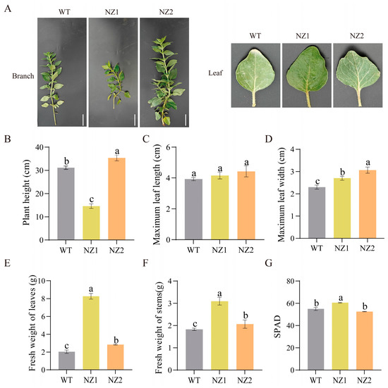
Figure 1.
Effect of 12C6+ heavy iron irradiation on growth characteristics of oregano. (A) Phenotypes of branch and leaves in oregano mutants NZ1 (middle), NZ2 (right), and WT (left); (B–G) plant height, maximum leaf length, maximum leaf width, fresh weight of leaves, fresh weight of stems, and SPAD. Bar = 5 cm. Different letters indicate significant differences between groups at p < 0.05 according to Duncan’s multiple range test.
3.2. Metabolomics Profiling of Volatile Compounds in Heavy-Ion-Induced Oregano Mutants (NZ1, NZ2) and the Wild-Type
The economic value of oregano is primarily determined by its essential oil yield, which consists mainly of phenolic monoterpenes, including carvacrol and thymol, along with their precursors. To analyze the volatile organic compounds in the leaves of the homozygous lines NZ1 and NZ2, generated through heavy-ion mutagenesis, as well as their WT counterpart, headspace solid-phase microextraction coupled with gas chromatography-tandem mass spectrometry (HS-SPME-GC-MS/MS) was employed. Pearson correlation analysis indicated strong correlations among biological replicates within the same line, while revealing weak correlations between different lines) (Figure S3). Principal component analysis (PCA) of the detected volatiles (Figure 2A) showed that the first two principal components accounted for 45.9% and 30.8% of the variance, respectively. Notably, NZ1, NZ2, and WT were distinctly separated, and the three biological replicates of each sample were in close proximity, thereby confirming the reliability of the experimental results and highlighting the significant disparities among the samples.
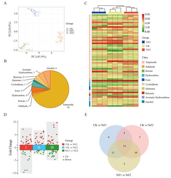
Figure 2.
Metabolite analysis of volatile compounds in heavy-ion-induced oregano mutants (NZ1, NZ2) and the wild-type plant. (A) Principal component analysis (PCA) of all 9 samples. (B) Classification and proportion of VOCs detected in the oregano leaves. (C) Heatmap analysis of all VOCs in three plants. (D) Volcano plots of DAVs in various comparison groups. (E) Venn diagram of DAVs.
A total of 62 volatile organic compounds were identified across the three materials and classified into 10 categories: terpenoids, aldehydes, ketones, hydrocarbons, esters, cycloalkenes, quinones, benzenes, aromatic hydrocarbons, and alcohols (Table S1). The analysis indicated that terpenoids were the most abundant volatile organic compounds, totaling 43 spcies. These were followed by alcohol (4), hydrocarbons (3), esters (3), aromatic hydrocarbons (3), benzenes (2), aldehydes (1), ketone (1), cycloalkene (1), and quinones (1) (Figure 2B). Additionally, a heatmap illustrating the relative content of volatile metabolites (Figure 2C) revealed that the three materials clustered into three distinct branches. The biological replicate samples of each variety were grouped together, highlighting clear differences among the materials, which aligns with the PCA results.
A comparison among three materials was conducted to further investigate the impact of heavy-ion irradiation on volatile organic compounds in oregano leaves. In the comparisons between WT vs. NZ1, WT vs. NZ2, and NZ1 vs. NZ2, 36, 32, and 39 differentially accumulated volatiles (DAVs) were, respectively, identified (Figure 2D). Across the three comparative analyses, 20 DAVs were up-regulated and 16 were down-regulated in WT vs. NZ1, 12 DAVs were up-regulated and 20 were down-regulated in WT vs. NZ2, and 17 DAVs were up-regulated and 22 were down-regulated in NZ1 vs. NZ2 (Figure 2E). Notably, 15 metabolites were identified as common DAVs in all three comparison groups. Analysis using the KEGG pathway revealed enrichment of these identified DAVs in biosynthetic pathways, particularly those associated with terpenoid synthesis. In conclusion, the results indicate significant variations in metabolites among the three materials, highlighting the substantial impact of heavy-ion irradiation on the volatile metabolic profile of oregano, particularly affecting terpenoid compounds.
3.3. Transcriptome Analysis Reveals Dynamic Changes in Gene Expression in Heavy-Ion Induced Oregano Mutants (NZ1, NZ2) and the Wild-Type (WT)
To further elucidate the effects of heavy-ion irradiation on gene expression in oregano leaves, a de novo transcriptome analysis was performed using RNA sequencing technology on leaves from NZ1, NZ2, and WT. The construction of nine libraries yielded 53.45 GB of clean data (Table S2), with average Q20, Q30, and GC content values of 98.12%, 94.88%, and 49.14%, respectively. Principal component analysis (PCA) of the nine oregano samples (Figure 3A) demonstrated that the first two principal components (PC1 and PC2) accounted for 35.8% and 22.9% of the variance, respectively. The samples clustered into three distinct groups, indicating high reproducibility within groups and significant differences between them. This validates their suitability for subsequent analysis. The clean data from the nine RNA-seq samples were assembled using Trinity software (version 2.15.2), resulting in the identification of 41,492 genes. These genes were annotated against the Nr, SwissProt, KEGG, and COG/KOG databases, yielding 26,183, 19,175, 25,551, and 15,225 annotated genes, respectively. Subsequently, three comparison groups (NZ1 vs. WT, NZ2 vs. WT, and NZ2 vs. NZ1) were employed to identify differentially expressed genes (DEGs) induced by heavy-ion irradiation. The comparisons revealed 4385 DEGs (2058 up-regulated and 2327 down-regulated), 1592 DEGs (651 up-regulated and 941 down-regulated), and 4033 DEGs (1600 up-regulated and 2433 down-regulated), as shown in Figure 3B,C (Tables S3–S5). Notably, out of the total 6204 DEGs identified, 551 were common across all three comparison groups.
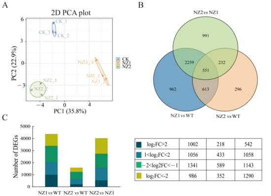
Figure 3.
Characterization of DEGs in oregano NZ1, NZ2 and WT. (A) PCA of 9 samples. (B) Venn diagram of DEGs. (C) Statistics of DEGs in NZ1, NZ2, and WT.
3.4. GO Annotation and KEGG Enrichment Analysis of DEGs
To elucidate the impact of heavy-ion irradiation, particularly the deposition of 12C6+ ions, on gene expression in oregano, we conducted Gene Ontology (GO) annotation analysis on the DEGs identified from the relevant pathways. The outcomes are presented in Figure S4. In the comparison between NZ1 and WT, the DEGs were predominantly associated with molecular function, biological process, and cellular component categories. Notably, under the Molecular Function category, the terms Binding (1815 genes, representing 41.39% of total DEGs) and Catalytic Activity (1501 genes, 34.23%) stood out. In the Cellular Component category, Cellular Anatomical Entity (1286 genes, 29.33%) exhibited significance. Regarding Biological Process, Cellular Process (1772 genes, 40.41%) and Metabolic Process (1598 genes, 36.44%) encompassed a substantial portion of the annotated genes. In the comparison between NZ2 and WT, DEGs were annotated according to Molecular Function and Biological Process. Notably, the terms Binding (629 genes, 39.51%) and Catalytic Activity (524 genes, 32.91%) were predominant in Molecular Function, while Cellular Process (565 genes, 35.49%) and Metabolic Process (510 genes, 32.04%) were highly represented in Biological Process. In the NZ2 versus NZ1 comparison, DEGs were annotated across Molecular Function, Biological Process, and Cellular Component. The most frequently annotated terms included Binding (1644 genes, 40.76%) and Catalytic Activity (1355 genes, 33.60%) in Molecular Function; Cellular Process (1589 genes, 39.40%) and Metabolic Process (1451 genes, 36.00%) in Biological Process; and Cellular Anatomical Entity (1142 genes, 28.32%) in Cellular Component.
KEGG pathway analysis was performed on the DEGs identified from all three comparison groups, revealing their association with various metabolic pathways. Screening the top 18 pathways based on FDR value showed that 10 metabolic pathways were common across the three comparison groups (Figure 4). Processes impacted by heavy-ion irradiation included biosynthesis of secondary metabolites, MAPK signaling pathway, flavonoid biosynthesis, phenylpropanoid biosynthesis, plant–pathogen interaction, stilbenoid, diarylheptanoid, and gingerol biosynthesis, isoquinoline alkaloid biosynthesis, diterpenoid biosynthesis, monoterpenoid biosynthesis, tryptophan metabolism, plant secondary metabolism, and sesquiterpenoid and triterpenoid biosynthesis. These results highlight the significant influence of heavy-ion irradiation on various physiological processes in the two selected typical mutants, particularly those related to terpenoids’ synthesis and metabolism among secondary metabolites.
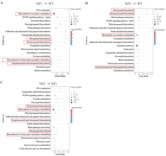
Figure 4.
KEGG enrichment of DEGs in NZ1 vs. WT (A), NZ2 vs. WT (B), and NZ2 vs. NZ1 (C), respectively.
3.5. Analysis of Transcription Factors Among DEGs
Plant transcription factors (TFs) are essential regulatory proteins that precisely control the transcriptional expression levels of target genes by binding specifically to their promoter regions. These proteins are crucial in various biological processes, including plant growth and development, stress responses, and the synthesis of secondary metabolites. To identify TFs potentially involved in heavy-ion-induced metabolic reprogramming, we first screened all differentially expressed genes (DEGs) from the comparisons of NZ1 vs. WT, NZ2 vs. WT, and NZ2 vs. NZ1 for TF annotation. This identified a total of 250 TFs derived from the combined DEG sets, belonging to 32 distinct families (Figure 5A). Comparing the two mutant lines separately to the wild-type, we observed distinct TF family profiles: NZ1 showed prominent changes in WRKY (28), bHLH (22), and ERF (20) families, while NZ2 was marked by alterations in MYB (15), HD-ZIP (15), and NAC (12) families, among others (Figure 5B).
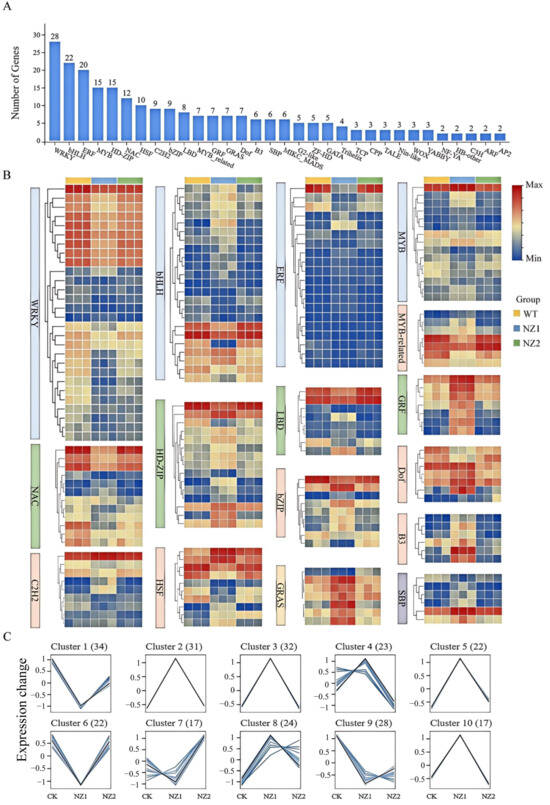
Figure 5.
Differential expression analysis of transcription factors in NZ1, NZ2, and WT. (A) Statistical analysis of transcription factors in DEGs. (B) Heat map of expression levels of 16 transcription factor families. (C) K-means clustering analysis of 250 differential expression of transcription factors.
To further uncover expression patterns associated with terpenoid accumulation, K-means clustering was performed on the 250 TFs across the three genotypes (WT, NZ1, NZ2). The TFs were grouped into 10 clusters (Figure 5C), comprising 34, 31, 32, 23, 22, 22, 17, 24, 28, and 17 TFs, respectively (Table S6). Notably, correlating with the content of carvacrol and thymol in these materials, it is suggested that TFs in Cluster 1 and Cluster 6 may play a role in the synthesis of carvacrol, while those in Cluster 9 may be linked to the synthesis of thymol.
3.6. Analysis of DEGs Involved in the Terpenoids Biosynthesis Pathways in Origanum vulgare L.
In plants, terpenoid compounds are synthesized via two distinct metabolic pathways: the mevalonate (MVA) pathway and the methylerythritol phosphate (MEP) pathway. To further investigate the molecular mechanisms underlying terpenoid biosynthesis in oregano leaves, we analyzed DEGs from the transcriptome. This analysis identified a total of 59 genes encoding enzymes associated with terpenoid biosynthesis that exhibited differential expression (Figure 6). Within the terpene backbone synthesis pathway, several genes in the MEP pathway exhibited notable sensitivity to heavy-ion irradiation, including DXS (Unigene0010384), HDR (Unigene0035540), GPPS (Unigene0004264, Unigene0044469), and GGPPS (Unigene0004264, Unigene0044469). Additionally, in the MVA pathway, genes such as ACAT (Unigene0039294), HMGS (Unigene0042224), HMGR (Unigene0031242, Unigene0047434, Unigene0016941), MVD (Unigene0012560), GPPS (Unigene0004264, Unigene0044469), GGPPS (Unigene0004264, Unigene0044469), and PCME (Unigene0034971) also exhibited significant changes in expression in response to the irradiation treatment (Table S7).
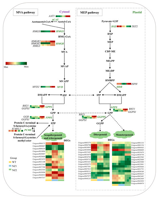
Figure 6.
Profiles of genes encoding enzymes involved in terpenoids biosynthesis. The left and right showed the MVA and MEP pathways in oregano, respectively. Key enzymes in DEGs include acetyl-CoA C-acetyltransferase (AACT), hydroxymethylglutaryl-CoA synthase (HMGS), hydroxymethylglutaryl-CoA reductase (HMGR), mevalonate-5-diphosphate decarboxylase (MVD), geranyl pyrophosphate synthase (GPPS), farnesyl pyrophosphate synthase (FPPS), 1-deoxy-D-xylulose-5-phosphate synthase (DXS), and geranylgeranyl pyrophosphate synthase (GGPPS). The color scale from green to red represents the abundance of DEGs ranging from low to high levels in WT, NZ1, and NZ2.
3.7. Identification of OvTPSs and OvCYP71Ds in the Thymol and Carvacrol Biosynthesis Pathway in Origanum vulgare L.
In oregano, the compounds thymol and carvacrol are synthesized via the monoterpenoid biosynthesis pathway. Terpene synthases (TPSs) serve as crucial enzymes in terpenoid biosynthesis, facilitating the final step in the formation of the terpenoid backbone. During monoterpene synthesis, γ-terpinene, produced through TPS catalysis, undergoes a hydroxylation reaction regulated by regioselective cytochrome P450 hydroxylases (CYP450s). This reaction introduces a hydroxyl group at distinct positions (C3 or C2) on the aromatic ring, resulting in the specific formation of carvacrol or thymol, respectively. In this study, the annotation and analysis of transcriptome results resulted in the identification of 13 TPSs and 7 CYP71Ds (Figure 7A). Notably, among the identified TPSs and CYP71Ds, the expression patterns of TPS6A (Unigene0020887), TPS6C (Unigene0021109), and TPS3 (Unigene0047317) at the transcriptional level aligned with the trend in γ-terpinene content observed in the oregano samples. Additionally, CYP71D178 (Unigene0040852) and CYP71D181 (Unigene0009943) displayed expression patterns that matched the accumulation trend of carvacrol, while CYP71D10B (Unigene0046282) corresponded to the trend in thymol content (Figure 7B,C). These findings indicate that these genes may facilitate the biosynthesis and accumulation of terpenoid compounds—particularly carvacrol and thymol—in oregano through positive regulation, thereby enhancing the levels of bioactive constituents.

Figure 7.
DEGs and DAVs involved in carvacrol and thymol biosynthesis. (A) Scheme of carvacrol and thymol biosynthesis in oregano. (B,C) Validation of OvTPSs and OvCYP71Ds using RT-qPCR. The data are presented as mean values ± SD, and statistical significance was analyzed using a one-way ANOVA. (* p < 0.05, ** p < 0.01, *** p < 0.001, ns: not significant).

4. Discussion
Heavy-ion beam irradiation is increasingly acknowledged as a potent physical mutagenesis method for enhancing Chinese medicinal materials and crops, characterized by a high mutation rate and consistent inheritance of mutant traits [31,32]. Its utilization in medicinal plants shows potential for improving the yield of valuable secondary metabolites. In this investigation, we effectively utilized 12C6+ heavy-ion irradiation to create two unique, phenotypically stable, homozygous mutant lines (NZ1 and NZ2) of Origanum vulgare L. Our comprehensive transcriptomic and metabolomic analysis systematically unraveled the molecular mechanisms behind the irradiation-induced changes, offering new insights into the regulation of terpenoid biosynthesis, specifically the production of the key compounds carvacrol and thymol.
The phenotypic characterization of the mutants revealed significant alterations in plant architecture and biomass accumulation. Previous studies have shown that among the plant mutants obtained through heavy-ion radiation mutagenesis technology, Arabidopsis mutants exhibit multiple variant characteristics in leaves, stems, flowers or throughout the life cycle [33,34,35], while potato and chili pepper mutants show phenotypic differences in structure, development, leaves, flowers and fruits [36,37]. In this study, the contrasting phenotypes of NZ1 (reduced height, increased leaf biomass) and NZ2 (slightly increased height) suggest that heavy-ion irradiation induced diverse genetic lesions affecting different aspects of growth regulation (Figure 1). Such morphological variations are common consequences of radiation-induced genomic alterations, which can disrupt phytohormone homeostasis or cell cycle genes. For instance, in lotus flowers, mutants that can delay aging have been created by using heavy ion mutagenesis technology. In fact, this is because it affects the regulation of multiple hormones, specially making them no longer sensitive to ethylene [38]. Furthermore, the significantly higher SPAD in NZ1 indicates potential improvement in photosynthetic pigment density, a trait often associated with improved metabolic efficiency. Just as in the case of Scenedesmus quadricauda mutants induced by heavy-ion beam irradiation, the photosynthetic properties are affected through changes in central carbon metabolism, ultimately leading to two mutants’ opposite biomass accumulation [39]. These divergent phenotypes provided a solid foundation for investigating the corresponding molecular and metabolic changes.
Terpenoids are a large and diverse family of secondary metabolites that have gained more attention from chemists and pharmacologists for their intriguing skeletons and broad biological activities, including anti-inflammatory, antitumoral, antimicrobial, and anti-viral effects [40,41]. In this study, metabolomic profiling unequivocally identified terpenoids as the most abundant class of volatile organic compounds, accounting for 69.35% of the total detected metabolites (Figure 2). This predominance underscores the central role of terpenoid metabolism in oregano’s chemical composition and its particular sensitivity to heavy-ion irradiation stress. The significant number of DAVs, especially those enriched in terpenoid biosynthesis pathways, directly links the physical mutagen to a reprogramming of secondary metabolism [42]. This observation aligns with findings in Astragalus mongholicus where mutagenic treatments significantly modulated amino acid metabolism and secondary metabolite biosynthesis pathways.
Previous studies have demonstrated that multiple signaling pathways, including the ubiquitin–proteasome pathway, MAPK signaling–plant, epigenomic regulatory pathways, and plant hormones, play crucial roles in regulating the accumulation of plant secondary metabolites. As essential components of regulatory networks, MAPK cascades initiate the synthesis of secondary metabolites by phosphorylating downstream substrates, such as transcription factors and key enzymes [43]. For instance, in Catharanthus roseus L., the CrMAPKKK1-CrMAPKK1-CrMAPKK3/6 cascade activates the ORCA gene cluster, thereby enhancing the synthesis of terpene indole alkaloids (TIAs) [44]. In Arabidopsis thaliana, MPK3 and MPK6 phosphorylate WRKY33, which in turn activates the expression of the phytochelatin camalexin synthesis gene (PAD3) [45]. In rice, the OsMKK4-OsMPK6 module regulates the chitin-induced synthesis of diterpenoid phytocannabinoids [46]. These findings underscore the essential role of the MAPK pathway in the accumulation of secondary metabolites. In this study, transcriptome analysis identified 6204 DEGs, with significant enrichment in critical pathways, including the biosynthesis of secondary metabolites and the MAPK signaling pathway in plants, which are vital to the genomic response to 12C6+ heavy-ion radiation. These results indicate that heavy ion radiation alters the synthesis of secondary metabolites in the mutants by affecting genes within the oregano MAPK signaling–plant pathway, consistent with the findings of previous studies. Furthermore, transcription factors are pivotal in regulating gene expression by binding to cis-acting elements within the promoters of key genes associated with secondary metabolism. They function as essential intermediaries that link upstream signaling pathways, including MAPK and hormonal signals, to downstream metabolic processes. For example, within the WRKY family, AaGSW1 binds to the W-box to activate CYP71AV1 and AaORA in Artemisia annua [47]. Additionally, WRKY33 facilitates sapogenin synthesis upon phosphorylation in Arabidopsis thaliana [48], while SmWRKY34 inhibits the production of salvianolic acid and tanshinone in Salvia miltiorrhiza [49]. In the bHLH family, the AtTT8-GL3/EGL3-TTG1 complex activates genes responsible for anthocyanin and proanthocyanidin synthesis in Arabidopsis thaliana [50]. Furthermore, SmMYC2a/b interacts with the E-box element to regulate the synthesis of tanshinone and tansyolic acid in Salvia miltiorrhiza [51]. In the AP2/ERF family, AaERF1/AaERF2 activates ADS and CYP71AV1 in Artemisia annua, thereby promoting artemisinin synthesis [52]. Within the MYB family, AtPAP1/PAP2 activates genes involved in anthocyanin synthesis [53]. Moreover, AaMYC2, a MYB-related factor, binds to the AaGSW1 promoter to enhance artemisinin production in Artemisia annua, while PgMYB1 inhibits ginsenoside synthesis in Panax ginseng [54]. Additionally, AtMYBL2 suppresses anthocyanin accumulation by disrupting the MBW complex [55]. This study identified several transcription factors via transcriptome analysis, including prominent members of the WRKY, bHLH, and ERF families among the DEGs. These transcription factors are acknowledged as regulators of plant specialized metabolism and stress responses, and their differential expression likely contributes to the observed alterations in metabolic pathways in response to heavy-ion irradiation.
A central finding of this study is the elucidation of the molecular framework that governs terpenoid biosynthesis in response to heavy-ion irradiation. We identified critical DEGs encoding enzymes in both the MVA and MEP pathways. Notably, HMGR and MVD are involved in the MVA pathway, while DXS and HDR are key components of the MEP pathway. These enzymes provide the universal precursors IPP and DMAPP essential for terpenoid synthesis [56,57]. The altered expression of these genes indicates a reconfiguration of metabolic flux toward terpenoid precursors. Additionally, the identification of critical downstream genes, particularly GPPS and GGPPS, which catalyze the synthesis of GPP and GGPP, highlights specific nodes where metabolic channeling may be affected [58,59]. Our analysis identified candidate genes directly involved in the biosynthesis of the valuable monoterpenoids carvacrol and thymol. The correlation between the expression of TPS3, TPS6A, and TPS6C and the content of γ-terpinene, the central precursor, strongly suggests their roles as γ-terpinene synthases [60,61]. We identified cytochrome P450 genes—CYP71D178 and CYP71D181 associated with carvacrol, and CYP71D10B associated with thymol—whose expression patterns closely mirrored the accumulation of these phenolic monoterpenoids. This finding provides compelling evidence for their proposed roles in the regioselective hydroxylation of γ-terpinene at the C3 and C2 positions, respectively, which represents a critical branching point that determines the final product outcome [62,63].
Despite these findings, it is imperative to acknowledge the limitations of the present study. Most notably, while pronounced phenotypic, transcriptomic, and metabolomic alterations were observed in the NZ1 and NZ2 mutants, the exact genomic mutations caused by 12C6+ irradiation remain unidentified. It is well established that heavy-ion beams are capable of inducing large-fragment deletions, insertions, and chromosomal rearrangements, which have the potential to disrupt gene function and regulatory networks. However, in the absence of whole-genome sequencing or detailed genomic structural analysis, it is not possible to identify the specific genetic lesions responsible for the observed changes in plant architecture, terpenoid accumulation, or gene expression patterns. This discrepancy hinders our capacity to establish direct causal relationships between specific genomic alterations and the reprogramming of metabolic pathways. It is imperative that future studies employ whole-genome sequencing or fine-mapping approaches in order to ascertain the precise mutations that underpin the mutant traits and to validate the regulatory mechanisms proposed in this study.
5. Conclusions
In conclusion, the present study establishes that 12C6+ heavy-ion irradiation is an effective means of inducing genetic and metabolic variation in Origanum vulgare L. By integrating multi-omics data, the present study has systematically characterized the morphological, metabolic, and transcriptional consequences of mutagenesis, revealing a complex reprogramming of terpenoid metabolism. The identification of key structural genes and transcription factors, and most notably, the candidate genes TPS3/6A/6C, CYP71D178, CYP71D181, and CYP71D10B as potential determinants of carvacrol and thymol composition, represents a significant advance in the field. These findings provide a robust molecular foundation and valuable genetic resources for future metabolic engineering and marker-assisted breeding strategies aimed at optimizing oregano for enhanced medicinal and commercial value.
Supplementary Materials
The following supporting information can be downloaded at https://www.mdpi.com/article/10.3390/cimb48010007/s1.
Author Contributions
Z.T. and H.L. designed the study; Z.T., L.L. and Y.L. wrote the original manuscript; Z.T., L.L., Y.L., C.L., X.S., D.L., Y.S., M.S., Y.C., L.W. and D.L. performed the experiments; Z.T., L.L. and Y.L. analyzed data and made the graphs; Z.T. and H.L. revised the manuscript. All authors have read and agreed to the published version of the manuscript.
Funding
This research was funded by China Agriculture Research System of MOF and MARA (CARS-21); Henan Agriculture Research System (HARS-22-11-G3); Key Research and Development Program of Henan (241111310200, 231111110800, 251111310600, 251111111800); Henan Academy of Agricultural Sciences Emerging Discipline Development Project (2025XK01); Henan Academy of Agricultural Sciences Outstanding Youth Science and Technology Fund (2024YQ15, 2024YQ16); Henan Center for Overseas Scientists (GZS2024025); Central Government Funds to Guide Local Science and Technology Development (Z20241471131); and Henan Province Science and Technology Research Project (252102110283).
Institutional Review Board Statement
Not applicable.
Informed Consent Statement
Not applicable.
Data Availability Statement
The original contributions presented in this study are included in the article/Supplementary Material. Further inquiries can be directed to the corresponding author.
Acknowledgments
The data used to support the funding for this study are available from the corresponding author upon request.
Conflicts of Interest
The authors declare no conflicts of interest.
References
- Vârban, D.; Zăhan, M.; Crișan, I.; Pop, C.R.; Gál, E.; Ștefan, R.; Rotar, A.M.; Muscă, A.S.; Meseșan, Ș.D.; Horga, V.; et al. Unraveling the Potential of Organic Oregano and Tarragon Essential Oils: Profiling Composition, FT-IR and Bioactivities. Plants 2023, 12, 4017. [Google Scholar] [CrossRef]
- Hao, Y.; Guo, X.; Yang, R.; Yan, Y.; Sun, M.; Li, H.; Bai, H.; Cui, H.; Li, J.; Shi, L. Unraveling the Biosynthesis of Carvacrol in Different Tissues of Origanum vulgare L. Int. J. Mol. Sci. 2022, 23, 13231. [Google Scholar] [CrossRef] [PubMed]
- Guo, X.; Hao, Y.; Zhang, W.; Xia, F.; Bai, H.; Li, H.; Shi, L. Comparison of Origanum Essential Oil Chemical Compounds and Their Antibacterial Activity against Cronobacter sakazakii. Molecules 2022, 27, 6702. [Google Scholar] [CrossRef]
- Kaurinovic, B.; Popovic, M.; Vlaisavljevic, S.; Trivic, S. Antioxidant Capacity of Ocimum basilicum L. and Origanum vulgare L. Extracts. Molecules 2011, 16, 7401–7414. [Google Scholar] [CrossRef]
- Hao, Y.; Kang, J.; Yang, R.; Li, H.; Cui, H.; Bai, H.; Tsitsilin, A.; Li, J.; Shi, L. Multidimensional exploration of essential oils generated via eight oregano cultivars: Compositions, chemodiversities, and antibacterial capacities. Food Chem. 2022, 374, 131629. [Google Scholar] [CrossRef]
- Hao, Y.; Li, J.; Shi, L. A Carvacrol-Rich Essential Oil Extracted from Oregano (Origanum vulgare “Hot & Spicy”) Exerts Potent Antibacterial Effects Against Staphylococcus aureus. Front. Microbiol. 2021, 12, 741861. [Google Scholar] [CrossRef] [PubMed]
- Khanam, S.; Mishra, P.; Faruqui, T.; Alam, P.; Albalawi, T.; Siddiqui, F.; Rafi, Z.; Khan, S. Plant-based secondary metabolites as natural remedies: A comprehensive review on terpenes and their therapeutic applications. Front. Pharmacol. 2025, 16, 1587215. [Google Scholar] [CrossRef] [PubMed]
- Choudhary, N.; Bawari, S.; Burcher, J.T.; Sinha, D.; Tewari, D.; Bishayee, A. Targeting Cell Signaling Pathways in Lung Cancer by Bioactive Phytocompounds. Cancers 2023, 15, 3980. [Google Scholar] [CrossRef]
- Lv, Z.; Zhang, L.; Tang, K. New insights into artemisinin regulation. Plant Signal Behav. 2017, 12, e1366398. [Google Scholar] [CrossRef] [PubMed]
- Yu, H.; Lan, F.; Zhuang, Y.; Li, Q.; Zhang, L.; Tian, H.; Bu, X.; Chen, R.; Gao, Y.; Wang, Z.; et al. Paclitaxel anti-cancer therapeutics: From discovery to clinical use. Chin. J. Nat. Med. 2025, 23, 769–789. [Google Scholar] [CrossRef]
- Węglarz, Z.; Kosakowska, O.; Przybył, J.; Pióro-Jabrucka, E.; Bączek, K. The Quality of Greek Oregano (O. vulgare L. Subsp. hirtum (Link) Ietswaart) and Common Oregano (O. vulgare L. Subsp. vulgare) Cultivated in the Temperate Climate of Central Europe. Foods 2020, 9, 1671. [Google Scholar] [CrossRef]
- Sharopov, F.; Braun, M.S.; Gulmurodov, I.; Khalifaev, D.; Isupov, S.; Wink, M. Antimicrobial, Antioxidant, and Anti-Inflammatory Activities of Essential Oils of Selected Aromatic Plants from Tajikistan. Foods 2015, 4, 645–653. [Google Scholar] [CrossRef] [PubMed]
- Li, C.; Zha, W.; Li, W.; Wang, J.; You, A. Advances in the Biosynthesis of Terpenoids and Their Ecological Functions in Plant Resistance. Int. J. Mol. Sci. 2023, 24, 11561. [Google Scholar] [CrossRef] [PubMed]
- Jiang, H.; Wang, X. Biosynthesis of monoterpenoid and sesquiterpenoid as natural flavors and fragrances. Biotechnol. Adv. 2023, 65, 108151. [Google Scholar] [CrossRef]
- Krause, S.; Liao, P.; Crocoll, C.; Boachon, B.; Förster, C.; Leidecker, F.; Wises, N.; Zhao, D.; Wood, J.; Buell, C.; et al. The biosynthesis of thymol, carvacrol, and thymohydroquinone in Lamiaceae proceeds via cytochrome P450s and a short-chain dehydrogenase. Proc. Natl. Acad. Sci. USA 2021, 118, e2110092118. [Google Scholar] [CrossRef]
- Tanvi, S.; Anantika, S.; Tabasum, K.; Arun, K. A bZIP type transcription factor from Picrorhiza kurrooa modulates the expression of terpenoid biosynthesis genes in Nicotiana benthamiana. S. Afr. J. Bot. 2024, 169, 210–218. [Google Scholar] [CrossRef]
- Liu, X.; Shao, Y.; Mu, D.; Zhou, Y.; He, J.; Zhao, H.; Wilson, I.; Zhang, Y.; Zhu, L.; Qiu, D.; et al. Genome-wide identification of TCP transcription factors and functional role of UrTCP4 in regulating terpenoid indole alkaloids biosynthesis in Uncaria rhynchophylla. Curr. Plant Biol. 2024, 40, 100406. [Google Scholar] [CrossRef]
- Wei, J.; Mu, X.; Wang, S.; Wei, Q.; Zhu, L.; Zhang, X.; Zhang, J.; Liu, X.; Wen, B.; Li, M.; et al. Integrated metabolome and transcriptome analysis provides insights into the mechanisms of terpenoid biosynthesis in tea plants (Camellia sinensis). Food Res. Int. 2025, 201, 115542. [Google Scholar] [CrossRef]
- Dong, Y.; Wei, Z.; Li, J.; Zhang, W.; Gao, F.; Xia, F.; Bai, H.; Li, H.; Shi, L. Exogenous application of methyl jasmonate affects the transcriptome of terpenoid biosynthesis genes in lavender and functional characterization of the candidate gene LabHLH74. Plant Physiol. Biochem. 2025, 227, 110085. [Google Scholar] [CrossRef]
- Ikegaya, T.; Shirasawa, K.; Fujino, K. Strategies to assess genetic diversity for crop breeding. Euphytica 2023, 219, 59. [Google Scholar] [CrossRef]
- Singh, H.; Raigar, O.; Chahota, R. Estimation of genetic diversity and its exploitation in plant breeding. Bot. Rev. 2022, 88, 413–435. [Google Scholar] [CrossRef]
- Guo, X.; Ren, J.; Zhou, X.; Zhang, M.; Lei, C.; Chai, R.; Zhang, L.; Lu, D. Strategies to improve the efficiency and quality of mutant breeding using heavy-ion beam irradiation. Crit. Rev. Biotechnol. 2024, 44, 735–752. [Google Scholar] [CrossRef]
- Hirano, T.; Kazama, Y.; Ishii, K.; Ohbu, S.; Shirakawa, Y.; Abe, T. Comprehensive identification of mutations induced by heavy-ion beam irradiation in Arabidopsis thaliana. Plant J. 2015, 82, 93–104. [Google Scholar] [CrossRef]
- Li, Y.; Gu, J.; Irshad, A.; Zhao, L.; Guo, H.; Xiong, H.; Xie, Y.; Zhao, S.; Ding, Y.; Zhou, L.; et al. Physiological and Differential Proteomic Analysis at Seedling Stage by Induction of Heavy-Ion Beam Radiation in Wheat Seeds. Front. Genet. 2022, 13, 942806. [Google Scholar] [CrossRef]
- Ren, H.; Zhang, B.; Zhang, C.; Liu, X.; Wang, X.; Zhang, F.; Zhao, K.; Yuan, R.; Abdelahany, A.; Lamlom, S. Uncovering molecular mechanisms of soybean response to 12C6+ heavy ion irradiation through integrated transcriptomic and metabolomic profiling. Ecotoxicol. Environ. Saf. 2025, 289, 117689. [Google Scholar] [CrossRef]
- Pan, W.; Zheng, L.; Tian, H.; Li, W.; Wang, J. Transcriptome responses involved in artemisinin production in Artemisia annua L. under UV-B radiation. J. Photochem. Photobiol. B 2014, 140, 292–300. [Google Scholar] [CrossRef] [PubMed]
- Chen, Y.; Shen, Q.; Lv, P.; Sun, C. Comparative metabolomic analyses of Dendrobium officinale Kimura et Migo responding to UV-B radiation reveal variations in the metabolisms associated with its bioactive ingredients. PeerJ 2020, 8, e9107. [Google Scholar] [CrossRef] [PubMed]
- Sasaki, N.; Watanabe, A.; Asakawa, T.; Sasaki, M.; Hoshi, N.; Naito, Z.; Furusawa, Y.; Shimokawa, T.; Nishihara, M. Biological effects of ion beam irradiation on perennial gentian and apple. Plant Biotechnol. 2018, 35, 249–257. [Google Scholar] [CrossRef]
- Livak, K.; Schmittgen, T. Analysis of Relative Gene Expression Data Using Real-Time Quantitative PCR and the 2(-Delta Delta C (T)) Method. Methods 2001, 25, 402–408. [Google Scholar] [CrossRef] [PubMed]
- Rawat, A.; Bhuj, B.; Srivastava, R.; Chand, S.; Singh, N.; Bisht, Y.; Dasila, H.; Bhatt, R.; Perveen, K.; Bukhari, N. Determination of lethal and mutation induction doses of gamma rays for gladiolus (Gladiolus grandifloras Hort.) genotypes. Heliyon 2024, 10, e37387. [Google Scholar] [CrossRef]
- Peng, D.; Liu, L.; Guo, H.; Li, Y.; Li, M.; Guo, X.; Wei, J. Research progress on radiation breeding of Chinese medicinal materials. Chin. Herb. Med. 2023, 54, 87–95. [Google Scholar] [CrossRef]
- Oladosu, Y.; Rafii, M.; Abdullah, N.; Hussin, G.; Ramli, A.; Rahim, H.; Miah, G.; Usman, M. Principle and application of plant mutagenesis in crop improvement: A review. Biotechnol. Biotechnol. Equip. 2016, 30, 1–16. [Google Scholar] [CrossRef]
- Chen, X.; Feng, H.; Du, Y.; Luo, S.; Li, W.; Yu, L.; Feng, Z.; Cui, T.; Zhou, L. Genetic polymorphisms in mutagenesis progeny of Arabidopsis thaliana irradiated by carbon-ion beams and gamma-rays irradiations. Int. J. Radiat. Biol. 2020, 96, 267–275. [Google Scholar] [CrossRef]
- Du, Y.; Li, W.; Yu, L.; Chen, G.; Liu, Q.; Luo, S.; Shu, Q.; Zhou, L. Mutagenic effects of carbon-ion irradiation on dry Arabidopsis thaliana seeds. Mutat. Res. Genet. Toxicol. Environ. Mutagen. 2014, 759, 28–36. [Google Scholar] [CrossRef]
- Du, Y.; Luo, S.; Li, X.; Yang, J.; Cui, T.; Li, W.; Yu, L.; Feng, H.; Chen, Y.; Mu, J.; et al. Identification of substitutions and small insertion-deletions induced by carbon-ion beam irradiation in Arabidopsis thaliana. Front. Plant Sci. 2017, 8, 1851. [Google Scholar] [CrossRef]
- Jo, Y.; Kim, S.; Hwang, J.; Kim, Y.; Kang, H.; Kwon, S.; Ryu, J.; Kim, J.; Kang, S. Construction of mutation populations by gamma-ray and carbon beam irradiation in chili pepper (Capsicum annuum L.). Hortic. Environ. Biotechnol. 2016, 57, 606–614. [Google Scholar] [CrossRef]
- Park, H.; Narasako, Y.; Abe, T.; Kunitake, H.; Hirano, T. Comprehensive effects of heavy-ion beam irradiation on sweet potato (Ipomoea batatas [L.] Lam.). Plant Biotechnol. 2022, 39, 311–316. [Google Scholar] [CrossRef] [PubMed]
- Du, Y.; Luo, S.; Zhao, J.; Feng, Z.; Chen, X.; Ren, W.; Liu, X.; Wang, Z.; Yu, L.; Li, W.; et al. Genome and transcriptome-based characterization of high energy carbon-ion beam irradiation induced delayed flower senescence mutant in Lotus japonicus. BMC Plant Biol. 2021, 21, 510. [Google Scholar] [CrossRef]
- Wang, J.; Li, X.; Wang, J.; Wei, W.; Jin, W.; Zhou, L. Comparative proteomics reveals energy and carbon metabolism changes in Scenedesmus quadricauda mutants induced by heavy-ion beam irradiation. Bioresour. Technol. 2024, 406, 130965. [Google Scholar] [CrossRef] [PubMed]
- Zang, Y.; Sun, R.; Feng, R.; Zhu, H.; Li, X. Recent Advances of Terpenoids with Intriguing Chemical Skeletons and Biological Activities. Chin. J. Chem. 2025, 43, 443–469. [Google Scholar] [CrossRef]
- Gu, X.; Hao, D.; Xiao, P. Research progress of Chinese herbal medicine compounds and their bioactivities: Fruitful 2020. Chin. Herb. Med. 2022, 14, 171–186. [Google Scholar] [CrossRef]
- Wang, Y.; Liu, X.; Wang, L.; Li, X.; Chen, F.; Li, P.; Ma, X.; Wang, F.; Li, L.; Xin, Z.; et al. Radiation-specific metabolic reprogramming drives yield-quality balance in Astragalus mongholicus: Comparative insights from gamma-ray and heavy-ion beam irradiation. Plant Stress 2025, 18, 101027. [Google Scholar] [CrossRef]
- Liu, S.; Zhang, Q.; Kollie, L.; Dong, J.; Liang, Z. Molecular networks of secondary metabolism accumulation in plants: Current understanding and future challenges. Ind. Crops Prod. 2023, 201, 116901. [Google Scholar] [CrossRef]
- Paul, P.; Singh, S.K.; Patra, B.; Sui, X.; Pattanaik, S.; Yuan, L. A differentially regulated AP2/ERF transcription factor gene cluster acts downstream of a MAP kinase cascade to modulate terpenoid indole alkaloid biosynthesis in Catharanthus roseus. New Phytol. 2017, 213, 1107–1123. [Google Scholar] [CrossRef]
- Mao, W.; Han, Y.; Chen, Y.; Sun, M.; Feng, Q.; Li, L.; Liu, L.; Zhang, K.; Wei, L.; Han, Z.; et al. Low temperature inhibits anthocyanin accumulation in strawberry fruit by activating FvMAPK3-induced phosphorylation of FvMYB10 and degradation of chalcone synthase 1. Plant Cell 2022, 34, 1226–1249. [Google Scholar] [CrossRef]
- Kishi-Kaboshi, M.; Okada, K.; Kurimoto, L.; Murakami, S.; Umezawa, T.; Shibuya, N.; Yamane, H.; Miyao, A.; Takatsuji, H.; Takahashi, A.; et al. A rice fungal MAMP-responsive MAPK cascade regulates metabolic flow to antimicrobial metabolite synthesis. Plant J. 2010, 63, 599–612. [Google Scholar] [CrossRef]
- Chen, M.; Yan, T.; Qian, S.; Lu, X.; Pan, Q.; Huang, Y.; Tang, Y.; Fu, X.; Liu, M.; Jiang, W.; et al. Glandular trichome-specific WRKY1 promotes artemisinin biosynthesis in Artemisia annua. New Phytol. 2017, 214, 304–316. [Google Scholar] [CrossRef]
- Mao, G.; Meng, X.; Liu, Y.; Zheng, Z.; Chen, Z.; Zhang, S. Phosphorylation of a WRKY transcription factor by two pathogen-responsive MAPKs drives phytoalexin biosynthesis in Arabidopsis. Plant Cell 2011, 23, 1639–1653. [Google Scholar] [CrossRef]
- Shi, M.; Zhu, R.; Zhang, Y.; Zhang, S.; Liu, T.; Li, K.; Liu, S.; Wang, L.; Wang, Y.; Zhou, W.; et al. A novel WRKY34-bZIP3 module regulates phenolic acid and tanshinone biosynthesis in Salvia miltiorrhiza. Metab. Eng. 2022, 73, 182–191. [Google Scholar] [CrossRef] [PubMed]
- Lloyd, A.; Brockman, A.; Aguirre, L.; Campbell, A.; Bean, A.; Cantero, A.; Gonzalez, A. Advances in the MYB-bHLH-WD repeat (MBW) pigment regulatory model: Addition of a WRKY factor and co-option of an anthocyanin MYB for betalain regulation. Plant Cell Physiol. 2017, 58, 1431–1441. [Google Scholar] [CrossRef]
- Zhou, Y.; Sun, W.; Chen, J.; Tan, H.; Xiao, Y.; Li, Q.; Ji, Q.; Gao, S.; Chen, L.; Chen, S.; et al. SmMYC2a and SmMYC2b played similar but irreplaceable roles in regulating the biosynthesis of tanshinones and phenolic acids in Salvia miltiorrhiza. Sci. Rep. 2016, 6, 22852, Erratum in Sci. Rep. 2020, 10, 7201.. [Google Scholar] [CrossRef] [PubMed]
- Yu, Z.; Li, J.; Yang, C.; Hu, W.; Wang, L.; Chen, X. The jasmonate-responsive AP2/ERF transcription factors AaERF1 and AaERF2 positively regulate artemisinin biosynthesis in Artemisia annua L. Mol. Plant 2012, 5, 353–365. [Google Scholar] [CrossRef]
- Xie, Y.; Tan, H.; Ma, Z.; Huang, J. DELLA proteins promote anthocyanin biosynthesis via sequestering MYBL2 and JAZ suppressors of the MYB/bHLH/WD40 complex in Arabidopsis thaliana. Mol. Plant 2016, 9, 711–721. [Google Scholar] [CrossRef]
- Afrin, S.; Jie, Z.; Cao, H.; Huang, J.; Xiu, H.; Luo, T.; Luo, Z. Molecular cloning and expression profile of an abiotic stress and hormone responsive MYB transcription factor gene from Panax ginseng. Acta Biochim. Biophys. Sin. 2015, 47, 267–276. [Google Scholar] [CrossRef]
- Matsui, K.; Umemura, Y.; Ohmetakagi, M. AtMYBL2, a protein with a single MYB domain, acts as a negative regulator of anthocyanin biosynthesis in Arabidopsis. Plant J. 2008, 55, 954–967. [Google Scholar] [CrossRef]
- Nagegowda, D. Plant volatile terpenoid metabolism: Biosynthetic genes, transcriptional regulation and subcellular compartmentation. FEBS Lett. 2010, 584, 2965–2973. [Google Scholar] [CrossRef]
- Vranová, E.; Coman, D.; Gruissem, W. Network analysis of the MVA and MEP pathways for isoprenoid synthesis. Annu. Rev. Plant Biol. 2013, 64, 665–700. [Google Scholar] [CrossRef]
- Nagegowda, D.; Gupta, P. Advances in biosynthesis, regulation, and metabolic engineering of plant specialized terpenoids. Plant Sci. 2020, 294, 110457. [Google Scholar] [CrossRef] [PubMed]
- Barja, M.; Rodriguez-Concepcion, M. Plant geranylgeranyl diphosphate synthases: Every (gene) family has a story. aBIOTECH 2021, 2, 289–298. [Google Scholar] [CrossRef] [PubMed]
- Tholl, D. Biosynthesis and biological functions of terpenoids in plants. Adv. Biochem. Eng. Biotechnol. 2015, 148, 63–106. [Google Scholar] [CrossRef] [PubMed]
- Chen, F.; Tholl, D.; Bohlmann, J.; Pichersky, E. The family of terpene synthases in plants: A mid-size family of genes for specialized metabolism that is highly diversified throughout the kingdom. Plant J. 2011, 66, 212–229. [Google Scholar] [CrossRef]
- Bathe, U.; Tissier, A. Cytochrome P450 enzymes: A driving force of plant diterpene diversity. Phytochemistry 2019, 161, 149–162. [Google Scholar] [CrossRef] [PubMed]
- Zheng, X.; Li, P.; Lu, X. Research advances in cytochrome P450-catalysed pharmaceutical terpenoid biosynthesis in plants. J. Exp. Bot. 2019, 70, 4619–4630. [Google Scholar] [CrossRef] [PubMed]


Disclaimer/Publisher’s Note: The statements, opinions and data contained in all publications are solely those of the individual author(s) and contributor(s) and not of MDPI and/or the editor(s). MDPI and/or the editor(s) disclaim responsibility for any injury to people or property resulting from any ideas, methods, instructions or products referred to in the content. |
© 2025 by the authors. Licensee MDPI, Basel, Switzerland. This article is an open access article distributed under the terms and conditions of the Creative Commons Attribution (CC BY) license.