Abstract
Background: Selective serotonin reuptake inhibitors (SSRIs) are widely prescribed, yet their direct impact on ovarian function remains poorly understood. While serotonin signaling is known to occur within the ovarian follicle, the specific molecular consequences of its disruption by SSRIs are unclear. This study aimed to elucidate the direct, intra-ovarian mechanisms by which fluoxetine, a common SSRI, affects follicular development and oocyte competence. Methods: We administered fluoxetine (20 mg/kg) or vehicle daily for seven days to both prepubertal and adult female mice to model short-term therapeutic exposure. Results: Fluoxetine treatment successfully blocked peripheral serotonin uptake, reducing serum levels by over 90%. Crucially, this occurred without altering circulating levels of estradiol, FSH, or LH and without disrupting the estrous cycle, indicating a mechanism independent of the central hypothalamic–pituitary–gonadal axis. Instead, we pinpoint a direct ovarian effect: fluoxetine inhibited serotonin transport activity in oocytes and significantly downregulated the expression of the pivotal oocyte-derived growth factor Gdf9. This was accompanied by reduced expression of genes crucial for granulosa cell function (Lhr, Fshr) and steroidogenesis (Cyp19a1). Functionally, these molecular changes manifested as a decline in oocyte quality and a significant reduction in ovulation rates in adult mice. Notably, these detrimental effects were more pronounced in prepubertal mice, indicating a heightened vulnerability during early follicular development. Conclusions: Our findings reveal a direct, intra-ovarian mechanism of fluoxetine-induced disruption. By inhibiting oocyte serotonin transport and downregulating GDF9, fluoxetine impairs critical oocyte–granulosa cell communication, thereby compromising oocyte competence and reducing fertility outcomes. This work identifies follicular development as a critical window of susceptibility to SSRI exposure, holding significant clinical implications for reproductive-aged and adolescent populations.
    1. Introduction
The female reproductive system is highly susceptible to external factors, which can impact not only maternal health but also the development and well-being of future offspring. In this context, the effects of widely prescribed psychotropic drugs are of particular concern. Women are twice as likely as men to experience depression, with a high incidence during their reproductive years []. Consequently, selective serotonin reuptake inhibitors (SSRIs) are commonly prescribed. However, their safety for the female reproductive system remains a subject of ongoing debate [,,,,,,], necessitating further study to understand their short- and long-term effects.
The primary antidepressant mechanism of SSRIs involves enhancing serotonergic neurotransmission by increasing serotonin (5-hydroxytryptamine, 5-HT) levels in the synaptic cleft []. Beyond the central nervous system (CNS), however, serotonin has a much broader physiological role. Notably, only 5% of the body’s serotonin is synthesized in the brain, with the remaining 95% produced in peripheral tissues [,]. A growing body of evidence indicates that serotonin is also a key signaling molecule in female reproduction, with its presence detected in the oviduct, uterus, ovaries, oocytes, and early mammalian embryos [,,,,,]. This has prompted investigations into the local ovarian serotonergic system, revealing the expression of key components: the serotonin synthesis enzymes Tph1, Tph2, and Ddc [,]; the degradation enzyme Mao []; vesicular transporters Vmat1 and Vmat2 [,,]; and various classes of serotonin receptors (Htr) [,,]. Intriguingly, the direct target of SSRIs, the serotonin reuptake transporter (Sert/Slc6A4), is also expressed and active in ovarian tissues, particularly in follicles []. Pharmacological or genetic inhibition of Sert has been shown to reduce aromatase (Cyp19a1) expression and subsequent 17β-estradiol (E2) secretion [,,]. In contrast, serotonin itself appears to stimulate steroidogenesis in mammalian follicles and granulosa cell cultures [,,]. However, the precise mechanisms by which SSRIs and serotonin modulate steroid hormone synthesis and overall ovarian function remain to be fully elucidated.
SSRIs are known to alter serotonin concentrations not only in the CNS but also in the blood and peripheral tissues [,], potentially leading to functional disturbances. Our previous work demonstrated that mouse oocytes accumulate serotonin via Sert [] and that oocyte Sert activity correlates with follicle survival and selection []. Moreover, fluoxetine treatment leads to tangible reproductive consequences, including reduced oocyte competence (the oocyte’s ability to resume meiosis, fertilize, and develop into a viable embryo) and lower ovulation rates []. This Sert-mediated serotonin uptake is prominent in growing follicles, a key process during both prepubertal development and adult cyclicity. Therefore, the aim of this study was to investigate the effects of short-term exposure to fluoxetine, one of the most commonly prescribed SSRIs, on ovarian function. We utilized two critical developmental windows: the first wave of folliculogenesis in prepubertal mice (7–14 days postpartum, dpp) and the established cyclicity in sexually mature adult females (2 months old).
2. Results
2.1. Short-Term Fluoxetine Administration Drastically Reduces Serum Serotonin Without Affecting Estradiol Levels
To establish the systemic effects of fluoxetine in our experimental models, we first measured serotonin (5-HT) concentrations in the blood serum of prepubertal (14 dpp) and adult (2-month-old) female mice after a 7-day treatment course. High-performance liquid chromatography (HPLC) analysis revealed a dramatic reduction in serum 5-HT in the fluoxetine-treated groups compared to vehicle controls. Specifically, 5-HT levels decreased by ~92% in prepubertal females and ~96% in adult females (Figure 1a,b, left panels). It was also noted that basal 5-HT levels in control prepubertal mice were approximately five-fold lower than in control adult mice.
      
    
    Figure 1.
      Establishment of the experimental model on mice and effects of 7 days of fluoxetine treatment on hormones. VH—vehicle treated, FLX—fluoxetine treated. (a,b) Serotonin and E2 levels in the blood serum of mature (a) and prepubertal (b) females in the experiment (M ± SEM). Significance levels are indicated as **—p < 0.01, ****—p < 0.0001 according to the Mann–Whitney U test. (c) Gonadotropin protein assessment in the blood serum of mature mice (M ± SEM). ns—not significant according to the Mann–Whitney U test. (d) Gonadotropins β-subunit gene expression in the pituitary gland of prepubertal mice. The relative quantity (RQ) was calculated using the 2−∆Ct method relative to the reference gene Rps18 (M ± SEM). ns—not significant according to the Mann–Whitney U test. (e) Estrous cycle of control (vehicle, n = 4) and fluoxetine treated (n = 8) mice. (f) Statistical evaluation of the length of the estrous cycle in mature mice (M ± SEM). ns—not significant according to the Mann–Whitney U test. (g) Comparison of the percentage of time spent by mice in each estrous cycle stage during 15 days of the experiment (M ± SEM). ns—not significant according to the Mann–Whitney U test. Figure created with BioRender.com (accessed on 10 September 2025).
  
Next, we measured serum 17β-estradiol (E2) levels by ELISA. Despite the profound drop in peripheral serotonin, the 7-day fluoxetine exposure had no significant effect on circulating E2 concentrations in either age group (Figure 1a,b, right panels). As expected, basal E2 levels were substantially lower in prepubertal mice than in adults. These results confirm that our administration protocol effectively depletes peripheral serotonin, providing a robust model to investigate the consequences of reduced systemic 5-HT on ovarian function, independent of acute changes in circulating E2.
2.2. Fluoxetine Administration Does Not Alter Gonadotropin Levels or Estrous Cyclicity
To determine if fluoxetine’s effects were mediated by the hypothalamic–pituitary–gonadal (HPG) axis, we assessed gonadotropin levels. In adult females, ELISA measurements showed no significant differences in the serum levels of follicle-stimulating hormone (FSH) or luteinizing hormone (LH) between fluoxetine-treated and control groups (Figure 1c). Similarly, in prepubertal mice, real-time PCR analysis of pituitary glands revealed no change in the mRNA expression of the beta-subunits of Fsh and Lh (Figure 1d).
Consistent with the stable gonadotropin levels, the estrous cycle in adult females was unaffected by fluoxetine treatment. We observed no significant differences in the overall length of the estrous cycle (Figure 1f) or the duration of its individual stages (Figure 1e,g). Together, these data strongly suggest that the effects of short-term fluoxetine exposure are not mediated by central gonadotropic regulation but likely stem from a direct action on the ovary.
2.3. Fluoxetine Disrupts Serotonin Accumulation in Oocytes of Growing Follicles Without Altering Follicular Morphology
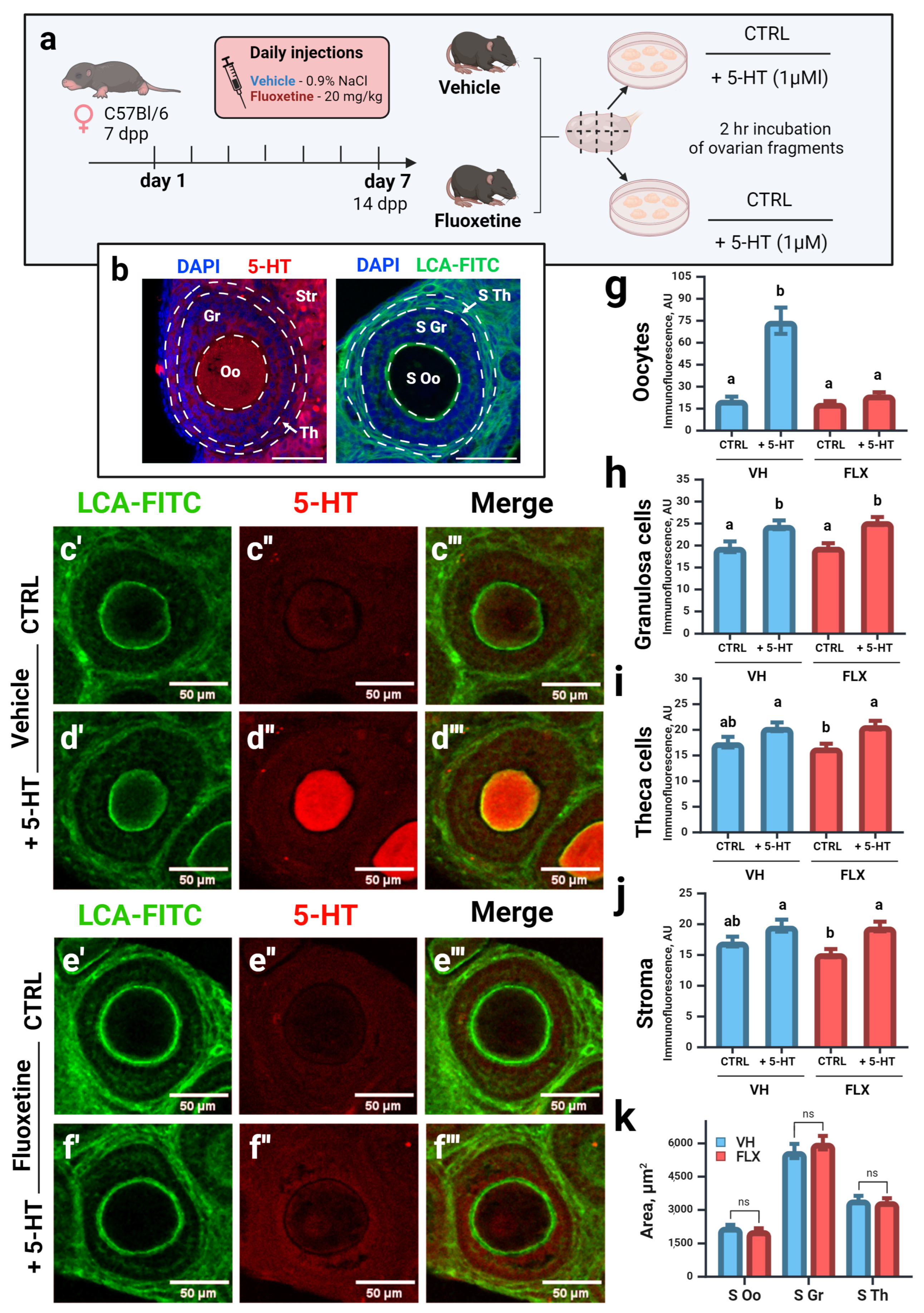
Given the evidence for a direct ovarian effect, we assessed the capacity for serotonin accumulation in ovarian compartments using an ex vivo culture system with ovaries from 14 dpp females, which are enriched with growing preantral follicles (Figure 2a). Ovarian fragments were incubated with 5-HT, and its uptake was visualized via immunostaining (Figure 2c–f).
      
    
    Figure 2.
      Effects of fluoxetine on serotonin accumulation in the ovary and follicular morphology. VH—vehicle treated, FLX—fluoxetine treated. (a) Experiment design. CTRL—control, incubation without extra chemicals, +5-HT—incubation with 1 μM of serotonin. n = 48 ovarian fragments for each group. (b) Schematic representation of the zones of interest. CLSM microphotographs of ovarian tissue (growing preantral follicles) labeled with antibodies against serotonin (5-HT) and with Lens Culinaris agglutinin conjugated with FITC (LCA-FITC), nuclei were marked with DAPI. Oo—oocyte, Gr—granulosa cells, Th—theca cells, Str—stromal tissue, S Oo—area of oocyte, S Gr—area of granulosa cell layer, S Th—area of theca cell layer. Scale bar 50 μm. (c′–f″′) Morphology and serotonin accumulation in ovarian tissue of experimental animals after incubation in the presence of 1μM of 5-HT. (g–j) Quantification of anti-serotonin immunoreactivity in different compartments of the ovarian follicle (M ± SEM)—in oocytes (g), granulosa cells (h), theca cells (i) and stroma (j). AU—arbitrary units of immunofluorescence. Letters a and b indicate significant differences (p < 0.05) between groups using the Kruskal–Wallis test with Dunn’s multiple comparisons test. (k) Quantification of the area of growing follicle compartments (M ± SEM). ns—not significant according to the Mann–Whitney U test. Figure created with BioRender.com (accessed on 10 September 2025).
  
As expected, oocytes from control animals exhibited robust 5-HT accumulation (Figure 2d″,g). In contrast, this accumulation was nearly abolished in oocytes from fluoxetine-treated females (Figure 2f″,g), consistent with both the systemic serotonin depletion and direct inhibition of the oocyte’s serotonin transporter (Sert). Other ovarian compartments, such as granulosa and theca cell layers, showed minimal 5-HT accumulation with no significant differences between groups (Figure 2h–j), suggesting the involvement of transport mechanisms insensitive to fluoxetine in these cells.
We then assessed follicular morphology in these preantral follicles (80–130 μm diameter). Analysis of the cross-sectional area of the oocyte, granulosa layer, and theca layer revealed no significant differences between the control and fluoxetine-treated groups (Figure 2b,k). This indicates that at this time point, fluoxetine impairs a key oocyte function—serotonin uptake—rather than inducing gross morphological changes in the follicle.
2.4. Fluoxetine Alters the Expression of Key Ovarian Genes
To dissect the molecular mechanisms underlying fluoxetine’s impact, we performed qPCR analysis on a curated set of genes that reflect the functional status of oocytes and somatic follicular cells.
2.4.1. Fluoxetine Downregulates the Expression of Key Oocyte-Secreted Factors and Sert
We first evaluated the expression of critical oocyte-derived factors that orchestrate folliculogenesis. Expression of Gdf9 was significantly downregulated in both adult and prepubertal mice following fluoxetine treatment, with a notable and consistent effect observed in both groups (Figure 3a,a′). Similarly, the expression of Bmp15 and Bmp6 was significantly reduced in prepubertal animals, although no significant change was observed in adults (Figure 3b,c,b′,c′).
      
    
    Figure 3.
      Expression of genes encoding oocyte factors after 7 days of fluoxetine treatment in the ovaries of mature (a–d) and prepubertal (a′–d′) females. VH—vehicle treated; FLX—fluoxetine treated. The relative quantity (RQ) was calculated using the 2−ΔCt method relative to the reference genes Tbp and Rps18 (M ± SEM). Significance levels are indicated as *—p < 0.05, **—p < 0.01, ***—p < 0.001, ns—not significant according to the Mann–Whitney U test. Figure created with BioRender.com (accessed on 10 September 2025).
  
We next examined the expression of the fluoxetine target gene itself, Sert (Slc6a4). Interestingly, Sert mRNA levels were significantly decreased in the ovaries of both adult and prepubertal mice after fluoxetine treatment (Figure 3d,d′). This suggests the existence of a positive feedback mechanism where Sert activity may regulate its own transcription.
2.4.2. Fluoxetine Alters the Expression of Genes for Steroidogenesis and Gonadotropin Response
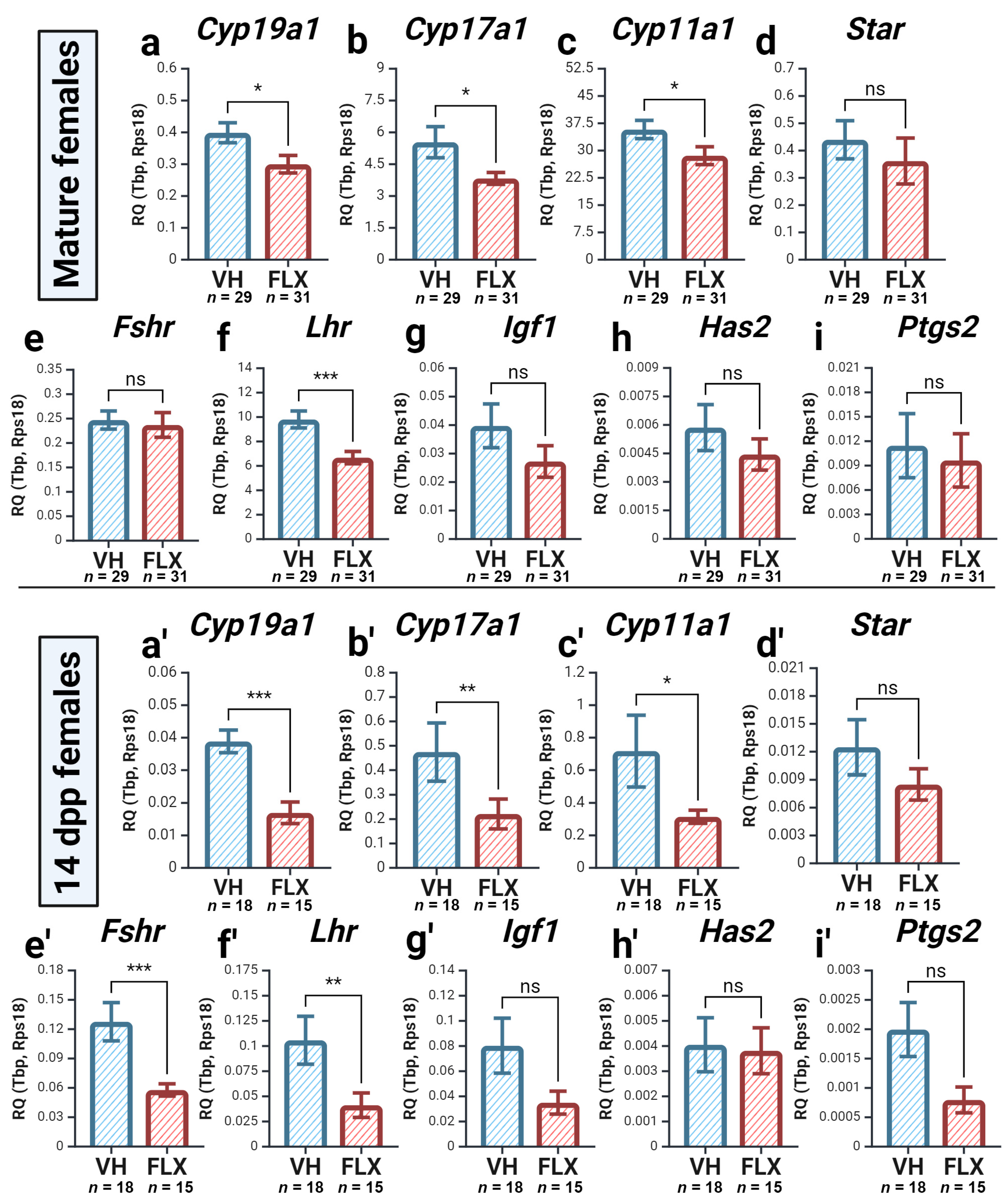
We then analyzed the expression of genes related to somatic cell function. The expression of genes crucial for both thecal androgen production (Cyp17a1) and granulosa cell estrogen synthesis (Cyp19a1) was significantly decreased in both age groups (Figure 4a,b,a′,b′). Likewise, the expression of Cyp11a1 and Star, involved in the initial steps of steroidogenesis, was also significantly downregulated (Figure 4c,d,c′,d′). This downregulation of steroidogenic gene expression is particularly noteworthy given the lack of change in systemic E2 levels, suggesting the presence of compensatory mechanisms that maintain hormonal homeostasis over this short-term exposure.
      
    
    Figure 4.
      Expression of genes encoding functional state of somatic cells and steroidogenic enzymes after 7 days of fluoxetine treatment in the ovaries of mature (a–i) and prepubertal (a′–i′) females. VH—vehicle treated, FLX—fluoxetine treated. The relative quantity (RQ) was calculated using the 2−ΔCt method relative to the reference genes Tbp and Rps18 (M ± SEM). Significance levels are indicated as *—p < 0.05, **—p < 0.01, and ***—p < 0.001, ns—not significant according to the Mann–Whitney U test. Figure created with BioRender.com (accessed on 10 September 2025).
  
Regarding gonadotropin sensitivity, the expression of Fshr was significantly reduced only in prepubertal mice, whereas Lhr expression was downregulated in both age groups (Figure 4e,f,e′,f′). Expression of Igf1 showed a non-significant decreasing trend in both groups (Figure 4g,g′). Finally, the expression of ovulation-associated genes, Has2 and Ptgs2, was not significantly affected in either model (Figure 4h,i,h′,i′). In summary, fluoxetine negatively impacts the transcriptional profile of genes essential for steroidogenesis and gonadotropin signaling, with effects often being more pronounced in the prepubertal ovary.
2.5. Fluoxetine Reduces Ovarian GDF9 Protein Levels
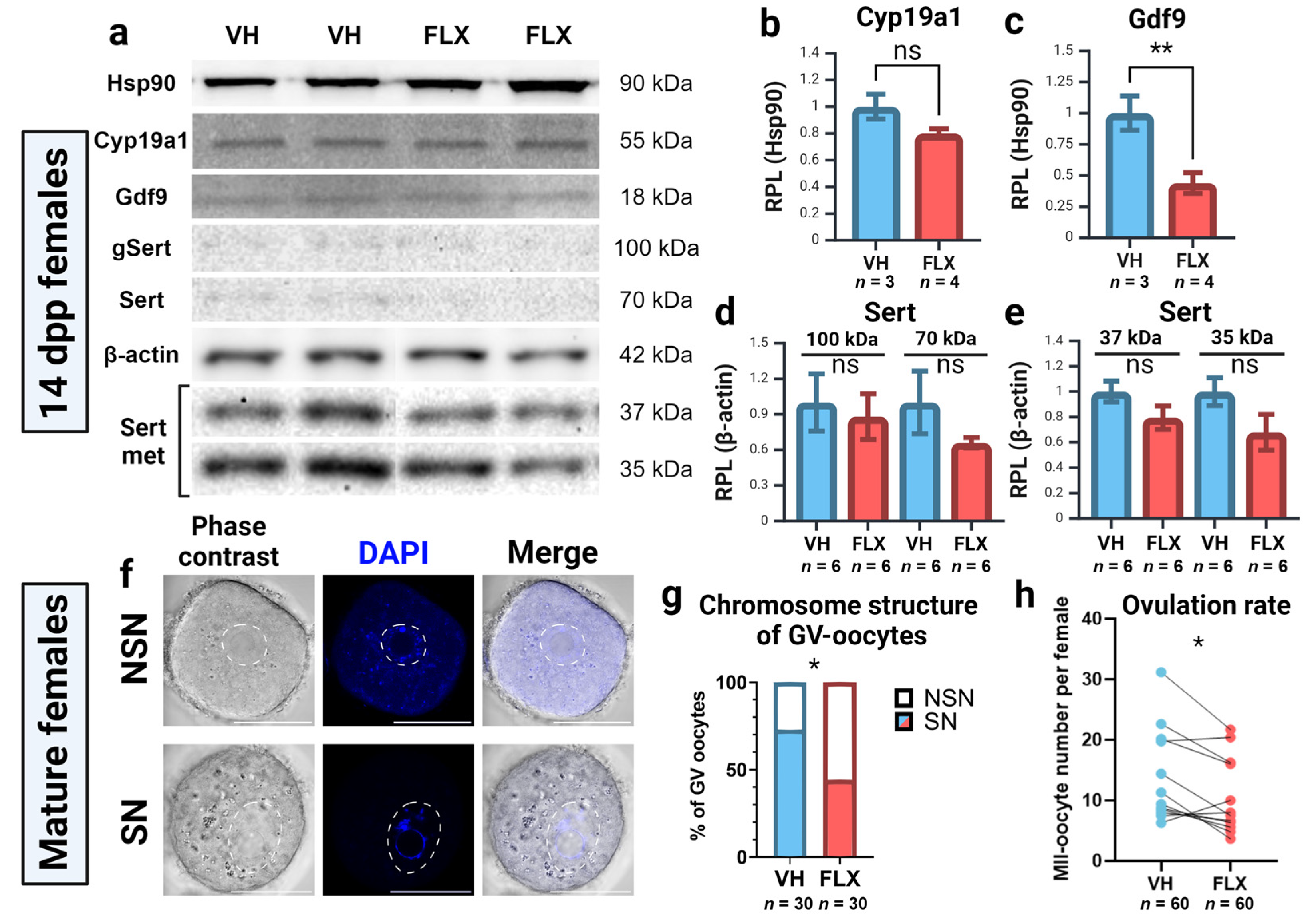
To validate our gene expression findings at the protein level, we performed Western blot analysis on ovarian lysates from prepubertal females for GDF9, Cyp19a1 (aromatase), and Sert (Figure 5a). Consistent with the stable systemic E2 levels, Cyp19a1 protein expression was unchanged by fluoxetine treatment (Figure 5b). In contrast, and in agreement with our qPCR data, GDF9 protein levels were significantly reduced in the ovaries of fluoxetine-treated mice (Figure 5c). For Sert, while a decreasing trend was observed, no statistically significant changes were detected in the abundance of its full-length (100 kDa), non-glycosylated (71 kDa), or fragmented forms (37–35 kDa) (Figure 5d,e). These results identify the downregulation of the critical oocyte-derived factor GDF9 as a key molecular consequence of fluoxetine exposure in the prepubertal ovary.
      
    
    Figure 5.
      Effects of 7-day fluoxetine treatment on protein expression and parameters of oocyte quality. VH—vehicle treated, FLX—fluoxetine treated. (a) Western blot analysis of Gdf9, Cyp19a1 and Sert protein expression in the ovaries of 14 dpp females in the experiment. gSert—glycosylated form of Sert protein, Sert met—immunopositive metabolites of Sert. (b–e) Quantification of the results of Western blot analysis. Relative protein expression level (RPL) was calculated in relation to reference proteins, Hsp90 and β-actin (M ± SEM). n—the number of samples analyzed per group. Significance levels are indicated as **—p < 0.01, ns—not significant according to the Mann–Whitney U test. (f) Visualization of chromatin conformation in nucleolus-like bodies of GV-oocytes. Scale bar 50 μm. (g) Analysis of NSN and SN-oocyte percentage obtained from the ovaries of vehicle and fluoxetine treated mature females. (h) Number of ovulated MII-oocytes obtained from oviducts of experimental females (M ± SEM). Paired values denote the quantity of oocytes from one experiment. Significance level is indicated as *—p < 0.05 according to the Wilcoxon test. Figure created with BioRender.com (accessed on 10 September 2025).
  
2.6. Fluoxetine Impairs Oocyte Competence and Reduces Ovulation Rate
To determine if the observed molecular changes translated to functional deficits, we assessed oocyte quality and ovulatory outcomes in adult mice. First, we evaluated germinal vesicle (GV)-stage oocytes retrieved after PMSG stimulation. Fluoxetine treatment led to a significant decrease in the proportion of developmentally competent oocytes with surrounded nucleolus (SN-type) and a corresponding increase in lower-quality non-surrounded nucleolus (NSN-type) oocytes (Figure 5f,g).
Furthermore, following a full superovulation protocol, fluoxetine-treated females yielded a significantly lower number of metaphase II (MII) oocytes compared to controls (Figure 5h). Collectively, these data demonstrate that the molecular and functional alterations induced by fluoxetine translate into a tangible decline in both oocyte quality and ovulatory potential.
2.7. Fluoxetine-Induced Oocyte Damage Is Independent of Oxidative Stress and Telomere Maintenance Pathways
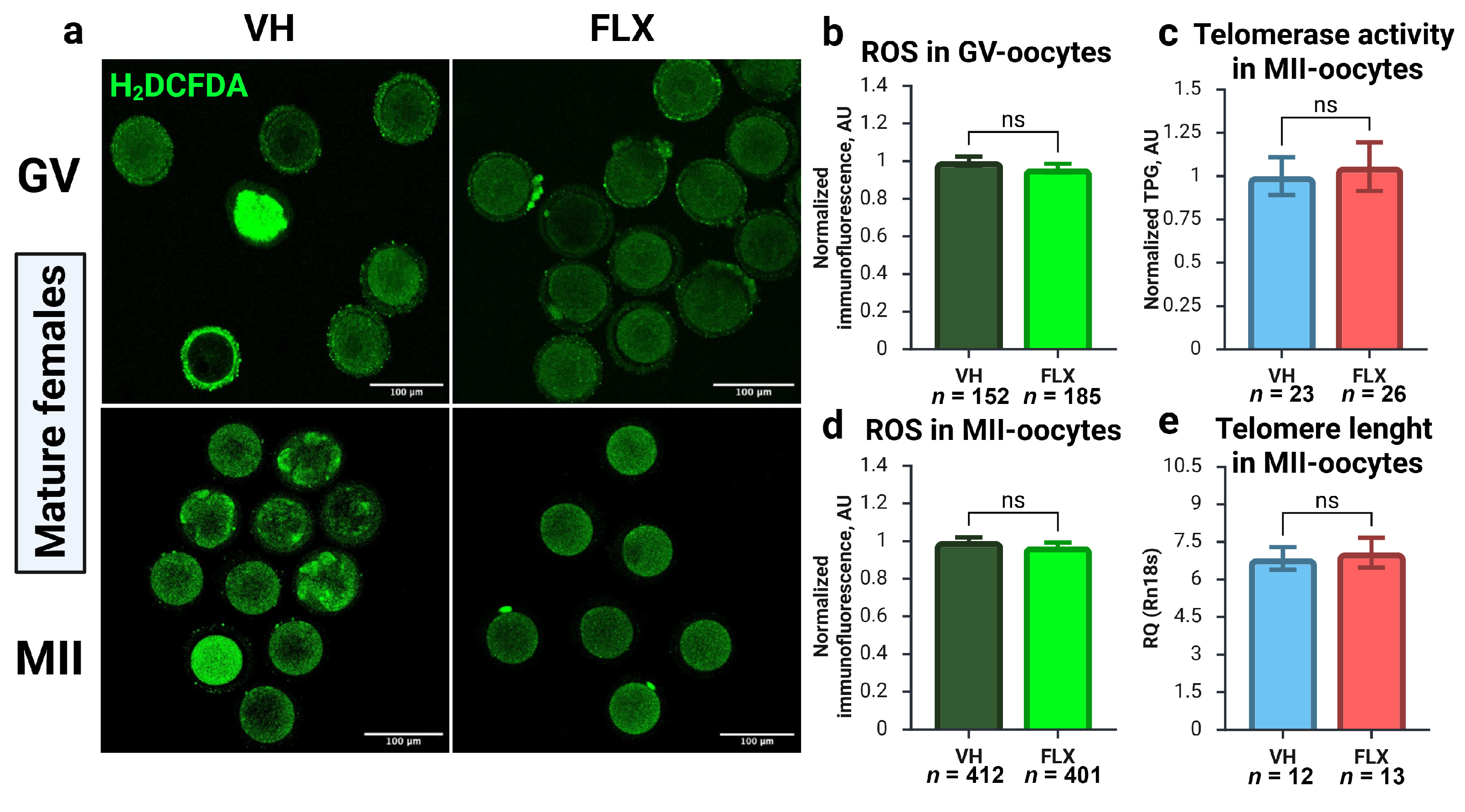
To elucidate the mechanisms underlying the decline in oocyte quality, we investigated two pathways known to impact gamete viability: oxidative stress and telomere maintenance (Figure 6). Quantification of reactive oxygen species (ROS) via H2O2 levels in GV-stage oocytes revealed no significant difference between fluoxetine-treated and control groups (Figure 6a,b). This was also true for MII oocytes matured in vivo (Figure 6a,d), indicating that fluoxetine does not induce a state of oxidative stress in the oocyte.
      
    
    Figure 6.
      Effects of 7-day fluoxetine treatment on ROS production and telomere maintenance machinery in GV- and MII-oocytes. VH—vehicle treated, FLX—fluoxetine treated. (a) CLSM microphotographs of GV- and MII-oocytes showing detection of intracellular ROS with a fluorescent probe 6-carboxy-H2DCFDA. (b,d) Quantification of ROS immunoreactivity in GV- and MII-oocytes obtained from experimental animals (M ± SEM). AU—arbitrary units of immunofluorescence, values are normalized to the mean value in the VH group. (c) Telomerase activity in MII-oocytes from experimental mice, analyzed by TRAP-qPCR and normalized to the values of the VH group. TPG—telomerase product generated, AU—arbitrary units. (e) Relative telomere length in MII-oocytes, calculated using the 2−ΔCt method relative to the reference gene Rn18s (M ± SEM). ns—not significant according to the Mann–Whitney U test. Figure created with BioRender.com (accessed on 10 September 2025).
  
Next, we assessed telomere integrity. Telomerase activity in MII oocytes was not significantly different between the treatment and control groups (Figure 6c). Moreover, qPCR analysis of telomere length also showed no evidence of fluoxetine-induced telomere shortening in MII oocytes (Figure 6e). Taken together, these results strongly suggest that the detrimental impact of fluoxetine on oocyte competence is not mediated by the induction of ROS-related oxidative stress or the disruption of telomere maintenance pathways.
3. Discussion
Research into the effects of various factors on ovarian function is essential to modern biology, medicine, and pharmacology. The ovary is not merely a component of the endocrine system; it houses the oocytes, which contain the molecular foundation for the development of a future organism and are themselves susceptible to external influences. Our recent work revealed that chronic fluoxetine exposure functionally impairs reproduction by reducing oocyte competence and ovulation rates []. However, that study raised a pivotal question: what are the direct, intra-ovarian molecular mechanisms driving this dysfunction? The present study serves as a direct mechanistic follow-up designed to answer this question. Our study, employing two distinct mouse models representing prepubertal and mature stages of sexual development, provides a comprehensive investigation into the effects of the widely used antidepressant fluoxetine on ovarian health, folliculogenesis, and oocyte quality.
The validity of any pharmacological model hinges on the precise calibration of its variables. We designed our experiments to specifically probe the effects of fluoxetine administration on folliculogenesis during distinct physiological periods. In the prepubertal model, the treatment window (7–14 dpp) was chosen to coincide with the first wave of follicular growth, initiated around 5–7 dpp [,]. This allowed us to isolate fluoxetine’s impact on the early, gonadotropin-independent stages of folliculogenesis, prior to antrum formation, active steroidogenesis, and ovulation. Conversely, the adult model targeted follicles recruited from the mature primordial follicle pool, which differ from those of the first wave []. In these animals, an 8-day exposure ensured that at least one full estrous cycle was affected and that both early-stage and preovulatory follicles were exposed []. Despite the fact that exposure was short-term, we already saw prominent effects on gene expression. Utilizing a short-term course of treatment also allowed us to isolate initial direct ovarian effects from potential long-term adaptive or central nervous system-mediated changes. To standardize the analysis, the cycle of adult mice was synchronized with PMSG prior to tissue collection.
The selected fluoxetine dosage of 20 mg/kg is another critical feature of our model, as it achieves plasma concentrations in mice that are comparable to the therapeutic levels observed in patients undergoing Prozac therapy (600–700 ng/mL), including adolescents [,,]. Although the dose of 20 mg/kg/day suggested by Dulawa [] can provoke high concentrations of fluoxetine and its metabolites in plasma, it can still be used in animal models, as the effective dose for anxiety and depression modeling, especially in juvenile mice []. The inclusion of a prepubertal model in our work is particularly relevant given the increasing use of SSRIs in pediatric and adolescent therapy [], as well as the risks associated with uncontrolled consumption.
A primary systemic effect observed in both models was a dramatic decrease in serum serotonin levels. This finding is consistent with studies of non-acute SSRI exposure in humans, which report similar reductions in plasma and platelets [,,,]. We hypothesize this occurs due to systemic Sert inhibition, including in gut enterocytes and platelets, leading to rapid hepatic metabolism of free serotonin. This indicates that our model captures not only the direct effects of fluoxetine but also the physiological consequences of peripheral serotonin depletion. Notably, and in contrast to some previous research, we observed no significant effect on circulating estradiol (E2) levels. To further dissect the mechanism, we analyzed FSH and LH levels and found them to be unchanged, a finding corroborated by the lack of alterations in estrous cyclicity. Even in prepubertal mice, where we anticipated potentially subtler effects and measured pituitary Fshb and Lhb expression, no changes were detected. Collectively, these data strongly suggest that the observed ovarian detriments are not mediated by the central hypothalamic–pituitary–gonadal axis but rather stem from direct effects within the ovary.
To assess whether these direct effects manifested as morphological changes, we analyzed growing follicles in prepubertal mice. The 14 dpp timepoint is ideal for such analysis due to the homogeneity of the follicular population, which predominantly consists of primordial and early growing follicles at a uniform, FSH-independent stage (80–130 μm diameter) []. This allows for standardized quantification of oocyte, granulosa, and theca volumes. However, our analysis revealed no differences in these morphological parameters between experimental groups. This lack of gross structural change suggested that the effects of fluoxetine and low serotonin are more subtle, impacting follicles at a molecular and functional level.
We have previously demonstrated the presence of active serotonin transport in ovarian tissue, primarily within growing oocytes [] and to a lesser extent in somatic follicular cells []. In the current study, we confirmed that in vivo fluoxetine treatment significantly ablates serotonin (5-HT) accumulation in the oocytes of growing follicles. This finding is of significant concern because these first-wave follicles are developmentally competent and contribute to the ovulatory pool []. Furthermore, serotonin-mediated signaling is a phylogenetically conserved mechanism involved in oocyte maturation across diverse species, including mammals [,,]. As we recently showed that serotonin accumulation capacity increases with oocyte maturation [], our current results confirm that SSRIs directly disrupt a fundamental physiological process within the oocyte itself.
Interestingly, fluoxetine treatment did not significantly affect Sert activity within the granulosa cell layer. While our previous short-term in vitro experiments did detect Sert and MAO activity in primary granulosa cells [], it is possible that during the longer in vivo exposure of our current model, any effects on somatic cells are mitigated by compensatory mechanisms or non-specific transport. The consistent and robust observation of specific Sert activity in oocytes across multiple experiments validates the concept that high-affinity serotonin transport is a specialized feature of the female gamete.
Even more striking than the inhibition of Sert activity was the downregulation of its own gene expression. We also observed a trend towards decreased protein expression for both major isoforms of Sert (100 and 70 kDa) and its metabolites [], indicating a reduction in its synthesis or an increase in its degradation. This aligns with clinical findings of reduced Sert binding sites on platelets during fluoxetine therapy [] and cellular studies showing that Sert can be internalized and metabolized []. Given that Sert phosphorylation and inhibition by fluoxetine are dependent on serotonin as a substrate [], our results may represent an unusual example of a disrupted positive feedback loop, where the transporter’s own activity is required for its sustained expression, a process unique to the oocyte’s physiology.
To identify the molecular consequences of Sert disruption, we analyzed the expression of key oocyte-derived factors: Gdf9, Bmp15, and Bmp6. GDF9 and BMP15 are critical for follicle maturation [,], with their expression increasing as follicles are recruited from the dormant pool []. GDF9, in particular, is essential for nearly all stages of follicular growth and ovulation [,], while BMP6 is another key oocyte-derived factor [] also found in granulosa cells [] and follicular fluid []. We observed a significant decrease in the expression of all three factors in prepubertal females. The more pronounced effect in the 14 dpp ovary is likely due to the higher oocyte-to-stroma ratio, which amplifies the oocyte’s transcriptional signal in whole-ovary lysates. Critically, Gdf9 expression declined in both age groups, a result we confirmed at the protein level, strongly suggesting a direct impact of fluoxetine and/or low serotonin on this pathway. This perfectly corroborates our previous in vitro work, where serotonin supplementation increased Gdf9 expression—an effect that was abolished by fluoxetine []. Given the dominant role of GDF9 in folliculogenesis in poly-ovulatory species like mice [], we propose that this disruption of the GDF9 signaling axis is a central mechanism underlying fluoxetine-induced ovarian dysfunction.
The central finding of GDF9 downregulation raises the question of its particular mechanism: whether it is a direct consequence of SERT inhibition within the oocyte or a secondary effect of systemic serotonin depletion. While our data cannot definitively separate these two interconnected events, several lines of evidence point to a critical role for intra-oocyte serotonin signaling. Serotonin itself can act as a signaling molecule, and its transport can influence cellular energy status and gene expression. A potential upstream regulator of Gdf9 that could be affected by disrupted serotonin signaling is NOBOX, which is vital for oocyte-specific gene expression of many genes, let alone GDF9 []. However, there is a lack of scientific data on SRRIs’ action on NOBOX and many other regulators of ovarian function. Future studies using oocyte-specific Sert knockout models or research on direct serotonin effects on NOBOX and other factors in ovaries and oocytes will be necessary to dissect this precise mechanism of fluoxetine action.
Initially, we hypothesized that fluoxetine would primarily impact steroidogenesis. However, we found no significant change in systemic E2 levels. Despite this, we observed a significant decline in the gene expression of key steroidogenic enzymes, including Cyp19a1, Cyp17a1, and Cyp11a1. We attribute the disconnect between gene expression and circulating hormone levels to two factors. First, the short duration of the experiment may have been insufficient to deplete existing enzyme reserves and impact systemic E2. Second, extra-ovarian sources such as the brain, bone, and adipose tissue can contribute to and compensate for total blood E2 levels []. We also acknowledge that directly measuring E2 secretion from ovarian explants or primary granulosa cell cultures would have provided more definitive evidence for a local intra-ovarian steroidogenic defect. The absence of such direct functional data is a limitation of our study, and this will be an important focus of our future in vitro investigations. The observed downregulation of genes responsible for androgen synthesis is also noteworthy, as SSRIs have been reported to reduce testosterone levels []. While beyond the scope of this study, a drop in ovarian androgen precursors could be a contributing factor to altered steroidogenesis.
Another crucial aspect of granulosa cell function is their sensitivity to gonadotropins. We observed a prominent decrease in the expression of gonadotropin receptors. Interestingly, Fshr expression was reduced in prepubertal mice but not in adults. This highlights the 7–14 dpp period as a potential window of vulnerability. Follicles acquire FSH susceptibility at early stages, even before they become fully dependent on it for antral development [,]. Fluoxetine exposure during this critical period of the first folliculogenesis wave may disrupt the establishment of the FSH receptor system. In contrast, the stable Fshr expression in adult mice may reflect a more established and resilient system. In terms of Lhr, expression decreased in both models. As both FSHR and LHR are critical for follicle selection, steroidogenesis, and the switch to LH-dependence pre-ovulation [,], these results indicate that fluoxetine can compromise follicular quality by impairing gonadotropin sensitivity, particularly in the still-developing prepubertal ovary.
The culmination of normal follicular development is ovulation, a process requiring functional changes in the cumulus-oocyte complex []. The expression of Has2, which produces the hyaluronic acid backbone of the expanded matrix [], and Ptgs2 (Cox2), essential for LH response [], are both induced by GDF9 signaling []. Despite our previous data showing serotonin stimulates these genes in vitro, we saw no changes in our current in vivo models. This is likely explained by the experimental design: prepubertal follicles lack a defined cumulus and are not yet competent for ovulation, while adult mice received only PMSG and not the ovulatory hCG trigger required to induce the expression of these periovulatory genes.
Higher vulnerability of prepubertal ovaries to fluoxetine is a critical finding. This increased sensitivity is likely multifactorial. Firstly, the higher oocyte-to-stroma ratio in prepubertal ovaries amplifies the transcriptional contribution of oocytes in whole-ovary analyses, making the downregulation of oocyte-specific genes like Gdf9 more pronounced. Secondly, the first wave of folliculogenesis represents a critical window where key signaling pathways, including the FSH receptor system, are being established. Disruption by fluoxetine during this period may have more profound and lasting effects compared to the mature, cyclically recruited follicles in adults, which operate within a more stable and resilient endocrine system. Finally, developmental differences in SERT expression, activity, or its associated regulatory networks warrant further investigation.
Ultimately, the most critical readout of any toxicant effect is the direct impact on oocyte quality and quantity. We evaluated oocytes at both the germinal vesicle (GV) and metaphase II (MII) stages. In adult mice, fluoxetine treatment caused a significant decrease in the proportion of high-quality GV-oocytes with a surrounded nucleolus (SN-type), which are known to have better developmental competence [,], and a corresponding increase in lower-quality NSN-type oocytes. Furthermore, we clearly demonstrated that the number of MII oocytes retrieved after superovulation was significantly reduced by fluoxetine exposure. Our analysis of ROS levels and telomere maintenance machinery suggests these negative effects are not mediated by oxidative stress or telomere disruption. Instead, these results demonstrate that even short-term fluoxetine exposure during the final stages of follicular growth has a direct, negative impact on oocyte competence and ovulatory success.
A key limitation of the current study is that we did not assess the ultimate functional endpoints of oocyte competence. While the observed decrease in SN-type oocytes and reduced ovulation rates are strong indicators of compromised fertility, a crucial next step will be to conduct in vitro fertilization studies. Assessing the fertilization capacity of fluoxetine-exposed oocytes and their subsequent potential for preimplantation embryonic development is essential to directly link the molecular disruptions we identified to tangible reproductive outcomes. It is also important to acknowledge that our study focused on a short-term exposure model to capture the initial molecular response within the ovary. This design revealed a transcriptional response in granulosa cells that was not yet reflected at the aromatase protein level or systemic estradiol levels. This suggests that the primary effect is a disruption of oocyte–granulosa cell signaling rather than an immediate impairment of steroidogenesis. This model, however, does not inform on the long-term consequences of chronic SSRI use or the potential for recovery after drug cessation. Future studies involving chronic administration and withdrawal periods are needed to fully understand the clinical implications of these findings. Finally, while our data point towards a SERT-mediated mechanism within the oocyte, it is critical to acknowledge that fluoxetine possesses known non-serotonergic activities, including interactions with sigma-1 receptors [], which are also expressed in ovarian tissue. Comparative experiments in a Sert genetic knockout mouse model would be crucial to definitively ascertain whether the observed effects on oocyte competence are exclusively mediated by SERT inhibition or involve these alternative pathways.
While our study identifies an intraovarian mechanism of fluoxetine action in mice, it is important to consider the limitations when extrapolating these findings to human fertility outcomes. Mice are polyovulatory with different follicular dynamics and endocrine profiles compared to humans. Furthermore, human reproductive cycles are longer and more complex. Therefore, while our data highlight a potentially significant risk to the ovarian reserve and oocyte quality, future studies using human ovarian cortical tissue, granulosa cell cultures, or correlative clinical data from IVF cycles are essential to confirm the translational relevance of these findings for women of reproductive age.
In conclusion, our findings identify follicular development as a critical period of vulnerability to fluoxetine, with the oocyte itself acting as the primary target of a disruptive molecular cascade.
4. Materials and Methods
4.1. Animals and Ethical Approval
Female C57BL/6J mice were obtained from the Stolbovaya breeding facility (Moscow region, Russia) and housed in the vivarium of the Koltzov Institute of Developmental Biology, Russian Academy of Sciences (IDB RAS). Animals were maintained under standard controlled conditions (22–24 °C, 14L:10D photoperiod) with ad libitum access to food and water. All experimental procedures were performed in accordance with the Council of the European Communities Directive 86/609/EEC and were approved by the Commission on Bioethics of IDB RAS (project identification code: № 68, date: 23 March 2023).
4.2. Experimental Design, Drug Administration, and Sample Collection
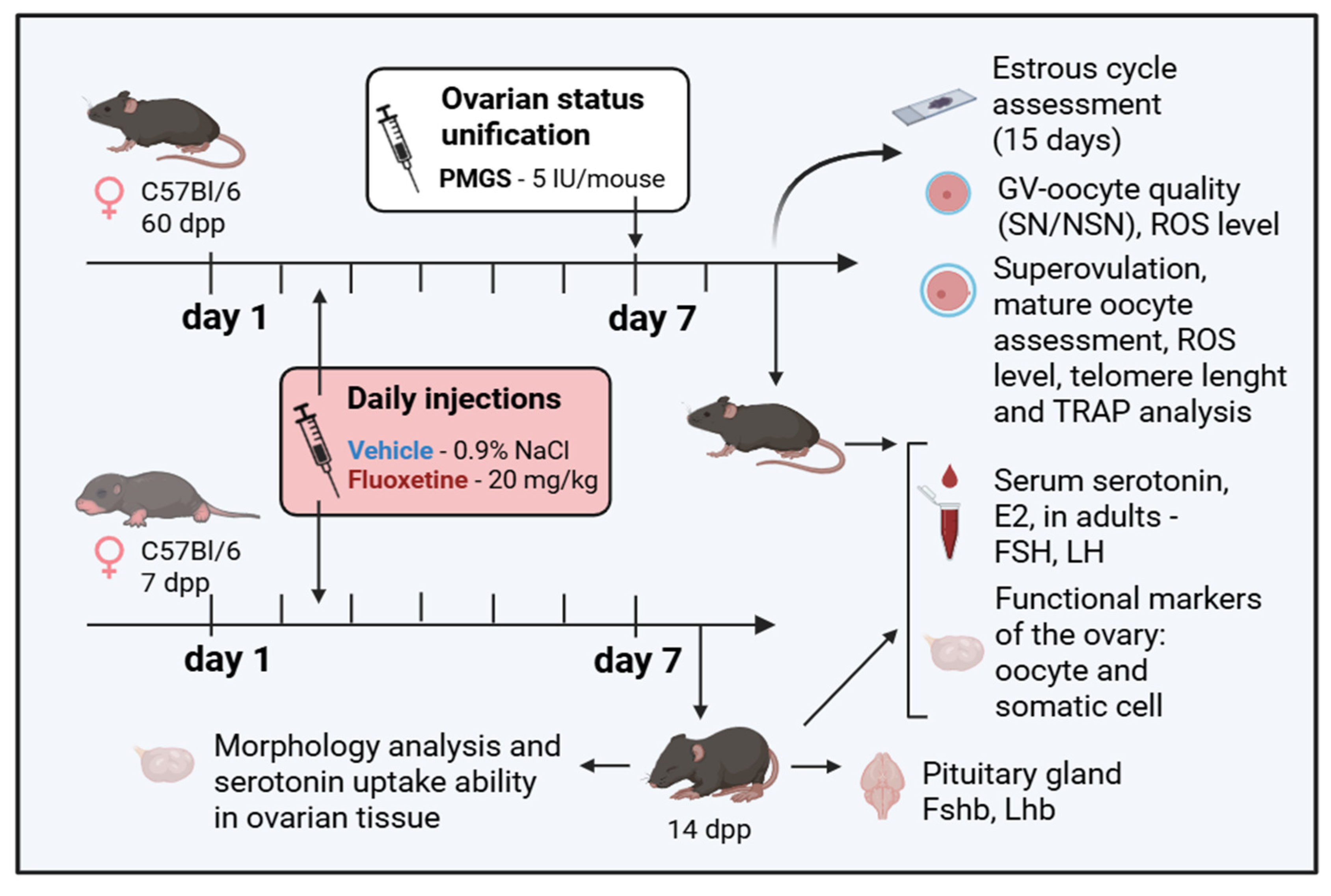
Two age-based models were used (Figure 7): a prepubertal model, with treatment starting on postnatal day 7 (7 dpp), and a sexually mature adult model (2 months old, ~60 dpp).
      
    
    Figure 7.
      The scheme of the experiment. Mature (60 dpp) and prepubertal (7 dpp) mice were treated with fluoxetine (FLX, 20 mg/kg/day) for 8 days and for 7 days, respectively, while control groups had saline injections (vehicle, VH, 0.9% NaCl). The next day after the final fluoxetine injection, biomaterials were obtained for multiple tests. Figure created with BioRender.com (accessed on 10 September 2025).
  
Female pups starting at 7 dpp received daily subcutaneous (s.c.) injections of fluoxetine hydrochloride (FLX, F132, Sigma-Aldrich, St. Louis, MO, USA) at a dose of 20 mg/kg or an equivalent volume of saline (0.9% NaCl, vehicle) for 7 consecutive days. Biomaterials were collected on day 8 (at 15 dpp). This time point coincides with the first wave of folliculogenesis, where follicles are largely synchronized and beginning to form early antrums, eliminating the need for hormonal synchronization [].
Mature females received daily s.c. injections of FLX (20 mg/kg) or vehicle for 8 consecutive days. To standardize the follicular stage for analysis, folliculogenesis was synchronized by a single s.c. injection of 5 IU pregnant mare serum gonadotropin (PMSG, Follimag®, Mosagrogen, Russia) on day 7 of treatment. Tissues and serum were collected 40 h post-PMSG injection. For the collection of ovulated metaphase II (MII) oocytes, the PMSG injection was followed 48 h later by an s.c. injection of 5 IU human chorionic gonadotropin (hCG, Chorulon®, Intervet International B.v., Boxmeer, Netherlands), and oocytes were collected from the oviducts 14–16 h post-hCG.
A total of 269 adult female mice and 70 prepubertal female mice were used in this study. Sample sizes were determined based on our previous work and published literature to ensure sufficient statistical power while adhering to the 3Rs principle of animal welfare. Within each age group (prepubertal and adult), animals were randomly assigned to either the control (vehicle) or experimental (fluoxetine) group. To minimize bias, experimenters performing sample collection and data analysis were blinded to the group allocations. Data points were excluded from the analysis only in cases of sample damage or processing errors.
On the day of collection, mice were humanely euthanized by cervical dislocation. Blood was collected via cardiac puncture for serum preparation, and ovaries and other tissues were harvested for subsequent analyses. The estrous cycle stage of adult mice was monitored daily by vaginal cytology as previously described [].
4.3. Serum Analysis: HPLC and ELISA
Serum serotonin concentration was measured using high-performance liquid chromatography with electrochemical detection (HPLC-ED), as previously described []. Briefly, 20 μL of serum was deproteinized with 200 μL of 0.1 N HClO4 containing 3,4-dihydroxybenzylamine as an internal standard.
Serum concentrations of 17β-estradiol (E2), follicle-stimulating hormone (FSH), and luteinizing hormone (LH) were quantified using commercial enzyme-linked immunosorbent assay (ELISA) kits following the manufacturer’s protocols: ImmunoFA-Estradiol (Immunotech, Moscow, Russia), Mouse FSH ELISA Kit (ELK8859, ELK Biotechnology, Wuhan, China), and Mouse LH ELISA Kit (ELK1327, ELK Biotechnology, Wuhan, China).
4.4. Oocyte Collection, Staining, and Analysis
MII-oocytes within cumulus-oocyte complexes were collected from the oviductal ampullae of superovulated females. Cumulus cells were removed by brief incubation in M2 medium containing hyaluronidase. Only morphologically normal MII oocytes were used for quantification and further analysis.
GV-stage oocytes were mechanically isolated from the antral follicles of adult ovaries in M2 medium. For chromatin configuration analysis, oocytes were fixed in 4% paraformaldehyde (PFA), permeabilized with 0.1% Triton X-100 in PBS (PBST), and stained with DAPI (D9542, Merck KGaA, Darmstadt, Germany). Oocytes were classified as surrounded nucleolus (SN) or non-surrounded nucleolus (NSN) type by confocal microscopy.
For ROS assessment, live GV and MII oocytes were incubated for 30 min in medium containing 10 µM 6-carboxy-2′,7′-dichlorodihydrofluorescein diacetate (carboxy-H2DCFDA, 3290, Lumiprobe, Moscow, Russia). Oocytes were then washed and imaged immediately in glass-bottom dishes using a confocal microscope to quantify fluorescence intensity.
4.5. Immunohistochemistry (IHC) and Image Analysis
Ovaries were fixed overnight in 4% PFA at 4 °C, cryoprotected in a sucrose gradient (15% then 30%), embedded in Tissue-Tek O.C.T. Compound (4583, Sakura, Torrance, CA, USA), and sectioned at 10 μm. For serotonin localization, sections were blocked with 5% fetal bovine serum (FBS, Pan-Eco, Moscow, Russia) and incubated overnight at 4 °C with a primary rabbit anti-serotonin antibody (1:1000, S5545, Merck KGaA, Darmstadt, Germany). To assess in vitro serotonin uptake capacity, ovaries from 14 dpp females were fragmented and incubated for 2 h in Leibovitz’s L-15 Medium (L0230, BioSera, Nuaille, France) with 1 μM serotonin before fixation and processing as above.
Sections were subsequently incubated with a secondary antibody (CF568-conjugated anti-rabbit IgG, 1:500, SAB4600085, Merck KGaA), FITC-conjugated Lens culinaris agglutinin (LCA, 1:1000, L32475, Thermo Fisher Scientific, Waltham, MA, USA) to visualize cell boundaries, and DAPI for nuclear staining. Images were acquired using a Zeiss LSM 880 Airyscan confocal microscope with consistent settings for all samples. Image analysis was performed using FIJI software (ImageJ 2.9.0/1.54f, open source, available at https://imagej.net/software/fiji/ (URL accessed on 29 March 2025)). Serotonin immunoreactivity was quantified by measuring the mean gray value (MGV) in regions of interest (oocyte, granulosa, theca). Follicle morphometry was assessed on LCA-stained images [].
4.6. Gene Expression Analysis
Total RNA was extracted from whole ovaries or pituitaries using ExtractRNA reagent (Evrogen, Moscow, Russia) and treated with DNase I (DNA-free™ Kit, AM1906, Thermo Fisher Scientific, Waltham, MA, USA). RNA was reverse transcribed using an MMLV RT kit (Evrogen). Quantitative PCR was performed on a 7500 Real-Time PCR System (Applied Biosystems, Foster City, CA, USA) using 5x qPCRmix-HS SYBR+LowRox (Evrogen). The specificity of each reaction was controlled by melting curve analysis. Relative gene expression was calculated using the 2−ΔCt method, with Rps18 and Tbp as reference genes. For this calculation, a uniform fluorescence threshold was applied to determine cycle threshold (Ct) values for all genes within a single experiment. The reference genes Tbp (TATA-box binding protein) and Rps18 (ribosomal protein S18) were selected for their stable expression in mouse ovarian tissue across different developmental stages and treatment conditions, as validated in our previous work and other literature [,]. All primer sequences are listed in Table 1.
       
    
    Table 1.
    Primer sequences used for qPCR analysis.
  
4.7. Telomerase Activity (TRAP) and Telomere Length Analysis
Telomerase activity in MII oocytes was assessed using the Telomeric Repeat Amplification Protocol (TRAP) via qPCR, as previously described []. For telomere length analysis, genomic DNA was extracted from pools of 50–100 MII oocytes using the QIAamp UCP DNA Micro Kit (56204, Qiagen, Hilden, Germany). Relative telomere length was determined via qPCR, comparing the amplification of telomeric repeats (Tel) to that of a reference gene (Rn18s), a multi-copy gene suitable for low-input samples []. Primer sequences are listed in Table 1.
4.8. Western Blotting
Ovarian protein lysates were prepared using RIPA buffer, and protein concentration was determined with a BCA Protein Assay Kit (23225, Thermo Fisher Scientific, Waltham, MA, USA). Proteins (20–30 µg) were denatured, separated by SDS-PAGE (10% or 12% gels), and transferred to a PVDF membrane. For the analysis of Cyp19a1 and Gdf9, 6 biological replicates from control mice and 7 biological replicates from fluoxetine-treated mice were initially analyzed. Similarly, for Sert, 6 biological replicates from control mice and 7 biological replicates from fluoxetine-treated mice were analyzed. Each biological sample was run in a single technical replicate. Membranes were blocked with 5% BSA and incubated with primary antibodies (Table 2). Following incubation with HRP-conjugated secondary antibodies (Table 2), chemiluminescence was detected using a Fusion-FX7 system (Vilber Lourmat, France). Band intensities were quantified using Image Lab software (Version 6.0.1, Bio-Rad Laboratories, Inc., Hercules, CA, USA) and normalized to β-actin or Hsp90 as loading controls. The final number of biological replicates included in the quantitative analysis for each protein is indicated in the respective figure legends (Figure 5).
       
    
    Table 2.
    List of antibodies used for membrane staining in Western blotting.
  
4.9. Statistical Analysis
Statistical analysis was performed using GraphPad Prism 9.0 (GraphPad Software, San Diego, CA, USA). Data were first tested for normality using the Shapiro–Wilk test. For comparison of two groups, an unpaired Student’s t-test or Mann–Whitney U test was used for normally or non-normally distributed data, respectively. For multiple group comparisons, one-way or two-way ANOVA followed by an appropriate post hoc test (e.g., Tukey’s or Sidak’s) was applied. Data are presented as the mean ± SEM. A p-value of < 0.05 was considered statistically significant.
5. Conclusions
Our research demonstrates that fluoxetine has a potential impact on ovarian function, and we propose a potential mechanism centered on the oocyte. We suggest a proposed mechanism: fluoxetine inhibits the oocyte’s serotonin transporter (SERT), leading to a significant downregulation of the pivotal oocyte-derived factor GDF9. This disruption, in turn, dysregulates gene expression in surrounding somatic cells, ultimately manifesting as compromised oocyte competence and reduced ovulatory potential. Critically, the heightened vulnerability observed in prepubertal females indicates that early follicular development is a window of particular susceptibility to SSRI-induced damage. These findings carry significant clinical weight, raising concerns about the potential impact of SSRI therapy on the establishment and preservation of the ovarian reserve in adolescents and underscoring the need for informed counseling when prescribing these medications to women of reproductive age.
Author Contributions
Conceptualization, D.A.N.; methodology, D.A.N., Y.O.N. and M.L.S.; investigation, N.M.A., M.V.B., M.D.T., Y.O.N., V.S.F., L.A.M., M.L.S., M.P.R. and D.A.N.; writing—original draft preparation, N.M.A. and M.V.B.; writing—review and editing, N.M.A. and D.A.N.; supervision, D.A.N.; project administration, D.A.N.; funding acquisition, D.A.N. and M.P.R. All authors have read and agreed to the published version of the manuscript.
Funding
This research was funded by the Russian Science Foundation, grant number 22-74-10009, for N.M.A., Y.O.N., V.S.F., and D.A.N. In the part of the work devoted to the evaluation of oocyte quality markers, this research was funded by the Government Program for Basic Research in the Koltzov Institute of Developmental Biology of the Russian Academy of Sciences in 2025, № 0088-2024-0012 for N.M.A., M.V.B., M.D.T., Y.O.N., L.A.M. and D.A.N. The part in which ROS level and telomerase activity were analyzed was funded by the Development Program of the MSU Interdisciplinary Scientific and Educational School “Molecular technologies of living systems and synthetic biology” at Lomonosov Moscow State University, grant № 23-SH04-20 for M.D.T., V.S.F., M.P.R. and D.A.N.
Institutional Review Board Statement
Experiments were performed in accordance with the Council of the European Communities Directive of 24 November 1986 (86/609/EEC). All protocols of animal experiments were approved by the Commission on Bioethics of the Koltzov Institute of Developmental Biology of the Russian Academy of Sciences (project identification code: № 68, date: 23 March 2023).
Informed Consent Statement
Not applicable.
Data Availability Statement
The original contributions presented in this study are included in the article. Further inquiries can be directed to the corresponding author.
Acknowledgments
The study was carried out using the equipment of the Core Centrum of Koltzov’s Institute of Developmental Biology RAS.
Conflicts of Interest
The authors declare no conflicts of interest.
Abbreviations
The following abbreviations are used in this manuscript:
      
| 5-HT | 5-hydroxytryptamine, serotonin | 
| dpp | days postpartum | 
| E2 | 17β-estradiol | 
| FLX | fluoxetine | 
| FSH | follicle-stimulating hormone | 
| GV | germinal vesicle | 
| hCG | human chorionic gonadotropin | 
| LH | luteinizing hormone | 
| MII | metaphase II | 
| NSN | non-surrounded nucleolus-like bodies | 
| PMSG | pregnant mare serum gonadotropin | 
| RPL | relative protein levels | 
| RQ | relative gene expression | 
| SN | surrounded nucleolus-like bodies | 
| SSRI | selective serotonin reuptake inhibitor | 
| VH | vehicle | 
References
- Albert, P.R. Why Is Depression More Prevalent in Women? J. Psychiatry Neurosci. 2015, 40, 219–221. [Google Scholar] [CrossRef]
 - Kaihola, H.; Yaldir, F.G.; Hreinsson, J.; Hörnaeus, K.; Bergquist, J.; Olivier, J.D.A.; Åkerud, H.; Sundström-Poromaa, I. Effects of Fluoxetine on Human Embryo Development. Front. Cell. Neurosci. 2016, 10, 160. [Google Scholar] [CrossRef] [PubMed]
 - Kim, C.-W.; Choe, C.; Kim, E.-J.; Lee, J.-I.; Yoon, S.-Y.; Cho, Y.-W.; Han, S.; Tak, H.-M.; Han, J.; Kang, D. Dual Effects of Fluoxetine on Mouse Early Embryonic Development. Toxicol. Appl. Pharmacol. 2012, 265, 61–72. [Google Scholar] [CrossRef] [PubMed]
 - Romero-Reyes, J.; Cárdenas, M.; Damián-Matsumura, P.; Domínguez, R.; Ayala, M.E. Inhibition of Serotonin Reuptake in the Prepubertal Rat Ovary by Fluoxetine and Effects on Ovarian Functions. Reprod. Toxicol. 2016, 59, 80–88. [Google Scholar] [CrossRef]
 - Reefhuis, J.; Devine, O.; Friedman, J.M.; Louik, C.; Honein, M.A. Specific SSRIs and Birth Defects: Bayesian Analysis to Interpret New Data in the Context of Previous Reports. BMJ 2015, 351, h3190. [Google Scholar] [CrossRef]
 - Ruiz-Santiago, C.; Rodríguez-Pinacho, C.V.; Pérez-Sánchez, G.; Acosta-Cruz, E. Effects of Selective Serotonin Reuptake Inhibitors on Endocrine System (Review). Biomed. Rep. 2024, 21, 1–13. [Google Scholar] [CrossRef]
 - Gök, S.; Gök, B.C.; Alataş, E.; Senol, H.; Topak, O.Z. Effects of Selective Serotonin Reuptake Inhibitor Treatment on Ovarian Reserves in Patients with Depression. Medicina 2023, 59, 517. [Google Scholar] [CrossRef]
 - Feng, Y.; Qu, X.; Hao, H. Progress in the Study of the Effects of Selective Serotonin Reuptake Inhibitors (SSRIs) on the Reproductive System. Front. Pharmacol. 2025, 16, 1567863. [Google Scholar] [CrossRef]
 - Schloss, P.; Williams, D.C. The Serotonin Transporter: A Primary Target for Antidepressant Drugs. J. Psychopharmacol. 1998, 12, 115–121. [Google Scholar] [CrossRef] [PubMed]
 - Berger, M.; Gray, J.A.; Roth, B.L. The Expanded Biology of Serotonin. Annu. Rev. Med. 2009, 60, 355–366. [Google Scholar] [CrossRef]
 - El-Merahbi, R.; Löffler, M.; Mayer, A.; Sumara, G. The Roles of Peripheral Serotonin in Metabolic Homeostasis. FEBS Lett. 2015, 589, 1728–1734. [Google Scholar] [CrossRef]
 - Dubé, F.; Amireault, P. Local Serotonergic Signaling in Mammalian Follicles, Oocytes and Early Embryos. Life Sci. 2007, 81, 1627–1637. [Google Scholar] [CrossRef]
 - Sheng, Y.; Wang, L.; Liu, X.S.J.S.; Montplaisir, V.; Tiberi, M.; Baltz, J.M.; Liu, X.S.J.S. A Serotonin Receptor Antagonist Induces Oocyte Maturation in Both Frogs and Mice: Evidence That the Same G Protein Ptor Is Responsible for Maintaining Meiosis Arrest in Both Species. J. Cell. Physiol. 2005, 202, 777–786. [Google Scholar] [CrossRef]
 - Bódis, J.; Sulyok, E.; Kőszegi, T.; Prémusz, V.; Várnagy, Á.; Koppán, M. Serum and Follicular Fluid Levels of Serotonin, Kisspeptin, and Brain-Derived Neurotrophic Factor in Patients Undergoing in Vitro Fertilization: An Observational Study. J. Int. Med. Res. 2020, 48, 030006051987933. [Google Scholar] [CrossRef] [PubMed]
 - Il’ková, G.; Rehák, P.; Veselá, J.; Cikos, S.; Fabian, D.; Czikková, S.; Koppel, J. Serotonin Localization and Its Functional Significance during Mouse Preimplantation Embryo Development. Zygote 2004, 12, 205–213. [Google Scholar] [CrossRef]
 - Basu, B.; Desai, R.; Balaji, J.; Chaerkady, R.; Sriram, V.; Maiti, S.; Panicker, M.M. Serotonin in Pre-Implantation Mouse Embryos Is Localized to the Mitochondria and Can Modulate Mitochondrial Potential. Reproduction 2008, 135, 657–669. [Google Scholar] [CrossRef] [PubMed]
 - Nikishin, D.A.; Alyoshina, N.M.; Semenova, M.L.; Shmukler, Y.B. Analysis of Expression and Functional Activity of Aromatic L-Amino Acid Decarboxylase (DDC) and Serotonin Transporter (SERT) as Potential Sources of Serotonin in Mouse Ovary. Int. J. Mol. Sci. 2019, 20, 3070. [Google Scholar] [CrossRef] [PubMed]
 - Alyoshina, N.M.; Tkachenko, M.D.; Malchenko, L.A.; Shmukler, Y.B.; Nikishin, D.A. Uptake and Metabolization of Serotonin by Granulosa Cells Form a Functional Barrier in the Mouse Ovary. Int. J. Mol. Sci. 2022, 23, 14828. [Google Scholar] [CrossRef]
 - Nikishin, D.A.; Khramova, Y.V.; Alyoshina, N.M.; Malchenko, L.A.; Shmukler, Y.B. Oocyte-Mediated Effect of Serotonin on the Functional Status of Granulosa Cells. Russ. J. Dev. Biol. 2021, 52, 97–104. [Google Scholar] [CrossRef]
 - Nikishin, D.A.; Alyoshina, N.M.; Semenova, M.L.; Shmukler, Y.B. Expression Dynamics of the Serotonergic System Components in Granulosa Cells of the Developing Ovarian Follicle and after Luteinization. Genes Cells 2017, XII, 33–38. [Google Scholar] [CrossRef]
 - Frolova, V.S.; Ivanova, A.D.; Konorova, M.S.; Shmukler, Y.B.; Nikishin, D.A. Spatial Organization of the Components of the Serotonergic System in the Early Mouse Development. Biochem. (Moscow) Suppl. Ser. A Membr. Cell Biol. 2023, 17, S59–S64. [Google Scholar] [CrossRef]
 - Zha, W.; Ho, H.T.B.; Hu, T.; Hebert, M.F.; Wang, J. Serotonin Transporter Deficiency Drives Estrogen-Dependent Obesity and Glucose Intolerance. Sci. Rep. 2017, 7, 1137. [Google Scholar] [CrossRef]
 - Zha, W.; Hu, T.; Hebert, M.F.; Wang, J. Effect of Pregnancy on Paroxetine-Induced Adiposity and Glucose Intolerance in Mice. J. Pharmacol. Exp. Ther. 2019, 371, 113–120. [Google Scholar] [CrossRef] [PubMed]
 - Hudon Thibeault, A.A.; Laurent, L.; Vo Duy, S.; Sauvé, S.; Caron, P.; Guillemette, C.; Sanderson, J.T.; Vaillancourt, C. Fluoxetine and Its Active Metabolite Norfluoxetine Disrupt Estrogen Synthesis in a Co-Culture Model of the Feto-Placental Unit. Mol. Cell. Endocrinol. 2017, 442, 32–39. [Google Scholar] [CrossRef]
 - Terranova, P.F.; Uilenbroek, J.T.; Saville, L.; Horst, D.; Nakamura, Y. Serotonin Enhances Oestradiol Production by Hamster Preovulatory Follicles in Vitro: Effects of Experimentally Induced Atresia. J. Endocrinol. 1990, 125, 433–438. [Google Scholar] [CrossRef]
 - Tanaka, E.; Baba, N.; Toshida, K.; Suzuki, K. Serotonin Stimulates Steroidogenesis in Rat Preovulatory Follicles: Involvement of 5-HT2 Receptor. Life Sci. 1993, 53, 563–570. [Google Scholar] [CrossRef]
 - Koppan, M.; Bodis, J.; Verzar, Z.; Tinneberg, H.-R.; Torok, A. Serotonin May Alter the Pattern of Gonadotropin-Induced Progesterone Release of Human Granulosa Cells in Superfusion System. Endocrine 2004, 24, 155–159. [Google Scholar] [CrossRef] [PubMed]
 - Holck, A.; Wolkowitz, O.M.; Mellon, S.H.; Reus, V.I.; Nelson, J.C.; Westrin, Å.; Lindqvist, D. Plasma Serotonin Levels Are Associated with Antidepressant Response to SSRIs. J. Affect. Disord. 2019, 250, 65–70. [Google Scholar] [CrossRef] [PubMed]
 - Alvarez, J.C.; Gluck, N.; Arnulf, I.; Quintin, P.; Leboyer, M.; Pecquery, R.; Launay, J.M.; Perez-Diaz, F.; Spreux-Varoquaux, O. Decreased Platelet Serotonin Transporter Sites and Increased Platelet Inositol Triphosphate Levels in Patients with Unipolar Depression: Effects of Clomipramine and Fluoxetine. Clin. Pharmacol. Ther. 1999, 66, 617–624. [Google Scholar] [CrossRef]
 - Alyoshina, N.M.; Tkachenko, M.D.; Nikishina, Y.O.; Nikishin, D.A.; Koltzov, N.K. Serotonin Transporter Activity in Mouse Oocytes Is a Positive Indicator of Follicular Growth and Oocyte Maturity. Int. J. Mol. Sci. 2023, 24, 11247. [Google Scholar] [CrossRef]
 - Tkachenko, M.D.; Alyoshina, N.M.; Nikishina, Y.O.; Frolova, V.S.; Nikishin, D.A. Impact of Chronic Fluoxetine Exposure on Oocyte Development and Reproductive Outcomes in a Mouse Model. Int. J. Mol. Sci. 2025, 26, 4858. [Google Scholar] [CrossRef]
 - Chen, Y.; Liu, Q.; Liu, R.; Yang, C.; Wang, X.; Ran, Z.; Zhou, S.; Li, X.; He, C. A Prepubertal Mice Model to Study the Growth Pattern of Early Ovarian Follicles. Int. J. Mol. Sci. 2021, 22, 5130. [Google Scholar] [CrossRef] [PubMed]
 - Richard, S.; Anderson, N.J.; Zhou, Y.; Pankhurst, M.W. Mouse Primary Follicles Experience Slow Growth Rates after Activation and Progressive Increases That Influence the Duration of the Primary Follicle Phase. Biol. Reprod. 2023, 109, 684–692. [Google Scholar] [CrossRef]
 - Zheng, W.; Zhang, H.; Liu, K. The Two Classes of Primordial Follicles in the Mouse Ovary: Their Development, Physiological Functions and Implications for Future Research. Mol. Hum. Reprod. 2014, 20, 286–292. [Google Scholar] [CrossRef]
 - Richard, S.; Zhou, Y.; Jasoni, C.L.; Pankhurst, M.W. Ovarian Follicle Size or Growth Rate Can Both Be Determinants of Ovulatory Follicle Selection in Mice. Biol. Reprod. 2024, 110, 130–139. [Google Scholar] [CrossRef] [PubMed]
 - Dulawa, S.C.; Holick, K.A.; Gundersen, B.; Hen, R. Effects of Chronic Fluoxetine in Animal Models of Anxiety and Depression. Neuropsychopharmacology 2004, 29, 1321–1330. [Google Scholar] [CrossRef] [PubMed]
 - Peters, M.A.; van Faassen, M.; de Jong, W.H.; Bouma, G.; Meijer, C.; Walenkamp, A.M.; de Vries, E.G.; Oosting, S.F.; Ruhé, H.G.; Kema, I.P. Use of Selective Serotonin Reuptake Inhibitors Is Associated with Very Low Plasma-Free Serotonin Concentrations in Humans. Ann. Clin. Biochem. Int. J. Lab. Med. 2020, 57, 59–63. [Google Scholar] [CrossRef]
 - Urbina, M.; Pineda, S.; Piñango, L.; Carreira, I.; Lima, L. [3H]Paroxetine Binding to Human Peripheral Lymphocyte Membranes of Patients with Major Depression before and after Treatment with Fluoxetine. Int. J. Immunopharmacol. 1999, 21, 631–646. [Google Scholar] [CrossRef]
 - Oh, J.; Zupan, B.; Gross, S.; Toth, M. Paradoxical Anxiogenic Response of Juvenile Mice to Fluoxetine. Neuropsychopharmacology 2009, 34, 2197–2207. [Google Scholar] [CrossRef]
 - Tiffin, P.A.; Mediavilla, J.L.; Close, H.; Kasim, A.S.; Welsh, P.; Paton, L.W.; Mason, J.M. What Were the Impacts of the Committee on Safety of Medicines Warning and Publication of the NICE Guidelines on Trends in Child and Adolescent Antidepressant Prescribing in Primary Care? A Population Based Study. BMJ Open 2019, 9, e028201. [Google Scholar] [CrossRef]
 - Dmitriev, A.D.; Factor, M.I.; Segal, O.L.; Pavlova, E.V.; Massino, Y.S.; Smirnova, M.B.; Yakovleva, D.A.; Dmitriev, D.A.; Kizim, E.A.; Kolyaskina, G.I.; et al. Western Blot Analysis of Human and Rat Serotonin Transporter in Platelets and Brain Using Site-Specific Antibodies: Evidence That Transporter Undergoes Endoproteolytic Cleavage. Clin. Chim. Acta 2005, 356, 76–94. [Google Scholar] [CrossRef]
 - Descarries, L.; Riad, M. Effects of the Antidepressant Fluoxetine on the Subcellular Localization of 5-HT 1A Receptors and SERT. Philos. Trans. R. Soc. B Biol. Sci. 2012, 367, 2416–2425. [Google Scholar] [CrossRef]
 - Ramamoorthy, S.; Blakely, R.D. Phosphorylation and Sequestration of Serotonin Transporters Differentially Modulated by Psychostimulants. Science 1999, 285, 763–766. [Google Scholar] [CrossRef]
 - Tavoulari, S.; Forrest, L.R.; Rudnick, G. Fluoxetine (Prozac) Binding to Serotonin Transporter Is Modulated by Chloride and Conformational Changes. J. Neurosci. 2009, 29, 9635–9643. [Google Scholar] [CrossRef]
 - Sanfins, A.; Rodrigues, P.; Albertini, D.F. GDF-9 and BMP-15 Direct the Follicle Symphony. J. Assist. Reprod. Genet. 2018, 35, 1741–1750. [Google Scholar] [CrossRef] [PubMed]
 - Reader, K.L.; Mottershead, D.G.; Martin, G.A.; Gilchrist, R.B.; Heath, D.A.; McNatty, K.P.; Juengel, J.L. Signalling Pathways Involved in the Synergistic Effects of Human Growth Differentiation Factor 9 and Bone Morphogenetic Protein 15. Reprod. Fertil. Dev. 2016, 28, 491–498. [Google Scholar] [CrossRef]
 - Otsuka, F.; McTavish, K.J.; Shimasaki, S. Integral Role of GDF-9 and BMP-15 in Ovarian Function. Mol. Reprod. Dev. 2011, 78, 9–21. [Google Scholar] [CrossRef] [PubMed]
 - Orisaka, M.; Orisaka, S.; Jiang, J.-Y.; Craig, J.; Wang, Y.; Kotsuji, F.; Tsang, B.K. Growth Differentiation Factor 9 Is Antiapoptotic during Follicular Development from Preantral to Early Antral Stage. Mol. Endocrinol. 2006, 20, 2456–2468. [Google Scholar] [CrossRef] [PubMed]
 - Gui, L.-M.; Joyce, I.M. RNA Interference Evidence That Growth Differentiation Factor-9 Mediates Oocyte Regulation of Cumulus Expansion in Mice. Biol. Reprod. 2005, 72, 195–199. [Google Scholar] [CrossRef]
 - Otsuka, F.; Moore, R.K.; Shimasaki, S. Biological Function and Cellular Mechanism of Bone Morphogenetic Protein-6 in the Ovary. J. Biol. Chem. 2001, 276, 32889–32895. [Google Scholar] [CrossRef]
 - Zhang, X.Y.; Chang, H.M.; Taylor, E.L.; Liu, R.Z.; Leung, P.C.K. BMP6 Downregulates GDNF Expression through SMAD1/5 and ERK1/2 Signaling Pathways in Human Granulosa-Lutein Cells. Endocrinology 2018, 159, 2926–2938. [Google Scholar] [CrossRef]
 - Liang, Y.; Cao, Q.; Gao, X.; Du, H. Increased Bone Morphogenetic Protein-6 in Follicular Fluidand Granulosa Cells May Correlate with Fertilization and Embryo Quality in Humans. Exp. Ther. Med. 2017, 14, 1171–1176. [Google Scholar] [CrossRef]
 - Choi, Y.; Rajkovic, A. Characterization of NOBOX DNA Binding Specificity and Its Regulation of Gdf9 and Pou5f1 Promoters. J. Biol. Chem. 2006, 281, 35747–35756. [Google Scholar] [CrossRef] [PubMed]
 - Stocco, C. Tissue Physiology and Pathology of Aromatase. Steroids 2012, 77, 27–35. [Google Scholar] [CrossRef]
 - Pavlidi, P.; Kokras, N.; Dalla, C. Antidepressants’ Effects on Testosterone and Estrogens: What Do We Know? Eur. J. Pharmacol. 2021, 899, 173998. [Google Scholar] [CrossRef]
 - Morton, A.J.; Candelaria, J.I.; McDonnell, S.P.; Zgodzay, D.P.; Denicol, A.C. Review: Roles of Follicle-Stimulating Hormone in Preantral Folliculogenesis of Domestic Animals: What Can We Learn from Model Species and Where Do We Go from Here? Animal 2023, 17 (Suppl. 1), 100743. [Google Scholar] [CrossRef] [PubMed]
 - Bhartiya, D.; Patel, H. An Overview of FSH-FSHR Biology and Explaining the Existing Conundrums. J. Ovarian Res. 2021, 14, 144. [Google Scholar] [CrossRef] [PubMed]
 - Hayashi, K.-G.; Ushizawa, K.; Hosoe, M.; Takahashi, T. Differential Genome-Wide Gene Expression Profiling of Bovine Largest and Second-Largest Follicles: Identification of Genes Associated with Growth of Dominant Follicles. Reprod. Biol. Endocrinol. 2010, 8, 11. [Google Scholar] [CrossRef]
 - Russell, D.L.; Robker, R.L. Molecular Mechanisms of Ovulation: Co-Ordination through the Cumulus Complex. Hum. Reprod. Update 2007, 13, 289–312. [Google Scholar] [CrossRef]
 - Diaz, F.; O’BRien, M.; Wigglesworth, K.; Eppig, J. The Preantral Granulosa Cell to Cumulus Cell Transition in the Mouse Ovary: Development of Competence to Undergo Expansion. Dev. Biol. 2006, 299, 91–104. [Google Scholar] [CrossRef]
 - Anderson, R.A.; Sciorio, R.; Kinnell, H.; Bayne, R.A.L.; Thong, K.J.; de Sousa, P.A.; Pickering, S. Cumulus Gene Expression as a Predictor of Human Oocyte Fertilisation, Embryo Development and Competence to Establish a Pregnancy. Reproduction 2009, 138, 629–637. [Google Scholar] [CrossRef] [PubMed]
 - Elvin, J.A.; Clark, A.T.; Wang, P.; Wolfman, N.M.; Matzuk, M.M. Paracrine Actions Of Growth Differentiation Factor-9 in the Mammalian Ovary. Mol. Endocrinol. 1999, 13, 1035–1048. [Google Scholar] [CrossRef] [PubMed]
 - Zuccotti, M.; Ponce, R.H.; Boiani, M.; Guizzardi, S.; Govoni, P.; Scandroglio, R.; Garagna, S.; Redi, C.A. The Analysis of Chromatin Organisation Allows Selection of Mouse Antral Oocytes Competent for Development to Blastocyst. Zygote 2002, 10, 73–78. [Google Scholar] [CrossRef]
 - Ma, J.-Y.; Li, M.; Luo, Y.-B.; Song, S.; Tian, D.; Yang, J.; Zhang, B.; Hou, Y.; Schatten, H.; Liu, Z.; et al. Maternal Factors Required for Oocyte Developmental Competence in Mice: Transcriptome Analysis of Non-Surrounded Nucleolus (NSN) and Surrounded Nucleolus (SN) Oocytes. Cell Cycle 2013, 12, 1928–1938. [Google Scholar] [CrossRef]
 - Safrany, S.T.; Brimson, J.M. Are Fluoxetine’s Effects Due to Sigma-1 Receptor Agonism? Pharmacol. Res. 2016, 113, 707–708. [Google Scholar] [CrossRef]
 - McLean, A.C.; Valenzuela, N.; Fai, S.; Bennett, S.A.L. Performing Vaginal Lavage, Crystal Violet Staining, and Vaginal Cytological Evaluation for Mouse Estrous Cycle Staging Identification. J. Vis. Exp. 2012, 67, e4389. [Google Scholar] [CrossRef]
 - Kim, A.R.; Nodel, M.R.; Pavlenko, T.A.; Chesnokova, N.B.; Yakhno, N.N.; Ugrumov, M.V. Tear Fluid Catecholamines as Biomarkers of the Parkinson’s Disease: A Clinical and Experimental Study. Acta Naturae 2019, 11, 99–103. [Google Scholar] [CrossRef] [PubMed]
 - Alyoshina, N.M.; Rousanova, V.R.; Malchenko, L.A.; Khramova, Y.V.; Nikishina, Y.O.; Konduktorova, V.V.; Evstifeeva, A.Y.; Nikishin, D.A. Analysis of the Ovarian Marker Genes Expression Revealed the Antagonistic Effects of Serotonin and Androstenedione on the Functional State of Mouse Granulosa Cells in Primary Culture. Russ. J. Dev. Biol. 2023, 54, 165–176. [Google Scholar] [CrossRef]
 - Filatov, M.A.; Nikishin, D.A.; Khramova, Y.V.; Semenova, M.L. Reference Genes Selection for Real-Time Quantitative PCR Analysis in Mouse Germinal Vesicle Oocytes. Zygote 2019, 27, 392–397. [Google Scholar] [CrossRef]
 - Nikishin, D.A.; Filatov, M.A.; Kiseleva, M.V.; Bagaeva, T.S.; Konduktorova, V.V.; Khramova, Y.V.; Malinova, I.V.; Komarova, E.V.; Semenova, M.L. Selection of Stable Expressed Reference Genes in Native and Vitrified/Thawed Human Ovarian Tissue for Analysis by QRT-PCR and Western Blot. J. Assist. Reprod. Genet. 2018, 35, 1851–1860. [Google Scholar] [CrossRef]
 - O’Callaghan, N.J.; Dhillon, V.S.; Thomas, P.; Fenech, M. A Quantitative Real-Time PCR Method for Absolute Telomere Length. Biotechniques 2008, 44, 807–809. [Google Scholar] [CrossRef] [PubMed]
 - Winstanley, Y.E.; Rose, R.D.; Sobinoff, A.P.; Wu, L.L.; Adhikari, D.; Zhang, Q.-H.; Wells, J.K.; Wong, L.H.; Szeto, H.H.; Piltz, S.G.; et al. Telomere Length in Offspring Is Determined by Mitochondrial-Nuclear Communication at Fertilization. Nat. Commun. 2025, 16, 2527. [Google Scholar] [CrossRef] [PubMed]
 
Disclaimer/Publisher’s Note: The statements, opinions and data contained in all publications are solely those of the individual author(s) and contributor(s) and not of MDPI and/or the editor(s). MDPI and/or the editor(s) disclaim responsibility for any injury to people or property resulting from any ideas, methods, instructions or products referred to in the content.  | 
© 2025 by the authors. Licensee MDPI, Basel, Switzerland. This article is an open access article distributed under the terms and conditions of the Creative Commons Attribution (CC BY) license (https://creativecommons.org/licenses/by/4.0/).