1. Introduction
Polyadenylation in bacterial cells plays a major role in RNA degradation, where polyA tails serve as a signal for RNAses-mediated cleavage. In E. coli, poly(A) talis are mostly synthesized in vivo by a specific poly(A) polymerase (PAP-1), and at least two other enzymes are also capable of producing them, including PAP-2 and polynucleotide phosphorylase. Together with closely related CCA-transferases, PAPs are included in the nucleotidyltransferase superfamily, where bacterial PAPs and eukaryotic CCA-transferases stand as class II enzymes, while eukaryotic PAPs and bacterial CCA-transferases are class I enzymes.
Despite early discovery in the 1960s, the details of polyadenylation and its role in cells remain not fully understood [
1]. Thus, only two bacterial PAPs were cloned and biochemically characterized, namely, PAP-1 from
E. coli and PAP from
Geobacter sulfurreducens [
2,
3,
4]. At least two other enzymes,
E. coli PAP-2 [
5] and PAP from
Pseudomonas putida [
6], have also been discovered, but these enzymes were purified directly from host organisms without cloning and their functions in cells were not directly studied. Bacterial PAPs were only recovered from
Gammaproteobacteria (
E. coli and
P. putida) and
Desulfuromonadia (
G. sulfurreducens). This lack of biochemical data is striking comparing to close counterparts of PAPs, bacterial CCA-transferases that were discovered in a wide range of bacterial taxa:
Gammaproteobacteria,
Bacilli,
Actinomycetes,
Aquificae,
Deinococci and
Cyanophyceae. While a few CCA-transferases and a few PAP from bacteria were characterized in vitro [
7,
8], their prediction in silico using amino acid sequences remains a challenging task. Homology of these nucleotidyltransferases is high and only one signature sequence has been reported for bacterial PAPs, which does not allow us to robustly distinguish PAPs and CCA-transferases [
9,
10].
The
pcnB gene encoding bacterial PAPs is thought to originate from a duplicated CCA-transferase gene and was inherited by
Gamma- and
Betaproteobacteria [
11]. In some other taxa including
Alphaproteobaceria,
Aquificae,
Baceroides, etc., the
pcnB gene could arise by horizontal transfer, while in other phyla, the gene could be transmitted vertically. Studying putative PAPs outside of
Gammaproteobacteria can shed light on how these enzymes changed after horizontal transfer. Being a sister class to
Gammaproteobacteria,
Alphaproteobacteria can be a good starting point for these studies because their PAPs are relatively close to
E. coli PAP-1 and can be identified in silico based on homology to this enzyme. Also, replication of
Alphaproteobacteria plasmids can be controlled by the same mechanism involving polyadenylation by PAP as ColE1-type plasmids from
Gammaproteobacteria [
12].
Recently, PAPs have become an important tool for biotechnology because of booming development of mRNA vaccines for treatment of various pathological conditions from viral infection to cancer [
13,
14]. Poly(A) tails can be attached to therapeutic mRNA during transcription, being encoded in the respective plasmid. However, this approach is complicated by insertions and deletions in a plasmid sequence encoding poly(A) tail. These altercations lead to length inconsistency of poly(A) tails, affecting the stability of mRNA. Usage of PAPs allows us to overcome this issue but a spectrum of available PAPs is narrow, including yeast PAP and
E. coli PAP-1. Other bacterial PAP can be superior considering thermal stability and reaction efficacy, yet these enzymes are not biochemically characterized. In vitro transcription coupled with polyadenylation at temperatures higher than 37 °C and in high-salt buffers can reduce dsRNA impurities and omit extra purification steps of the intendent mRNA products [
15]. Thus, the manufacturing cost and immunogenicity of mRNAs may be reduced, facilitating scaled-up production of these therapeutics.
Thus, the absence of biochemical data not only hampers studying of novel PAPs, but also restricts usage of bacterial PAPs in practical applications.
In the present work, we have cloned and biochemically characterized a novel poly(A)-polymerase from Marinobacter lipolyticus, a halophil discovered in saline soil in Spain. This enzyme, being the first PAP discovered from Alphaproteobacteria, was compared with known E. coli PAP-1 regarding optimal temperature, thermal stability, salt concentration and cofactors.
3. Discussion
Polyadenylation in E. coli was discovered in the early 1960s; however, unlike many other enzymes involved in nucleic acid metabolism, PAPs remain understudied. To date, only two bacterial PAPs have been cloned and biochemically characterized, and one additional PAP has been isolated directly from P. putida. This scarcity of experimental data makes in silico prediction of new PAPs a non-trivial task, given the high sequence similarity between tRNA nucleotidyltransferases and PAPs. As a result, despite the rapid development of mRNA therapeutics, modern biotechnology currently relies on only two PAPs for in vitro polyadenylation: E. coli PAP-1 and yeast PAP. In this study, we selected Alphaproteobacteria as a source of new PAPs because members of this class are believed to have acquired PAPs through horizontal gene transfer from Gammaproteobacteria, reducing the likelihood of inadvertently cloning a tRNA nucleotidyltransferase instead of a PAP.
E. coli PAP is known to be toxic to host cells, and its overexpression can lead to cell death. However, we did not observe any abnormalities in cell growth following induction of either E. coli PAP-1 or Mli PAP. It should be noted that we did not specifically assess host–cell viability, and potential toxicity of Mli PAP toward E. coli may not manifest as cell lysis. In this context, more detailed studies, including complementation experiments using a pcnB-knockout E. coli strain, would be informative and could clarify the impact of a heterologous bacterial PAP on E. coli growth.
Previously,
E. coli PAP-1 was reported to be prone to aggregation and to require relatively high ionic strength for stability in solution [
3,
4]. In our experiments,
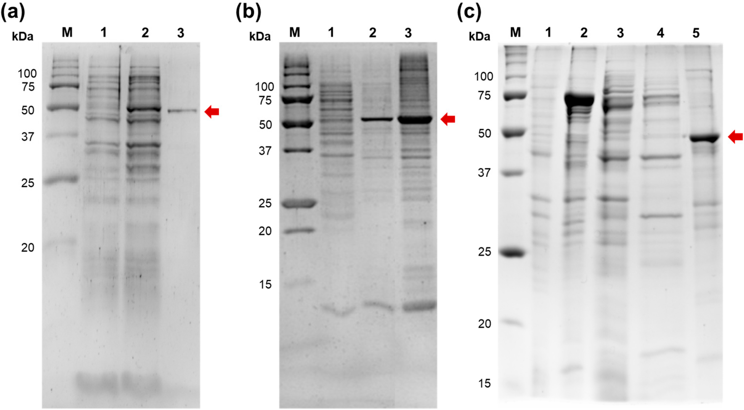
E. coli PAP-1 was also largely insoluble after cell lysis and rapidly lost activity when stored in low-salt buffers. A similar effect was observed for Mli PAP, suggesting that poor solubility may be a common characteristic of bacterial PAPs, as their related enzymes, tRNA nucleotidyltransferases, have also been reported to be insoluble [
17]. In the study by Shi et al.,
E. coli tRNA nucleotidyltransferase was successfully purified and retained activity after urea denaturation followed by renaturation on Ni–NTA resin. However, neither
E. coli PAP-1 nor Mli PAP retained activity after a denaturation–renaturation cycle in our study; so, we attempted to purify both enzymes under non-denaturing conditions. A sufficient amount of soluble
E. coli PAP-1 was available for successful purification. In contrast, Mli PAP expressed in
E. coli appeared to be completely insoluble. Both PAPs retained detectable activity in inclusion bodies, and we used this observation as the basis for attempts to purify Mli PAP by metal-chelate, ion-exchange, and affinity chromatography. However, Mli PAP solubilized from inclusion bodies using a high-salt buffer failed to bind to any of the tested resins despite extensive variation in pH and ionic strength. We managed to purify Mli PAP using only a solubilization tag, the DsbA protein, N-terminally fused to Mli PAP. The fused protein, while also being mostly insoluble, remained in a soluble fraction in a sufficient amount for purification and further analysis. It also should be emphasized that no poly(A) polymerase activity was detected in
E. coli cells prior to Mli PAP induction, indicating that the results obtained were robust and that no detectable background activity from host poly(A) polymerases interfered with our assays.
Regarding biochemical properties,
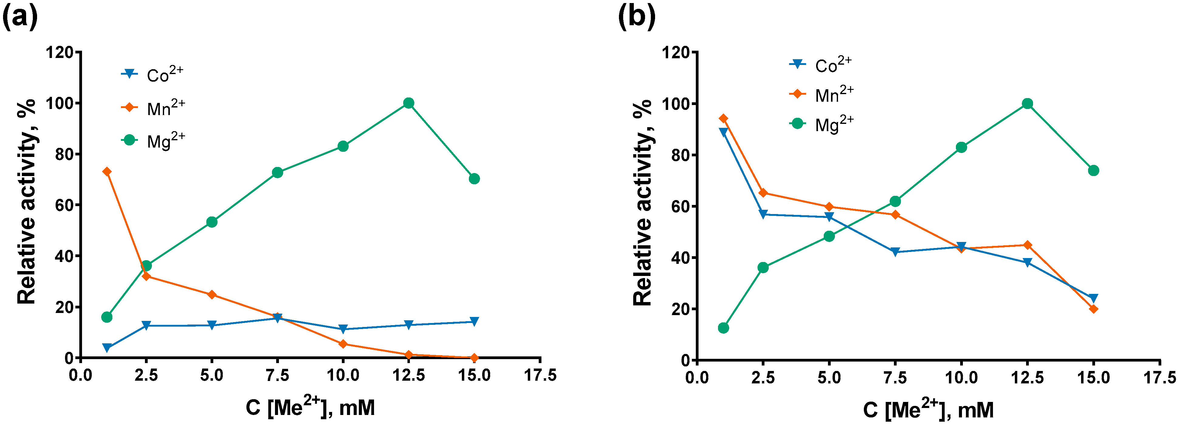
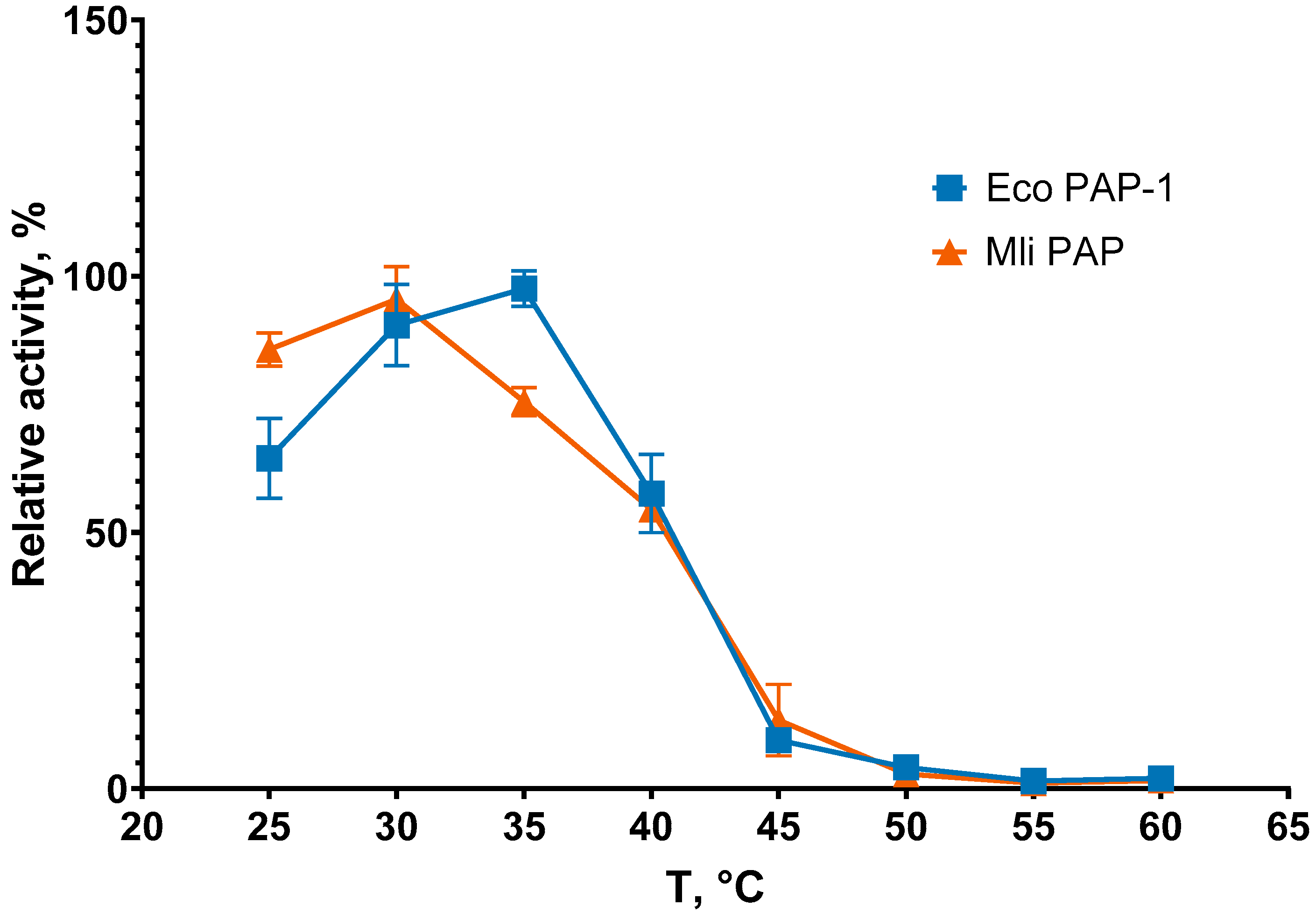
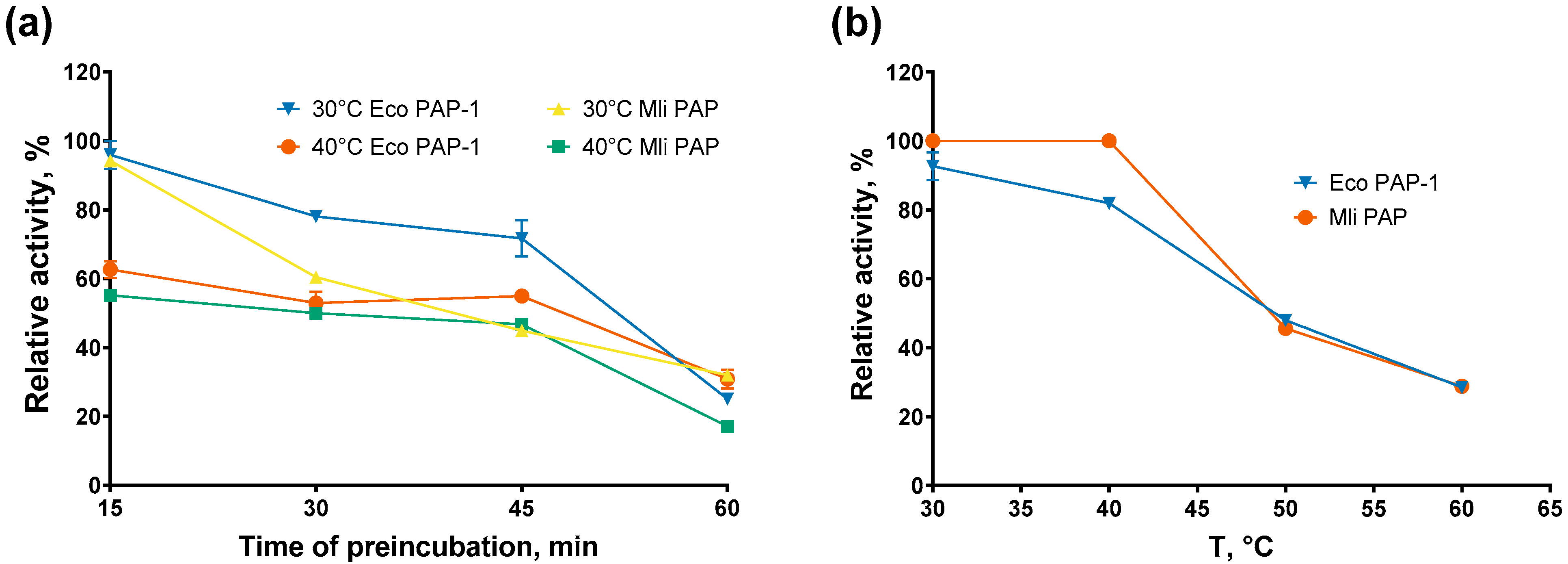
E. coli PAP-1 and Mli PAP demonstrated similar thermostability and optimal temperatures, although their cofactor and salt preferences differed slightly. Both host organisms are mesophiles with optimal growth temperatures near 37 °C; therefore, the observed optimal temperature and thermostability of the PAPs are consistent with expectations for enzymes from mesophilic bacteria. However,
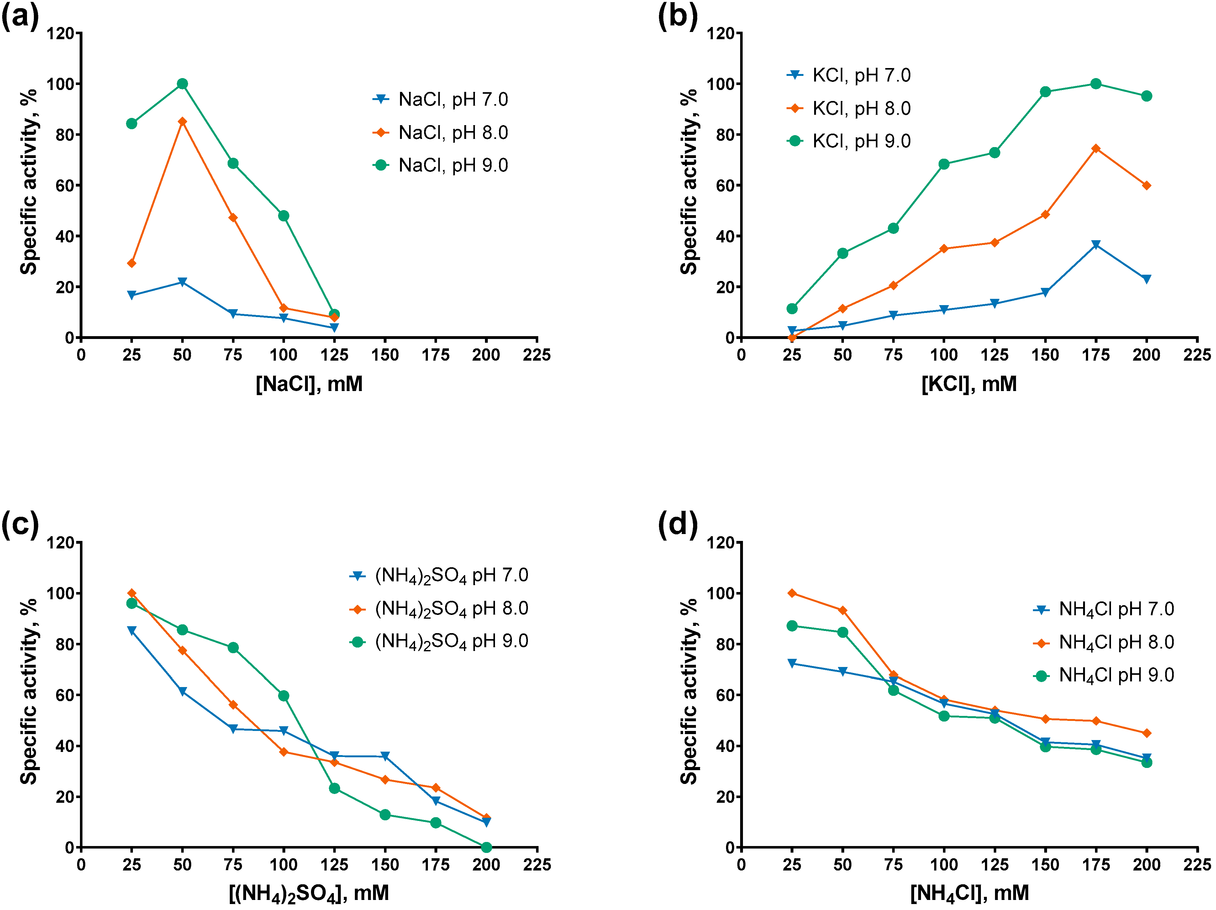
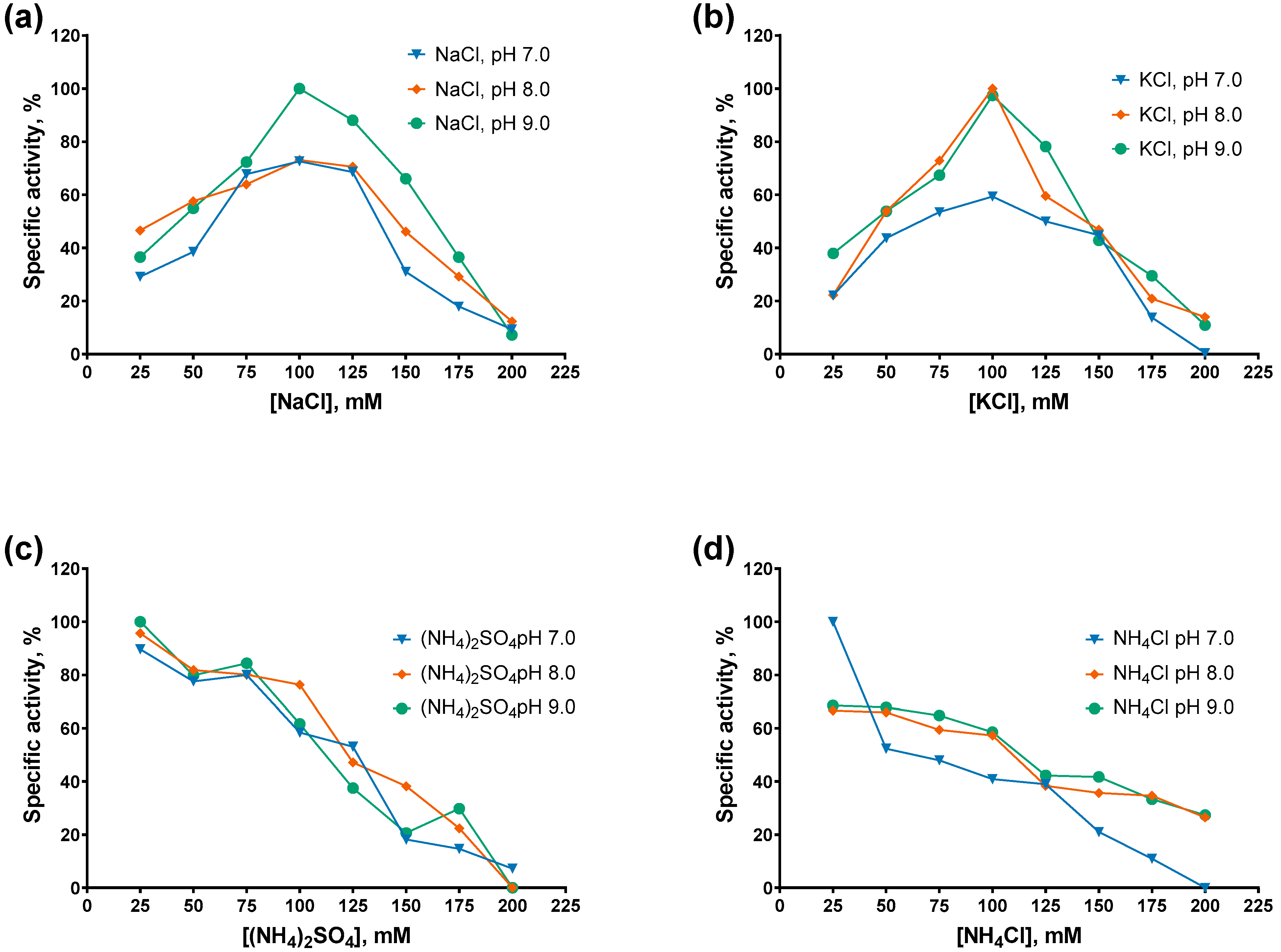
E. coli PAP-1 showed higher activity in the presence of Mn
2+ and Co
2+ and retained more activity in NaCl compared with Mli PAP. Unexpectedly, Mli PAP was strongly inhibited by NaCl but not by KCl, despite its native host,
M. lipolyticus, tolerating much higher NaCl concentrations than
E. coli (up to 25% vs. 7–11%
w/
v, respectively) [
16,
18]. This discrepancy may hint toward stabilization of Mli PAP through interactions with other
M. lipolyticus proteins that are absent in
E. coli. The nearly linear inhibition by NH
4+ may be related to the Hofmeister series, in which NH
4+ is among the strongest kosmotropic cations. Given the aggregation tendency of PAPs, inhibition by NH
4+ may reflect salt-induced precipitation. However, this explanation is preliminary and requires direct experimental validation. Structural studies would also provide an insight into the difference between the two studied PAPs, demonstrating possibly different binding with an RNA substrate or altered structure of active sites.
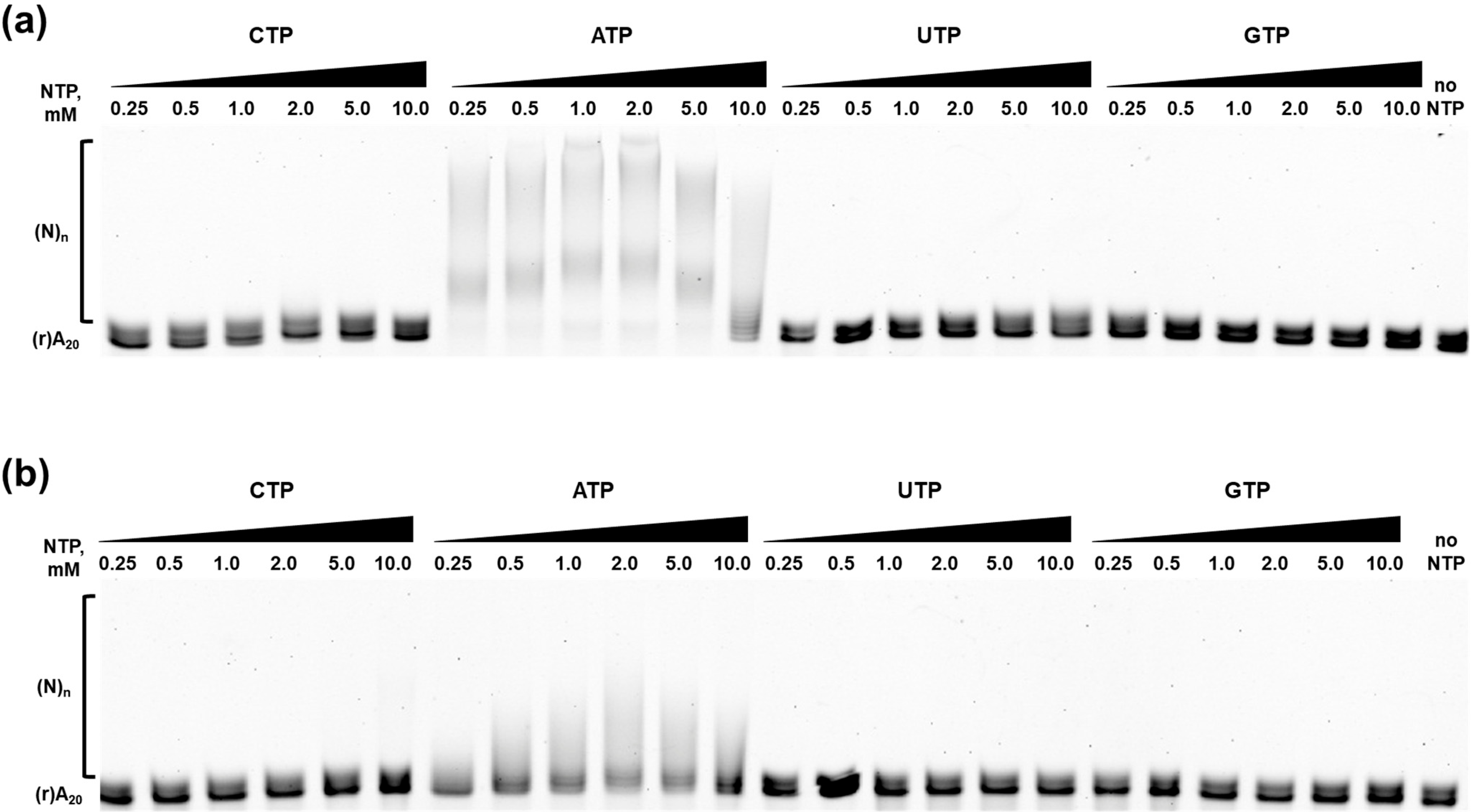
The nucleotide specificity of both PAPs was identical: ATP was the preferred substrate, while CMP and UMP were incorporated with much lower efficiency, and GMP incorporation was not detected. This observation partially contradicts previous reports suggesting that
E. coli PAP-1 can utilize not only ATP but also other NTPs as substrates for RNA elongation [
19]. In the study by Yehudai-Resheff and Schuster, the incorporation rates of non-ATP nucleotides were higher than those observed in our experimental system. This discrepancy may be attributed to differences in used reaction buffer compositions and substrates, as we used a short (r)A
20 oligonucleotide rather than a longer in vitro-transcribed RNA. Relaxed substrate specificity may be an in vitro artifact caused by somehow suboptimal reaction conditions. In the presence of Mn
2+, DNA polymerases incorporate NTPs much more efficiently than when Mg
2+ is used as a cofactor. For
E. coli DNA polymerase I, increasing pH from 7.6 to 9.1 results in increased misincorporation rate of ribonucleotides [
20]. Nevertheless, both
E. coli PAP-1 and Mli PAP exhibited relaxed, but not absolute, nucleotide specificity, suggesting the existence of mechanisms that enhance PAP NTPs’ selectivity in vivo.
Several limitations of the present study should be mentioned. First, we did not use long RNA substrates, although RNA’s secondary structure is known to affect the activity of
E. coli PAP-1 [
21]. Whether the RNA structure modulates poly(A) tail synthesis by Mli PAP remains an open question requiring further investigation. Second, NTP specificity was evaluated qualitatively but not quantitatively, leaving room for speculation regarding subtle differences in substrate preference between the two PAPs. Nevertheless, the similar K
m(ATP) values measured for
E. coli PAP-1 and Mli PAP suggest that both enzymes may incorporate alternative NTPs in a comparable manner. Third, the efficacy of Mli PAP in actual practical applications remains unknown since the respective comparison with
E. coli PAP-1 and the yeast PAP requires extensive additional experiments. However, this study reveals the true potential of various PAPs for usage in biotechnology, including not only efficacy of polyadenylation but also their stability and tolerance to various inhibitors that can appear in reaction mixes.
In summary, we cloned and characterized the biochemical properties of the first poly(A) polymerase identified in Alphaproteobacteria, specifically, Marinobacter lipolyticus. The newly characterized PAP exhibited properties broadly similar to those of E. coli PAP-1, providing a foundation for further studies on the diversity and mechanistic features of bacterial PAPs.
4. Materials and Methods
4.1. Search and Selection of Mli PAP Coding Sequence
The amino acid sequence of
E. coli PAP-1 was used as a reference for the search of potential bacterial PAPs using Protein BLAST 2.17.0 against proteins from
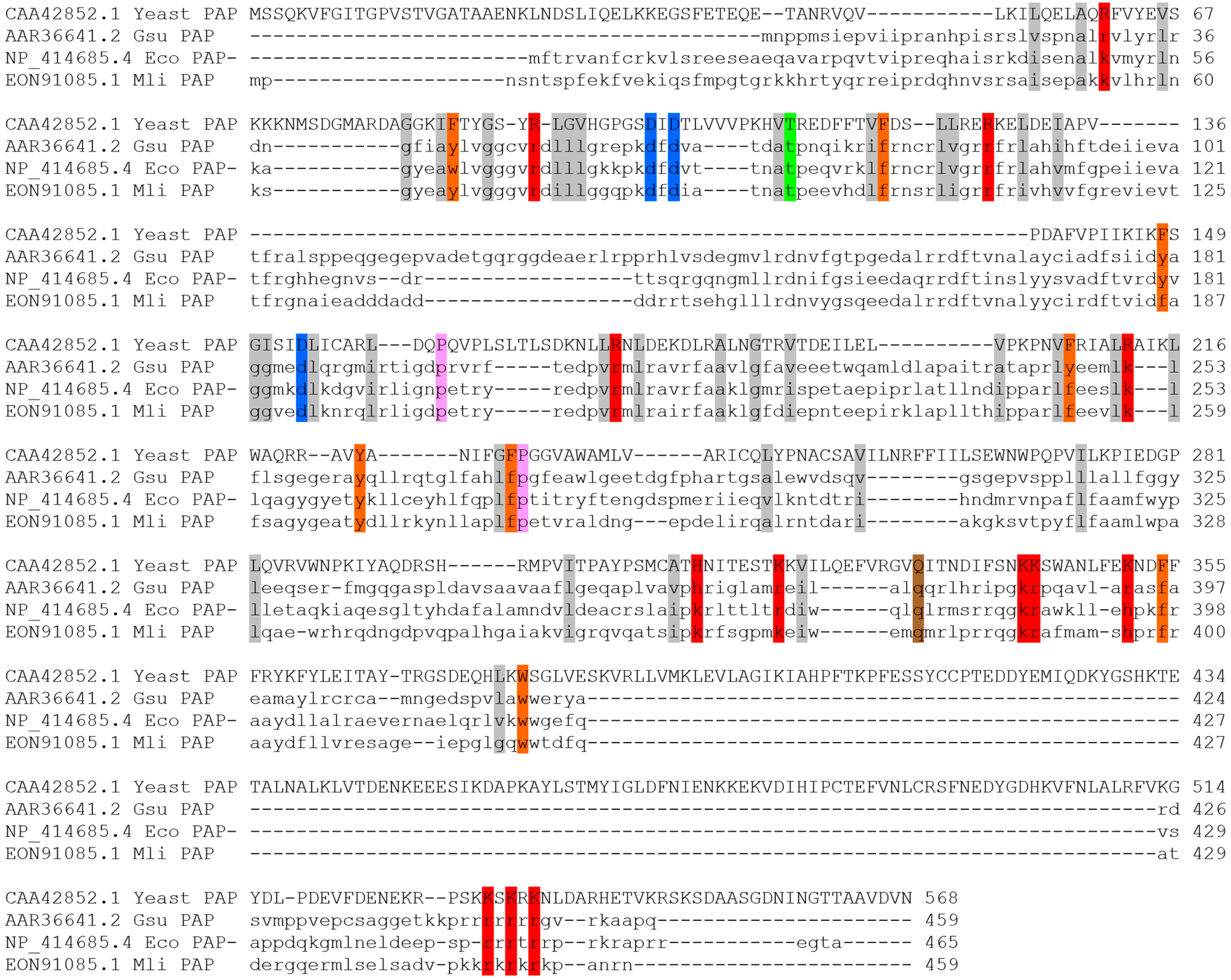
Alphaproteobacteria. Criteria for cloning were length of 400–600 a.a., the presence of all PAP structural domains (head, neck, body and leg), conservative positions corresponding to D69, D71, and E108 involved in catalysis in PAP I
E. coli [
11], and the characteristic signature of bacterial PAPs [LIV][LIV]G[R/K][R/K]Fx-[LIV]h[HQL][LIV] [
9]. The retrieved putative PAPs were organized in a range based on their similarity to
E. coli PAP-1. The selected coding sequence of a putative PAP from
Marinobacter lipolyticus (GenBank: EON91085.1) was synthesized and cloned into the pET23a vector by Shanghai RealGene Bio-tech, Inc. (Shanghai, China), resulting in a plasmid pPAP-Mli followed by cloning into pBADM52 vector, resulting in a plasmid pPAP-Mli2.
4.2. Expression and Purification of PAP from Marinobacter Lipolyticus
A starter culture of E. coli BL21 (DE3) pLysS (Promega, Madison, WI, USA) strain harboring either the plasmids pPAP-Mli or pPAP-Mli2 was grown to OD600 = 0.8 in LB medium with 100 μg/mL ampicillin at 37 °C. In a LiFlus GX fermenter (Biotron Inc., Bucheon, Republic of Korea), 4 L of LB with 100 μg/mL ampicillin was inoculated with 20 mL of the starter culture, and the cells were grown to OD600 = 0.6 at 37 °C. The expression of Mli PAP was induced by adding IPTG up to 1 mM concentration. After induction for 12 h at 18 °C, the cells were harvested by centrifugation at 4000× g and stored at −70 °C.
For partial Mli PAP purification, the cell pellet with pPAP-Mli was resuspended in the lysis buffer (50 mM Tris-HCl pH 8.0, 300 mM NaCl, 1 mM PMSF, 10 mM DTT, 10% glycerol, 1% Triton X-100, 1 mg/mL lysozyme), and incubated for 30 min at 37 °C, followed by sonication. After lysis, the soluble fraction was separated by two consequent centrifugation steps at 20,000× g for 30 min. The insoluble fraction was resuspended in the resuspension buffer (50 mM Tris-HCl pH 8.0, 1 M NaCl, 1 mM PMSF), incubated at 4 °C for 14 h and clarified by centrifugation at 25,000× g for 30 min. The resulting supernatant was supplemented by 0.01% NaN3 and stored at +4 °C. The protein concentration of was measured using a standard Bradford assay.
For full Mli PAP purification, after the lysis of the cell pellet with pPAP-Mli2 under the above-described conditions, the soluble proteins were precipitated ON by 60% (NH4)2SO4, followed by centrifugation at 20,000× g for 30 min. The resulting pellet was suspended in the resuspension buffer (50 mM NaPO4, pH 7.0, 1 M NaCl, 5 mM β-mercaptoethanol) and loaded onto a 2 mL IMAC column (Bio-Rad, Hercules, CA, USA) pre-equilibrated with buffer A1 (50 mM NaPO4, pH 7.0, 0.5 M NaCl, 5 mM β-mercaptoethanol), followed by washing the column with 25 mL of the buffer A with 1 M NaCl. Bound proteins were eluted using 10 column volumes and a 0–100% linear gradient of buffer B1 (buffer A1 with 0.5 M imidazole). After affinity chromatography, the fractions with the DsbA-Mli PAP fusion protein were pooled and loaded onto a 2 mL Macro-Prep DEAE Resin (Bio-Rad, column, Hercules, CA, USA) pre-equilibrated with buffer C1 (50 mM NaPO4, pH 7.0, 0.3 M NaCl, 5 mM β-mercaptoethanol). The column was washed with 10 mL of buffer C, and bound proteins were eluted by 10 column volumes and a 0–100% linear gradient of buffer D (50 mM NaPO4 pH 7.0, 1.5 M NaCl, 5 mM β-mercaptoethanol). The fractions with DsbA-Mli PAP were pooled and the fusion protein was hydrolyzed by TEV protease (Biolabmix, Novosibirsk, Russia), followed by further affinity chromatography under the same conditions for removal of His-tagged TEV protease and DsbA. Purified Mli PAP remained in the flow-throw and was dialyzed against a storage buffer (20 mM Tris-HCl, 300 mM NaCl, 0.1 mM EDTA, 1 mM DTT, 0.1% Triton X-100, 50% glycerol, pH 7.5), and stored at −20 °C. All the fractions from each step were analyzed by SDS-PAGE. The purity of the isolated Mli PAP was not lower than 95%. The concentration of purified Mli PAP was measured using a standard Bradford assay.
4.3. Expression and Purification of PAP-1 from E. coli
E. coli PAP-1 was cloned into the pET36b vector (Novagen, Madison, WI, USA) using the restriction–ligation method and NheI and XhoI restriction sites, resulting in the plasmid pET-PAP-Eco as described in our previous work [
22]. A starter culture of
E. coli BL21 (DE3) pLysS harboring the plasmid pET-PAP-Eco was grown to OD600 = 0.6 in LB medium with 100 μg/mL kanamycin at 37 °C. In a LiFlus GX fermenter (Biotron Inc., Bucheon, Republic of Korea), 4 L of LB with 100 μg/mL kanamycin was inoculated with 20 mL of the starter culture, and the cells were grown to OD600 = 0.6 at 37 °C. The expression of
E. coli PAP-1 was induced by adding IPTG up to 1 mM concentration. After induction for 12 h at 18 °C, the cells were harvested by centrifugation at 4000×
g and stored at −70 °C.
For protein purification, the cell pellet was resuspended in a lysis buffer (50 mM Tris-HCl pH 8.0, 500 mM NaCl, 1 mM PMSF, 2 M urea, 5 mM β-Mercaptoethanol, 1 mg/mL lysozyme), and incubated for 30 min on ice, followed by sonication. After lysis, the soluble fraction was separated by two consequent centrifugation steps at 20,000× g for 30 min. Soluble proteins were precipitated ON by 60% (NH4)2SO4, followed by centrifugation at 20,000× g for 30 min. The resulting pellet was suspended in the lysis buffer and loaded onto a 5 mL IMAC column (Bio-Rad, Hercules, CA, USA) pre-equilibrated with buffer A (50 mM NaPO4, pH 7.0, 0.5 M NaCl, 5 mM β-mercaptoethanol), followed by washing the column with 25 mL of the buffer A with 1 M NaCl. Bound proteins were eluted using 10 column volumes and a 0–100% linear gradient of buffer B (buffer A with 0.5 M imidazole). After affinity chromatography, the fractions with E. coli PAP-1 were pooled and loaded onto a 2 mL Macro-Prep HighS Resin (Bio-Rad, column, Hercules, CA, USA) pre-equilibrated with buffer C (50 mM NaPO4, pH 7.0, 0.3 M NaCl, 5 mM β-mercaptoethanol). The column was washed with 10 mL of buffer C, and bound proteins were eluted by 10 column volumes and a 0–100% linear gradient of buffer D (50 mM NaPO4, pH 7.0, 1.5 M NaCl, 5 mM β-mercaptoethanol). The fractions with E. coli PAP-1 were pooled, dialyzed against a storage buffer (20 mM Tris-HCl, 300 mM NaCl, 0.1 mM EDTA, 1 mM DTT, 0.1% Triton X-100, 50% glycerol, pH 7.5), and stored at −20 °C. All the fractions from each step were analyzed by SDS-PAGE. The purity of the isolated E. coli PAP-1 was not lower than 95%. The concentration of purified E. coli PAP-1 was measured using a standard Bradford assay.
4.4. Polyadenylation Assay
The specific activity of poly(A) polymerase was analyzed using elongation of the fluorescently labeled (r)A20 oligonucleotide. The reaction mixes (10 μL) contained a 1× reaction buffer for E. coli PAP-1 (50 mM Tris-HCl, 250 mM NaCl, 10 mM MgCl2, pH 8.0), 1 mM ATP, 10 pmol of (r)A20-oligonucleotide with 5′-terminal FAM, and an indicated amount of PAP. The reactions were started by the addition of the enzyme and immediately transferred to a preheated thermocycler, followed by incubation for 25 min at 37 °C. After incubation, the reactions were quenched by the addition of 10 μL of formamide and denatured by heating for 5 min at 95 °C. The reaction products were analyzed using denaturing PAGE in a 18% acrylamide gel with 7 M urea and quantified using the ImageLab software version 6.0.1 (Bio-Rad, Hercules, CA, USA).
4.5. Thermal Stability, Optimal Temperature, Ion Concentration and Nucleotide Specificity
The thermal stability of Mli PAP and E. coli PAP-1 was studied by heating the enzyme and then assaying the poly(A)-polymerase activity. The aliquots of the polymerase activity reaction buffer (described above) without ATP containing an identical amount of Mli PAP were incubated at temperatures from 30 °C to 60 °C, increasing by 10 °C per step for 15–60 min. The reactions were chilled on ice, and poly(A) polymerase activity was measured as mentioned above.
The temperature optimum of Mli PAP and E. coli PAP-1 was defined by measuring the poly(A)-polymerase activity at temperatures from 25 °C to 60 °C, increasing by 5 °C per step, with other conditions identical to those described above for the poly(A) polymerase activity assay. The reactions were initiated by adding the aliquots of enzymes to mixes containing all other components, including the primed template. The mixes were preheated for 5 min before the addition of enzymes.
The optimal ion concentrations were examined as in the poly(A) polymerase activity assay using 25–200 mM NH4Cl, KCl, NaCl, (NH4)2SO4 or 1–15 mM MgCl2, MnCl2, CoCl2.
After optimization of reaction conditions, nucleotide specificity of Mli PAP was defined in the optimized reaction buffer (50 mM Tris-HCl, 150 mM KCl, 10 mM MgCl2, pH 8.0) by titration of NTPs in reaction mixes for the poly(A) polymerase activity assay in the range of 0.25–10.0 mM. The polyadenylation assay was conducted for 30 min at 37 °C.
4.6. Data Analysis
The relative amount of the polyadenylated substrate was calculated—Ratio = I(elongated products)/(I(substrate) + I(elongated products))—and used to estimate by a linear regression model an enzyme amount necessary to elongate 50% of the substrate initially added to the reaction. All calculations were performed in the GraphPad Prism 8.0.1 software (Insight Venture Management, New York, NY, USA).