Abstract
Calcific aortic valve disease (CAVD) is characterized by progressive valve remodeling and calcification. Moreover, microRNAs (miRNAs) are emerging as key regulators of cardiovascular pathology and potential circulating biomarkers. We performed high-throughput miRNA profiling in calcified aortic valve tissue and matched patient serum samples using an array that included 98 human miRNAs. Expression data were log10-transformed and filtered to identify biologically relevant miRNAs. Shared miRNAs between tissue and serum were further validated by quantitative real-time polymerase chain reaction (qRT-PCR) in patients and healthy controls. Of the 49 actively expressed miRNAs, 18 were shared between valve tissue and serum. Thus, qRT-PCR validation revealed significant downregulation of miR-17-5p and miR-29a-3p in CAVD patient serum compared to controls. These results indicate that disease-associated miRNA alterations in calcified valves are mirrored in circulation. miR-17-5p and miR-29a-3p represent promising circulating biomarkers for CAVD, reflecting underlying pathological remodeling and extracellular matrix dysregulation. Our findings provide a framework for non-invasive monitoring of valve calcification and highlight miRNA-mediated pathways as potential therapeutic targets.
1. Introduction
Calcific aortic valve disease (CAVD) represents the most common valvular heart disorder in industrialized countries, characterized by progressive fibro-calcific remodeling of the aortic valve leaflets leading to stenosis and impaired cardiac function []. Once considered a passive, degenerative process associated with aging, valve calcification is now recognized as an active and highly regulated biological process, involving osteogenic trans-differentiation of valvular interstitial cells, chronic inflammation, lipid infiltration, extracellular matrix remodeling, and apoptosis [,,,,]. Despite its high prevalence and clinical impact, effective pharmacological strategies to prevent or slow disease progression are currently lacking, making the identification of novel biomarkers and molecular targets a crucial research priority. In recent years, microRNAs (miRNAs), small non-coding RNAs that regulate gene expression post-transcriptionally, have emerged as key players in cardiovascular pathophysiology []. Specific miRNAs have been implicated in endothelial dysfunction, inflammation, and osteogenic differentiation, all processes that contribute to valvular calcification [,]. Importantly, miRNAs can be detected not only in diseased valve tissue but also in circulating blood, suggesting their potential as minimally invasive biomarkers for disease diagnosis, monitoring, and risk stratification []. Several studies have reported dysregulated expression of distinct miRNAs in patients with valvular heart disease, highlighting their dual role as mediators and indicators of pathological remodeling []. However, the relationship between miRNA expression in valvular tissue and their circulating levels in patients with calcific valve disease remains poorly understood. Therefore, a better understanding of this relationship may help elucidate the molecular mechanisms driving disease progression and identify clinically relevant biomarker signatures. The present study aims to compare miRNA expression profiles in calcified valve tissue and serum samples from patients with CAVD. By correlating tissue and circulating miRNA patterns, we should provide new insights into the molecular pathways underlying CAVD and explore the feasibility of circulating miRNAs as potential biomarkers reflecting local pathological processes in the valve.
2. Results
2.1. MiRNA Expression Profiling in Calcified Aortic Valves and Patients Sera
To obtain a comprehensive overview of miRNA expression associated with valvular calcification, RNA was extracted from both calcified aortic valve tissue and corresponding serum samples from patients undergoing aortic valve replacement surgery. Thus, a high-throughput miRNA array including 98 unique human miRNAs was performed. Given the broad dynamic range of expression values observed across the two sample types, raw data were log10-transformed to facilitate inter-sample comparison and visualization. The heatmap including all 98 miRNAs revealed distinct expression clusters separating valve tissue from serum, suggesting the presence of tissue-enriched as well as systemically detectable miRNA populations. In particular, several miRNAs showed high expression levels specifically in valve tissue, whereas others displayed a more homogeneous distribution across both tissue and serum samples (Figure 1A). Therefore, to focus the analysis on biologically meaningful miRNAs, we applied a filtering threshold based on expression levels >1 in at least one of the two experimental conditions (valve or serum). This selection criterion reduced the dataset from 98 to 49 expressed miRNAs, which were represented in a dedicated heatmap (Figure 1A,B). This intermediate filtering step allowed for a more precise identification of miRNAs actively expressed in the pathological context of calcific aortic valve disease, excluding l and 1 Bow-abundance signals and background noise.
Figure 1.
Schemes follow the same formatting. (A) Heatmap showing the expression levels of all 98 miRNAs identified in calcified aortic valve tissue and corresponding serum samples from patients. Expression values were log10-transformed to normalize for differences in signal intensity across samples. Each row represents a single miRNA, and each column corresponds to an individual sample (valve or serum). (B) Heatmap showing the expression profiles of the 49 miRNAs retained after applying an expression threshold (log10-transformed values > 1 in at least one condition) to the initial dataset of 98 miRNAs. The color scale represents expression values transformed using base-10 logarithms (lighter colors = higher values, darker colors = lower values).
2.2. Shared miRNAs Between Valve Tissue and Serum
Among the 49 expressed miRNAs, 18 miRNAs were shared between calcified valve tissue and serum samples, indicating a molecular overlap between local valvular processes and systemic circulation. These shared miRNAs were: miR-17, miR-20a, miR-22, miR-24, miR-25, miR-26a, miR-26b, miR-27a, miR-29a, miR-92a, miR-106b, miR-125b, miR-140, miR-143, miR-145, miR-148a, miR-423, and miR-451a (Supplemental Table S1). The presence of these miRNAs in both samples supports the hypothesis that disease-associated miRNA alterations in the valve are reflected at the systemic level, potentially enabling their use as circulating biomarkers for disease detection and monitoring.
2.3. Pathway Enrichment Analysis of Shared Differentially Expressed miRNAs
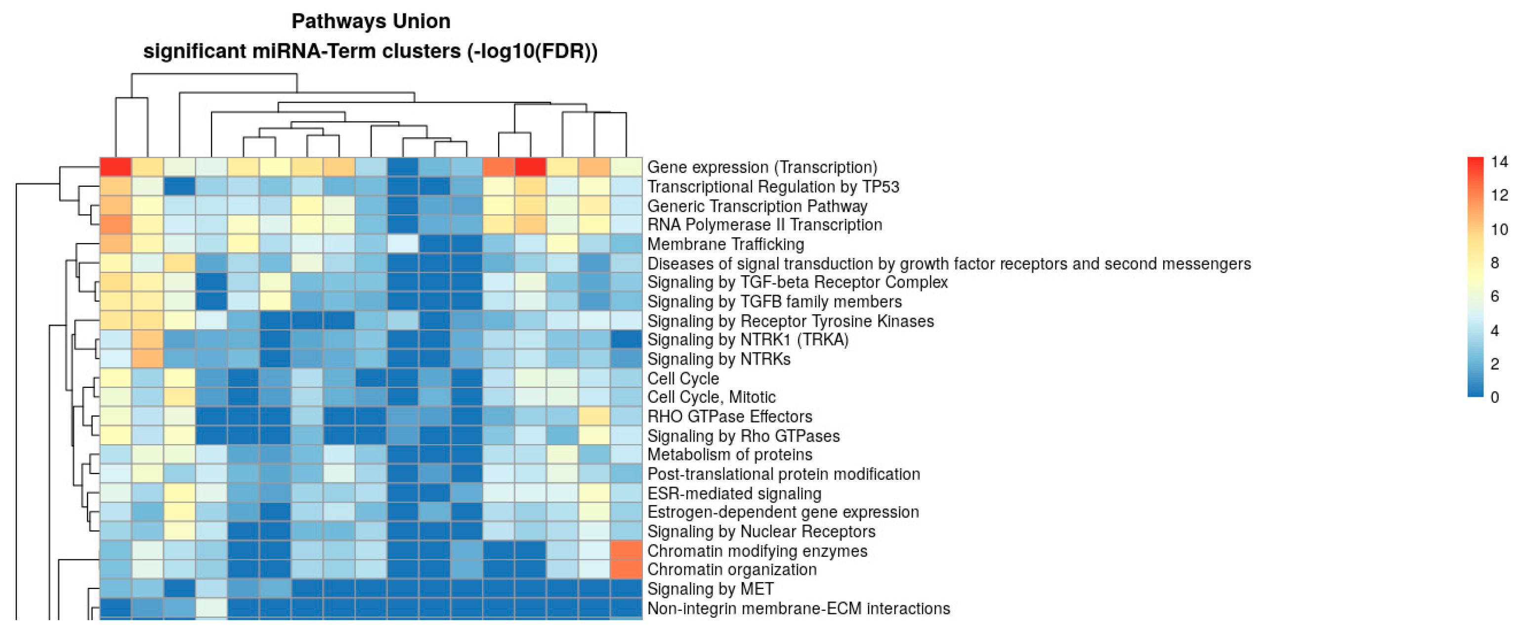
To explore the biological functions potentially regulated by the identified miRNAs, we performed pathway enrichment analysis using the DIANA-miRPath v4.0 platform (TarBase v8 and Reactome Pathway Union database, miRBase release v22.1, accessed on 20 August 2025, at http://62.217.122.229:3838/app/miRPathv4). Experimentally validated miRNA–target interactions were retrieved and mapped to Reactome pathways (miRBase release v22.1). Enrichment significance was assessed using FDR-corrected p-values with a threshold of 0.05. The analysis revealed significant enrichment of multiple pathways, including gene expression (transcription), transcriptional regulation by TP53, TGF-β signaling, receptor tyrosine kinase signaling, cell cycle regulation, Rho GTPase effectors, chromatin organization, and membrane trafficking (Figure 2). The heatmap shows the union of enriched pathways across significant miRNA-term clusters, with enrichment scores represented as −log10(FDR).
Figure 2.
Heatmap showing the union of significantly enriched pathways based on experimentally validated miRNA–target interactions analyzed with the DIANA-miRPath v4.0 platform (TarBase v8 and Reactome Pathway Union database, miRBase release v22.1, accessed on 20 August 2025, at http://62.217.122.229:3838/app/miRPathv4). Pathway enrichment significance was calculated using FDR-corrected p-values (threshold 0.05) and expressed as −log10(FDR). Hierarchical clustering was performed on significant miRNA–term associations to identify shared biological processes. Red indicates the highest level of significance, while blue represents lower significance levels.
Moreover, KEGG pathway enrichment analysis using the pathways union method in DIANA-miRPath v4.0 revealed several biological processes involving differentially expressed miRNAs (Table 1).
Table 1.
Kyoto encyclopedia of genes and genomes and gene ontology pathways regulated by the differentially expressed microRNAs. The list of target genes of the analyzed miRNAs is provided in Supplementary Table S2.
Table 1.
Kyoto encyclopedia of genes and genomes and gene ontology pathways regulated by the differentially expressed microRNAs. The list of target genes of the analyzed miRNAs is provided in Supplementary Table S2.
| Term Name | Term Genes | miRNAs (n) | miRNA Names | Merged p-Value | Merged FDR |
|---|---|---|---|---|---|
| Signaling by TGFB family members | 104 | 11 | hsa-miR-17-5p hsa-miR-20a-5p hsa-miR-22-3p hsa-miR-24-3p hsa-miR-25-3p hsa-miR-26a-5p hsa-miR-26b-5p hsa-miR-27a-3p hsa-miR-92a-3p hsa-miR-125b-5p hsa-miR-126b-5p | 7.5 × 10−61 | 4.89 × 10−59 |
| Signaling by WNT | 366 | 7 | hsa-miR-17-5p hsa-miR-20a-5p hsa-miR-22-3p hsa-miR-24-3p hsa-miR-29a-3p hsa-miR-92a-3p hsa-miR-125b-5p | 1.68 × 10⁻²⁰ | 1.68 × 10⁻¹⁸ |
| PI3K-Akt signaling pathway | 372 | 10 | hsa-miR-17-5p hsa-miR-20a-5p hsa-miR-22-3p hsa-miR-24-3p hsa-miR-25-3p hsa-miR-26a-5p hsa-miR-26b-5p hsa-miR-27a-3p hsa-miR-29a-3p hsa-miR-145-5p | 1.59 × 10⁻²⁹ | 1.06 × 10⁻²⁸ |
| Signaling pathway regulating pluripotency of Stem Cells | 156 | 10 | hsa-miR-17-5p hsa-miR-22-3p hsa-miR-24-3p hsa-miR-26a-5p hsa-miR-26b-5p hsa-miR-27a-3p hsa-miR-29a-3p hsa-miR-92a-3p hsa-miR-125b-5p hsa-miR-106b-5p | 7.16 × 10⁻²⁷ | 4.08 × 10⁻²⁶ |
To further characterize the biological processes regulated by the identified miRNAs, enrichment analysis was also performed using the Gene Ontology (GO) terms Pathway Union database through miRPath v4 (TarBase v8). Among the significantly enriched GO terms, heart development showed a strong association (FDR = 2.40 × 10⁻²⁹; p-value = 2.53 × 10⁻³⁰), involving 7 miRNAs: hsa-miR-17-5p, hsa-miR-22-3p, hsa-miR-24-3p, hsa-miR-27a-3p, hsa-miR-29a-3p, hsa-miR-125b-5p, and hsa-miR-106b-5p (Table 2).
Table 2.
GO terms enrichment analysis of identified miRNAs.
2.4. Downregulation of miR-17-5p and miT-29a-3p in Patient Serum as Potential Circulating Biomarkers for Detection of Valve Calcification
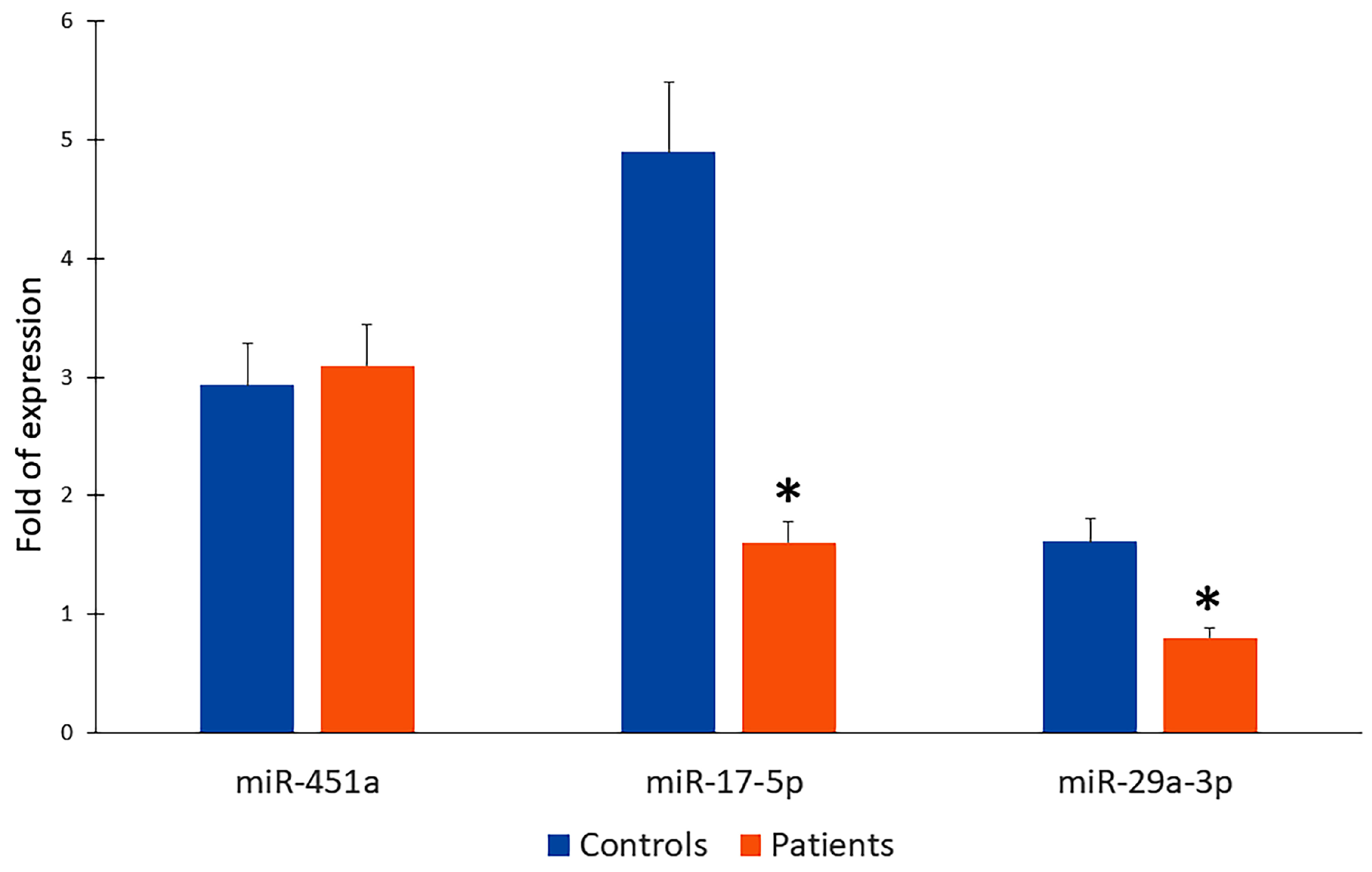
Based on the array profiling data and the pathway association miR-17-5p and miR-29a-3p were selected for independent validation by quantitative real-time polymerase chain reaction (qRT-PCR) in serum samples from patients with calcified aortic valves and healthy control subjects. We also analysed miR-451a which was express in array profiling but no KEGG pathways were enriched by genes targeted by miR-451a alone or in combination with other studied miRs. Thus, the expression levels of miR-451a were comparable between patients and healthy controls, showing mean fold changes of 3.20 and 2.91, respectively, and no statistically significant difference (p = 1) (Figure 3). These results suggest that miR-451a may not be directly involved in the pathological mechanisms of valve calcification or may act as a stable circulating miRNA.
Figure 3.
MiR-451a, miR-17-5p, and miR-29a-3p qRT-PCR in controls and patients; * p < 0.005.
In contrast, miR-17-5p expression was significantly downregulated in patient serum compared to controls (1.70 vs. 4.72-fold; p = 0.014) (Figure 3). Similarly, miR-29a-3p was also significantly reduced in patients compared with controls (0.86 vs. 1.79-fold; p = 0.014) (Figure 3). Overall, these findings suggest that miR-17-5p and miR-29a-3p could be considered potential circulating biomarkers for CAVD characterization.
3. Discussion
In this study, we performed a comprehensive analysis of miRNA expression in calcified aortic valve tissue and corresponding patient serum, aiming to identify molecular signatures reflective of valvular pathology and potential circulating biomarkers. Our high-throughput profiling revealed 98 miRNAs expressed across tissue and serum, with clear clustering distinguishing valve-enriched from systemically detectable miRNAs. Application of an expression threshold allowed refinement of the dataset to 49 actively expressed miRNAs, facilitating the identification of biologically relevant candidates while minimizing background noise. Among these, 18 miRNAs were consistently shared between valve tissue and serum, including miR-17, miR-20a, miR-22, miR-24, miR-25, miR-26a/b, miR-27a, miR-29a, miR-92a, miR-106b, miR-125b, miR-140, miR-143, miR-145, miR-148a, miR-423, and miR-451a. The presence of these miRNAs in both compartments suggests that alterations occurring within the calcified valve are reflected in the systemic circulation, supporting their potential as minimally invasive biomarkers for disease detection and monitoring. Pathway enrichment analysis revealed that several key signaling cascades are regulated by the differentially expressed miRNAs identified in this study. In particular, the most significantly enriched pathways included the TGF-β signaling pathway (FDR = 4.89 × 10−59), Wnt signaling pathway (FDR = 1.68 × 10⁻¹⁸), and PI3K-Akt signaling pathway (FDR = 1.06 × 10⁻²⁸). These pathways are known to play fundamental roles in cardiovascular homeostasis, tissue remodeling, and pathological calcification processes [,,,,]. Several miRNAs—including hsa-miR-17-5p, hsa-miR-20a-5p, hsa-miR-22-3p, hsa-miR-24-3p, hsa-miR-25-3p, hsa-miR-26a/b-5p, hsa-miR-27a-3p, hsa-miR-29a-3p, hsa-miR-92a-3p, and hsa-miR-125b-5p—were found to be commonly associated with these pathways, suggesting that they may act as central regulatory nodes in disease-relevant molecular networks. Of particular interest, the pathway regulating pluripotency of stem cells was also significantly enriched (FDR = 4.08 × 10⁻²⁶), highlighting the potential involvement of miRNA-mediated mechanisms in modulating cellular differentiation and plasticity during disease progression. Although the pathway enrichment analysis strongly supports the involvement of the identified miRNAs in signaling pathways relevant to aortic valve calcification, experimental validation of their predicted target genes will be required to fully elucidate their mechanistic contribution. Due to the limited availability of biological material in the current study, this analysis could not be performed and will be prioritized in future investigations using additional patient samples. However, our findings are in line with previous reports indicating that reactivation of developmental signaling cascades contributes to the osteogenic trans-differentiation of valvular interstitial cells and extracellular matrix remodeling in calcific valve disease [,,]. Complementary enrichment analysis using GO terms further strengthened these observations. “Heart development” emerged as the most significantly associated biological process (FDR = 2.40 × 10⁻²⁹), involving seven miRNAs (hsa-miR-17-5p, hsa-miR-22-3p, hsa-miR-24-3p, hsa-miR-27a-3p, hsa-miR-29a-3p, hsa-miR-125b-5p, and hsa-miR-106b-5p). The strong association with this GO term suggests that the dysregulated miRNAs may participate in developmental programs that are aberrantly reactivated during the pathological calcification of aortic valves [,,,]. Based on their enrichment in key biological pathways, miR-17-5p and miR-29a-3p were selected for validation as potential circulating biomarkers. Specifically, both miRNAs presented strong enrichment in pathways relevant to disease pathogenesis (including pluripotency regulation in stem cells, WNT signalling, and heart development). Therefore, their validation was prioritized due to the combination of strong biological relevance and potential clinical utility. Quantitative real-time PCR confirmed a significant downregulation of both miR-17-5p (1.70-fold vs. 4.72-fold in controls, p = 0.004) and miR-29a-3p (0.86-fold vs. 1.79-fold in controls, p = 0.002) in patient serum. These findings support their potential role as non-invasive biomarkers for disease detection and characterization. Accordingly, it has been demonstrated that miR-17-5p can attenuate osteogenic differentiation and that vascular smooth muscle cells (VSMCs) transfected with a miR-17-5p mimic—thus enhancing its expression—exhibit a significant reduction in calcium deposition []. A decreased expression of miR-17-5p has also been reported in patients with bicuspid aortic valve (BAV), raising questions about its regulatory role in VSMC proliferation and valve development []. A reduction in miR-17a-5p could lead to enhanced calcification of the aortic valve by failing to suppress osteogenic pathways []. miR-17-5p is part of the miR-17-92 cluster which influences cell cycle progression and Notch signaling []. Reduced miR-17-5p could affect the expression of NOTCH1, a gene implicated in BAV pathogenesis []. By downregulating miR-17-5p, there may be increased expression of pro-calcific genes or dysregulated VSMC phenotypic switching, favoring a calcific environment or abnormal vascular remodeling []. In a chronic kidney disease (CKD) rat model, miR-17-5p has also been identified as a calcification-protective miRNAs that targets vascular endothelial growth factor A (VEGFA) signaling in CKD-driven vascular calcification []. Therefore, the reduced expression levels of miR-17-5p observed in CAVD may contribute to the development of calcification, suggesting a potential protective role for miR-17-5p in counteracting these processes. Regarding miR-29, it has been reported in a rat model that a miR-29a-3p mimic induces osteogenic differentiation, as demonstrated by the upregulation of RUNX2, COL1A1, and ALP compared to a negative control mimic. This suggests that miR-29a-3p enhances osteogenesis by activating key osteogenic genes []. In vitro studies showed that miR-29a-3p overexpression significantly attenuated VSMC calcification under high phosphate conditions, while VEGFA overexpression exacerbated calcification. Notably, miR-29a-3p counteracted calcification even in VEGFA-overexpressing cells, underscoring its protective role in vascular integrity []. However, miR-29 isoforms display pleiotropic and context-dependent functions, with their effects varying according to the cellular context (e.g., osteogenic versus inflammatory compartments), disease stage, and compensatory regulatory mechanisms. Notably, miR-29-3p has also been shown to suppress macrophage lineage commitment and exert anti-inflammatory effects []. Thus, in serum of CAVD patients, the observed downregulation of miR-29-3p may therefore reflect alterations in cellular composition, inflammatory signaling, or feedback regulation rather than a simple loss of a pro-osteogenic factor. Taken together, these findings highlight distinct and potentially complementary roles of miR-17-5p and miR-29-3p in the pathophysiology of valvular calcification, suggesting that their regulation is intricately linked to both osteogenic and inflammatory processes. In contrast, miR-451a, which was detected in the profiling data but not linked to any significantly enriched pathway, showed no significant difference between groups, suggesting a limited or indirect involvement in the disease process.
Overall, these results point to a coordinated dysregulation of miRNAs affecting multiple signaling pathways central to cardiovascular development and pathological remodeling. The identification of miR-17-5p and miR-29a-3p as significantly downregulated in patient serum further underscores their potential as biomarkers for CAVD [,,,].
4. Materials and Methods
This study was conducted in accordance with the Declaration of Helsinki and approved by the Ethics Committee of the Azienda Ospedaliera Universitaria Integrata of Verona (approval number: 13371; approval date: 13 March 2016). Informed consent was obtained from all participants in the study.
4.1. Aortic Valves and Sera
Human tricuspid aortic valves were obtained from four adult patients undergoing isolated surgical aortic valve replacement (SAVR) for severe calcific aortic stenosis. Inclusion criteria were: male sex, age < 75 years, tricuspid aortic valve, severe aortic stenosis requiring isolated SAVR, e-GFR ≥ 50 mL/min; exclusion criteria were: female sex, age > 75 years, BAV, concomitant coronary artery disease, mitral valve disease or aortic aneurysm, atrial fibrillation, e-GFR < 50 mL/min, diabetes, priori or ongoing neoplasia, prior or ongoing infective endocarditis, rheumatic disease, autoimmune disease, therapy with warfarin, DOACs, levothyroxine or steroids, enteral or parenteral nutrition. Aortic valve leaflets were collected during surgery immediately after valve leaflets removal, rinsed with saline solution (NaCl 0.9%) under sterile conditions and kept overnight in RNAlater® (Qiagen, Hilden, Germany) at 4 °C and then frozen and stored at −80 °C until further use. Blood samples were collected from the same patients before surgery and from four age-matched healthy controls in serum separator tubes without anticoagulant. Samples were centrifuged at 1800× g for 15 min. The serum was aliquoted and stored at −80 °C until analysis.
4.2. Tissue Processing and Homogenization
Cardiac valve tissues were processed under sterile and RNase-free conditions. Before miRNA extraction, the tissues were homogenized using a mechanical disruption system- Precellys 24 (Bertin Technologies, Montigny-le-Bretonneux, France), to ensure complete tissue lysis. The homogenates were subsequently processed for downstream molecular analyses.
4.3. miRNA Extraction and Quantification
Circulating miRNAs were isolated from human serum samples using the miRNeasy® Serum/Plasma Kit (Cat. Number 217204, Qiagen, Hilden, Germany) according to the manufacturer’s instructions. For cardiac valve tissues, miRNA was extracted using the miRNeasy® Mini kit (Cat. Number 217004 Qiagen, Hilden, Germany) following the manufacturer’s protocol. miRNAs were quantified by NanoDrop™ spectrophotometer (LifeReal, Aurogene, Hangzhou Biotechnology, Hangzhou, China) and then 10 ng of the extracted miRNAs were reverse transcribed with a TaqMan™ Advanced miRNA cDNA Synthesis Kit (Cat. Number A28007, Thermo Fisher Corporation, Waltham, MA, USA) following the manufacturer’s instructions.
4.4. TaqMan Gene Expression Array
TaqMan Gene Expression Array analysis was performed using the TaqMan™ Advanced miRNA Human A 96-well Plates, standard-Panel 1,3,4 (Ref: A32026, Applied Biosystems, Thermo Fisher Scientific, Waltham, MA, USA) for both cDNA derived from serum and cardiac valve tissue. cDNA solution was prepared according to the manufacturer’s protocol. Then, the 96-well array Plate was centrifuged and sealed using the proper membrane (Opti-seal, AB analitica). The PCRs protocol was then performed on the LineGene 9620 Real-Time PCR System (Aurogene, Hangzhou Bioer Technology, Hangzhou, China) using the parameters listed in Table 3 below.
Table 3.
PCR conditions.
4.5. Gene Expression Analysis/ Real Time-PCR
The Real-Time PCR was performed using the TaqMan Fast Advanced Master Mix (Cat. Number 4444557, Thermo Fisher Scientific, Waltham, MA, USA) with the following commercially pre-designed probes-TaqMan Advanced miRNA assay (Cat. Number A25576, Thermo Fisher Scientific, Waltham, MA, USA) (Table 4).
Table 4.
List of pre-designed probes-TaqMan Advanced miRNA assay.
The MicroAmp Optical 96-Well Reaction Plate (Applied Biosystems, Thermo Fisher Scientific, Waltham, MA, USA) was used to load 15 µL of PCR mix into the designed wells. Next, 5 µL (10 ng) of cDNA was added. Three copies of each sample were loaded. The plate was covered and sealed using the proper membrane (Opti-seal, AB analitica, Padova, Italy) following a brief centrifugation. The thermal protocol was performed on the LineGene 9620 Real-Time PCR System (Aurogene, Hangzhou Bioer Technology, China) using the parameters listed in Table 3. Each analysis was performed in triplicate and with Ct values averaged. miR-191-5p was used as housekeeping gene based on its stable expression across all samples. In addition, the choice of miR-191-5p is supported by published studies in mesenchymal stromal cells and human serum samples [,].
4.6. Pathway Enrichment Analysis
To explore the biological significance of the shared set of 18 differentially expressed miRNAs identified in calcified aortic valves and patient sera, pathway enrichment and functional annotation analyses were performed using publicly available bioinformatics tools. Experimentally validated miRNA–target interactions were analyzed using miRPath v4 DIANA-miRPath v4.0 [], which integrates TarBase v8 for validated miRNA–gene interactions. The list of miRNAs identified in this study was submitted to miRPath v4 for pathway enrichment analysis using the Reactome Pathway Union database. miRBase release v22.1 was used as the reference for miRNA annotation. Enrichment significance was assessed using a false discovery rate (FDR)-corrected p-value threshold of 0.05. Results were expressed as −log10(FDR) values. Hierarchical clustering of significant miRNA–term associations was performed to identify shared biological processes among the input miRNAs. A heatmap was generated to visualize the most significantly enriched pathways miRNA-specific.
4.7. Statistical Analysis
Expression levels of miRNAs in healthy controls and patients were compared using the Mann–Whitney U test for independent samples, as the groups were independent and sample sizes were small (n = 4 per group). Data are presented as mean ± standard deviation (SD). A p-value of less than 0.05 was considered statistically significant. Statistical evaluations were conducted using GraphPad Prism (version 9).
5. Conclusions
In this study we demonstrate a clear overlap between miRNA signatures in calcified valve tissue and patient serum, with miR-17-5p and miR-29a-3p emerging as promising circulating biomarkers. These findings enhance our understanding of the molecular underpinnings of CAVD, pave the way for the development of non-invasive diagnostic and prognostic tools based on miRNA profiling, and highlight miRNA-mediated pathways as potential therapeutic targets.
Our study has some limitations. The sample size, while sufficient to identify robust changes in miRNA expression, warrants expansion in future studies to validate the utility of these miRNAs as predictive biomarkers across diverse patient cohorts. Additionally, functional studies are needed to delineate the mechanistic roles of miR-17-5p and miR-29a-3p in valve calcification and systemic cardiovascular remodeling.
Supplementary Materials
The following supporting information can be downloaded at: https://www.mdpi.com/article/10.3390/ijms262210813/s1.
Author Contributions
Conceptualization, A.G. and M.T.V.; methodology, A.G. and M.T.V.; validation, A.G., M.B., L.D.C. and M.T.V.; formal analysis, A.M.; investigation, A.G., A.M., M.B. and M.C.; resources: L.D.C.; data curation, A.G., M.G.R. and M.T.V.; writing—original draft preparation, A.G. and M.T.V.; writing—review and editing, L.D.C., G.F. and G.B.L.; visualization, M.C.; supervision, M.T.V.; project administration, A.G. All authors have read and agreed to the published version of the manuscript.
Funding
This research was partially funded by Fondo Universitario per la Ricerca (FUR) of the Department of Engineering for the Innovation Medicine, University of Verona (L.D.C.), FUR of the Department of Neurosciences, Biomedicine and Movement Sciences, University of Verona (M.T.V.), and FUR of the Department of Surgery, Dentistry, Pediatrics and Gynecology, Division of Cardiac Surgery, University of Verona (G.F.).
Institutional Review Board Statement
This study was conducted in accordance with the Declaration of Helsinki and approved by the Ethics Committee of the Azienda Ospedaliera Universitaria Integrata of Verona (approval number: 13371; approval date: 13 March 2016).
Informed Consent Statement
Informed consent was obtained from all subjects involved in the study.
Data Availability Statement
The original contributions presented in this study are included in the article/Supplementary Materials. Further inquiries can be directed to the corresponding author.
Acknowledgments
We would like to thank the Centre for Technological Platforms, University of Verona, Italy, for their support.
Conflicts of Interest
The authors declare no conflicts of interest.
Abbreviations
The following abbreviations are used in this manuscript:
| BAV | Bicuspid Aortic Valve |
| CAVD | Calcific Aortic Valve Disease |
| DOACs | Direct Oral Anticoagulants |
| e-GRF | Estimated Glomerular Filtration Rate |
| GO | Gene Ontology |
| PCR | Polymerase Chain Reaction |
| miRNA | Micro RNA |
| qRT-PCR | Quantitative Real-Time Polymerase Chain Reaction |
| SAVR | Surgical Aortic Valve Replacement |
| VEGFA | Vascular Endothelial Growth Factor A |
| VSMC | Vascular Smooth Muscular Cell |
References
- Moncla, L.M.; Briend, M.; Bossé, Y.; Mathieu, P. Calcific aortic valve disease: Mechanisms, prevention and treatment. Nat. Rev. Cardiol. 2023, 20, 546–559. [Google Scholar] [CrossRef]
- Rutkovskiy, A.; Malashicheva, A.; Sullivan, G.; Bogdanova, M.; Kostareva, A.; Stensløkken, K.O.; Fiane, A.; Vaage, J. Valve Interstitial Cells: The Key to Understanding the Pathophysiology of Heart Valve Calcification. J. Am. Heart Assoc. 2017, 6, e006339. [Google Scholar] [CrossRef] [PubMed]
- Galeone, A.; Paparella, D.; Colucci, S.; Grano, M.; Brunetti, G. The role of TNF-α and TNF superfamily members in the pathogenesis of calcific aortic valvular disease. Sci. World J. 2013, 2013, 875363. [Google Scholar] [CrossRef] [PubMed]
- Di Vito, A.; Donato, A.; Presta, I.; Mancuso, T.; Brunetti, F.S.; Mastroroberto, P.; Amorosi, A.; Malara, N.; Donato, G. Extracellular Matrix in Calcific Aortic Valve Disease: Architecture, Dynamic and Perspectives. Int. J. Mol. Sci. 2021, 22, 913. [Google Scholar] [CrossRef] [PubMed]
- Galeone, A.; Brunetti, G.; Oranger, A.; Greco, G.; Di Benedetto, A.; Mori, G.; Colucci, S.; Zallone, A.; Paparella, D.; Grano, M. Aortic valvular interstitial cells apoptosis and calcification are mediated by TNF-related apoptosis-inducing ligand. Int. J. Cardiol. 2013, 169, 296–304. [Google Scholar] [CrossRef]
- Abbas, M.; Gaye, A. Emerging roles of noncoding RNAs in cardiovascular pathophysiology. Am. J. Physiol. Heart Circ. Physiol. 2025, 328, H603–H621. [Google Scholar] [CrossRef]
- Goettsch, C.; Hutcheson, J.D.; Aikawa, E. MicroRNA in cardiovascular calcification: Focus on targets and extracellular vesicle delivery mechanisms. Circ. Res. 2013, 112, 1073–1084, Erratum in Circ. Res. 2013, 113, e106. [Google Scholar] [CrossRef]
- Sivan, S.; Vijayakumar, G.; Pillai, I.C. Non-coding RNAs mediating the regulation of genes and signaling pathways in aortic valve calcification. Gene 2025, 936, 149117. [Google Scholar] [CrossRef]
- Tijsen, A.J.; Pinto, Y.M.; Creemers, E.E. Circulating microRNAs as diagnostic biomarkers for cardiovascular diseases. Am. J. Physiol. Heart Circ. Physiol. 2012, 303, H1085–H1095. [Google Scholar] [CrossRef]
- Oury, C.; Servais, L.; Bouznad, N.; Hego, A.; Nchimi, A.; Lancellotti, P. MicroRNAs in Valvular Heart Diseases: Potential Role as Markers and Actors of Valvular and Cardiac Remodeling. Int. J. Mol. Sci. 2016, 17, 1120. [Google Scholar] [CrossRef]
- Chen, J.; Simmons, C.A. Cell-matrix interactions in the pathobiology of calcific aortic valve disease: Critical roles for matricellular, matricrine, and matrix mechanics cues. Circ. Res. 2011, 108, 1510–1524. [Google Scholar] [CrossRef] [PubMed]
- Clark-Greuel, J.N.; Connolly, J.M.; Sorichillo, E.; Narula, N.R.; Rapoport, H.S.; Mohler, E.R., 3rd; Gorman, J.H., 3rd; Gorman, R.C.; Levy, R.J. Transforming growth factor-beta1 mechanisms in aortic valve calcification: Increased alkaline phosphatase and related events. Ann. Thorac. Surg. 2007, 83, 946–953. [Google Scholar] [CrossRef]
- Albanese, I.; Yu, B.; Al-Kindi, H.; Barratt, B.; Ott, L.; Al-Refai, M.; de Varennes, B.; Shum-Tim, D.; Cerruti, M.; Gourgas, O.; et al. Role of Noncanonical Wnt Signaling Pathway in Human Aortic Valve Calcification. Arterioscler. Thromb. Vasc. Biol. 2017, 37, 543–552. [Google Scholar] [CrossRef]
- Ghafouri-Fard, S.; Khanbabapour Sasi, A.; Hussen, B.M.; Shoorei, H.; Siddiq, A.; Taheri, M.; Ayatollahi, S.A. Interplay between PI3K/AKT pathway and heart disorders. Mol. Biol. Rep. 2022, 49, 9767–9781. [Google Scholar] [CrossRef] [PubMed]
- Wang, Y.; Xiao, X.; Zhou, T.; Han, D.; Dong, N. Novel mechanisms for osteogenic differentiation of human aortic valve interstitial cells. J. Thorac. Cardiovasc. Surg. 2020, 159, 1742–1753.e7. [Google Scholar] [CrossRef]
- Huang, Y.; Xu, K.; Zhou, T.; Zhu, P.; Dong, N.; Shi, J. Comparison of Rapidly Proliferating, Multipotent Aortic Valve-Derived Stromal Cells and Valve Interstitial Cells in the Human Aortic Valve. Stem Cells Int. 2019, 2019, 7671638. [Google Scholar] [CrossRef]
- New, S.E.; Aikawa, E. Role of extracellular vesicles in de novo mineralization: An additional novel mechanism of cardiovascular calcification. Arterioscler. Thromb. Vasc. Biol. 2013, 33, 1753–1758. [Google Scholar] [CrossRef]
- Bartel, D.P. MicroRNAs: Target recognition and regulatory functions. Cell 2009, 136, 215–233. [Google Scholar] [CrossRef]
- Wang, H.; Shi, J.; Li, B.; Zhou, Q.; Kong, X.; Bei, Y. MicroRNA Expression Signature in Human Calcific Aortic Valve Disease. BioMed Res. Int. 2017, 2017, 4820275. [Google Scholar] [CrossRef] [PubMed]
- Ni, W.J.; Wu, Y.Z.; Ma, D.H.; Leng, X.M. Noncoding RNAs in Calcific Aortic Valve Disease: A Review of Recent Studies. J. Cardiovasc. Pharmacol. 2018, 71, 317–323. [Google Scholar] [CrossRef]
- Nigam, V.; Srivastava, D. Notch1 represses osteogenic pathways in aortic valve cells. J. Mol. Cell Cardiol. 2009, 47, 828–834. [Google Scholar] [CrossRef] [PubMed]
- Baba, I.; Matoba, T.; Katsuki, S.; Koga, J.I.; Kawahara, T.; Kimura, M.; Akita, H.; Tsutsui, H. EVs-miR-17-5p attenuates the osteogenic differentiation of vascular smooth muscle cells potentially via inhibition of TGF-β signaling under high glucose conditions. Sci. Rep. 2024, 14, 16323. [Google Scholar] [CrossRef] [PubMed]
- Sanchez-Garcia, A.d.J.; Soule-Egea, M.; Fuentevilla-Alvarez, G.; Vargas-Alarcon, G.; Hernández-Mejia, B.I.; Martínez-Hernández, H.; Mora-Canela, S.L.; Santibanez-Escobar, F.; Ávila-Martinez, V.; Castrejón-Tellez, V.; et al. Role of miRNAs in Regulating Ascending Aortic Dilation in Bicuspid Aortic Valve Patients Operated for Aortic Stenosis. Int. J. Mol. Sci. 2025, 26, 779. [Google Scholar] [CrossRef]
- Zhang, Y.; Jing, X.; Li, Z.; Tian, Q.; Wang, Q.; Chen, X. Investigation of the role of the miR17-92 cluster in BMP9-induced osteoblast lineage commitment. J. Orthop. Surg. Res. 2021, 16, 652. [Google Scholar] [CrossRef]
- Bai, X.; Hua, S.; Zhang, J.; Xu, S. The MicroRNA Family Both in Normal Development and in Different Diseases: The miR-17-92 Cluster. BioMed Res. Int. 2019, 2019, 9450240. [Google Scholar] [CrossRef] [PubMed]
- Girdauskas, E.; Petersen, J.; Neumann, N.; Ungelenk, M.; Kurth, I.; Reichenspurner, H.; Zeller, T. MiR-145 expression and rare NOTCH1 variants in bicuspid aortic valve-associated aortopathy. PLoS ONE 2018, 13, e0200205. [Google Scholar] [CrossRef]
- Sabatino, J.; Wicik, Z.; De Rosa, S.; Eyileten, C.; Jakubik, D.; Spaccarotella, C.; Mongiardo, A.; Postula, M.; Indolfi, C. MicroRNAs fingerprint of bicuspid aortic valve. J. Mol. Cell. Cardiol. 2019, 134, 98–106. [Google Scholar] [CrossRef]
- Koide, T.; Mandai, S.; Kitaoka, R.; Matsuki, H.; Chiga, M.; Yamamoto, K.; Yoshioka, K.; Yagi, Y.; Suzuki, S.; Fujiki, T.; et al. Circulating Extracellular Vesicle-Propagated microRNA Signature as a Vascular Calcification Factor in Chronic Kidney Disease. Circ. Res. 2023, 132, 415–431. [Google Scholar] [CrossRef]
- Luo, B.; Liang, Z.; Lin, W.; Li, Y.; Zhong, W.; Bai, D.; Hu, X.; Xie, J.; Li, Y.; Wang, P.; et al. Aqueous extract of Rehmanniae Radix Praeparata improves bone health in ovariectomized rats by modulating the miR-29a-3p/NFIA/Wnt signaling pathway axis. J. Ethnopharmacol. 2025, 344, 119549. [Google Scholar] [CrossRef]
- Fu, C.; Liang, Q.; Ma, L.; Liu, W.; Guo, W.; Wang, G. miR-29a-3p/Vegfa axis modulates high phosphate-induced vascular smooth muscle cell calcification. Ren. Fail. 2025, 47, 2489712. [Google Scholar] [CrossRef]
- Shin, B.; Hrdlicka, H.C.; Karki, S.; Fraser, B.; Lee, S.K.; Delany, A.M. The miR-29-3p family suppresses inflammatory osteolysis. J. Cell. Physiol. 2024, 239, e31299. [Google Scholar] [CrossRef] [PubMed]
- Ejaz, M.; Usman, S.M.; Amir, S.; Khan, M.J. Holistic expression of miR-17-92 cluster in obesity, kidney diseases, cardiovascular diseases, and diabetes. Mol. Biol. Rep. 2023, 50, 6913–6925. [Google Scholar] [CrossRef]
- Cao, Y.; Zheng, M.; Sewani, M.A.; Wang, J. The miR-17-92 cluster in cardiac health and disease. Birth Defects Res. 2024, 116, e2273. [Google Scholar] [CrossRef] [PubMed]
- Roncarati, R.; Viviani Anselmi, C.; Losi, M.A.; Papa, L.; Cavarretta, E.; Da Costa Martins, P.; Contaldi, C.; Saccani Jotti, G.; Franzone, A.; Galastri, L.; et al. Circulating miR-29a, among other up-regulated microRNAs, is the only biomarker for both hypertrophy and fibrosis in patients with hypertrophic cardiomyopathy. J. Am. Coll. Cardiol. 2014, 63, 920–927. [Google Scholar] [CrossRef]
- Torres-Do Rego, A.; Barrientos, M.; Ortega-Hernández, A.; Modrego, J.; Gómez-Gordo, R.; Álvarez-Sala, L.A.; Cachofeiro, V.; Gómez-Garre, D. Identification of a Plasma Microrna Signature as Biomarker of Subaneurysmal Aortic Dilation in Patients with High Cardiovascular Risk. J. Clin. Med. 2020, 9, 2783. [Google Scholar] [CrossRef] [PubMed]
- Costé, É.; Rouleux-Bonnin, F. The crucial choice of reference genes: Identification of miR-191-5p for normalization of miRNAs expression in bone marrow mesenchymal stromal cell and HS27a/HS5 cell lines. Sci. Rep. 2020, 10, 17728. [Google Scholar] [CrossRef]
- Zheng, G.; Wang, H.; Zhang, X.; Yang, Y.; Wang, L.; Du, L.; Li, W.; Li, J.; Qu, A.; Liu, Y.; et al. Identification and validation of reference genes for qPCR detection of serum microRNAs in colorectal adenocarcinoma patients. PLoS ONE 2013, 8, e83025. [Google Scholar] [CrossRef]
- Tastsoglou, S.; Skoufos, G.; Miliotis, M.; Karagkouni, D.; Koutsoukos, I.; Karavangeli, A.; Kardaras, F.S.; Hatzigeorgiou, A.G. DIANA-miRPath v4.0: Expanding target-based miRNA functional analysis. Nucleic Acids Res. 2023, 51, W154–W159. [Google Scholar] [CrossRef]
Disclaimer/Publisher’s Note: The statements, opinions and data contained in all publications are solely those of the individual author(s) and contributor(s) and not of MDPI and/or the editor(s). MDPI and/or the editor(s) disclaim responsibility for any injury to people or property resulting from any ideas, methods, instructions or products referred to in the content. |
© 2025 by the authors. Licensee MDPI, Basel, Switzerland. This article is an open access article distributed under the terms and conditions of the Creative Commons Attribution (CC BY) license (https://creativecommons.org/licenses/by/4.0/).