Abstract
In recent years, amyloid proteins that perform vital functions in the brain have been characterized. The question of why some amyloids are neurotoxic while others are harmless remains open. Here, we provide a brief overview of pathological and functional brain amyloids and present a comparative analysis of their amino acid sequences based on the percentage of hydrophobic and charged residues, as well as their enrichment in glutamine, asparagine, serine, and glycine. We demonstrate that pathological and functional brain amyloid proteins, along with their amyloidogenic fragments, do not differ in amino acid composition, contrary to previous assumptions. The ability of an amyloid to cause toxicity can instead be explained by the concept of “available targets”. Evidence from studies of pathological amyloids demonstrate that their toxicity is determined not only by a loss of function but also by aberrant interactions with specific targets, such as PrPC or mitochondrial membranes. Binding to these targets triggers pathological cascades that ultimately lead to cell death. In contrast, such targets are inaccessible to functional amyloids, either because of localized translation and protein sequestration within specialized cellular structures, or because their interactions with physiological partners prevent binding to dangerous targets.
1. Introduction
Amyloids are protein fibrils with a cross-β structure. While intramolecular β-sheets are common structural motifs in proteins, the formation of intermolecular cross-β structures is a rare event that leads to protein aggregation. Often, the appearance of amyloid oligomers and fibrils is associated with incurable diseases known as amyloidoses. Pathological amyloids formed by proteins such as PrP, Aβ, tau, α-synuclein, TDP-43, and mutant huntingtin are cytotoxic and cause neuronal cell death [1]. However, the etiology of neurodegenerative diseases is complex, and amyloid formation is not always the primary cause. Accordingly, pathologies such as Alzheimer’s, Parkinson’s, and Huntington’s diseases, as well as tauopathies and amyotrophic lateral sclerosis, are not typically classified as amyloidoses.
Until the early 21st century, amyloid fibrils were considered exclusively pathological. However, over the past quarter-century, numerous proteins have been described that function normally in the amyloid form. These functional amyloids have been discovered in bacteria, fungi, plants, and animals, including humans [2,3,4,5,6,7,8,9,10,11,12]. Amyloids represent a special form of protein folding, and it is not surprising that, during evolution, some proteins have acquired the ability to form cross-β fibrils that provide adaptive advantages. Thus, the view of amyloids as exclusively cytotoxic structures is now considered outdated. Several proteins that are stored or function in the amyloid form are present in the brain. For example, the amyloid form of the FXR1 protein binds RNA [7], while amyloid fibrils of MBP stabilize the myelin structure [10] in the vertebrate brain. Amyloid oligomers of the Orb2 protein regulate translation in the fruit fly brain neurons [8]. Furthermore, several mammalian peptide and protein hormones are stored in amyloid form within the secretory granules of the pituitary gland [11].
The question of why some amyloids are cytotoxic to neuronal cells while others are not remains unresolved. In this review, we perform a comparative analysis of functional and pathological brain amyloids and discuss the factors that determine the cytotoxicity of the latter. We focus only on brain proteins whose amyloid properties have been clearly established. Over the past two years, advances in cryo-electron microscopy have enabled the structural characterization of several pathological amyloids associated with familial forms of amyotrophic lateral sclerosis (ALS) and frontotemporal lobar degeneration (FTLD) [13,14]. However, the molecular mechanisms underlying the pathogenesis of these amyloids have not yet been elucidated. Therefore, we did not include these amyloids in our comparative analysis. Some studies have shown that a protein or its fragment can form amyloid fibrils in vitro or when overproduced in a heterologous system. This alone does not prove that the protein forms such fibrils under physiological conditions. In recent decades, there has been a growing tendency to label proteins as “amyloid-like” if they share sequence similarity with known amyloids or have been characterized only in vitro or in heterologous systems. While such data may be useful for identifying potential amyloids, they are not considered in this review. In this work, we compared functional and pathological amyloids based on parameters such as amino acid composition, localization, and their interaction with various molecular targets. A comparative analysis of brain proteins with well-established amyloid properties allows us to propose a concept explaining why some amyloids are toxic to brain cells while others are not.
2. Functional Amyloids
2.1. Myelin Basic Protein
Myelin basic protein (MBP) is a key structural and functional component of the central nervous system (CNS) myelin, constituting nearly one-third of its total protein mass [15]. It is encoded by the Golli/Mbp locus, which, through alternative splicing, produces both classic MBP isoforms, restricted to myelinating oligodendrocytes, and Golli proteins, which are more broadly expressed [16,17]. The mRNA encoding classical MBP isoforms is transported to the oligodendrocyte processes, where local translation occurs [18]. Translation is actively repressed during transport to prevent premature MBP accumulation [19,20]. This spatial regulation ensures that MBP is produced exactly at sites where the new myelin membrane needs to be compacted. The N- and C-termini of MBP molecules bind to lipids on opposite membranes in the flattened oligodendrocyte processes [21]. This event is accompanied by the displacement of other proteins located in this region [22]. Following membrane attachment, MBP becomes compacted, resulting in the formation of an electron-dense line 3 nm thick between the membranes [23].
It was previously shown that the corpus callosum in wild-type mice was stained with the amyloid-specific dye Thioflavin S (ThS), whereas the corresponding brain region in mice with MBP deletion was not [22]. Another study found that MBP is present in the rat brain in the form of SDS-resistant amyloid-like aggregates [7]. Recently, we obtained direct evidence that MBP is present in the vertebrate brain in amyloid form [10]. This protein clearly colocalizes with Congo Red (CR) and ThS on brain sections of the chicken Gallus gallus domesticus, the common frog Rana temporaria, the red-eared slider Trachemys scripta, and the rat Rattus norvegicus. Purified MBP fibrils obtained from brain tissue are detectable by transmission electron microscopy (TEM) and exhibit apple-green birefringence after CR staining [10]. In Figure 1, protofibrils of immunoprecipitated MBP labeled with gold are shown; their thickness is ~3 nm, indicating that the main dense line between the oligodendrocyte membranes corresponds to MBP amyloid protofibrils.
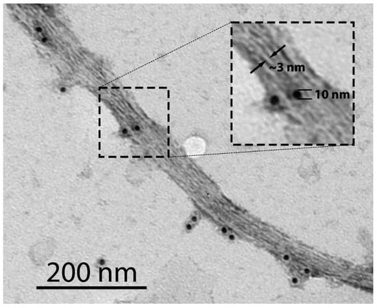
Figure 1.
TEM image of MBP amyloid fibrils immunoprecipitated from the brain of the rat Ratus norvegicus. PAA539Mi01 was used as a primary rabbit anti-MBP antibody. A goat anti-rabbit gold-conjugated antibody with a gold particle (10 nm) was used as a secondary antibody. Immunoprecipitation was performed as described previously [10]. The inset shows a magnified region of the image, highlighting individual protofibrils with a thickness of ~3 nm (indicated by arrows).
Importantly, the central domain of MBP (residues 60–119) is essential for amyloid formation: this region confers aggregation capacity both in a yeast model system and in a cell-free system, where it forms twisted amyloid fibrils. Based on these findings, we proposed a model in which MBP not only bridges opposing cytoplasmic membranes via its N- and C-termini but also forms longitudinal “amyloid stitches” that reinforce the multilamellar myelin sheath, thereby providing mechanical stability to compact myelin [10].
While MBP is protective within compact myelin, mislocalized MBP can become neurotoxic. Experimental studies have shown that extracellular MBP binds to neuronal plasma membranes, depolarizes them, increases Ca2+ influx, and ultimately induces neuronal death [24]. In lipid vesicle assays, MBP disrupted bilayer integrity, suggesting a direct membrane-permeabilizing mechanism [24]. These findings indicate that MBP, once displaced from its physiological context, can act as a membrane-active toxin. Also, it has been shown that in Alzheimer’s disease (AD), against the background of neuronal death and myelin sheath destruction, MBP is present within extracellular Aβ plaques [25].
2.2. FXR1 and Orb2
The FMR1 autosomal homolog 1 (FXR1) is an RNA-binding protein that belongs to the Fragile X-related (FXR) family [26]. Although FXR1 is broadly expressed across different tissues, the highest levels are found in muscle and brain, particularly in neurons and Purkinje cells [27]. Within cells, FXR1 has predominantly cytoplasmic localization, where it forms ribonucleoprotein (RNP) particles. The FXR1 protein is critical for development and survival, as FXR1 knockout mice die at the neonatal stage, most likely due to cardiac or respiratory failure resulting from impaired mRNA transport and translational control in muscle [28]. Conditional FXR1 knockout in the excitatory forebrain neurons of mice demonstrated selective enhancement of long-term spatial memory storage, hippocampal long-lasting synaptic plasticity, and de novo GluA2 synthesis [29].
Under normal conditions, FXR1 is associated with translational silencing through binding to AU-rich elements (AREs) located in the 3′ untranslated regions (UTRs) of many oncogenes, cytokine, and growth factor mRNAs [30]. FXR1 has been identified as a negative regulator of mRNAs encoding inflammatory proteins, including TNFα, ICAM-1, IL-1β, and MCP-1 [31]. This protein may also stabilize and activate translation of its targets. For example, it binds to AREs within cMYC mRNA, enhances transcript stability, and promotes the recruitment of the eIF4F complex to translation initiation sites, thereby facilitating cMYC translation [32].
The RNA-binding activity of FXR1 is mediated by three arginine-rich motifs (RG, RGG, and R) that interact with nucleic acid structures such as G-quadruplexes [33], as well as by two KH domains that bind RNAs containing pseudoknots or kissing loops [34]. In addition to RNA-binding domains, FXR1 harbors multiple protein–protein interaction domains. These include the KH domains and RG/RGG motifs, which are multifunctional and bind both RNA and proteins, as well as two Tudor domains, a KH0 domain, and coiled-coil regions [35]. Tudor domains mediate dimerization and recognize methylated arginines [36,37]. Since the KH0 domain lacks the GXXG motif required for RNA binding, it likely functions exclusively in protein–protein interactions [38]. Beyond its homologues, FXR1 has been shown to interact with a wide range of partners, including cytoskeletal proteins such as Arp2 and CYFIP1 [39], the Argonaute 2 protein involved in microRNA-mediated decay [40], as well as many other protein partners [41].
FXR1 has previously been shown to function in amyloid form in the cytoplasm of cortical neurons across multiple vertebrate species [7,42]. Immunohistochemical assays of brain slices revealed clear colocalization of FXR1 with amyloid-specific dyes CR and ThS in the neuronal cytoplasm. FXR1 fibrils immunoprecipitated from brain lysates exhibit characteristic yellow-green birefringence after CR staining. The amyloidogenic region of FXR1 corresponds to its N-terminal fragment (residues 1–379) [7].
FXR1-containing RNP particles were shown to stabilize associated mRNAs, as transcripts colocalized with FXR1 were resistant to high concentrations of RNase A [7]. Experiments using the human neuroblastoma cell line SH-SY5Y provided higher-resolution insights into the subcellular localization and aggregation level of FXR1 [43]. Normally, FXR1 forms small cytoplasmic oligomers or grains (Figure 2A). These observations suggest that interactions with RNA molecules and RNA-binding proteins limit the growth of FXR1 amyloid fibrils. Upon oxidative stress or heat shock, small FXR1-containing particles are recruited into stress granules—transient, membraneless RNP complexes that assemble through protein/RNA phase separation and promote cell survival (Figure 2B). Importantly, the amyloid properties of FXR1 were shown to be preserved during its incorporation into stress granules [43]. However, FXR1 recruitment into stress granules appears to occur through non-amyloid protein–protein interactions, consistent with the highly dynamic and reversible nature of these structures that disassemble upon stress resolution.

Figure 2.
FXR1 localization in SH-SY5Y cells under physiological and stress conditions. (A) FXR1 forms cytoplasmic oligomers (small grains) in the human neuroblastoma SH-SY5Y cell line under physiological conditions. (B) FXR1 is recruited into large stress granules upon treatment of cells with 3 mM sodium arsenite for 1 h. Rabbit anti-FXR1 antibodies DF12402 was used as the primary antibody, and goat anti-rabbit IgG(H+L) antibody conjugated with Alexa Fluor 488 as the secondary (green fluorescence). Nuclei were stained with Hoechst 33342 (blue fluorescence). The immunocytochemistry assay was performed as described previously [43].
The Drosophila melanogaster protein Orb2, like vertebrate FXR1, is an RNA-binding protein that regulates translation [44]. Orb2 is a member of the cytoplasmic polyadenylation element-binding (CPEB) family. This evolutionarily conserved group of RNA-binding proteins regulates mRNA transport and local translation, processes essential for early embryonic development, synaptic plasticity, and long-term memory [45,46]. While mammalian CPEB protein fragments have been shown to form amyloid fibrils in vitro, clear evidence for amyloid properties in the brain has been demonstrated conclusively only for the fruit fly Orb2 protein [8]. The authors of that study isolated Orb2 from approximately three million fruit fly heads and determined the structure of its native fibrils using cryo-electron microscopy. The amyloidogenic core of Orb2 has been shown to comprise amino acid residues 176 to 206 and to be enriched in glutamine residues. Orb2 binds to the UTRs of various target mRNAs via its RNA recognition motifs to regulate their translation [47]. The monomeric form of Orb2 represses translation and shortens mRNA poly(A) tails, whereas the amyloid oligomeric form enhances translation and elongates poly(A) tails [44]. Orb2 has also been shown to interact with several proteins involved in translation initiation, mRNA binding, and synaptic activity [48].
2.3. Peptide and Protein Hormones in Secretory Granules
Neuroendocrine cells, as well as peptidergic neurons, are known to form large vesicles with electron-dense cores, commonly referred to as secretory granules. These membrane-coated granules serve as inert storage sites for specific secretory proteins and peptides over extended periods, allowing the cell to release them rapidly and massively by regulated exocytosis [49,50]. Growth hormone, prolactin, adrenocorticotropic hormone (ACTH), β-endorphin, oxytocin, and vasopressin have been shown to form storage granules in the pituitary gland [51,52,53,54]. Proteins and peptides concentrated in secretory granules exist as reversible aggregates formed by self-association [55]. The degree of concentration is remarkable: in the case of prolactin, its level in granules is approximately 200 times higher than in the endoplasmic reticulum [56]. Upon stimulation of neuroendocrine cells, the granule content is released in soluble form into the extracellular space [49].
The aggregation state of proteins in secretory granules ranges from amorphous to crystalline form, ensuring their stable and dense packaging for regulated secretion [49,50]. Direct evidence for the presence of amyloid material in the secretory granules of pituitary neuroendocrine cells has been obtained. Maji and colleagues purified secretory granules from the mouse pituitary tumor neuroendocrine cell line AtT20 and analyzed their amyloid properties ex vivo [11]. AtT20 cells are known to synthesize, correctly glycosylate, and process precursors of ACTH and β-endorphin, thereby producing the mature forms of these hormones and storing them properly in secretory granules [57].
It was shown that purified protein contents from AtT20 secretory granules bind conformation-dependent amyloid-specific antibodies OC, whereas monomeric ACTH and β-endorphin do not. Moreover, purified granules from AtT20 cells were demonstrated to bind Thioflavin T, CR, and exhibit birefringence under cross-polarized light upon CR binding. Finally, X-ray fiber diffraction of purified membraneless secretory granules provided strong evidence for amyloid fibrils. A preparation of granules without membrane was employed, since membrane lipids produce a pronounced reflection at 4.1 Å, which is close to the 4.7 Å reflection characteristic of a cross-β-sheet structure. Thus, it can be concluded that at least ACTH and β-endorphin included in secretory granules are of amyloid nature. Similar results were obtained by analysis of secretory granules purified from rat pituitary using the same methods. Immunohistochemical studies of colocalization of hormone-specific antibodies with ThS or OC antibodies on mouse pituitary cryosections confirmed that neuroendocrine hormones such as ACTH, β-endorphin, prolactin, growth hormone, oxytocin, and vasopressin display amyloid properties [11]. However, it is worth noting that these molecules perform their physiological functions in the monomeric form, whereas the amyloid conformation serves exclusively for storage.
Protein/peptide hormones do not exhibit toxicity because they lack the ability to interact with other proteins within the densely packed, membrane-enclosed secretory granule. Amyloid aggregation of these hormones during secretory granule formation is highly regulated by processing events and seeding requirements, including high local concentration, acidic pH, elevated Ca2+ levels, and helper molecules such as granins or glycosaminoglycans. This aggregation is strongly localized to the trans-Golgi network, preventing cytotoxic effects [55,58,59]. Disruption of localized aggregation or improper sorting, however, can give rise to cytotoxicity. For instance, in diabetes insipidus, folding-deficient mutants of provasopressin are retained in the endoplasmic reticulum and produce cytotoxic fibrillar aggregates [60].
3. Pathological Amyloids
3.1. Prion Protein
The cellular prion protein (PrPC) is anchored to the neuronal membrane via glycosylphosphatidylinositol and can act as a receptor or transducer of extracellular signals [61,62,63]. In neurons, PrPC is predominantly localized in the pre- and postsynaptic compartments of nerve terminals. It binds divalent cations such as copper and zinc and interacts with various cellular receptors, including the NMDA receptor [64,65]. PrPC is involved in regulating neuritogenesis, neuronal homeostasis, cell signaling, cell adhesion, and stress responses [66].
The pathological amyloid form of PrP is designated Prion Protein Scrapie (PrPSc) [67]. The formation of PrPSc fibrils occurs sporadically or results from aggregation-promoting mutations. PrPSc particles are infectious and can be transmitted through contact with infected animals or consumption of contaminated feed. These fibrils exhibit remarkable resistance to various treatments, including boiling, certain autoclaving conditions, and degradation by gastrointestinal proteases [67,68]. Following ingestion, PrPSc particles from the gastrointestinal tract enter the lymphatic system and penetrate follicular dendritic cells (FDCs), which produce their own PrPC. Attachment of the exogenous PrPSc particles to endogenous PrPC induces conformational conversion of the latter into the amyloid form [69]. Although PrPSc replicates within FDCs, it does not cause toxicity. As FDCs mature, they migrate to the spleen, where they contact cells of the peripheral nervous system [70]. Subsequently, PrPSc particles are internalized by cells of the peripheral nervous system and later spread to the central nervous system. Through intracellular transport, PrPSc reaches the brain, bypassing the blood–brain barrier (BBB), and is released into the extracellular space.
Prion infections can undergo interspecies transmission. For instance, prion disease can be contracted by consuming contaminated meat, as seen in variant Creutzfeldt-Jakob Disease (vCJD) resulting from bovine spongiform encephalopathy (BSE, or “mad cow disease”) [71]. Cattle, in turn, became infected through feed containing meat and bone meal derived from sheep affected by scrapie [72]. However, prion infection is not transmitted directly from infected sheep to humans [73]. This phenomenon is known as the “species barrier” for prion transmission. The presence or absence of this barrier is determined by similarities and differences in the PrP sequences among mammalian species. Variations in PrP amino acid sequences can prevent the conversion of PrPC to its pathological counterpart, PrPSc, when infectious prion particles from another species enter an organism. PrPSc particles arising sporadically, through mutation, or via infectious transmission, cause various forms of incurable prion diseases in humans and animals, all of which lead to neurodegeneration and fatal outcomes [74]. These forms differ in the brain regions predominantly affected, incubation periods, and clinical symptoms.
Experimental data indicate that PrPSc oligomers exhibit the highest toxicity [75]. Current understanding of PrPSc toxicity mechanisms is discussed in a recent review [65]. PrPSc interacts with the C-terminal domain of PrPC, inducing its conversion into the amyloid form [76]. This interaction liberates the N-terminal domain, enabling it to initiate toxic activity at the cell surface [77]. The conversion of PrPC to PrPSc triggers the opening of NMDA receptors, leading to an influx of Ca2+. This event activates the MAPK p38 pathway and its downstream effectors MK2/3, ultimately resulting in the collapse of the actin cytoskeleton and dendritic spines [78]. Furthermore, this conversion triggers translation repression via activation of the unfolded protein response, mediated by PERK-dependent phosphorylation of eukaryotic initiation factor 2α [79]. Importantly, PERK inhibition prevented neuronal death and behavioral symptoms despite ongoing PrPSc accumulation [80]. Scrapie infection also induces mitochondrial reactive oxygen species (mtROS) production and loss of mitochondrial membrane potential. These events promote abnormal mitochondrial fission and mitophagy, leading to caspase-3 activation and apoptosis [81]. The authors attribute apoptosis initiation to altered mitochondrial Ca2+ concentration and increased mtROS, concurrent with the conversion of PrPC to PrPSc on the cell plasma membrane.
Prion infection additionally activates microglia and astrocytes [82,83,84]. In the early stages, this activation can transiently delay PrPSc proliferation and spread, but prolonged activation leads to chronic neuroinflammation. In advanced disease, high levels of pro-inflammatory cytokines and persistent astrocyte activation contribute to synaptic disruption and neurodegeneration [85]. Despite the diversity of downstream mechanisms, all prion-induced neurotoxic pathways are associated with direct or indirect interactions of the prion protein with targets such as cellular receptors, microglia, and astrocytes.
3.2. Aβ Peptide
Amyloid-β (Aβ) peptide oligomers are the primary cytotoxic factor in AD. Although the etiology of AD is complex, the formation of extracellular Aβ amyloid conformers and intracellular aggregates of hyperphosphorylated tau protein is a defining pathological hallmark. AD is the most common neurodegenerative disorder, with a strong correlation between incidence and age. Currently, over 50 million people worldwide are affected by AD [86]. Most cases of AD are sporadic, whereas less than 5% exhibit a hereditary predisposition, most often associated with mutations in the APP, PSEN1/2, and APOE genes [87]. These mutations directly or indirectly promote Aβ peptide accumulation in the brain. Aβ is generated by proteolytic processing of the transmembrane amyloid precursor protein (APP) by β- and γ-secretases [88]. Numerous Aβ species exist, with Aβ40 being the most abundant (80–90%) and Aβ42 accounting for 5–10%. Aβ production is a normal physiological process that does not inherently cause pathology. Aβ acts as a modulator of synaptic activity and a component of the brain’s innate immunity by regulating neurotransmitter release and neuronal receptor function [89,90,91]. It also possesses antimicrobial properties by binding to and neutralizing pathogens in the brain [92].
The excessive accumulation of the Aβ peptide in the extracellular space can induce the formation of cytotoxic amyloid conformers. Among them, Aβ42 oligomers demonstrate the highest toxicity [93]. The accumulation of neurotoxic Aβ oligomers (AβOs) clearly underlies hereditary AD forms, whereas the causes of sporadic AD remain unclear. In sporadic cases, the formation of neurotoxic Aβ likely represents one stage of a pathological cascade. Besides age, risk factors for sporadic AD include brain trauma, vascular disorders, metabolic syndromes, and aluminum accumulation [87,94]. However, only a small percentage of people under 65 suffer from AD, whereas its prevalence among individuals aged ≥95 in the USA reaches about 50% [95]. Given this strict age dependence, AD onset may be linked to changes in the level of an uncharacterized aging-dependent hormonal factor responsible for Aβ clearance. Aβ clearance occurs through both enzymatic and non-enzymatic pathways [96]. Key non-enzymatic clearance is mediated by the glymphatic system, which eliminates Aβ during sleep [97], as well as by transport across the BBB via LRP1 (efflux) and RAGE (influx) receptors [98]. Enzymatic clearance depends on Aβ degradation by enzymes such as neprilysin (NEP) and insulin-degrading enzyme (IDE) [99]. Impairment of any of these mechanisms promotes Aβ accumulation in the brain.
The toxicity of AβOs primarily involves mitochondrial damage, impairment of the endosome-lysosome system, cellular membrane disruption, hyperphosphorylation and polymerization of tau protein into neurofibrillary tangles, ultimately leading to neuronal death. Aβ amyloid conformers interact with the N-terminal domain of PrPC on cortical neuron membranes [100]. This interaction induces tau hyperphosphorylation, resulting in synaptic loss [101]. Additionally, this interaction activates Fyn kinase, which phosphorylates the NR2B subunit of the NMDA receptor, transiently elevating surface NR2B levels and leading to excitotoxicity and destabilization of dendritic spines [102]. Some evidence suggests that AβOs directly interact with NMDA receptors containing GluN2B subunits and with metabotropic glutamate receptor 1 in primary cortical neurons [103]. Binding to these receptors likely induces various pathological responses that impair synaptic function. Aβ42 oligomers can also insert into cellular membranes, forming large ion-channel pores that disrupt intracellular Ca2+ homeostasis [104]. Mitochondria represent another direct target of AβOs [105]. AβOs can be internalized and imported into mitochondria via the translocase of the outer membrane (TOM) complex, accumulating within mitochondrial cristae [106]. This interaction disrupts mitochondrial morphology, interferes with electron transport chain processes, induces oxidative stress, and impairs mitochondrial fission-fusion dynamics, ultimately disrupting mitochondrial function [107]. Similarly to PrPSc, AβOs accumulation activates microglia and astrocytes, which attempt to clear the toxic conformers [108]. However, this activation becomes chronic in AD, accelerating neurodegeneration. In conclusion, amyloid oligomers bind to multiple targets, triggering various cytotoxic cascades.
3.3. Tau
Tau is a microtubule-associated protein highly expressed in neurons, where it plays a critical role in stabilizing microtubules [109], regulating neurite outgrowth [110], and supporting the transport of organelles and vesicles [111]. Beyond its structural role, tau is involved in neuronal development, dendritic and synaptic plasticity, and modulation of intracellular signaling pathways [112]. Under pathological conditions, however, tau undergoes conformational changes and self-assembles into amyloid fibrils [113], which accumulate in neurofibrillary tangles (NFTs), a defining feature of AD and some other disorders. The amyloid structure of hyperphosphorylated tau filaments isolated from the brain was confirmed using cryo-electron microscopy [113]. Notably, accumulation of Aβ, particularly in its oligomeric form, has been shown to promote tau hyperphosphorylation and aggregation, acting upstream in the pathological cascade of AD [114,115].
Tau pathology is central to a group of neurodegenerative diseases collectively known as tauopathies. These include AD, progressive supranuclear palsy (PSP), corticobasal degeneration (CBD), and Pick’s disease, among others [116]. Clinically, tauopathies present with diverse phenotypes depending on the distribution and extent of tau pathology. AD primarily manifests as progressive memory impairment and difficulties with higher-order cognitive functions [116]. PSP and CBD are characterized by Parkinsonian features, including bradykinesia and rigidity [116]. Pick’s disease presents as frontotemporal dementia, with profound changes in personality, language, and behavior [116].
Post-translational modifications play key roles in tau toxicity. Hyperphosphorylation disrupts microtubule binding and promotes tau aggregation [117], while acetylation at Lys174 delays protein turnover and exacerbates accumulation [118]. Proteolytic processing generates truncated tau fragments that further enhance aggregation propensity and toxicity [119]. Notably, full-length tau in amyloid form can activate calpain-2, leading to degradation of the nicotinic acetylcholine receptor subunit 4 and disruption of cholinergic signaling, thereby creating a feedback loop that promotes tau fragmentation [120]. Pathological tau also perturbs neuronal physiology by altering calcium homeostasis, inducing dendritic spine loss, impairing axonal transport, and disrupting mitochondrial dynamics [121]. Soluble tau oligomers are particularly neurotoxic: they act as seeds for misfolding, impair synaptic plasticity, and cause mitochondrial dysfunction and memory impairment in animal models [122,123]. Phosphorylated tau interacts with mitochondria, impairing their function and contributing to neuronal toxicity [124,125]. This interaction leads to mitochondrial membrane depolarization and increased production of reactive oxygen species, thereby exacerbating cellular stress and neuronal damage [126]. Mislocalization of tau to dendrites contributes to AMPA receptor downregulation, further exacerbating synaptic failure [127]. Tau is also found in the neuronal nucleus, where it binds DNA and associates with nuclear lamina components such as Lamin B1, contributing to chromatin remodeling and nuclear envelope deformation. These nuclear alterations have been linked to neuronal vulnerability in tauopathies [128].
Tau pathology affects not only neurons but also glial cells. Microglia can internalize extracellular tau and contribute to its propagation by releasing tau-containing vesicles [129], while also promoting neuroinflammation through activation of the NLRP3 inflammasome [115]. Astrocytes can likewise take up extracellular tau [130], and its accumulation has been shown to impair astrocytic support of synapses and to exacerbate neuronal dysfunction [131]. In neuronal cell models, tau oligomers were shown to bind to PrPC and muscarinic receptors, disturbing intracellular Ca2+ signaling [132]. Importantly, data suggest that it is the dephosphorylated, rather than hyperphosphorylated, form of extracellular tau that acts as an agonist of muscarinic M1 and M3 receptors. This interaction, mediated by tissue-nonspecific alkaline phosphatase (TNAP), provokes sustained intracellular calcium elevation and ultimately leads to neuronal death [133]. It has further been demonstrated that extracellular tau, particularly when associated with extracellular vesicles isolated from AD brains, is efficiently taken up by neurons, where it seeds aggregation of endogenous tau and propagates tau pathology in vivo [134]. This prion-like mechanism is thought to underlie the stereotypical spread of tau pathology across interconnected brain regions in tauopathies.
In summary, tau is a physiologically essential protein that maintains neuronal structure and function. However, under pathological conditions, it adopts amyloid conformations that drive neurodegeneration. Misfolded or extracellular tau becomes toxic when it interacts with vulnerable cellular components, including receptors, organelles, and signaling pathways, leading to calcium dysregulation, synaptic failure, and neuronal death.
3.4. α-Synuclein
α-Synuclein (α-syn) is a small, intrinsically disordered protein enriched at presynaptic terminals, where it regulates synaptic vesicle trafficking and neurotransmitter release. Under pathological conditions, α-syn misfolds into cross-β structures ranging from soluble oligomers to fibrils [135]. These aggregates accumulate in Lewy bodies and Lewy neurites, which are the histopathological hallmarks of Parkinson’s disease (PD) and related synucleinopathies. In these disease contexts, α-syn behaves as a pathological amyloid, acquiring conformations that confer cytotoxic properties [136]. Most cases of PD are sporadic, typically occurring in people over the age of 60. The disease can be triggered by various risk factors, including brain injuries, exposure to environmental toxins, and oxidative stress, which promote α-syn misfolding and the formation of toxic oligomers [137,138]. Amyloid conformers of α-syn contribute to the degeneration of dopaminergic neurons in the substantia nigra, leading to the characteristic motor and cognitive symptoms of PD.
A central debate concerns which α-syn species are most toxic. Although inclusions are a defining neuropathological feature, evidence from post-mortem human tissue and model systems suggests that soluble oligomeric species, rather than mature fibrils, are primarily responsible for cytotoxicity [139]. Different oligomeric “strains” may also account for the heterogeneity of clinical phenotypes across synucleinopathies [140].
At the synapse, pathological α-syn perturbs vesicle trafficking and SNARE-complex function. Aggregated α-syn binds synaptobrevin-2, blocking SNARE complex assembly and vesicle docking [141], while clusters of oligomer-bound vesicles impair recycling and neurotransmitter release [142]. α-syn in amyloid form also interferes with dopaminergic neurotransmission by inhibiting tyrosine hydroxylase, the rate-limiting enzyme in dopamine biosynthesis [143], and by binding the dopamine transporter, thereby reducing dopamine reuptake [144]. Moreover, α-syn oligomers can insert into vesicle membranes and form pore-like structures, causing neurotransmitter leakage, which in the case of dopaminergic neurons exacerbates oxidative stress [145].
Mitochondrial dysfunction represents another critical pathway of α-syn toxicity. α-syn has been identified within mitochondria in patient tissue and transgenic models [146]. Its association with mitochondrial membranes disrupts organellar architecture, reduces complex I activity, and promotes abnormal fragmentation [139]. These changes impair ROS handling and can lead to mtDNA damage and defective mitophagy. Notably, SNCA knockout mice exhibit reduced ROS production and resistance to mitochondrial toxins [147], underscoring the role of α-syn in oxidative stress.
Pathological interactions with the cytoskeleton and nucleus further broaden α-syn’s toxic profile. α-syn binds α- and β-tubulin, co-aggregating with tubulin and impairing microtubule polymerization, vesicle trafficking, and neurite integrity [148]. Within the nucleus, α-syn binds histones and DNA, inhibiting histone acetylation and impairing DNA repair [149]. These interactions may underlie transcriptional dysregulation observed in synucleinopathies.
Beyond neurons, α-syn toxicity involves glial activation, contributing to neuroinflammation. Pathological α-syn species activate microglia, promoting the release of pro-inflammatory cytokines and exosomes that can facilitate prion-like α-syn propagation and exacerbate dopaminergic neuron loss [150,151]. The microglial responses vary depending on activation state and α-syn burden, with overloaded or pro-inflammatory microglia amplifying oligomer transmission, while resting microglia can partially restrain it [150,152]. Similarly, astrocytes internalize oligomeric and fibrillar α-syn, leading to mitochondrial dysfunction, oxidative stress, and pro-inflammatory cytokine production [153]. α-syn activated astrocytes reduce their trophic support for neurons, which contribute to neuronal death [153]. Together, these glial responses amplify neurotoxicity and promote disease progression.
These findings demonstrate that α-syn toxicity is multifaceted, spanning synaptic dysfunction, membrane permeabilization, mitochondrial stress, cytoskeletal disruption, and nuclear dysregulation. These harmful effects occur when α-syn, normally confined to its physiological locations, misfolds and interacts with cellular components it does not normally encounter. The pleiotropic mechanisms of α-syn toxicity act in parallel, collectively contributing to progressive neuronal death in PD and related disorders.
3.5. Huntingtin
Huntington’s disease (HD) is the most common and well-studied polyglutamine neurodegenerative disease with the onset of first symptoms between the ages of 35 and 50 years. Early neurological symptoms of HD include subtle motor impairments such as clumsiness, awkward involuntary movements, bradykinesia, and rigidity. As the disease progresses, voluntary motor coordination deteriorates while involuntary movements intensify, ultimately leading to loss of mobility and communication. Death usually results from heart failure or aspiration pneumonia. Cognitive decline is also characteristic, with profound dementia in late stages [154]. The major neuropathological hallmark of HD is progressive neuronal degeneration in the striatum (caudate nucleus and putamen) and cerebral cortex, resulting from the accumulation of intranuclear inclusions (NIIs) and protein aggregates in dystrophic neurites [155,156].
Although the clinical features of HD were first described in the 19th century, its genetic basis was identified only in 1993. HD is caused by an unstable expansion of a CAG trinucleotide repeat in exon 1 of the huntingtin gene (HTT), leading to an elongated polyglutamine tract in the N-terminus of the protein [157]. In healthy individuals, the repeat length ranges from 6 to 35. Alleles with 27–35 repeats do not affect neurological functions but confer risk to offspring [158]. Pathogenic expansions exceeding 35 repeats cause HD, with the most severe cases containing more than 100 glutamine residues [157].
Wild-type Htt interacts with numerous proteins—234 high-confidence partners have been identified [159]—and regulates vesicle transport, cytoskeleton assembly, endocytosis, postsynaptic signaling, and transcription. It also promotes neuronal survival by enhancing expression of BDNF and NeuroD [160,161] and by inhibiting caspase-mediated apoptosis through interference with apoptosome and HIP1-HIP1-interactor protein complex formation [162,163]. Mutant Htt with an expanded polyglutamine tract disrupts these interactions, impairing neuronal function. Although mutant HTT is expressed throughout the body, striatal neurons—particularly medium-sized projection spiny neurons—are most vulnerable. These neurons receive dopaminergic and glutamatergic inputs from the substantia nigra, cortex, and thalamus, and produce the inhibitory transmitter γ-Aminobutyric acid (GABA). Therefore, their loss contributes to the uncontrolled movements observed in HD patients [164].
Mutant huntingtin (mHtt) is more susceptible to proteolysis than the normal protein, being cleaved by calpains, matrix metalloproteinases, and caspases [165,166,167], which generate toxic polyQ-containing fragments that further activate proteases. In both HD patients and mice, caspase-derived fragments can be detected before striatal neurodegeneration, and their production depends on the length of the polyQ tract [168,169]. In HD models, cleaved fragments of mHtt have been shown to promote cytotoxicity, excitotoxicity, and aggregate formation [170].
The intracellular aggregates of huntingtin observed in samples from HD patients are of a true amyloid nature, exhibiting a cross-β structure, as demonstrated by CR staining with green birefringence in samples from the HD frontal cortex [171] and by fibrils generated in vitro [172]. Furthermore, the antiparallel β-sheet organization of polyglutamine aggregates of huntingtin has been confirmed by numerous in vitro structural analyses [173,174,175].
Accumulation of mHtt aggregates in nuclei and neurites has been proposed to trigger neuronal death by sequestering proteins and disrupting key intracellular pathways [154]. However, inclusions may represent a protective mechanism, isolating toxic soluble N-terminal fragments or oligomers that would otherwise cause greater damage [176]. Supporting this, in vitro studies show that suppressing nuclear inclusions increases neuronal death, whereas inclusion formation reduces diffuse mHtt and enhances survival [177,178].
mHtt impairs axonal transport by altering interactions between motor proteins and microtubules, reducing delivery of neurotrophic factors such as BDNF [155,156,179]. mHtt sequesters key synaptic proteins, including Complexin II, Synaptobrevin-2, Rabphilin 3A, and PACSIN 1/Syndapin, while also disrupting neurotransmitter receptor expression and function [179,180,181]. Beyond synaptic effects, mHtt induces mitochondrial dysfunction by disrupting mitochondrial dynamics, including fission and fusion, and through interactions with key proteins such as Drp1, Mfn1/2, and OPA1. This results in altered mitochondrial morphology and defective mitophagy [182]. mHtt also reduces electron transport chain and TCA cycle activity, lowering ATP production and increasing reactive oxygen species (ROS) [182]. In addition, mHtt may activate both caspase-dependent and caspase-independent apoptotic pathways, contributing to neuronal cell death [183,184].
Thus, mutations in the HTT gene cause intracellular dysfunction in two ways: loss of normal wild-type huntingtin functions due to interference by the mutant protein, and a toxic gain of function resulting from altered localization and aggregation. Together, these effects disrupt multiple intracellular pathways by sequestering key components into aggregates, ultimately leading to neuronal dysfunction and death.
3.6. TDP-43
Amyotrophic lateral sclerosis (ALS) and frontotemporal lobar degeneration (FTLD) are two related neurodegenerative diseases that overlap clinically, morphologically, and genetically. ALS is a motor neuron disorder (MND) characterized by the premature degeneration of motor neurons in the spinal cord, brainstem, and motor cortex, leading to muscle weakness, hyperreflexia, spasticity of the limbs, and respiratory failure [185]. FTLD is a clinically and pathologically heterogeneous group of dementias, defined by degeneration of the frontal and anterior temporal lobes, resulting in progressive changes in behavior, personality, and language skills [186]. While ALS and FTLD were traditionally considered distinct disorders, they are now recognized as representing opposite ends of a spectrum of clinically, pathologically, and genetically overlapping conditions [187]. Pure forms of FTLD and ALS are connected in FTLD-MND syndromes [188].
Although most cases of ALS are sporadic, approximately 10% are familial, predominantly with autosomal dominant mutations [189]. In FTLD, roughly 30% of cases are familial [190]. Both ALS and FTLD are histopathologically characterized by abnormal accumulation of misfolded protein aggregates in affected regions of the nervous system [188]. Due to their heterogeneity, ALS and FTLD can be classified according to pathological subtypes and the presence of inclusions of specific aggregated proteins: for ALS, these include superoxide dismutase 1 (SOD1), fused in sarcoma (FUS), and optineurin (OPTN) [191]; for FTLD, tau and FUS are common. Both disorders are also associated with TAR DNA-binding protein 43 (TDP-43)-positive inclusions [186].
Direct evidence for the amyloid nature of misfolded TDP-43 aggregates in ALS-FTLD has been obtained via cryo-electron microscopy. The brain samples of patients who had ALS and FTLD with type B cortical TDP-43 pathology were characterized by the presence of round neuronal cytoplasmic inclusions of amyloid fibrils of TDP-43 in the motor cortex and spinal cord [192].
With the development of cryo-electron microscopy, growing evidence indicates that amyloid fibrils of various proteins contribute to intracellular inclusions in ALS-FTLD pathology. Amyloid filament structures of TATA-binding protein-associated factor 15 (TAF15) have been resolved from the prefrontal and temporal cortices of individuals with FTLD-FUS [13]. Additionally, it has been shown that annexin A11 (ANXA11) co-assembles with TDP-43 into heteromeric amyloid filaments in FTLD-TDP type C [14]. However, the mechanism of cytotoxicity of these amyloids has not yet been characterized. Therefore, in this review we focus on the protein TDP-43, which represents the major component of intracellular ALS-FTLD pathological inclusions.
TDP-43 is encoded by the TARDBP gene and belongs to the heterogenous nuclear ribonucleoprotein (hnRNPs) family, a complex and functionally diverse group of RNA-binding proteins. Under physiological conditions, TDP-43 is ubiquitously expressed and predominantly localized in the nucleus, where it regulates gene expression and multiple aspects of RNA metabolism, including transcriptional regulation, RNA trafficking and splicing, mRNA stabilization and turnover, and microRNA biogenesis [193,194,195,196]. TDP-43 is estimated to regulate more than 4000 different mRNA transcripts [197], including its own mRNA and tau transcripts [198,199]. Binding to these transcripts promotes their destabilization, leading to downregulation of tau and TDP-43 protein levels. The neurotoxicity of pathological TDP-43 is believed to arise from both a gain of toxic cytoplasmic function and a loss of physiological function associated with nuclear depletion of TDP-43. Cytoplasmic oligomers and aggregates of TDP-43 have been shown to be cytotoxic in vivo [200,201].
TDP-43 amyloid fibrils are transported into the cytoplasm and incorporated into stress granules, which leads to their irreversibility. Also, cytoplasmic amyloid conformers of TDP-43 impair axonal transport and axon growth. The nuclear depletion of TDP-43 caused by its cytoplasmic mislocalization likely produces loss-of-function effects through dysregulation of RNA metabolism, alternative splicing, and transport of dozens of transcripts [202]. TDP-43 interacts with mitochondrial outer membrane protein prohibitin 2 (PHB2) and voltage-dependent anion channel 1 (VDAC1), which are crucial receptors for Parkin-mediated mitophagy [203]. Overexpression of either wild-type or mutant TDP-43 induces mitochondrial structural and functional abnormalities, including swollen and degenerated cristae, the complete lack of cristae, reduced membrane potential, increased ROS, reduced mitochondrial ATP production, and activation of the mitochondrial unfolded protein response. Similar abnormalities have been observed in patient samples via electron microscopy. Additionally, TDP-43 pathology has been linked to disrupted Ca2+ homeostasis, activation of glycogen synthase kinase 3β (GSK-3β), and autophagy dysregulation [202].
4. Comparative Analysis of Pathological and Functional Brain Amyloids Based on Their Amino Acid Composition
Previously, a comparative analysis of the amino acid composition of the Orb2 and Aβ40 proteins suggested that the amyloidogenic cores of pathological proteins are enriched in hydrophobic residues, whereas those of functional amyloids are predominantly hydrophilic [204]. In our opinion, drawing broad conclusions from the comparison of only two proteins seems premature. We conducted a comparative analysis of the pathological and functional brain amyloids discussed in this review (Table 1 and Table 2 and Figure 3A,B). With the exception of the fruit fly Orb2 protein, all analyzed sequences correspond to human proteins. We included β-endorphin as a representative of the large group of amyloid secretory granule proteins. The ability of β-endorphin to form amyloid fibrils has been demonstrated in at least two independent studies [3,205]. The comparison was based on several parameters such as hydrophobicity/hydrophilicity, the proportions of positively and negatively charged amino acids, and enrichment in asparagine/glutamine (Q/N) or serine/glycine (G/S) residues. The analysis was performed for both full-length proteins and their amyloidogenic core sequences, the boundaries of which were determined based on previously obtained experimental data [7,8,10,113,175,205,206,207,208,209].

Table 1.
Comparative analysis of the amino acid composition of full-length pathological and functional brain amyloids. The table shows the percentages of hydrophobic, charged (+/−), positively or negatively charged, and Q/N- and G/S-enriched residues. The percentages were calculated using the Prot pi Tool, version 2.2.29.152 (https://www.protpi.ch/Calculator/ProteinTool, accessed on 15 September 2025).
Table 2.
Comparative analysis of the amino acid composition of experimentally defined amyloidogenic core fragments of pathological and functional brain amyloids. The table shows the percentages of hydrophobic, charged (+/−), positively or negatively charged, and Q/N- and G/S-enriched residues. The percentages were calculated using the Prot pi Tool, version 2.2.29.152 (https://www.protpi.ch/Calculator/ProteinTool, accessed on 15 September 2025).

Figure 3.
Comparative analysis of amino acid composition of pathological and functional brain amyloids. (A) Distribution of hydrophobic, charged (+/−), positively or negatively charged, and Q/N- and G/S-enriched residues in the full-length sequences of pathological and functional brain amyloid proteins. The percentages of amino acid residues with the corresponding characteristics are plotted along the vertical axis. (B) The same parameters calculated for their experimentally defined amyloidogenic core fragments. The names of pathological amyloids are marked with red lines. The names of functional amyloids are marked with green lines.
The results presented in Table 1 and Table 2, as well as in Figure 3, convincingly show that the amino acid sequences of pathological and functional amyloids, as well as their amyloidogenic cores, do not differ in any of the parameters examined. For instance, the amyloidogenic core of α-syn is less hydrophobic than the cores of β-endorphin and FXR1, but more hydrophobic than those of MBP and Orb2 (Table 2 and Figure 3B). The amyloidogenic core of the mHtt protein is the most hydrophilic (Table 2 and Figure 3B), as it consists almost entirely of glutamine residues with the exception of a single phenylalanine [175]. Thus, the differences between pathological and functional amyloids are not determined by their amino acid composition, contrary to previous assumptions.
5. Factors Determining the Toxicity and Functionality of Amyloids
In this review, we have characterized pathological and functional brain amyloids and demonstrated that their toxicity or lack thereof cannot be explained by differences in amino acid composition. Theoretically, toxicity could be determined by the length, orientation, or location of amyloidogenic tracts within the protein. However, available experimental data contradict this hypothesis. For example, the amyloidogenic sequence is located in the N-terminal part of the mHtt protein, while in the PrP protein it is in the C-terminal part. Moreover, many pathological amyloids share a parallel in-register β-sheet structure, which is also characteristic of the functional amyloid Orb2 [8]. The length of the amyloidogenic core in the Aβ peptide is roughly similar to that of the functional amyloid Orb2. These findings challenge the assumption that toxicity is determined solely by the structural features of amyloid conformers. At the same time, the data presented in this review indicate that the presence or absence of toxicity may be determined by the localization of amyloid proteins and/or their interaction with functional partners.
5.1. Amyloids’ Localization
As discussed above, extracellular oligomers of PrPSc and Aβ interact with PrPC on the cell surface, initiating cytotoxic cascades [76,100]. Moreover, amyloid conformers of the yeast Sup35NM protein and a synthetic amyloid peptide also bind PrPC in cell models, leading to cell death [76]. These data suggest that PrPC is a universal target for a wide range of extracellular amyloids, making them toxic to neuronal cells. Unlike extracellular amyloids, not all intracellular amyloids are toxic. Their subcellular localization plays a decisive role in determining whether they can access specific targets and trigger cytotoxicity. For example, all amyloid hormones in the pituitary gland are isolated from the cytosol by secretory granule membranes, preventing interaction with external targets [11]. The pH of secretory granules is lower than the intracellular pH. The acidic environment inside these granules promotes amyloid stability, but upon granule fusion with the cell membrane, the pH shift facilitates rapid fibril disassembly, releasing the hormone in its monomeric, functional form [11]. Amyloid fibrils of MBP are not isolated in membrane compartments, but are located exclusively within specialized cellular structures. As noted earlier, MBP mRNA is transported to and translated within oligodendrocyte processes. MBP molecules anchor via their N- and C-termini to the opposing membranes of these flattened processes, displacing other proteins [15]. The central fragments of the anchored MBP molecules come into close proximity and form a cross-β structure. [10]. Thus, MBP amyloid fibrils are effectively isolated within oligodendrocyte processes. Collectively, these examples indicate that the specific localization of many functional amyloids within cells prevents access to potentially dangerous targets.
5.2. Interaction of Amyloids with Pathological or Functional Partners
In the case of pathological amyloidogenesis, conformational changes lead not only to protein mislocalization but also to the disruption of interactions with functional partners, as well as to the appearance of interactions with new targets. All known pathological intracellular amyloids bind specific targets that trigger cell death pathways. Amyloid conformers of mHtt, α-syn, tau, and TDP-43 directly interact with mitochondrial membranes, leading to mitochondrial fragmentation and dysfunction, excessive ROS production, oxidative stress, and apoptosis [210,211,212,213].
Functional amyloid FXR1 in vertebrates and Orb2 in the fruit fly are not isolated within membrane-bound compartments or other distinct cellular structures. They are distributed throughout the cytoplasm and could theoretically interact with a wide variety of harmful targets, yet they do not exhibit toxicity. FXR1 has been shown to function as oligomers that bind numerous RNA molecules and other RNA-binding proteins [43,214]. Similarly, Orb2 forms functional oligomers that associate with RNA-binding proteins and RNA [47,48]. Apparently, specific interaction with functional partners prevents the uncontrolled growth of amyloid fibrils and sterically hinders their binding to targets that could provoke cell death. For example, expression of truncated FXR1 variants (residues 1–379 and 1–399) lacking the C-terminal RG/RGG motifs in HeLa cells drastically altered granule size and distribution of FXR1 [215]. These findings suggest that minor changes in the repertoire of FXR1’s partners strongly affect its aggregation propensity. Under physiological conditions, the complex domain architecture of FXR1 likely ensures tight regulation of its amyloidogenic potential by molecular interactors, thereby preventing cytotoxic amyloid accumulation.
5.3. The Concept of “Available Targets”
Based on data analyzed, we propose the “available targets” concept to explain why some amyloids are toxic while others are not. The toxicity of pathological amyloids arises from their ability to bind universal or specific targets. Interactions with key targets, such as PrPC or mitochondria, initiate pathological cascades leading to cell death. Functional amyloids remain non-toxic because these targets are inaccessible to them. This inaccessibility is ensured either by localized translation and amyloids sequestration within specialized cellular structures, or by interaction with functional partners, which sterically blocks binding to harmful targets.
6. Conclusions
We suggest that the concept of “available targets” provides a universal explanation for the fundamental differences between toxic and functional amyloids not only in the brain. For example, the functional amyloid Pmel17 in melanocytes is stringently sequestered from the cytoplasm within melanosomes [2]. The seeds of garden pea contain amyloid aggregates of storage protein Vicilin that accumulate in vacuoles. In vitro, fibrils of this protein exhibit toxicity in mammalian cell culture. [9]. Different systemic amyloidoses are associated with the accumulation of proteins in the blood plasma, which, due to mislocalization and disrupted interactions with functional partners, form amyloid conformers and affect the cells of various organs and tissues [216,217]. Isolation of amyloids in specialized cellular compartments or their interaction with functional partners prevents amyloid toxicity. Identifying specific factors that block the interaction of amyloids with dangerous targets and limit the growth of amyloid fibrils may potentially facilitate the development of new approaches to the treatment of amyloidosis.
Author Contributions
Conceptualization, A.P.G.; formal analysis, E.I.S.; writing—A.P.G.—Abstract, Introduction, Prion Protein, Comparative Analysis of Pathological and Functional Brain Amyloids Based on Their Amino Acid Composition and Conclusion; V.A.M. and A.A.M.—amyloid beta peptide section; A.A.V.—FXR1 and Orb2, Peptide and Protein Hormones in Secretory Granules, Huntingtin and TDP-43 sections; E.I.S.—Myelin Basic Protein, tau and α-Synuclein sections; review and editing, A.P.G., A.A.V. and E.I.S.; visualization, E.I.S. and A.A.V.; supervision, A.P.G.; funding acquisition, A.P.G. and A.A.M. All authors have read and agreed to the published version of the manuscript.
Funding
This research was funded by the Russian Science, grant number 24-14-00233 to A.G and by the Russian Science Foundation, grant number 19-74-30007 to A.M.
Institutional Review Board Statement
Not applicable.
Informed Consent Statement
Not applicable.
Data Availability Statement
No new data were created or analyzed in this study. Data sharing is not applicable to this article.
Conflicts of Interest
The authors declare no conflicts of interest.
References
- Wolfe, K.J.; Cyr, D.M. Amyloid in Neurodegenerative Diseases: Friend or Foe? Semin. Cell Dev. Biol. 2011, 22, 476–481. [Google Scholar] [CrossRef] [PubMed]
- Fowler, D.M.; Koulov, A.V.; Alory-Jost, C.; Marks, M.S.; Balch, W.E.; Kelly, J.W. Functional Amyloid Formation within Mammalian Tissue. PLoS Biol. 2006, 4, e6. [Google Scholar] [CrossRef] [PubMed]
- Sønderby, T.V.; Najarzadeh, Z.; Otzen, D.E. Functional Bacterial Amyloids: Understanding Fibrillation, Regulating Biofilm Fibril Formation and Organizing Surface Assemblies. Molecules 2022, 27, 4080. [Google Scholar] [CrossRef]
- Lipke, P.N.; Klotz, S.A.; Dufrene, Y.F.; Jackson, D.N.; Garcia-Sherman, M.C. Amyloid-Like β-Aggregates as Force-Sensitive Switches in Fungal Biofilms and Infections. Microbiol. Mol. Biol. Rev. 2017, 82, e00035-17. [Google Scholar] [CrossRef] [PubMed]
- Ryzhova, T.A.; Sopova, J.V.; Zadorsky, S.P.; Siniukova, V.A.; Sergeeva, A.V.; Galkina, S.A.; Nizhnikov, A.A.; Shenfeld, A.A.; Volkov, K.V.; Galkin, A.P. Screening for Amyloid Proteins in the Yeast Proteome. Curr. Genet. 2018, 64, 469–478. [Google Scholar] [CrossRef]
- Sergeeva, A.V.; Sopova, J.V.; Belashova, T.A.; Siniukova, V.A.; Chirinskaite, A.V.; Galkin, A.P.; Zadorsky, S.P. Amyloid Properties of the Yeast Cell Wall Protein Toh1 and Its Interaction with Prion Proteins Rnq1 and Sup35. Prion 2019, 13, 21–32. [Google Scholar] [CrossRef]
- Sopova, J.V.; Koshel, E.I.; Belashova, T.A.; Zadorsky, S.P.; Sergeeva, A.V.; Siniukova, V.A.; Shenfeld, A.A.; Velizhanina, M.E.; Volkov, K.V.; Nizhnikov, A.A.; et al. RNA-Binding Protein FXR1 Is Presented in Rat Brain in Amyloid Form. Sci. Rep. 2019, 9, 18983. [Google Scholar] [CrossRef]
- Hervas, R.; Rau, M.J.; Park, Y.; Zhang, W.; Murzin, A.G.; Fitzpatrick, J.A.J.; Scheres, S.H.W.; Si, K. Cryo-EM Structure of a Neuronal Functional Amyloid Implicated in Memory Persistence in Drosophila. Science 2020, 367, 1230–1234. [Google Scholar] [CrossRef]
- Antonets, K.S.; Belousov, M.V.; Sulatskaya, A.I.; Belousova, M.E.; Kosolapova, A.O.; Sulatsky, M.I.; Andreeva, E.A.; Zykin, P.A.; Malovichko, Y.V.; Shtark, O.Y.; et al. Accumulation of Storage Proteins in Plant Seeds Is Mediated by Amyloid Formation. PLoS Biol. 2020, 18, e3000564. [Google Scholar] [CrossRef]
- Sysoev, E.I.; Shenfeld, A.A.; Belashova, T.A.; Valina, A.A.; Zadorsky, S.P.; Galkin, A.P. Amyloid Fibrils of the Myelin Basic Protein Are an Integral Component of Myelin in the Vertebrate Brain. Sci. Rep. 2025, 15, 29053. [Google Scholar] [CrossRef]
- Maji, S.K.; Perrin, M.H.; Sawaya, M.R.; Jessberger, S.; Vadodaria, K.; Rissman, R.A.; Singru, P.S.; Nilsson, K.P.R.; Simon, R.; Schubert, D.; et al. Functional Amyloids as Natural Storage of Peptide Hormones in Pituitary Secretory Granules. Science 2009, 325, 328–332. [Google Scholar] [CrossRef]
- Valina, A.A.; Siniukova, V.A.; Belashova, T.A.; Kanapin, A.A.; Samsonova, A.A.; Masharsky, A.E.; Lykholay, A.N.; Galkina, S.A.; Zadorsky, S.P.; Galkin, A.P. Amyloid Fibrils of the S36 Protein Modulate the Morphogenesis of Drosophila Melanogaster Eggshell. Int. J. Mol. Sci. 2024, 25, 12499. [Google Scholar] [CrossRef] [PubMed]
- Tetter, S.; Arseni, D.; Murzin, A.G.; Buhidma, Y.; Peak-Chew, S.Y.; Garringer, H.J.; Newell, K.L.; Vidal, R.; Apostolova, L.G.; Lashley, T.; et al. TAF15 Amyloid Filaments in Frontotemporal Lobar Degeneration. Nature 2024, 625, 345–351. [Google Scholar] [CrossRef] [PubMed]
- Arseni, D.; Nonaka, T.; Jacobsen, M.H.; Murzin, A.G.; Cracco, L.; Peak-Chew, S.Y.; Garringer, H.J.; Kawakami, I.; Suzuki, H.; Onaya, M.; et al. Heteromeric Amyloid Filaments of ANXA11 and TDP-43 in FTLD-TDP Type C. Nature 2024, 634, 662–668. [Google Scholar] [CrossRef] [PubMed]
- Boggs, J.M. Myelin Basic Protein: A Multifunctional Protein. Cell. Mol. Life Sci. 2006, 63, 1945–1961. [Google Scholar] [CrossRef]
- Landry, C.F.; Ellison, J.A.; Pribyl, T.M.; Campagnoni, C.; Kampf, K.; Campagnoni, A.T. Myelin Basic Protein Gene Expression in Neurons: Developmental and Regional Changes in Protein Targeting within Neuronal Nuclei, Cell Bodies, and Processes. J. Neurosci. 1996, 16, 2452–2462. [Google Scholar] [CrossRef]
- Campagnoni, A.T.; Skoff, R.P. The Pathobiology of Myelin Mutants Reveal Novel Biological Functions of the MBP and PLP Genes. Brain Pathol. 2001, 11, 74–91. [Google Scholar] [CrossRef]
- Müller, C.; Bauer, N.M.; Schäfer, I.; White, R. Making Myelin Basic Protein—From MRNA Transport to Localized Translation. Front. Cell. Neurosci. 2013, 7, 169. [Google Scholar] [CrossRef]
- Kosturko, L.D.; Maggipinto, M.J.; Korza, G.; Lee, J.W.; Carson, J.H.; Barbarese, E. Heterogeneous Nuclear Ribonucleoprotein (HnRNP) E1 Binds to HnRNP A2 and Inhibits Translation of A2 Response Element MRNAs. Mol. Biol. Cell 2006, 17, 3521–3533. [Google Scholar] [CrossRef]
- Bauer, N.M.; Moos, C.; Van Horssen, J.; Witte, M.; Van Der Valk, P.; Altenhein, B.; Luhmann, H.J.; White, R. Myelin Basic Protein Synthesis Is Regulated by Small Non-Coding RNA 715. EMBO Rep. 2012, 13, 827–834. [Google Scholar] [CrossRef]
- Kattnig, D.R.; Bund, T.; Boggs, J.M.; Harauz, G.; Hinderberger, D. Lateral Self-Assembly of 18.5-KDa Myelin Basic Protein (MBP) Charge Component-C1 on Membranes. Biochim. Biophys. Acta 2012, 1818, 2636–2647. [Google Scholar] [CrossRef]
- Aggarwal, S.; Snaidero, N.; Pähler, G.; Frey, S.; Sánchez, P.; Zweckstetter, M.; Janshoff, A.; Schneider, A.; Weil, M.T.; Schaap, I.A.T.; et al. Myelin Membrane Assembly Is Driven by a Phase Transition of Myelin Basic Proteins into a Cohesive Protein Meshwork. PLoS Biol. 2013, 11, e1001577. [Google Scholar] [CrossRef]
- Raasakka, A.; Ruskamo, S.; Kowal, J.; Barker, R.; Baumann, A.; Martel, A.; Tuusa, J.; Myllykoski, M.; Bürck, J.; Ulrich, A.S.; et al. Membrane Association Landscape of Myelin Basic Protein Portrays Formation of the Myelin Major Dense Line. Sci. Rep. 2017, 7, 4974. [Google Scholar] [CrossRef]
- Zhang, J.; Sun, X.; Zheng, S.; Liu, X.; Jin, J.; Ren, Y.; Luo, J. Myelin Basic Protein Induces Neuron-Specific Toxicity by Directly Damaging the Neuronal Plasma Membrane. PLoS ONE 2014, 9, e108646. [Google Scholar] [CrossRef] [PubMed]
- Zhan, X.; Jickling, G.C.; Ander, B.P.; Stamova, B.; Liu, D.; Kao, P.F.; Zelin, M.A.; Jin, L.W.; DeCarli, C.; Sharp, F.R. Myelin basic protein associates with AβPP, Aβ1-42, and amyloid plaques in cortex of Alzheimer’s disease brain. J. Alzheimer’s Dis. 2015, 44, 1213–1229. [Google Scholar] [CrossRef]
- Majumder, M.; Johnson, R.H.; Palanisamy, V. Fragile X-Related Protein Family: A Double-Edged Sword in Neurodevelopmental Disorders and Cancer. Crit. Rev. Biochem. Mol. Biol. 2020, 55, 409–424. [Google Scholar] [CrossRef] [PubMed]
- Tamanini, F.; Willemsen, R.; Van Unen, L.; Bontekoe, C.; Galjaard, H.; Oostra, B.A.; Hoogeveen, A.T. Differential Expression of FMR1, FXR1 and FXR2 Proteins in Human Brain and Testis. Hum. Mol. Genet. 1997, 6, 1315–1322. [Google Scholar] [CrossRef] [PubMed]
- Meintjes, E.J.; Willemsen, R.; Kirkpatrick, L.L.; Niuewenhuizen, I.M.; Hoogeveen-Westerveld, M.; Verweij, M.; Reis, S.; Bardoni, B.; Hoogeveen, A.T.; Oostra, B.A.; et al. Fxr1 Knockout Mice Show a Striated Muscle Phenotype: Implications for Fxr1p Function In Vivo. Hum. Mol. Genet. 2004, 13, 1291–1302. [Google Scholar] [CrossRef]
- Cook, D.; Nuro, E.; Jones, E.V.; Altimimi, H.F.; Farmer, W.T.; Gandin, V.; Hanna, E.; Zong, R.; Barbon, A.; Nelson, D.L.; et al. FXR1P Limits Long-Term Memory, Long-Lasting Synaptic Potentiation, and de Novo GluA2 Translation. Cell Rep. 2014, 9, 1402–1416. [Google Scholar] [CrossRef]
- Garnon, J.; Lachance, C.; Di Marco, S.; Hel, Z.; Marion, D.; Ruiz, M.C.; Newkirk, M.M.; Khandjian, E.W.; Radzioch, D. Fragile X-Related Protein FXR1P Regulates Proinflammatory Cytokine Tumor Necrosis Factor Expression at the Post-Transcriptional Level. J. Biol. Chem. 2005, 280, 5750–5763. [Google Scholar] [CrossRef]
- Herman, A.B.; Vrakas, C.N.; Ray, M.; Kelemen, S.E.; Sweredoski, M.J.; Moradian, A.; Haines, D.S.; Autieri, M.V. FXR1 Is an IL-19-Responsive RNA-Binding Protein That Destabilizes Pro-Inflammatory Transcripts in Vascular Smooth Muscle Cells. Cell Rep. 2018, 24, 1176–1189. [Google Scholar] [CrossRef]
- George, J.; Li, Y.; Kadamberi, I.P.; Parashar, D.; Tsaih, S.W.; Gupta, P.; Geethadevi, A.; Chen, C.; Ghosh, C.; Sun, Y.; et al. RNA-Binding Protein FXR1 Drives CMYC Translation by Recruiting EIF4F Complex to the Translation Start Site. Cell Rep. 2021, 37, 109934. [Google Scholar] [CrossRef]
- Schaeffer, C.; Bardoni, B.; Mandel, J.L.; Ehresmann, B.; Ehresmann, C.; Moine, H. The Fragile X Mental Retardation Protein Binds Specifically to Its MRNA via a Purine Quartet Motif. EMBO J. 2001, 20, 4803–4813. [Google Scholar] [CrossRef] [PubMed]
- Darnell, J.C.; Fraser, C.E.; Mostovetsky, O.; Darnell, R.B. Discrimination of Common and Unique RNA-Binding Activities among Fragile X Mental Retardation Protein Paralogs. Hum. Mol. Genet. 2009, 18, 3164–3177. [Google Scholar] [CrossRef] [PubMed]
- Winograd, C.; Ceman, S. Fragile X Family Members Have Important and Non-Overlapping Functions. Biomol. Concepts 2011, 2, 343–352. [Google Scholar] [CrossRef] [PubMed]
- Adinolfi, S.; Ramos, A.; Martin, S.R.; Dal Piaz, F.; Pucci, P.; Bardoni, B.; Mandel, J.L.; Pastore, A. The N-Terminus of the Fragile X Mental Retardation Protein Contains a Novel Domain Involved in Dimerization and RNA Binding. Biochemistry 2003, 42, 10437–10444. [Google Scholar] [CrossRef]
- Adams-Cioaba, M.A.; Guo, Y.; Bian, C.B.; Amaya, M.F.; Lam, R.; Wasney, G.A.; Vedadi, M.; Xu, C.; Min, J. Structural Studies of the Tandem Tudor Domains of Fragile X Mental Retardation Related Proteins FXR1 and FXR2. PLoS ONE 2010, 5, e13559. [Google Scholar] [CrossRef]
- Myrick, L.K.; Hashimoto, H.; Cheng, X.; Warren, S.T. Human FMRP Contains an Integral Tandem Agenet (Tudor) and KH Motif in the Amino Terminal Domain. Hum. Mol. Genet. 2015, 24, 1733–1740. [Google Scholar] [CrossRef]
- St. Paul, A.; Corbett, C.; Peluzzo, A.; Kelemen, S.; Okune, R.; Haines, D.S.; Preston, K.; Eguchi, S.; Autieri, M.V. FXR1 Regulates Vascular Smooth Muscle Cell Cytoskeleton, VSMC Contractility, and Blood Pressure by Multiple Mechanisms. Cell Rep. 2023, 42, 112381. [Google Scholar] [CrossRef]
- Vasudevan, S.; Steitz, J.A. AU-Rich-Element-Mediated Upregulation of Translation by FXR1 and Argonaute 2. Cell 2007, 128, 1105–1118. [Google Scholar] [CrossRef]
- Yang, J.; Chen, Z.; He, J.; Zou, B.; Si, Y.; Ma, Y.; Yu, J. Modular RNA Interactions Shape FXR1 Condensates Involved in MRNA Localization and Translation. Nat. Commun. 2025, 16, 8589. [Google Scholar] [CrossRef]
- Velizhanina, M.E.; Galkin, A.P. Amyloid Properties of the FXR1 Protein Are Conserved in Evolution of Vertebrates. Int. J. Mol. Sci. 2022, 23, 7997. [Google Scholar] [CrossRef]
- Valina, A.A.; Belashova, T.A.; Yuzman, A.K.; Zadorsky, S.P.; Sysoev, E.I.; Mitkevich, V.A.; Makarov, A.A.; Galkin, A.P. Functional Amyloid Protein FXR1 Is Recruited into Neuronal Stress Granules. Prion 2025, 19, 1–16. [Google Scholar] [CrossRef] [PubMed]
- Khan, M.R.; Li, L.; Pérez-Sánchez, C.; Saraf, A.; Florens, L.; Slaughter, B.D.; Unruh, J.R.; Si, K. Amyloidogenic Oligomerization Transforms Drosophila Orb2 from a Translation Repressor to an Activator. Cell 2015, 163, 1468–1483. [Google Scholar] [CrossRef]
- Charlesworth, A.; Meijer, H.A.; De Moor, C.H. Specificity Factors in Cytoplasmic Polyadenylation. Wiley Interdiscip. Rev. RNA 2013, 4, 437–461. [Google Scholar] [CrossRef] [PubMed]
- Kozlov, E.; Shidlovskii, Y.V.; Gilmutdinov, R.; Schedl, P.; Zhukova, M. The Role of CPEB Family Proteins in the Nervous System Function in the Norm and Pathology. Cell Biosci. 2021, 11, 64. [Google Scholar] [CrossRef] [PubMed]
- Stepien, B.K.; Oppitz, C.; Gerlach, D.; Dag, U.; Novatchkova, M.; Krüttner, S.; Stark, A.; Keleman, K. RNA-Binding Profiles of Drosophila CPEB Proteins Orb and Orb2. Proc. Natl. Acad. Sci. USA 2016, 113, E7030–E7038. [Google Scholar] [CrossRef]
- White-Grindley, E.; Li, L.; Mohammad Khan, R.; Ren, F.; Saraf, A.; Florens, L.; Si, K. Contribution of Orb2A Stability in Regulated Amyloid-Like Oligomerization of Drosophila Orb2. PLoS Biol. 2014, 12, e1001786. [Google Scholar] [CrossRef]
- Dannies, P.S. Concentrating Hormones into Secretory Granules: Layers of Control. Mol. Cell Endocrinol. 2001, 177, 87–93. [Google Scholar] [CrossRef]
- Kim, T.; Gondré-Lewis, M.C.; Arnaoutova, I.; Loh, Y.P. Dense-Core Secretory Granule Biogenesis. Physiology 2006, 21, 124–133. [Google Scholar] [CrossRef]
- Tesar, J.T.; Koenig, H.; Hughes, C. Hormone Storage Granules in the Beef Anterior Pituitary. I. Isolation, Ultrastructure, and Some Biochemical Properties. J. Cell Biol. 1969, 40, 225–235. [Google Scholar] [CrossRef]
- Perdue, J.F.; McShan, W.H. Association of Adrenocorticotropic Hormone Activity with Small Secretory Granules from Rat Anterior Pituitary Glands. Endocrinology 1966, 78, 406–408. [Google Scholar] [CrossRef] [PubMed]
- Tasso, F.; Picard, D.; Dreifuss, J.J. Ultrastructural Identification of Granules Containing Oxytocin and Vasopressin. Nature 1976, 260, 621–622. [Google Scholar] [CrossRef] [PubMed]
- Weber, E.; Martin, R.; Voigt, K.H. Corticotropin/β-Endorphin Precursor: Concomitant Storage of Its Fragments in the Secretory Granules of Anterior Pituitary Corticotropin/Endorphin Cells. Life Sci. 1979, 25, 1111–1118. [Google Scholar] [CrossRef] [PubMed]
- Tooze, S.A. Biogenesis of Secretory Granules in the Trans-Golgi Network of Neuroendocrine and Endocrine Cells. Biochim. Biophys. Acta Mol. Cell Res. 1998, 1404, 231–244. [Google Scholar] [CrossRef]
- Farquhar, M.G.; Reid, J.A.J.; Daniell, L.W. Intracellular Transport and Packaging of Prolactin: A Quantitative Electron Microscope Autoradiographic Study of Mammotrophs Dissociated from Rat Pituitaries. Endocrinology 1978, 102, 296–311. [Google Scholar] [CrossRef]
- Gumbiner, B.; Kelly, R.B. Secretory Granules of an Anterior Pituitary Cell Line, AtT-20, Contain Only Mature Forms of Corticotropin and β-Lipotropin. Proc. Natl. Acad. Sci. USA 1981, 78, 318–322. [Google Scholar] [CrossRef]
- Dannies, P.S. Prolactin and Growth Hormone Aggregates in Secretory Granules: The Need to Understand the Structure of the Aggregate. Endocr. Rev. 2012, 33, 254–270. [Google Scholar] [CrossRef]
- Chanat, E.; Huttner, W.B. Milieu-Induced, Selective Aggregation of Regulated Secretory Proteins in the Trans-Golgi Network. J. Cell Biol. 1991, 115, 1505–1519. [Google Scholar] [CrossRef]
- Birk, J.; Friberg, M.A.; Prescianotto-Baschong, C.; Spiess, M.; Rutishauser, J. Dominant Pro-Vasopressin Mutants That Cause Diabetes Insipidus Form Disulfide-Linked Fibrillar Aggregates in the Endoplasmic Reticulum. J. Cell Sci. 2009, 221, 3994–4002. [Google Scholar] [CrossRef]
- Bendheim, P.E.; Brown, H.R.; Rudelli, R.D.; Scala, L.J.; Goller, N.L.; Wen, G.Y.; Kascsak, R.J.; Cashman, N.R.; Bolton, D.C. Nearly Ubiquitous Tissue Distribution of the Scrapie Agent Precursor Protein. Neurology 1992, 42, 149–156. [Google Scholar] [CrossRef]
- McDonald, A.J.; Leon, D.R.; Markham, K.A.; Wu, B.; Heckendorf, C.F.; Schilling, K.; Showalter, H.D.; Andrews, P.C.; McComb, M.E.; Pushie, M.J.; et al. Altered Domain Structure of the Prion Protein Caused by Cu2+ Binding and Functionally Relevant Mutations: Analysis by Cross-Linking, MS/MS, and NMR. Structure 2019, 27, 907–922. [Google Scholar] [CrossRef] [PubMed]
- Wu, G.R.; Mu, T.C.; Gao, Z.X.; Wang, J.; Sy, M.S.; Li, C.Y. Prion Protein Is Required for Tumor Necrosis Factor α (TNFα)-Triggered Nuclear Factor Κb (NF-ΚB) Signaling and Cytokine Production. J. Biol. Chem. 2017, 292, 18747–18759. [Google Scholar] [CrossRef]
- Brown, D.R.; Clive, C.; Haswell, S.J. Antioxidant Activity Related to Copper Binding of Native Prion Protein. J. Neurochem. 2001, 76, 69–76. [Google Scholar] [CrossRef] [PubMed]
- Mercer, R.C.C.; Harris, D.A. Mechanisms of Prion-Induced Toxicity. Cell Tissue Res. 2023, 392, 81–96. [Google Scholar] [CrossRef] [PubMed]
- Legname, G. Elucidating the Function of the Prion Protein. PLoS Pathog. 2017, 13, e1006458. [Google Scholar] [CrossRef]
- Prusiner, S.B.; Scott, M.R.; DeArmond, S.J.; Cohen, F.E. Prion Protein Biology. Cell 1998, 93, 337–348. [Google Scholar] [CrossRef]
- Sakudo, A.; Ano, Y.; Onodera, T.; Nitta, K.; Shintani, H.; Ikuta, K.; Tanaka, Y. Fundamentals of Prions and Their Inactivation (Review). Int. J. Mol. Med. 2011, 27, 483–489. [Google Scholar] [CrossRef]
- Kujala, P.; Raymond, C.R.; Romeijn, M.; Godsave, S.F.; van Kasteren, S.I.; Wille, H.; Prusiner, S.B.; Mabbott, N.A.; Peters, P.J. Prion Uptake in the Gut: Identification of the First Uptake and Replication Sites. PLoS Pathog. 2011, 7, e1002449. [Google Scholar] [CrossRef]
- Prinz, M.; Helkenwalder, M.; Junt, T.; Schwarz, P.; Glatzel, M.; Heppner, F.L.; Fu, Y.X.; Lipp, M.; Aguzzi, A. Positioning of Follicular Dendritic Cells within the Spleen Controls Prion Neuroinvasion. Nature 2003, 425, 957–962. [Google Scholar] [CrossRef]
- Aguzzi, A.; Weissmann, C. Spongiform encephalopathies: A suspicious Signature. Nature 1996, 383, 666–667. [Google Scholar] [CrossRef]
- Ferguson, N.M.; Donnelly, C.A.; Woolhouse, M.E.J.; Anderson, R.M. The Epidemiology of BSE in Cattle Herds in Great Britain. II. Model Construction and Analysis of Transmission Dynamics. Philos. Trans. R. Soc. Lond. B Biol. Sci. 1997, 352, 803–838. [Google Scholar] [CrossRef] [PubMed]
- Moore, R.A.; Vorberg, I.; Priola, S.A. Species Barriers in Prion Diseases–Brief Review. Arch. Virol. Suppl. 2005, 19, 187–202. [Google Scholar] [CrossRef]
- Johnson, R.T. Prion Diseases. Lancet Neurol. 2005, 4, 635–642. [Google Scholar] [CrossRef] [PubMed]
- Simoneau, S.; Rezaei, H.; Salès, N.; Kaiser-Schulz, G.; Lefebvre-Roque, M.; Vidal, C.; Fournier, J.G.; Comte, J.; Wopfner, F.; Grosclaude, J.; et al. In Vitro and in Vivo Neurotoxicity of Prion Protein Oligomers. PLoS Pathog. 2007, 3, e125. [Google Scholar] [CrossRef]
- Resenberger, U.K.; Harmeier, A.; Woerner, A.C.; Goodman, J.L.; Müller, V.; Krishnan, R.; Vabulas, R.M.; Kretzschmar, H.A.; Lindquist, S.; Hartl, F.U.; et al. The Cellular Prion Protein Mediates Neurotoxic Signalling of Î 2-Sheet-Rich Conformers Independent of Prion Replication. EMBO J. 2011, 30, 2057–2070. [Google Scholar] [CrossRef]
- Goold, R.; Rabbanian, S.; Sutton, L.; Andre, R.; Arora, P.; Moonga, J.; Clarke, A.R.; Schiavo, G.; Jat, P.; Collinge, J.; et al. Rapid Cell-Surface Prion Protein Conversion Revealed Using a Novel Cell System. Nat. Commun. 2011, 2, 281. [Google Scholar] [CrossRef]
- Fang, C.; Wu, B.; Le, N.T.T.; Imberdis, T.; Mercer, R.C.C.; Harris, D.A. Prions Activate a P38 MAPK Synaptotoxic Signaling Pathway. PLoS Pathog. 2018, 14, e1007283. [Google Scholar] [CrossRef]
- Moreno, J.A.; Radford, H.; Peretti, D.; Steinert, J.R.; Verity, N.; Martin, M.G.; Halliday, M.; Morgan, J.; Dinsdale, D.; Ortori, C.A.; et al. Sustained Translational Repression by EIF2α-P Mediates Prion Neurodegeneration. Nature 2012, 485, 507–511, Erratum in: Nature 2014, 511, 370. [Google Scholar] [CrossRef]
- Moreno, J.A.; Halliday, M.; Molloy, C.; Radford, H.; Verity, N.; Axten, J.M.; Ortori, C.A.; Willis, A.E.; Fischer, P.M.; Barrett, D.A.; et al. Oral Treatment Targeting the Unfolded Protein Response Prevents Neurodegeneration and Clinical Disease in Prion-Infected Mice. Sci. Transl. Med. 2013, 5, 206ra138. [Google Scholar] [CrossRef]
- Kim, M.J.; Kim, H.J.; Jang, B.; Kim, H.J.; Mostafa, M.N.; Park, S.J.; Kim, Y.S.; Choi, E.K. Impairment of Neuronal Mitochondrial Quality Control in Prion-Induced Neurodegeneration. Cells 2022, 11, 2744. [Google Scholar] [CrossRef]
- Carroll, J.A.; Striebel, J.F.; Rangel, A.; Woods, T.; Phillips, K.; Peterson, K.E.; Race, B.; Chesebro, B. Prion Strain Differences in Accumulation of PrPSc on Neurons and Glia Are Associated with Similar Expression Profiles of Neuroinflammatory Genes: Comparison of Three Prion Strains. PLoS Pathog. 2016, 12, e1005551. [Google Scholar] [CrossRef]
- Makarava, N.; Chang, J.C.Y.; Molesworth, K.; Baskakov, I.V. Region-Specific Glial Homeostatic Signature in Prion Diseases Is Replaced by a Uniform Neuroinflammation Signature, Common for Brain Regions and Prion Strains with Different Cell Tropism. Neurobiol. Dis. 2020, 137, 104783. [Google Scholar] [CrossRef] [PubMed]
- Makarava, N.; Mychko, O.; Chang, J.C.Y.; Molesworth, K.; Baskakov, I.V. The Degree of Astrocyte Activation Is Predictive of the Incubation Time to Prion Disease. Acta Neuropathol. Commun. 2021, 9, 87. [Google Scholar] [CrossRef] [PubMed]
- Makarava, N.; Kushwaha, R.; Baskakov, I.V. Reactive Astrocytes in Prion Diseases: Friend or Foe? PLoS Pathog. 2024, 20, e1012286. [Google Scholar] [CrossRef] [PubMed]
- Luo, J.; Thomassen, J.Q.; Bellenguez, C.; Grenier-Boley, B.; De Rojas, I.; Castillo, A.; Parveen, K.; Küçükali, F.; Nicolas, A.; Peters, O.; et al. Genetic Associations between Modifiable Risk Factors and Alzheimer Disease. JAMA Netw. Open 2023, 6, e2313734. [Google Scholar] [CrossRef]
- Armstrong, R.A. Risk Factors for Alzheimer’s Disease. Folia Neuropathol. 2019, 57, 87–105. [Google Scholar] [CrossRef]
- Chen, G.F.; Xu, T.H.; Yan, Y.; Zhou, Y.R.; Jiang, Y.; Melcher, K.; Xu, H.E. Amyloid Beta: Structure, Biology and Structure-Based Therapeutic Development. Acta Pharmacol. Sin. 2017, 38, 1205–1235. [Google Scholar] [CrossRef]
- Palop, J.J.; Mucke, L. Amyloid-Β-Induced Neuronal Dysfunction in Alzheimer’s Disease: From Synapses toward Neural Networks. Nat. Neurosci. 2010, 13, 812–818. [Google Scholar] [CrossRef]
- Petrushanko, I.Y.; Tverskoi, A.M.; Barykin, E.P.; Petrovskaya, A.V.; Strelkova, M.A.; Leonova, O.G.; Anashkina, A.A.; Tolstova, A.P.; Adzhubei, A.A.; Bogdanova, A.Y.; et al. Na,K-ATPase Acts as a Beta-Amyloid Receptor Triggering Src Kinase Activation. Cells 2022, 11, 2753. [Google Scholar] [CrossRef]
- Kamenetz, F.; Tomita, T.; Hsieh, H.; Seabrook, G.; Borchelt, D.; Iwatsubo, T.; Sisodia, S.; Malinow, R. APP Processing and Synaptic Function. Neuron 2003, 37, 925–937. [Google Scholar] [CrossRef]
- Kumar, D.K.V.; Choi, H.S.; Washicosky, K.J.; Eimer, W.A.; Tucker, S.; Ghofrani, J.; Lefkowitz, A.; McColl, G.; Goldstein, L.E.; Tanzi, R.E.; et al. Amyloid-β Peptide Protects against Microbial Infection in Mouse and Worm Models of Alzheimer’s Disease. Sci. Transl. Med. 2016, 8, 340ra72. [Google Scholar] [CrossRef]
- Pike, C.J.; Burdick, D.; Walencewicz, A.J.; Glabe, C.G.; Cotman, C.W. Neurodegeneration Induced by β-Amyloid Peptides in Vitro: The Role of Peptide Assembly State. J. Neurosci. 1993, 13, 1676–1687. [Google Scholar] [CrossRef]
- Ezkurdia, A.; Ramírez, M.J.; Solas, M. Metabolic Syndrome as a Risk Factor for Alzheimer’s Disease: A Focus on Insulin Resistance. Int. J. Mol. Sci. 2023, 24, 4354. [Google Scholar] [CrossRef]
- Hou, Y.; Dan, X.; Babbar, M.; Wei, Y.; Hasselbalch, S.G.; Croteau, D.L.; Bohr, V.A. Ageing as a Risk Factor for Neurodegenerative Disease. Nat. Rev. Neurol. 2019, 15, 565–581. [Google Scholar] [CrossRef]
- Yoon, S.S.; Jo, S.A. Mechanisms of Amyloid-β Peptide Clearance: Potential Therapeutic Targets for Alzheimer’s Disease. Biomol. Ther. 2012, 20, 245–255. [Google Scholar] [CrossRef]
- Nedergaard, M.; Goldman, S.A. Glymphatic Failure as a Final Common Pathway to Dementia. Science 2020, 370, 50–56. [Google Scholar] [CrossRef]
- Sweeney, M.D.; Zhao, Z.; Montagne, A.; Nelson, A.R.; Zlokovic, B.V. Blood-Brain Barrier: From Physiology to Disease and Back. Physiol. Rev. 2019, 99, 21–78. [Google Scholar] [CrossRef] [PubMed]
- Sikanyika, N.L.; Parkington, H.C.; Smith, A.I.; Kuruppu, S. Powering Amyloid Beta Degrading Enzymes: A Possible Therapy for Alzheimer’s Disease. Neurochem. Res. 2019, 44, 1289–1296. [Google Scholar] [CrossRef] [PubMed]
- Laurén, J.; Gimbel, D.A.; Nygaard, H.B.; Gilbert, J.W.; Strittmatter, S.M. Cellular Prion Protein Mediates Impairment of Synaptic Plasticity by Amyloid-Β Oligomers. Nature 2009, 457, 1128–1132. [Google Scholar] [CrossRef] [PubMed]
- Stoner, A.; Fu, L.; Nicholson, L.S.; Zheng, C.; Toyonaga, T.; Spurrier, J.; Laird, W.; Cai, Z.; Strittmatter, S.M. Neuronal Transcriptome, Tau and Synapse Loss in Alzheimer’s Knock-in Mice Require Prion Protein. Alzheimer’s Res. Ther. 2023, 15, 201. [Google Scholar] [CrossRef] [PubMed]
- Um, J.W.; Stephen, M.S. Amyloid-β Induced Signaling by Cellular Prion Protein and Fyn Kinase in Alzheimer Disease. Prion 2013, 7, 37–41. [Google Scholar] [CrossRef]
- Taniguchi, K.; Yamamoto, F.; Amano, A.; Tamaoka, A.; Sanjo, N.; Yokota, T.; Kametani, F.; Araki, W. Amyloid-β Oligomers Interact with NMDA Receptors Containing GluN2B Subunits and Metabotropic Glutamate Receptor 1 in Primary Cortical Neurons: Relevance to the Synapse Pathology of Alzheimer’s Disease. Neurosci. Res. 2022, 180, 90–98. [Google Scholar] [CrossRef]
- Bode, D.C.; Baker, M.D.; Viles, J.H. Ion Channel Formation by Amyloid-Β42 Oligomers but Not Amyloid-Β40 in Cellular Membranes. J. Biol. Chem. 2017, 292, 1404–1413. [Google Scholar] [CrossRef]
- Lustbader, J.W.; Cirilli, M.; Lin, C.; Xu, H.W.; Takuma, K.; Wang, N.; Caspersen, C.; Chen, X.; Pollak, S.; Chaney, M.; et al. ABAD Directly Links Aβ to Mitochondrial Toxicity in Alzheimer’s Disease. Science 2004, 304, 448–452. [Google Scholar] [CrossRef]
- Hansson Petersen, C.A.; Alikhani, N.; Behbahani, H.; Wiehager, B.; Pavlov, P.F.; Alafuzoff, I.; Leinonen, V.; Ito, A.; Winblad, B.; Glaser, E.; et al. The Amyloid β-Peptide Is Imported into Mitochondria via the TOM Import Machinery and Localized to Mitochondrial Cristae. Proc. Natl. Acad. Sci. USA 2008, 105, 13145–13150. [Google Scholar] [CrossRef]
- Meng, X.; Song, Q.; Liu, Z.; Liu, X.; Wang, Y.; Liu, J. Neurotoxic β-Amyloid Oligomers Cause Mitochondrial Dysfunction—The Trigger for PANoptosis in Neurons. Front. Aging Neurosci. 2024, 16, 1400544. [Google Scholar] [CrossRef]
- Deng, Q.; Wu, C.; Parker, E.; Liu, T.C.Y.; Duan, R.; Yang, L. Microglia and Astrocytes in Alzheimer’s Disease: Significance and Summary of Recent Advances. Aging Dis. 2024, 15, 1537–1564. [Google Scholar] [CrossRef]
- Weingarten, M.D.; Lockwood, A.H.; Hwo, S.Y.; Kirschner, M.W. A Protein Factor Essential for Microtubule Assembly. Proc. Natl. Acad. Sci. USA 1975, 72, 1858–1862. [Google Scholar] [CrossRef] [PubMed]
- Johnson, G.V.W.; Stoothoff, W.H. Tau Phosphorylation in Neuronal Cell Function and Dysfunction. J. Cell Sci. 2004, 117, 5721–5729. [Google Scholar] [CrossRef] [PubMed]
- Trinczek, B.; Ebneth, A.; Mandelkow, E.M.; Mandelkow, E. Tau Regulates the Attachment/Detachment but Not the Speed of Motors in Microtubule-Dependent Transport of Single Vesicles and Organelles. J. Cell Sci. 1999, 112, 2355–2367. [Google Scholar] [CrossRef]
- Aranda-Abreu, G.E.; Rojas-Durán, F.; Hernández-Aguilar, M.E.; Herrera-Covarrubias, D.; García-Hernández, L.I.; Toledo-Cárdenas, M.R.; Chi-Castañeda, D. The Role of Tau in Neuronal Function and Neurodegeneration. Neurol. Int. 2025, 17, 75. [Google Scholar] [CrossRef] [PubMed]
- Fitzpatrick, A.W.P.; Falcon, B.; He, S.; Murzin, A.G.; Murshudov, G.; Garringer, H.J.; Crowther, R.A.; Ghetti, B.; Goedert, M.; Scheres, S.H.W. Cryo-EM Structures of Tau Filaments from Alzheimer’s Disease. Nature 2017, 547, 185–190. [Google Scholar] [CrossRef] [PubMed]
- Ferrari, A.; Hoerndli, F.; Baechi, T.; Nitsch, R.M.; Götz, J. β-Amyloid Induces Paired Helical Filament-like Tau Filaments in Tissue Culture. J. Biol. Chem. 2003, 278, 40162–40168. [Google Scholar] [CrossRef] [PubMed]
- Ising, C.; Venegas, C.; Zhang, S.; Scheiblich, H.; Schmidt, S.V.; Vieira-Saecker, A.; Schwartz, S.; Albasset, S.; McManus, R.M.; Tejera, D.; et al. NLRP3 Inflammasome Activation Drives Tau Pathology. Nature 2019, 575, 669–673. [Google Scholar] [CrossRef]
- Zhang, Y.; Wu, K.M.; Yang, L.; Dong, Q.; Yu, J.T. Tauopathies: New Perspectives and Challenges. Mol. Neurodegener. 2022, 17, 28. [Google Scholar] [CrossRef]
- Rodríguez-Martín, T.; Cuchillo-Ibáñez, I.; Noble, W.; Nyenya, F.; Anderton, B.H.; Hanger, D.P. Tau Phosphorylation Affects Its Axonal Transport and Degradation. Neurobiol. Aging 2013, 34, 2146–2157. [Google Scholar] [CrossRef]
- Min, S.W.; Chen, X.; Tracy, T.E.; Li, Y.; Zhou, Y.; Wang, C.; Shirakawa, K.; Minami, S.S.; Defensor, E.; Mok, S.A.; et al. Critical Role of Acetylation in Tau-Mediated Neurodegeneration and Cognitive Deficits. Nat. Med. 2015, 21, 1154–1162. [Google Scholar] [CrossRef]
- Zhang, H.; Cao, Y.; Ma, L.; Wei, Y.; Li, H. Possible Mechanisms of Tau Spread and Toxicity in Alzheimer’s Disease. Front. Cell Dev. Biol. 2021, 9, 707268. [Google Scholar] [CrossRef]
- Yin, Y.; Wang, Y.; Gao, D.; Ye, J.; Wang, X.; Fang, L.; Wu, D.; Pi, G.; Lu, C.; Zhou, X.W.; et al. Accumulation of Human Full-Length Tau Induces Degradation of Nicotinic Acetylcholine Receptor A4 via Activating Calpain-2. Sci. Rep. 2016, 6, 27283. [Google Scholar] [CrossRef]
- Parra Bravo, C.; Naguib, S.A.; Gan, L. Cellular and Pathological Functions of Tau. Nat. Rev. Mol. Cell Biol. 2024, 25, 845–864. [Google Scholar] [CrossRef] [PubMed]
- Berger, Z.; Roder, H.; Hanna, A.; Carlson, A.; Rangachari, V.; Yue, M.; Wszolek, Z.; Ashe, K.; Knight, J.; Dickson, D.; et al. Accumulation of Pathological Tau Species and Memory Loss in a Conditional Model of Tauopathy. J. Neurosci. 2007, 27, 3650–3662. [Google Scholar] [CrossRef] [PubMed]
- Lasagna-Reeves, C.A.; Castillo-Carranza, D.L.; Sengupta, U.; Clos, A.L.; Jackson, G.R.; Kayed, R. Tau Oligomers Impair Memory and Induce Synaptic and Mitochondrial Dysfunction in Wild-Type Mice. Mol. Neurodegener. 2011, 6, 39. [Google Scholar] [CrossRef] [PubMed]
- Manczak, M.; Reddy, P.H. Abnormal Interaction between the Mitochondrial Fission Protein Drp1 and Hyperphosphorylated Tau in Alzheimer’s Disease Neurons: Implications for Mitochondrial Dysfunction and Neuronal Damage. Hum. Mol. Genet. 2012, 21, 2538–2547. [Google Scholar] [CrossRef]
- Manczak, M.; Reddy, P.H. Abnormal Interaction of VDAC1 with Amyloid Beta and Phosphorylated Tau Causes Mitochondrial Dysfunction in Alzheimer’s Disease. Hum. Mol. Genet. 2012, 21, 5131–5146. [Google Scholar] [CrossRef]
- Eckert, A.; Nisbet, R.; Grimm, A.; Götz, J. March Separate, Strike Together—Role of Phosphorylated TAU in Mitochondrial Dysfunction in Alzheimer’s Disease. Biochim. Biophys. Acta Mol. Basis Dis. 2014, 1842, 1258–1266. [Google Scholar] [CrossRef]
- Hoover, B.R.; Reed, M.N.; Su, J.; Penrod, R.D.; Kotilinek, L.A.; Grant, M.K.; Pitstick, R.; Carlson, G.A.; Lanier, L.M.; Yuan, L.L.; et al. Tau Mislocalization to Dendritic Spines Mediates Synaptic Dysfunction Independently of Neurodegeneration. Neuron 2010, 68, 1067–1081. [Google Scholar] [CrossRef]
- Younas, N.; Saleem, T.; Younas, A.; Zerr, I. Nuclear Face of Tau: An inside Player in Neurodegeneration. Acta Neuropathol. Commun. 2023, 11, 196. [Google Scholar] [CrossRef]
- Bolós, M.; Llorens-Martín, M.; Jurado-Arjona, J.; Hernández, F.; Rábano, A.; Avila, J. Direct Evidence of Internalization of Tau by Microglia in Vitro and in Vivo. J. Alzheimer’s Dis. 2016, 50, 77–87. [Google Scholar] [CrossRef]
- Perea, J.R.; López, E.; Diéz-Ballesteros, J.C.; Ávila, J.; Hernández, F.; Bolós, M. Extracellular Monomeric Tau Is Internalized by Astrocytes. Front. Neurosci. 2019, 13, 442. [Google Scholar] [CrossRef]
- Piacentini, R.; Li Puma, D.D.; Mainardi, M.; Lazzarino, G.; Tavazzi, B.; Arancio, O.; Grassi, C. Reduced Gliotransmitter Release from Astrocytes Mediates Tau-Induced Synaptic Dysfunction in Cultured Hippocampal Neurons. Glia 2017, 65, 1302–1316. [Google Scholar] [CrossRef]
- Corbett, G.T.; Wang, Z.; Hong, W.; Colom-Cadena, M.; Rose, J.; Liao, M.; Asfaw, A.; Hall, T.C.; Ding, L.; DeSousa, A.; et al. PrP Is a Central Player in Toxicity Mediated by Soluble Aggregates of Neurodegeneration-Causing Proteins. Acta Neuropathol. 2020, 139, 503–526. [Google Scholar] [CrossRef]
- Wysocka, A.; Palasz, E.; Steczkowska, M.; Niewiadomska, G. Dangerous Liaisons: Tau Interaction with Muscarinic Receptors. Curr. Alzheimer Res. 2020, 17, 224–237. [Google Scholar] [CrossRef]
- Ruan, Z.; Pathak, D.; Venkatesan Kalavai, S.; Yoshii-Kitahara, A.; Muraoka, S.; Bhatt, N.; Takamatsu-Yukawa, K.; Hu, J.; Wang, Y.; Hersh, S.; et al. Alzheimer’s Disease Brain-Derived Extracellular Vesicles Spread Tau Pathology in Interneurons. Brain 2021, 144, 288–309, Erratum in: Brain 2021, 144, e42. [Google Scholar] [CrossRef]
- Bigi, A.; Cascella, R.; Cecchi, C. α-Synuclein Oligomers and Fibrils: Partners in Crime in Synucleinopathies. Neural Regen. Res. 2023, 18, 2332–2342. [Google Scholar] [CrossRef]
- Guerrero-Ferreira, R.; Taylor, N.M.I.; Mona, D.; Ringler, P.; Lauer, M.E.; Riek, R.; Britschgi, M.; Stahlberg, H. Cryo-EM Structure of Alpha-Synuclein Fibrils. eLife 2018, 7, e36402. [Google Scholar] [CrossRef]
- Brieger, K.; Schiavone, S.; Miller, F.J.; Krause, K.H. Reactive Oxygen Species: From Health to Disease. Swiss Med. Wkly. 2012, 142, w13659. [Google Scholar] [CrossRef]
- Zhang, P.; Tian, B. Metabolic Syndrome: An Important Risk Factor for Parkinson’s Disease. Oxid. Med. Cell. Longev. 2014, 2014, 729194. [Google Scholar] [CrossRef]
- Brás, I.C.; Xylaki, M.; Outeiro, T.F. Mechanisms of Alpha-Synuclein Toxicity: An Update and Outlook. Prog. Brain Res. 2020, 252, 91–129. [Google Scholar] [CrossRef]
- Bousset, L.; Pieri, L.; Ruiz-Arlandis, G.; Gath, J.; Jensen, P.H.; Habenstein, B.; Madiona, K.; Olieric, V.; Böckmann, A.; Meier, B.H.; et al. Structural and Functional Characterization of Two Alpha-Synuclein Strains. Nat. Commun. 2013, 4, 2575. [Google Scholar] [CrossRef]
- Choi, B.K.; Choi, M.G.; Kim, J.Y.; Yang, Y.; Lai, Y.; Kweon, D.H.; Lee, N.K.; Shin, Y.K. Large α-Synuclein Oligomers Inhibit Neuronal SNARE-Mediated Vesicle Docking. Proc. Natl. Acad. Sci. USA 2013, 110, 4087–4092. [Google Scholar] [CrossRef]
- Wang, L.; Das, U.; Scott, D.A.; Tang, Y.; McLean, P.J.; Roy, S. α-Synuclein Multimers Cluster Synaptic Vesicles and Attenuate Recycling. Curr. Biol. 2014, 24, 2319–2326. [Google Scholar] [CrossRef]
- Perez, R.G.; Waymire, J.C.; Lin, E.; Liu, J.J.; Guo, F.; Zigmond, M.J. A Role for α-Synuclein in the Regulation of Dopamine Biosynthesis. J. Neurosci. 2002, 22, 3090–3099. [Google Scholar] [CrossRef]
- Wersinger, C.; Sidhu, A. Attenuation of Dopamine Transporter Activity by α-Synuclein. Neurosci. Lett. 2003, 340, 189–192. [Google Scholar] [CrossRef]
- Bro̷chner, B.V.; Zhang, X.; Nielsen, J.; Kjems, J.; Otzen, D.E.; Malle, M.G. Single-Vesicle Tracking of α-Synuclein Oligomers Reveals Pore Formation by a Three-Stage Model. ACS Nano 2025, 19, 32108–32122. [Google Scholar] [CrossRef]
- Pozo Devoto, V.M.; Falzone, T.L. Mitochondrial Dynamics in Parkinson’s Disease: A Role for α-Synuclein? Dis. Model. Mech. 2017, 10, 1075–1087. [Google Scholar] [CrossRef]
- Klivenyi, P.; Siwek, D.; Gardian, G.; Yang, L.; Starkov, A.; Cleren, C.; Ferrante, R.J.; Kowall, N.W.; Abeliovich, A.; Beal, M.F. Mice Lacking Alpha-Synuclein Are Resistant to Mitochondrial Toxins. Neurobiol. Dis. 2006, 21, 541–548. [Google Scholar] [CrossRef]
- Koch, J.C.; Bitow, F.; Haack, J.; Hedouville, Z.; Zhang, J.N.; Tönges, L.; Michel, U.; Oliveira, L.M.A.; Jovin, T.M.; Liman, J.; et al. Alpha-Synuclein Affects Neurite Morphology, Autophagy, Vesicle Transport and Axonal Degeneration in CNS Neurons. Cell Death Dis. 2015, 6, e1811. [Google Scholar] [CrossRef]
- Saramowicz, K.; Siwecka, N.; Galita, G.; Kucharska-Lusina, A.; Rozpędek-Kamińska, W.; Majsterek, I. Alpha-Synuclein Contribution to Neuronal and Glial Damage in Parkinson’s Disease. Int. J. Mol. Sci. 2024, 25, 360. [Google Scholar] [CrossRef]
- Guo, M.; Wang, J.; Zhao, Y.; Feng, Y.; Han, S.; Dong, Q.; Cui, M.; Tieu, K. Microglial Exosomes Facilitate A-Synuclein Transmission in Parkinson’s Disease. Brain 2020, 143, 1476–1497. [Google Scholar] [CrossRef]
- Boza-Serrano, A.; Reyes, J.F.; Rey, N.L.; Leffler, H.; Bousset, L.; Nilsson, U.; Brundin, P.; Venero, J.L.; Burguillos, M.A.; Deierborg, T. The Role of Galectin-3 in α-Synuclein-Induced Microglial Activation. Acta Neuropathol. Commun. 2014, 2, 156. [Google Scholar] [CrossRef]
- George, S.; Rey, N.L.; Tyson, T.; Esquibel, C.; Meyerdirk, L.; Schulz, E.; Pierce, S.; Burmeister, A.R.; Madaj, Z.; Steiner, J.A.; et al. Microglia Affect α-Synuclein Cell-to-Cell Transfer in a Mouse Model of Parkinson’s Disease. Mol. Neurodegener. 2019, 14, 34. [Google Scholar] [CrossRef]
- Chavarría, C.; Rodríguez-Bottero, S.; Quijano, C.; Cassina, P.; Souza, J.M. Impact of Monomeric, Oligomeric and Fibrillar Alpha-Synuclein on Astrocyte Reactivity and Toxicity to Neurons. Biochem. J. 2018, 475, 3153–3169. [Google Scholar] [CrossRef]
- Gil, J.M.; Rego, A.C. Mechanisms of Neurodegeneration in Huntington’s Disease. Eur. J. Neurosci. 2008, 27, 2803–2820. [Google Scholar] [CrossRef]
- DiFiglia, M.; Sapp, E.; Chase, K.O.; Davies, S.W.; Bates, G.P.; Vonsattel, J.P.; Aronin, N. Aggregation of Huntingtin in Neuronal Intranuclear Inclusions and Dystrophic Neurites in Brain. Science 1997, 277, 1990–1993. [Google Scholar] [CrossRef]
- Sapp, E.; Penney, J.; Young, A.; Aronin, N.; Vonsattel, J.P.; DiFiglia, M. Axonal Transport of N-Terminal Huntingtin Suggests Early Pathology of Corticostriatal Projections in Huntington Disease. J. Neuropathol. Exp. Neurol. 1999, 58, 165–173. [Google Scholar] [CrossRef]
- MacDonald, M.E.; Ambrose, C.M.; Duyao, M.P.; Myers, R.H.; Lin, C.; Srinidhi, L.; Barnes, G.; Taylor, S.A.; James, M.; Groot, N.; et al. A Novel Gene Containing a Trinucleotide Repeat That Is Expanded and Unstable on Huntington’s Disease Chromosomes. Cell 1993, 72, 971–983. [Google Scholar] [CrossRef]
- Ranen, N.G.; Stine, O.C.; Abbott, M.H.; Sherr, M.; Codori, A.M.; Franz, M.L.; Chao, N.I.; Chung, A.S.; Pleasant, N.; Callahan, C.; et al. Anticipation and Instability of IT-15 (CAG)N Repeats in Parent-Offspring Pairs with Huntington Disease. Am. J. Hum. Genet. 1995, 57, 593–602. [Google Scholar]
- Kaltenbach, L.S.; Romero, E.; Becklin, R.R.; Chettier, R.; Bell, R.; Phansalkar, A.; Strand, A.; Torcassi, C.; Savage, J.; Hurlburt, A.; et al. Huntingtin Interacting Proteins Are Genetic Modifiers of Neurodegeneration. PLoS Genet. 2007, 3, e82. [Google Scholar] [CrossRef]
- Zuccato, C.; Tartari, M.; Crotti, A.; Goffredo, D.; Valenza, M.; Conti, L.; Cataudella, T.; Leavitt, B.R.; Hayden, M.R.; Timmusk, T.; et al. Huntingtin Interacts with REST/NRSF to Modulate the Transcription of NRSE-Controlled Neuronal Genes. Nat. Genet. 2003, 35, 76–83. [Google Scholar] [CrossRef]
- Marcora, E.; Gowan, K.; Lee, J.E. Stimulation of NeuroD Activity by Huntingtin and Huntingtin-Associated Proteins HAP1 and MLK2. Proc. Natl. Acad. Sci. USA 2003, 100, 9578–9583. [Google Scholar] [CrossRef]
- Rigamonti, D.; Bauer, J.H.; De-Fraja, C.; Conti, L.; Sipione, S.; Sciorati, C.; Clementi, E.; Hackam, A.; Hayden, M.R.; Li, Y.; et al. Wild-Type Huntingtin Protects from Apoptosis Upstream of Caspase-3. J. Neurosci. 2000, 20, 3705–3713. [Google Scholar] [CrossRef]
- Gervais, F.G.; Singaraja, R.; Xanthoudakis, S.; Gutekunst, C.A.; Leavitt, B.R.; Metzler, M.; Hackam, A.S.; Tam, J.; Vaillancourt, J.P.; Houtzager, V.; et al. Recruitment and Activation of Caspase-8 by the Huntingtin-Interacting Protein Hip-1 and a Novel Partner Hippi. Nat. Cell Biol. 2002, 4, 95–105. [Google Scholar] [CrossRef]
- Joel, D. Open Interconnected Model of Basal Ganglia-Thalamocortical Circuitry and Its Relevance to the Clinical Syndrome of Huntington’s Disease. Mov. Disord. 2001, 16, 407–423. [Google Scholar] [CrossRef]
- Cowan, C.M.; Fan, M.M.Y.; Fan, J.; Shehadeh, J.; Zhang, L.Y.J.; Graham, R.K.; Hayden, M.R.; Raymond, L.A. Polyglutamine-Modulated Striatal Calpain Activity in YAC Transgenic Huntington Disease Mouse Model: Impact on NMDA Receptor Function and Toxicity. J. Neurosci. 2008, 28, 12725–12735. [Google Scholar] [CrossRef]
- Miller, J.P.; Holcomb, J.; Al-Ramahi, I.; de Haro, M.; Gafni, J.; Zhang, N.; Kim, E.; Sanhueza, M.; Torcassi, C.; Kwak, S.; et al. Matrix Metalloproteinases Are Modifiers of Huntingtin Proteolysis and Toxicity in Huntington’s Disease. Neuron 2010, 67, 199–212. [Google Scholar] [CrossRef]
- Martin, D.D.O.; Schmidt, M.E.; Nguyen, Y.T.; Lazic, N.; Hayden, M.R. Identification of a Novel Caspase Cleavage Site in Huntingtin That Regulates Mutant Huntingtin Clearance. FASEB J. 2019, 33, 3190–3197. [Google Scholar] [CrossRef]
- Wellington, C.L.; Ellerby, L.M.; Gutekunst, C.A.; Rogers, D.; Warby, S.; Graham, R.K.; Loubser, O.; Van Raamsdonk, J.; Singaraja, R.; Yang, Y.Z.; et al. Caspase Cleavage of Mutant Huntingtin Precedes Neurodegeneration in Huntington’s Disease. J. Neurosci. 2002, 22, 7862–7872. [Google Scholar] [CrossRef]
- Goldberg, Y.P.; Nicholson, D.W.; Rasper, D.M.; Kalchman, M.A.; Koide, H.B.; Graham, R.K.; Bromm, M.; Kazemi-Esfarjani, P.; Thornberry, N.A.; Vaillancourt, J.P.; et al. Cleavage of Huntingtin by Apopain, a Proapoptotic Cysteine Protease, Is Modulated by the Polyglutamine Tract. Nat. Genet. 1996, 13, 442–449. [Google Scholar] [CrossRef]
- Bano, D.; Zanetti, F.; Mende, Y.; Nicotera, P. Neurodegenerative Processes in Huntington’s Disease. Cell Death Dis. 2011, 2, e228. [Google Scholar] [CrossRef]
- Huang, C.C.; Faber, P.W.; Persichetti, F.; Mittal, V.; Vonsattel, J.P.; MacDonald, M.E.; Gusella, J.F. Amyloid Formation by Mutant Huntingtin: Threshold, Progressivity and Recruitment of Normal Polyglutamine Proteins. Somat. Cell Mol. Genet. 1998, 24, 217–233. [Google Scholar] [CrossRef]
- Scherzinger, E.; Lurz, R.; Turmaine, M.; Mangiarini, L.; Hollenbach, B.; Hasenbank, R.; Bates, G.P.; Davies, S.W.; Lehrach, H.; Wanker, E.E. Huntingtin-Encoded Polyglutamine Expansions Form Amyloid-like Protein Aggregates In Vitro and In Vivo. Cell 1997, 90, 549–558. [Google Scholar] [CrossRef]
- Xiong, K.; Punihaole, D.; Asher, S.A. UV Resonance Raman Spectroscopy Monitors Polyglutamine Backbone and Side Chain Hydrogen Bonding and Fibrillization. Biochemistry 2012, 51, 5822–5830. [Google Scholar] [CrossRef]
- Buchanan, L.E.; Carr, J.K.; Fluitt, A.M.; Hoganson, A.J.; Moran, S.D.; De Pablo, J.J.; Skinner, J.L.; Zanni, M.T. Structural Motif of Polyglutamine Amyloid Fibrils Discerned with Mixed-Isotope Infrared Spectroscopy. Proc. Natl. Acad. Sci. USA 2014, 111, 5796–5801. [Google Scholar] [CrossRef]
- Bagherpoor Helabad, M.; Matlahov, I.; Kumar, R.; Daldrop, J.O.; Jain, G.; Weingarth, M.; van der Wel, P.C.A.; Miettinen, M.S. Integrative Determination of Atomic Structure of Mutant Huntingtin Exon 1 Fibrils Implicated in Huntington Disease. Nat. Commun. 2024, 15, 10793. [Google Scholar] [CrossRef]
- Kuemmerle, S.; Gutekunst, C.A.; Klein, A.M.; Li, X.J.; Li, S.H.; Beal, M.F.; Hersch, S.M.; Ferrante, R.J. Huntingtin Aggregates May Not Predict Neuronal Death in Huntington’s Disease. Ann. Neurol. 1999, 46, 842–849. [Google Scholar] [CrossRef]
- Saudou, F.; Finkbeiner, S.; Devys, D.; Greenberg, M.E. Huntingtin Acts in the Nucleus to Induce Apoptosis but Death Does Not Correlate with the Formation of Intranuclear Inclusions. Cell 1998, 95, 55–66. [Google Scholar] [CrossRef]
- Arrasate, M.; Mitra, S.; Schweitzer, E.S.; Segal, M.R.; Finkbeiner, S. Inclusion Body Formation Reduces Levels of Mutant Huntingtin and the Risk of Neuronal Death. Nature 2004, 431, 805–810. [Google Scholar] [CrossRef]
- Gauthier, L.R.; Charrin, B.C.; Borrell-Pagès, M.; Dompierre, J.P.; Rangone, H.; Cordelières, F.P.; De Mey, J.; MacDonald, M.E.; Leßmann, V.; Humbert, S.; et al. Huntingtin Controls Neurotrophic Support and Survival of Neurons by Enhancing BDNF Vesicular Transport along Microtubules. Cell 2004, 118, 127–138. [Google Scholar] [CrossRef]
- Morton, A.J.; Edwardson, J.M. Progressive Depletion of Complexin II in a Transgenic Mouse Model of Huntington’s Disease. J. Neurochem. 2001, 76, 166–172. [Google Scholar] [CrossRef]
- Modregger, J.; DiProspero, N.A.; Charles, V.; Tagle, D.A.; Plomann, M. PACSIN 1 Interacts with Huntingtin and Is Absent from Synaptic Varicosities in Presymptomatic Huntington’s Disease Brains. Hum. Mol. Genet. 2002, 11, 2547–2558. [Google Scholar] [CrossRef]
- Jurcau, A. Molecular Pathophysiological Mechanisms in Huntington’s Disease. Biomedicines 2022, 10, 1432. [Google Scholar] [CrossRef]
- Maglione, V.; Cannella, M.; Gradini, R.; Cislaghi, G.; Squitieri, F. Huntingtin Fragmentation and Increased Caspase 3, 8 and 9 Activities in Lymphoblasts with Heterozygous and Homozygous Huntington’s Disease Mutation. Mech. Ageing Dev. 2006, 127, 213–216. [Google Scholar] [CrossRef]
- Schapira, A.H.V.; Olanow, C.W.; Greenamyre, J.T.; Bezard, E. Slowing of Neurodegeneration in Parkinson’s Disease and Huntington’s Disease: Future Therapeutic Perspectives. Lancet 2014, 384, 545–555. [Google Scholar] [CrossRef]
- Zarei, S.; Carr, K.; Reiley, L.; Diaz, K.; Guerra, O.; Altamirano, P.F.; Pagani, W.; Lodin, D.; Orozco, G.; Chinea, A. A Comprehensive Review of Amyotrophic Lateral Sclerosis. Surg. Neurol. Int. 2015, 6, 171. [Google Scholar] [CrossRef]
- Rabinovici, G.D.; Miller, B.L. Frontotemporal Lobar Degeneration: Epidemiology, Pathophysiology, Diagnosis and Management. CNS Drugs 2010, 24, 375–398. [Google Scholar] [CrossRef]
- Lillo, P.; Hodges, J.R. Frontotemporal Dementia and Motor Neurone Disease: Overlapping Clinic-Pathological Disorders. J. Clin. Neurosci. 2009, 16, 1131–1135. [Google Scholar] [CrossRef]
- Van Langenhove, T.; Van Der Zee, J.; Van Broeckhoven, C. The Molecular Basis of the Frontotemporal Lobar Degeneration-Amyotrophic Lateral Sclerosis Spectrum. Ann. Med. 2012, 44, 817–828. [Google Scholar] [CrossRef]
- Andersen, P.M.; Al-Chalabi, A. Clinical Genetics of Amyotrophic Lateral Sclerosis: What Do We Really Know? Nat. Rev. Neurol. 2011, 7, 603–615. [Google Scholar] [CrossRef]
- Logroscino, G.; Piccininni, M.; Graff, C.; Hardiman, O.; Ludolph, A.C.; Moreno, F.; Otto, M.; Remes, A.M.; Rowe, J.B.; Seelaar, H.; et al. Incidence of Syndromes Associated With Frontotemporal Lobar Degeneration in 9 European Countries. JAMA Neurol. 2023, 80, 279–286. [Google Scholar] [CrossRef]
- Tzeplaeff, L.; Jürs, A.V.; Wohnrade, C.; Demleitner, A.F. Unraveling the Heterogeneity of ALS—A Call to Redefine Patient Stratification for Better Outcomes in Clinical Trials. Cells 2024, 13, 452. [Google Scholar] [CrossRef]
- Arseni, D.; Hasegawa, M.; Murzin, A.G.; Kametani, F.; Arai, M.; Yoshida, M.; Ryskeldi-Falcon, B. Structure of Pathological TDP-43 Filaments from ALS with FTLD. Nature 2022, 601, 139–143. [Google Scholar] [CrossRef]
- Alami, N.H.; Smith, R.B.; Carrasco, M.A.; Williams, L.A.; Winborn, C.S.; Han, S.S.W.; Kiskinis, E.; Winborn, B.; Freibaum, B.D.; Kanagaraj, A.; et al. Axonal Transport of TDP-43 MRNA Granules Is Impaired by ALS-Causing Mutations. Neuron 2014, 81, 536–543. [Google Scholar] [CrossRef]
- Higashi, S.; Kabuta, T.; Nagai, Y.; Tsuchiya, Y.; Akiyama, H.; Wada, K. TDP-43 Associates with Stalled Ribosomes and Contributes to Cell Survival during Cellular Stress. J. Neurochem. 2013, 126, 288–300. [Google Scholar] [CrossRef]
- Kawahara, Y.; Mieda-Sato, A. TDP-43 Promotes MicroRNA Biogenesis as a Component of the Drosha and Dicer Complexes. Proc. Natl. Acad. Sci. USA 2012, 109, 3347–3352. [Google Scholar] [CrossRef]
- Fukushima, M.; Hosoda, N.; Chifu, K.; Hoshino, S. ichi TDP-43 Accelerates Deadenylation of Target MRNAs by Recruiting Caf1 Deadenylase. FEBS Lett. 2019, 593, 277–287. [Google Scholar] [CrossRef]
- Sephton, C.F.; Cenik, C.; Kucukural, A.; Dammer, E.B.; Cenik, B.; Han, Y.H.; Dewey, C.M.; Roth, F.P.; Herz, J.; Peng, J.; et al. Identification of Neuronal RNA Targets of TDP-43-Containing Ribonucleoprotein Complexes. J. Biol. Chem. 2011, 286, 1204–1215. [Google Scholar] [CrossRef]
- Ayala, Y.M.; De Conti, L.; Avendaño-Vázquez, S.E.; Dhir, A.; Romano, M.; D’Ambrogio, A.; Tollervey, J.; Ule, J.; Baralle, M.; Buratti, E.; et al. TDP-43 Regulates Its MRNA Levels through a Negative Feedback Loop. EMBO J. 2011, 30, 277–288. [Google Scholar] [CrossRef]
- Gu, J.; Wu, F.; Xu, W.; Shi, J.; Hu, W.; Jin, N.; Qian, W.; Wang, X.; Iqbal, K.; Gong, C.X.; et al. TDP-43 Suppresses Tau Expression via Promoting Its MRNA Instability. Nucleic Acids Res. 2017, 45, 6177–6193. [Google Scholar] [CrossRef]
- Fang, Y.S.; Tsai, K.J.; Chang, Y.J.; Kao, P.; Woods, R.; Kuo, P.H.; Wu, C.C.; Liao, J.Y.; Chou, S.C.; Lin, V.; et al. Full-Length TDP-43 Forms Toxic Amyloid Oligomers That Are Present in Frontotemporal Lobar Dementia-TDP Patients. Nat. Commun. 2014, 5, 4824. [Google Scholar] [CrossRef]
- Johnson, B.S.; Snead, D.; Lee, J.J.; McCaffery, J.M.; Shorter, J.; Gitler, A.D. TDP-43 Is Intrinsically Aggregation-Prone, and Amyotrophic Lateral Sclerosis-Linked Mutations Accelerate Aggregation and Increase Toxicity. J. Biol. Chem. 2009, 284, 20329–20339. [Google Scholar] [CrossRef]
- Wood, A.; Gurfinkel, Y.; Polain, N.; Lamont, W.; Lyn Rea, S. Molecular Mechanisms Underlying TDP-43 Pathology in Cellular and Animal Models of ALS and FTLD. Int. J. Mol. Sci. 2021, 22, 4705. [Google Scholar] [CrossRef]
- Davis, S.A.; Itaman, S.; Khalid-Janney, C.M.; Sherard, J.A.; Dowell, J.A.; Cairns, N.J.; Gitcho, M.A. TDP-43 interacts with mitochondrial proteins critical for mitophagy and mitochondrial dynamics. Neurosci. Lett. 2018, 678, 8–15. [Google Scholar] [CrossRef]
- Ulamec, S.M.; Radford, S.E. Spot the Difference: Function versus Toxicity in Amyloid Fibrils. Trends Biochem. Sci. 2020, 45, 635–636. [Google Scholar] [CrossRef]
- Seuring, C.; Verasdonck, J.; Gath, J.; Ghosh, D.; Nespovitaya, N.; Wälti, M.A.; Maji, S.K.; Cadalbert, R.; Güntert, P.; Meier, B.H.; et al. The Three-Dimensional Structure of Human β-Endorphin Amyloid Fibrils. Nat. Struct. Mol. Biol. 2020, 27, 1178–1184. [Google Scholar] [CrossRef]
- Sun, C.; Zhou, K.; DePaola, P.; Shin, W.S.; Hillyer, T.; Sawaya, M.R.; Zhu, R.; Peng, C.; Zhou, Z.H.; Jiang, L. Cryo-EM Structure of Amyloid Fibril Formed by α-Synuclein Hereditary A53E Mutation Reveals a Distinct Protofilament Interface. J. Biol. Chem. 2023, 299, 104566. [Google Scholar] [CrossRef]
- Manka, S.W.; Zhang, W.; Wenborn, A.; Betts, J.; Joiner, S.; Saibil, H.R.; Collinge, J.; Wadsworth, J.D.F. 2.7 Å Cryo-EM Structure of Ex Vivo RML Prion Fibrils. Nat. Commun. 2022, 13, 4004. [Google Scholar] [CrossRef]
- Arseni, D.; Chen, R.; Murzin, A.G.; Peak-Chew, S.Y.; Garringer, H.J.; Newell, K.L.; Kametani, F.; Robinson, A.C.; Vidal, R.; Ghetti, B.; et al. TDP-43 Forms Amyloid Filaments with a Distinct Fold in Type A FTLD-TDP. Nature 2023, 620, 898–903. [Google Scholar] [CrossRef]
- Yang, Y.; Arseni, D.; Zhang, W.; Huang, M.; Lövestam, S.; Schweighauser, M.; Kotecha, A.; Murzin, A.G.; Peak-Chew, S.Y.; MacDonald, J.; et al. Cryo-EM Structures of Amyloid-b 42 Filaments from Human Brains. Science 2022, 375, 167–172. [Google Scholar] [CrossRef]
- Joshi, D.C.; Chavan, M.B.; Gurow, K.; Gupta, M.; Dhaliwal, J.S.; Ming, L.C. The Role of Mitochondrial Dysfunction in Huntington’s Disease: Implications for Therapeutic Targeting. Biomed. Pharmacother. 2025, 183, 117827. [Google Scholar] [CrossRef]
- Lurette, O.; Martín-Jiménez, R.; Khan, M.; Sheta, R.; Jean, S.; Schofield, M.; Teixeira, M.; Rodriguez-Aller, R.; Perron, I.; Oueslati, A.; et al. Aggregation of Alpha-Synuclein Disrupts Mitochondrial Metabolism and Induce Mitophagy via Cardiolipin Externalization. Cell Death Dis. 2023, 14, 729. [Google Scholar] [CrossRef]
- Needs, H.I.; Wilkinson, K.A.; Henley, J.M.; Collinson, I. Aggregation-Prone Tau Impairs Mitochondrial Import, Which Affects Organelle Morphology and Neuronal Complexity. J. Cell Sci. 2023, 136, jcs260993. [Google Scholar] [CrossRef] [PubMed]
- Chong, Z.Z.; Souayah, N. Pathogenic TDP-43 in Amyotrophic Lateral Sclerosis. Drug Discov. Today 2025, 30, 104351. [Google Scholar] [CrossRef] [PubMed]
- Khan, F.A.; Fang, N.; Zhang, W.; Ji, S. The Multifaceted Role of Fragile X-Related Protein 1 (FXR1) in Cellular Processes: An Updated Review on Cancer and Clinical Applications. Cell Death Dis. 2024, 15, 72. [Google Scholar] [CrossRef] [PubMed]
- Chen, X.; Fansler, M.M.; Janjoš, U.; Ule, J.; Mayr, C. The FXR1 Network Acts as a Signaling Scaffold for Actomyosin Remodeling. Cell 2024, 187, 5048–5063.e25. [Google Scholar] [CrossRef]
- Nuvolone, M.; Merlini, G. Systemic amyloidosis: Novel therapies and role of biomarkers. Nephrol. Dial. Transplant. 2017, 32, 770–780. [Google Scholar] [CrossRef][Green Version]
- Galkin, A.P.; Sysoev, E.I. Stress Response Is the Main Trigger of Sporadic Amyloidoses. Int. J. Mol. Sci. 2021, 22, 4092. [Google Scholar] [CrossRef]


Disclaimer/Publisher’s Note: The statements, opinions and data contained in all publications are solely those of the individual author(s) and contributor(s) and not of MDPI and/or the editor(s). MDPI and/or the editor(s) disclaim responsibility for any injury to people or property resulting from any ideas, methods, instructions or products referred to in the content. |
© 2025 by the authors. Licensee MDPI, Basel, Switzerland. This article is an open access article distributed under the terms and conditions of the Creative Commons Attribution (CC BY) license (https://creativecommons.org/licenses/by/4.0/).