Article

Insights into Complex Compounds of Ampicillin: Potentiometric and Spectroscopic Studies

1
Faculty of Chemistry, Adam Mickiewicz University in Poznan, Uniwersytetu Poznańskiego 8, 61-614 Poznan, Poland
2
Eurecat, Centre Tecnològic de Catalunya, Unitat de Tecnologia Química, Marcellí Domingo 2, 43007 Tarragona, Spain
3
Department of Clinical Neuropsychology, Faculty of Health Science, Ludwik Rydygier Collegium Medicum in Bydgoszcz, Nicolaus Copernicus University in Torun, ul. Sklodowskiej Curie 9, 85-094 Bydgoszcz, Poland
*
Author to whom correspondence should be addressed.
Int. J. Mol. Sci. 2025, 26(15), 7605; https://doi.org/10.3390/ijms26157605
Submission received: 19 June 2025 / Revised: 1 August 2025 / Accepted: 4 August 2025 / Published: 6 August 2025
(This article belongs to the Section Physical Chemistry and Chemical Physics)

Abstract

Metal ions, including Mg(II), Ca(II), Sr(II), Co(II), Ni(II), Cu(II), Nd(III), Eu(III), and Tb(III), were investigated in binary systems alongside ampicillin at molar ratios of 1:1 and 1:2. These investigations were carried out in aqueous solutions, and the formation of complexes was verified through meticulous computational analysis. Detailed stability constants for the formed complexes and equilibrium constants for the involved reactions were meticulously determined. Furthermore, a comprehensive examination of the impact of ligand concentration on the configuration of the central metal atom’s coordination sphere was conducted. This investigation was complemented by spectroscopic measurements, which effectively confirmed the observed changes in the coordination sphere of the metal ions.

1. Introduction

Ampicillin (Amp) (Figure 1), (2S,5R,6R)-6-([(2R)-2-amino-2-phenylacetyl]amino)-3,3-dimethyl-7-oxo-4-thia-1-azabicyclo [3.2.0]heptane-2-carboxylic acid, is a β-lactam antibiotic belonging to the group of aminopenicillins, derivatives of penicillin, and has been widely recognized for its efficacy in the treatment of bacterial infections [1,2,3,4,5,6]. The effect of this antibiotic is to combat bacterial infections by inhibiting the production of bacterial cell walls, leading to their disintegration. Its mechanism involves blocking bacterial cell wall synthesis by binding to penicillin-binding proteins, which ultimately compromises the structure of the bacterial cell wall and leads to cell lysis [7,8]. Ampicillin, a semisynthetic derivative of penicillin, is a cornerstone in the arsenal of antibiotics because of its broad-spectrum activity against a wide range of bacterial pathogens. It is often recommended for respiratory and urinary tract infections, specific forms of meningitis, and similar diseases [5,9,10,11]. Adherence to the prescribed dose and completion of the duration of treatment are crucial to maximize effectiveness and decrease the development of potential antibiotic resistance [12,13,14,15,16].
However, despite its early effectiveness, extensive utilization and incorrect use of ampicillin have resulted in increased antimicrobial resistance, which presents a notable concern for public health on a global scale [17,18,19,20,21]. Bacteria have devised various strategies to overcome the impact of antibiotics. Among these is the creation of β-lactamase enzymes that break down the β-lactam ring of ampicillin, making it incapable of effectively fighting bacterial infections. These advances underscore the importance of judicious antibiotic use and the exploration of substitute therapies for microbial infections [22,23,24,25,26].
In addition to the challenge of antimicrobial resistance, recent research has focused on exploring the complex interactions of ampicillin with metal ions. Metal ions play a diverse role in biological systems, influencing enzyme activity, redox reactions, and cellular processes. Furthermore, the coordination of ampicillin with metal ions can affect its pharmacological properties by altering its stability, bioavailability, and antimicrobial activity [27,28,29,30,31,32].
The study of the interactions between ampicillin and metal ions may be crucial in pharmacology, providing valuable information on how this antibiotic behaves in living organisms or in the environment. Investigating the impact of metal ions on the effectiveness of ampicillin could lead to new approaches to improve its efficacy and address resistance mechanisms, ultimately significantly advancing antibiotic treatment options [33,34,35,36]. We selected s-block (Mg(II), Ca(II), Sr(II)) and d-block (Co(II), Ni(II), Cu(II)) ions that are both essential in human physiology and commonly encountered in environmental matrices. The f-block lanthanides (Nd(III), Eu(III), Tb(III)) were chosen due to their growing use in luminescent probes and theranostic applications.
In this paper, we present the results of potentiometric and spectroscopic studies on complex formation in systems involving ampicillin and selected metal ions. By gaining insight into molecular-level interactions and exploring the structural and functional characteristics of complexes formed between ampicillin and metal ions, we aim to improve our understanding of antibiotic mechanisms and lay the groundwork for the advancement of new antimicrobial treatments for addressing AMR while improving antibiotic therapy.

2. Results and Discussion

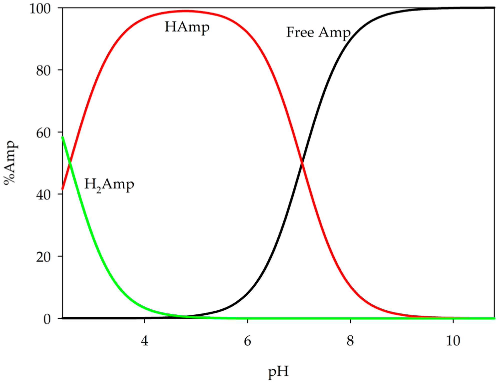
This research involved the examination of binary systems comprising ampicillin and metal ions using potentiometric titration and spectroscopy methods. The first stage of this experiment was to determine successive protonation constants for ampicillin, which were found to be logβ = 7.06 for H(Amp) and logβ = 9.61 for the form H2(Amp) (pKa1 = 2.55; pKa2 = 7.06). Literature pKa values for ampicillin are 2.54 and 7.30 [27,31]; however, our values differ due to the use of a different temperature and ionic strength (I = 0.10 mol·dm−3 KNO3). Analysis of these values of ampicillin protonation constants revealed deprotonation of the carboxyl group at relatively low pH values, followed by deprotonation of the amino group at pH values above 5.0 (Figure 2). Subsequently, the obtained values of the protonation constant of ampicillin and the hydrolysis constant of metal ions previously determined were used as a set of data for theoretical computations [36,37,38,39].

2.1. Equilibrium Study of S-Block Elements

A series of equimolar titrations and a two-fold excess of ampicillin were performed. Then, on the basis of the data obtained, the overall stability constants (logβ) of the complexes formed in the system and the equilibrium constants of the formation of the complex (logKe) were determined, as shown in Table 1.
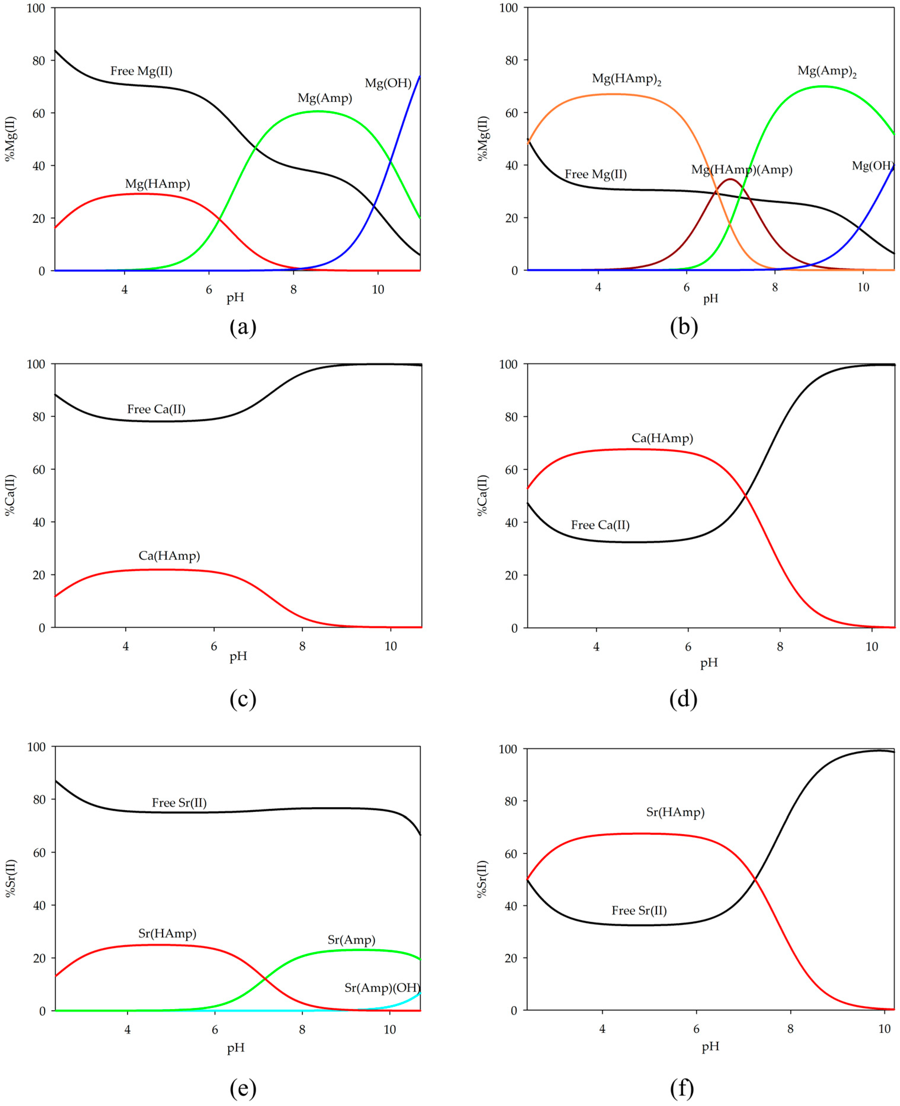
The distribution diagrams of the complex forms were plotted using the obtained stability constant values (Figure 3). In the equimolar system, only a small amount of the ions participate in the complexation process and form complexes with one molecule of ampicillin in the inner coordination sphere. At a low pH value, when the complexation process begins and the ampicillin molecule becomes partially deprotonated, the M(HAmp) form is observed as the only form in each of the tested systems for the s-block elements. For the system with magnesium ions, as the pH increases within the range of 7.0 to 9.8, the Mg(Amp) complex becomes dominant and binds over 60% of the magnesium ions.
In a ligand-excess system involving magnesium ions, various forms with two molecules of ampicillin were observed. Initially, both molecules are partially deprotonated; then, within a narrow pH range (6.7–7.2), fully and partially deprotonated molecular forms dominate until pH 7.2, where Mg(Amp)2 becomes dominant up to pH 10.7.
In the case of calcium ions, the Ca(HAmp) form is the sole species present in both the equimolar system and the two-fold excess of ampicillin. In the equimolar system, this form binds only 20% of the ions, whereas, with an excess of ampicillin, 65% of the studied ligand participates in metal coordination. A similar situation is observed for strontium ions, where the same proportion of ions engage in complexation processes. However, in the equimolar system, a complex with a fully deprotonated ligand was also observed for strontium ions, although only approximately 20% of the metal ions participated in coordination. In addition, the formation of a hydroxy complex was observed in this system.

2.2. Equilibrium Study of D-Block Elements

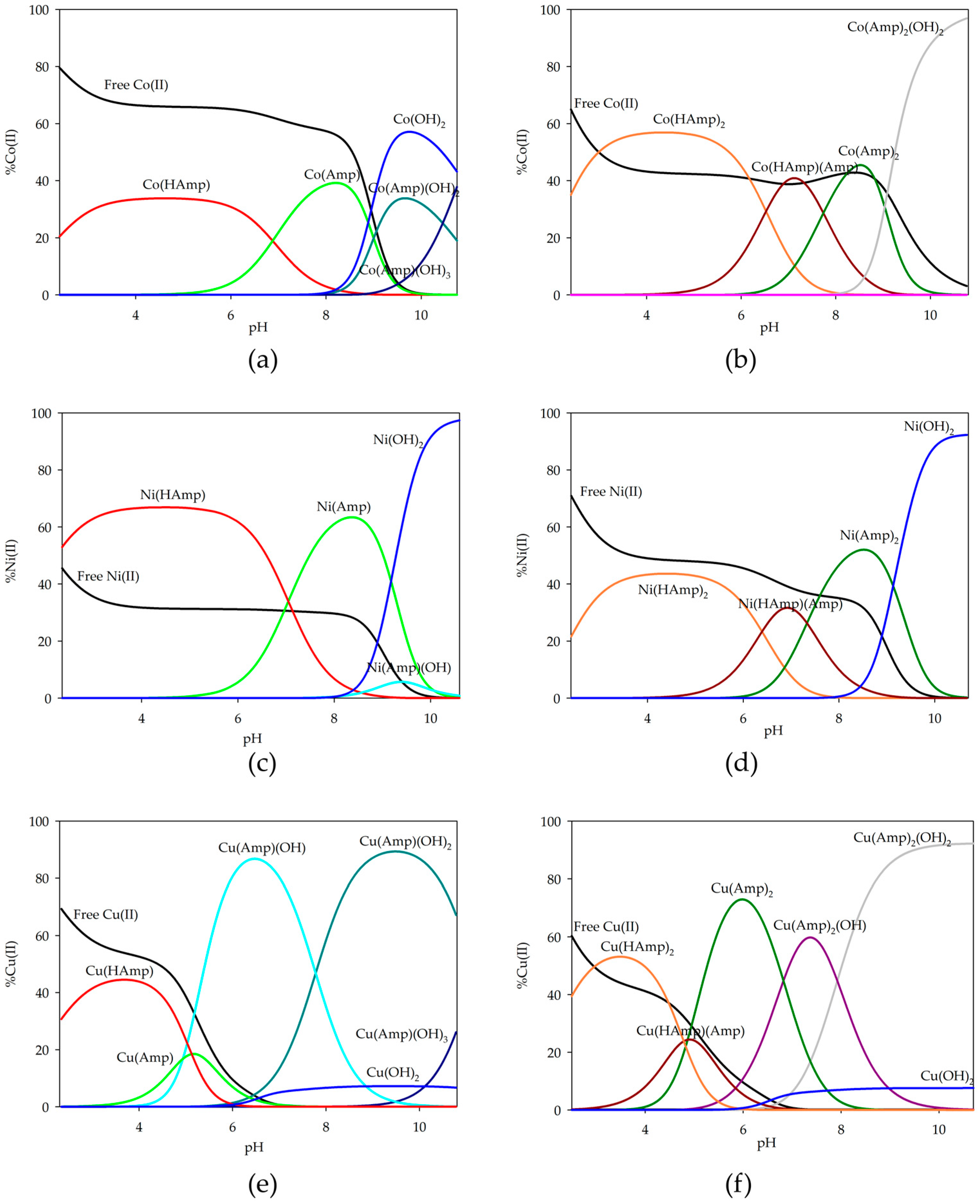
For d-block metal ions (Co(II), Ni(II), Cu(II)), a series of potentiometric titrations were performed, and the overall stability constants (logβ) and the equilibrium constants of formation (logKe) were calculated (Table 2). Distribution diagrams were plotted as illustrated in Figure 4. In the case of equilibrium studies with d-block metal ions, the presence of a larger number of various complex forms in the systems can be observed. In the equimolar systems, only complexes with one coordinated ampicillin molecule were observed, while an excess of antibiotic led to forms with two ampicillin molecules in the inner coordination sphere of the metal ion. Notable differences compared to systems with s-block ions include the presence of a greater number of complexes and a higher percentage of ion participation in the complexation process.
In the equimolar system, the complex with a partially deprotonated form of ampicillin occurs at lower pH values. For cobalt(II) ions, the M(HAmp) form binds about 40% of the ions; for nickel(II) ions, it is the dominant form and binds over 60% of the ions, while for copper(II), it binds about 44%. As the pH value increases, the predominant form in the system becomes the M(Amp) form. At higher pH values, hydroxyl complexes begin to form M(Amp)(OH)x, where x = 1–3. In the systems with cobalt and nickel ions, they are not dominant due to the dominance of metal hydroxides. However, for the copper(II) ion system, the dominant is M(Amp)(OH)2 binding about 90% of the present ions.
In systems with an excess of ampicillin, forms with two ligand molecules coordinated to the inner coordination sphere were identified. Similarly to what was observed in magnesium-complexed ampicillin systems at lower pH values, complexation begins with the formation of a complex with a two-molecule incompletely deprotonated ligand, which binds approximately half of the available metal ions. At around pH 7.0, when the fully deprotonated ligand is present in the system, the M(HAmp)(Amp) complex obtains the maximum concentration.
It should be noted that for both copper-based systems, ligand deprotonation occurs at lower pH values, causing the Cu(HAmp)(Amp) species to dominate at lower pH compared to other metals. The dominant form in the systems and reaching its maximum concentration at pH around 7.0 is the Cu(Amp)2 form. At pH above 8.0, for copper and cobalt systems, dihydroxyl complexes appear, binding almost 90% of the ions present in the solution. However, under the same conditions for the system with nickel ions, the dominant form is hydroxide.

2.3. Equilibrium Study of F-Block Elements

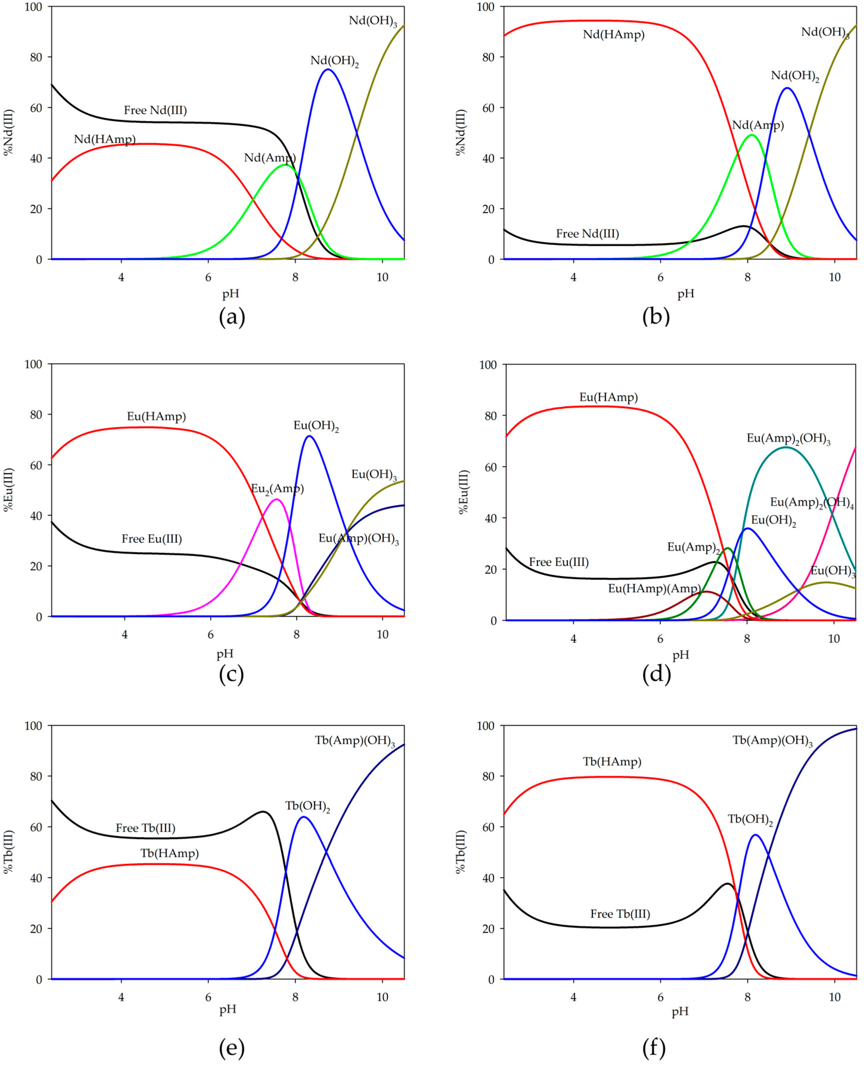
The complexation process for f-block metals was also examined. The overall stability constants (logβ) and the equilibrium constants of formation (logKe) of the complexes are presented in Table 3, and the distribution diagrams are shown in Figure 5.
In f-block metal ion systems, a different coordination method for europium ions is observed. In equimolar systems with neodymium ions, there is a partially deprotonated ampicillin form that binds about 40% of metal ions. As the pH increases, only the Nd(Amp) form occurs. However, neither of these forms dominates the system. Above pH 8.0, only neodymium hydroxides occur. Similar forms are present in excess ampicillin systems, with the notable difference being the significant quantitative participation of neodymium ions in the complexation process. The Nd(HAmp) form binds over 90% of the ions in solution, and the Nd(Amp) form binds approximately 60%. Subsequently, as seen in equimolar systems, hydroxides are predominant.
In equimolar systems with europium ions, the initial form to appear is similar to that of the neodymium system. Subsequently, the next form that occurs involves two europium ions coordinated to a single ampicillin molecule. This particular form reaches its peak concentration at around pH 7.3. Following this, there are formations of europium hydroxides and a hydroxy complex; however, these do not dominate within the system. However, in systems with excess ampicillin, there is a wider range of forms of complex compounds. At lower pH values, Eu(HAmp) initially appears, which binds about 80% of the europium ions. Then, due to the deprotonation of ampicillin and the involvement of another molecule in the coordination species containing two ampicillin molecules, a high pH level above 8.0 is reached in the case of hydroxyl complexes and the corresponding europium hydroxides, while these also formed, but without domination, as observed in other systems.
In both systems, terbium ions exist in the same forms but with varying degrees of involvement in the complexation process. In an equimolar system, around 50% of terbium ions bind to the initial Tb(HAmp) form, while this increases to about 80% in a system with a two-fold excess. Subsequently, terbium hydroxide is formed, and from pH 9.0 onwards, the predominant form present in solution is the hydroxy complex Tb(Amp)(OH)3, and almost 100% of the metal ions are involved in coordination at pH 11.0.
All spectroscopic measurements (UV–Vis, FTIR, EPR, CD, luminescence) were performed in solution under conditions where multiple species are known to coexist in equilibrium, as determined by potentiometric titrations. The formation of coordination compounds in systems containing metal ions was confirmed by spectroscopic techniques. These measurements were carried out at selected pH values, based on speciation diagrams, to ensure the highest possible proportion of the desired complex form in solution.

2.4. UV–Vis Spectroscopy

d-electron elements, such as cobalt(II), nickel(II), and copper(II), are characterized by the presence of electrons in the d subshell. Electron transitions within this subshell (e.g., d-d transitions) cause absorption of radiation in the visible and ultraviolet range. These transitions are often well defined and lead to characteristic absorption bands in ultraviolet-visible (UV–Vis) spectra. The confirmation of the coordination of the transition metal ions studied with ampicillin is provided by the characteristic shifts and changes in the absorption spectra. The observed changes are shown in Figure 6, and the spectroscopic parameters of the systems studied are placed in Table 4. Due to the solubility limits of the studied systems, the absorbance values remained below 0.1 AU in several cases. As a result, the molar absorptivity coefficients reported in Table 4 should be considered as approximate values. Changes in absorbance are interpreted cautiously and serve as qualitative indicators of coordination changes, supported by complementary data (e.g., EPR, FTIR).
In systems with cobalt ions, the d-d parameters suggest a similar coordination in the equimolar and two-fold excess of ligand systems. To account for the presence of uncomplexed Co2+ in the UV–Vis spectra of metal–ligand systems, the spectrum of free Co2+ (recorded under identical conditions) was subtracted from the mixture spectrum. The subtraction was qualitative and did not involve scaling by molar absorptivity or estimated proportions, but speciation data were considered during the interpretation to identify the dominant absorbing species. In this case of the CoL and CoL2 complex, a slight shift towards higher wavelengths was observed, indicating a change in the metal ions’ internal coordination sphere. For hydroxy complexes, spectral recording was impossible due to precipitate formation. Although identical concentrations were used in potentiometric studies, the dynamic potentiometric cell conditions (slow titrant dosing, continuous stirring, inert atmosphere) prevented visible M(OH)2 precipitation that was encountered in static cuvette measurements. For nickel-ion complexes, slight shifts toward longer wavelengths were observed along with a decrease in the molar absorption coefficient for the maximum wavelength as pH increased. In copper ion systems, the absorbance shifted toward lower wavelengths, and there was an increase in the molar absorption coefficient for the maximum wavelength as the pH increased. UV–Vis measurements at higher pH values were not performed due to precipitate formation. These results overall imply changes in the internal coordination sphere of the metal ion with increasing pH.

2.5. EPR Spectroscopy

EPR measurements confirmed that only monomorphic forms occurred in systems with copper(II) ions. At low pH values, characteristic spectra for copper ions were observed (Figure 7). In the equimolar system, the form of Cu(HAmp) indicates coordination of the oxygen atom from partially deprotonated ampicillin. However, in the system with a twofold excess of ampicillin, Cu(HAmp)2 is formed at low pH and also indicates coordination by oxygen atoms. As the pH increases, there is an observable shift toward higher gII values in the EPR spectra, indicating a change in the coordination environment around the copper (II) ions. The spectrum of the Cu(II)-Amp system at pH 4.0 (Figure 7b) indicates the presence of multiple species in equilibrium, as evidenced by more than four hyperfine lines in the parallel region. The g∥ and A∥ values reported correspond to the most intense signal components. Although the coordination mode is tentatively assigned to involve oxygen donor atoms based on the position of these parameters in the Peisach–Blumberg diagram [40].

2.6. Luminescence Spectroscopy

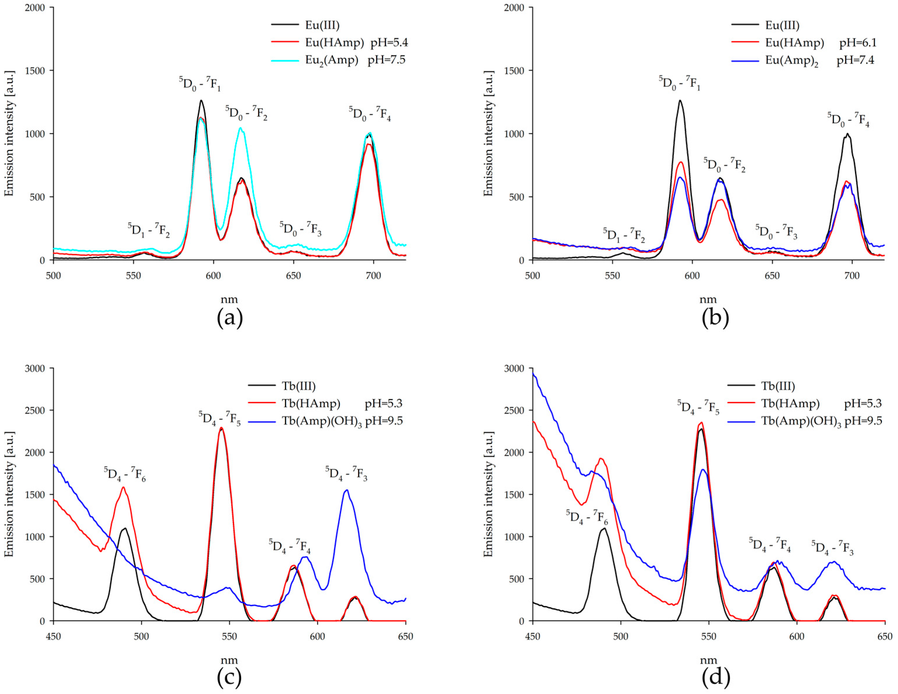
Drawing on the fundamental concepts and applications of lanthanide luminescence [41,42], emission spectra were measured for ampicillin binary systems with europium(III) ions and terbium(III) ions at the pH of the dominant complex forms (Figure 8). For europium(III) ion complexes with higher pH values, no emission measurements were carried out due to the formation of europium(III) hydroxide precipitate. When europium (III) ions were excited, emissions from the 5D0 state to 7Fj (j = 1, 2, 3, 4) levels and from the 5D1 state to the 7F2 level were observed. The emission band was quenched for formed complexes, while a two-fold increase in emission of the 5D0-7F2 band was observed for the Eu2(Amp) complex. Additionally, when a two-fold excess of ampicillin was used in one system, narrower emission bands were observed from the excited nondegenerate state.
Excitation of terbium(III) ions resulted in emission from the excited nondegenerate state 5D4 to the energy level of 7Fj (j = 3, 4, 5, 6) of the ground state. The observed bands showed an increase in intensity and band shifts for complexes with a partially deprotonated ampicillin molecule, Tb(HAmp), and the equimolar system of the hydroxy complex (Tb(Amp)(OH)3). Furthermore, there was a sharp decrease in the intensity of the 5D4 -7F5 band. In cases with excess ampicillin, this band was also extinguished, but to a lesser extent, while showing only a small increase in intensity. The baseline rise at high pH likely reflects light scattering by incipient Tb(OH)3 precipitate, observed as faint turbidity. Emission intensities should thus be treated qualitatively.

2.7. IR Spectroscopy

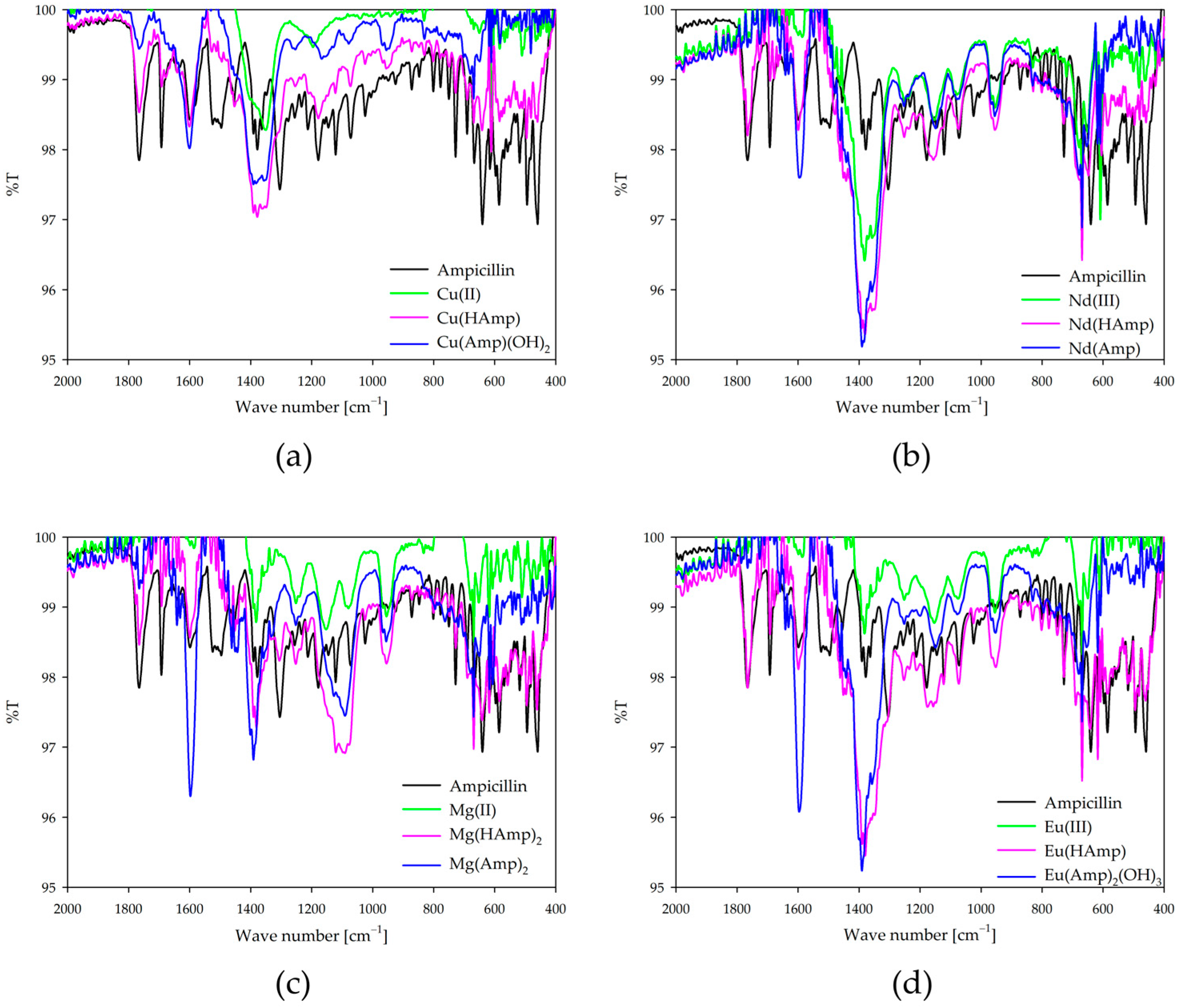
Infrared spectra of the metal complexes with ampicillin confirm the involvement of the oxygen and nitrogen donor atoms in coordination. Spectra were recorded for dominant complexes and compared to free ampicillin and free metal ions under the same pH conditions (Figure 9). FTIR spectra were recorded for complex solutions prepared in D2O. To suppress background signals from the solvent, the instrument was calibrated using pure D2O as the reference background prior to each measurement. The observed changes in the intensity of the bands and their shifts suggest participation in the coordination of the carboxyl group, the carbonyl group of the amide, and the β-lactam ring, as well as the amine groups. Participation in the coordination of the carboxyl group is evidenced, among other things, by band differences in the 1390 cm−1 region for complexes compared to free ampicillin. This band corresponds to the symmetric stretching vibration (νS COO) of the carboxylate group, which is known to occur in this region in aqueous solution environments [43,44]. In addition, confirmation of coordination through the donor oxygen atoms is provided by changes in the characteristic bands near 1685 cm−1, 1766 cm−1, and 3383 cm−1, which correspond, respectively, to coordination through the carbonyl group of the amide, the carbonyl group of the β-lactam ring, and the hydroxyl group (from the carbonyl group). The confirmation of the formation of coordination bonds with the central atom of the amino groups is provided by the changes found for the deformation vibrations of the amino groups (N-H, ~1505 cm−1) and the stretching vibrations (~3383 cm−1).
IR spectra of ampicillin (Figure S1) and all IR spectra for equimolar systems (Figure S2) and those with a two-fold excess of ampicillin (Figure S3) are included in the Supplementary Materials.

2.8. CD Spectroscopy

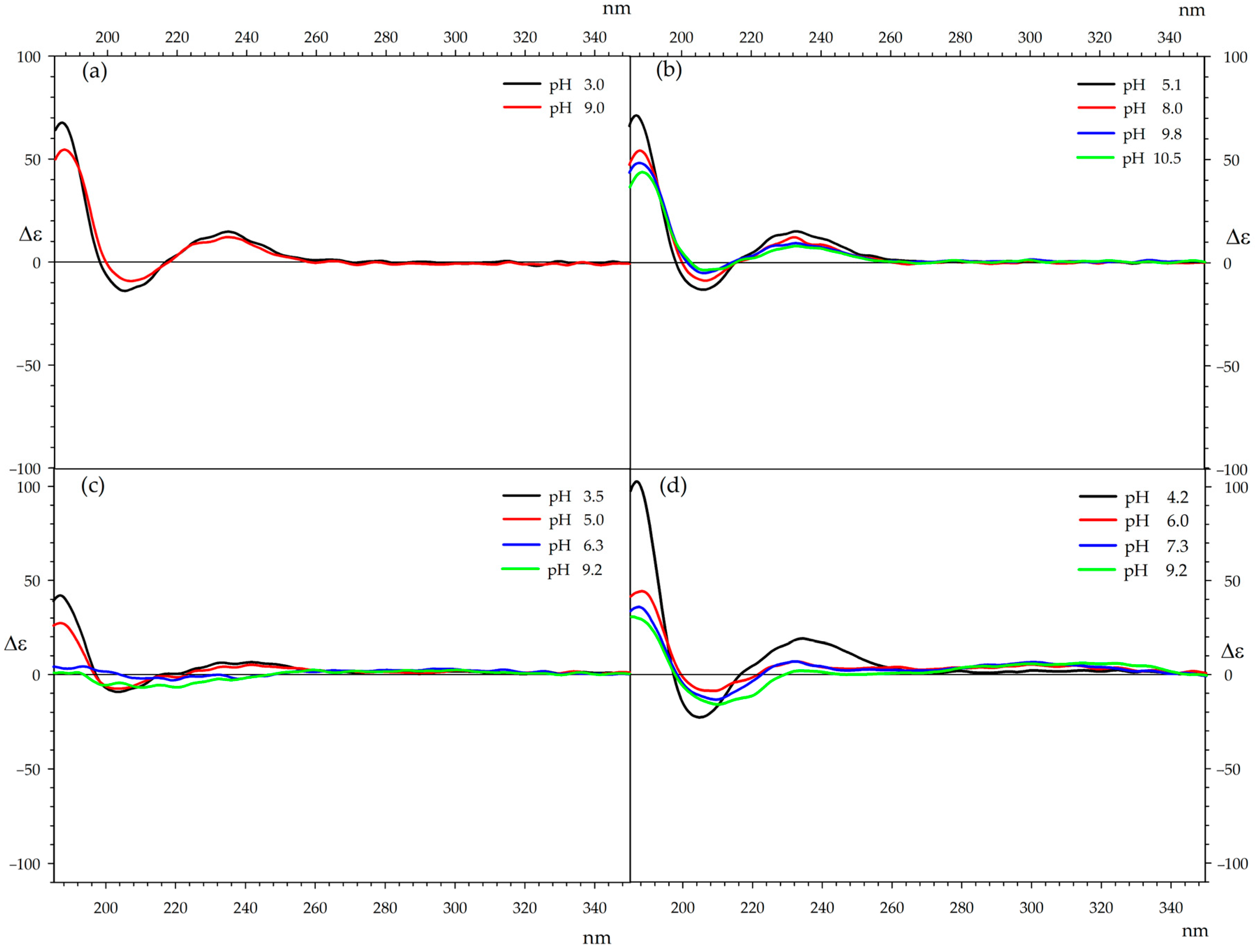
To determine the conformational preferences of ampicillin complexes, circular dichroism (CD) spectra analysis was initiated by measuring the CD spectra of ampicillin at different pH levels. This approach was intended to distinguish the changes related to pH from those associated with interactions with metal cations when analyzing the CD spectra of ampicillin complexes. The CD spectra of ampicillin in water, measured at the same concentrations as the complex spectra, exhibit a long-wavelength Cotton effect (Δε) 14.8 at 235 nm, and two additional Cotton effects, Δε = −14.1 at 205 nm and Δε = 67.7 at 187 nm, at pH 3.0. At an alkaline pH 9.0, these effects are Δε = 12.0 at 235 nm, Δε = −9.3 at 207 nm, and Δε = 54.5 at 188 nm (Figure 10a). Thus, with increasing pH, a decrease of approximately 20% in the Cotton effects is observed, with no significant changes in their positions, indicating that the antibiotic’s conformation remains unchanged regardless of pH. The CD spectra of the ampicillin complexes were recorded at pH values previously determined by potentiometric titration. In all cases, the measurements were conducted for both equimolar complexes and those with a twofold excess of the antibiotic (Table 5, Table 6 and Table 7).
Al-Khodir’s group synthesized Amp complexes with iron(III), palladium(II), and gold(III) [45]. In the complexes with iron(III) and gold(III), coordination occurs through the amide carbonyl group, the β-lactam carbonyl group, the amino group, and the carboxylate group of Amp. In contrast, the palladium(II) complex is coordinated via the β-lactam carbonyl group and the carboxylate group. The Abbas group synthesized Schiff bases of Amp with acetylacetone, along with their complexes containing cobalt(II), nickel(II), copper(II), europium(III), and gadolinium(III) [46]. In these complexes, coordination occurs through the amide nitrogen atom, the β-lactam carbonyl group, and the amino group. The spatial structure of the studied compounds appears to be similar to that obtained by Abbas, which explains the results observed during circular dichroism (CD) measurements. Due to the relatively high rigidity of the ampicillin structure, the CD spectra of its complexes do not undergo significant changes. An example can be the CD spectra recorded over a wide pH range for cobalt complexes with ampicillin in a 1:1 molar ratio (Figure 10b).
When the pH changes, the amino group undergoes protonation in acidic conditions, while the carboxyl group ionizes in alkaline conditions. The aromatic chromophore is positioned away from the complexation sites, and its orientation relative to the rest of the molecule remains largely unchanged with pH variations.
The only deviation from the general trend is observed in the CD spectra of copper complexes. In the case of the Cu-Amp 1:1 complex under alkaline conditions, there is a reversal of the long-wavelength Cotton effect and a disappearance of the short-wavelength effect located around 188 nm (Figure 10c). Interestingly, in the system with a twofold excess of ampicillin, these effects are reduced by approximately half, suggesting that the second molecule does not participate in complexation in the same manner as the first molecule (Figure 10d). The changes observed in the CD spectra for copper complexes are reflected in the distribution diagrams. For alkaline solutions, a significant presence of Cu-antibiotic complexes is still observed, while, compared to other systems, the amount of metal in the form of hydroxide is relatively low.
All CD spectra for equimolar systems (Figure S4) and those with a two-fold excess of ampicillin (Figure S5) are included in the Supplementary Materials.

3. Materials and Methods

3.1. Materials

Metal nitrates (Mg(NO3)2·2H2O, Ca(NO3)2·4H2O, Sr(NO3)2, Co(NO3)2·6H2O, Ni(NO3)2·6H2O, Cu(NO3)2·3H2O, Nd(NO3)3·6H2O, Eu(NO3)3·5H2O, Tb(NO3)3·5H2O) were purchased from Merck (Darmstadt, Germany) (>99% purity) and used as supplied. Ampicillin, anhydrous basis (96.0–102.0%) obtained from Merck (Darmstadt, Germany), was used without further purification.

3.2. Equilibrium Studies

The equilibrium studies were obtained by potentiometric measurements, which were performed using a Titrando 905 Metrohm equipped with an autoburette with an electrode Metrohm 6.0233.100 (Metrohm AG, Herisau, Switzerland). This equipment was calibrated in terms of hydrogen ion concentration prior to each series of titrations [37,38,39,47,48,49].
Before each series of measurements, the glass electrode (Metrohm AG, Herisau, Switzerland) was calibrated daily using two standard buffer solutions of pH 4.00 and pH 9.00, and the water ionization constant (pKw = 13.77 at 20 °C, I = 0.1 mol·dm−3) was determined in situ by titrating acidified KNO3 solutions. The conditions under which the measurements were taken were constant and clearly defined: temperature 20 ± 1 °C, constant ionic strength of 0.1 mol·dm−3 (KNO3), neutral gas atmosphere (helium—Ultra High Purity 5.0; Linde Gaz, Krakow, Poland), and a CO2-free NaOH titrant. Titrations used 30 mL samples and burette increments of 0.004 mL of base at a concentration of approximately 0.2 mol·dm−3. Furthermore, the concentration of metal ions was 0.001 mol·dm−3, and the metal:ligand ratio was 1:1 and 1:2 in binary systems.
Calculations were performed using the HYPERQUAD 2008 program (Hyperquad Limited, Leeds, UK) to determine the overall stability constants (logβ) and the equilibrium constants of formation (logKe) of the complexes. On the basis of the calculations obtained from the program, models of the formation of complexes in the studied systems were proposed. To verify the precision of the assumed model, the Hamilton test and the chi-squared test [47,48,49] were used to examine the standard deviation values, and the experimental curve converged with the model. Additionally, the following equilibria were evaluated as stability constants of oM + pL + qH ⇆ MoLpHq (where M = metal ion, L = ligand), and calculated using the following equation:
β = M o L p H q [ M ] o [ L ] p H q
The hydrolysis constants of the metal ions were included in the computer analysis of the potentiometric data. These constants were taken from our previous article, and their values were the ionic product of water was pKw = 13.77 [38,39,47,48]. The HySS (Hyperquad Simulation and Speciation) program was used to generate distribution diagrams of particular forms [37].

3.3. UV–Vis Spectroscopy

The UV–Vis absorption spectra were collected at room temperature using Evolution 300 UV–Vis ThermoFisher Scientific equipment (Thermo Electron Scientific Instruments LLC, Madison, WI, USA) with a xenon lamp and a Plastibrand PMMA cell (Brand, Wertheim, Germany) with a path length of 1 cm. Measurements covered wavelengths ranging from 340 to 1000 nm, while the concentration of metal ions was maintained at 0.001 mol·dm−3, with metal-to-ligand molar ratios of 1:1 and 1:2. Measurements were performed at pH values selected based on speciation diagrams, corresponding to conditions under which the highest proportion of the metal ion is involved in complex formation.

3.4. EPR Spectroscopy

EPR measurements were carried out at X-band frequency (ν = 8.9 GHz) and at a temperature of −196 °C (the temperature of liquid nitrogen) using glass capillary tubes with a volume of 130 µm3, and the data were recorded on an SE/X 2457 Radiopan spectrometer (Radiopan, Poznan, Poland). This study focused on copper ion systems with a metal ion concentration of 0.005 mol·dm−3 in a water:glycol mixture (3:1), examining both 1:1 and 1:2 metal to ligand ratios. All experimental spectra were simulated using the EPRsim32 program (version 0.3 alpha, Kraków, Poland) [50]. Measurements were performed at pH values selected based on speciation diagrams, corresponding to conditions under which the highest proportion of the metal ion is involved in complex formation.

3.5. Luminescence Spectroscopy

The luminescence studies were recorded on an RF−6000 spectrofluorophotometer (Shimadzu, Kyoto, Japan) using 5/5 nm slit widths. The concentration of metal ions was 0.001 mol·dm−3. The samples for europium ions were excited at 395 nm, and those for terbium ions were excited at 370 nm. The absorbance of the solutions at the excitation wavelength was <0.2 absorbance units. The samples were prepared using ultra-high-quality deionized and purified water from a Simplicity Ultrapure Water System (Millipore, Darmstadt, Germany). Measurements were performed at pH values selected based on speciation diagrams, corresponding to conditions under which the highest proportion of the metal ion is involved in complex formation.

3.6. Infrared Spectroscopy (FT-IR)

Infrared studies were recorded on an FT-IR INVENIO R spectrophotometer (Bruker, Bremen, Germany). Samples were prepared by dissolving the relevant ligand species (ampicillin and appropriate metal nitrates) in D2O. The metal concentration for the IR studies was 0.1 mol·dm−3, with metal:ligand molar ratios of 1:1 and 1:2; the concentration was higher to ensure sufficient absorbance in transmission. The pH values were adjusted by the addition of NaOD or DCl. The pH values were corrected according to the formula pD = pH meter reading + 0.4 [49]. Measurements were performed at pH values selected based on speciation diagrams, corresponding to conditions under which the highest proportion of the metal ion is involved in complex formation.

3.7. CD Spectroscopy

The circular dichroism (CD) studies were recorded on the J810 spectropolarimeter (JASCO, Tokyo, Japan) in the range of 180–400 nm in water solutions. Spectra were recorded at ambient temperature and accumulated with 4 scans, both for free ampicillin and ampicillin complexes. The measurements were made in nitrogen gas (flow 10 L/min) with an optical path length of 0.5 mm. The concentrations of the metals in solutions were 1 × 10⁻⁴ mol·dm−3. Measurements were performed at pH values selected based on speciation diagrams, corresponding to conditions under which the highest proportion of the metal ion is involved in complex formation.

4. Conclusions

The formation of binary complexes in binary systems of ampicillin with selected metal ions has been thoroughly investigated and established. In the equimolar system, the researchers found that only complex forms with one coordinated ampicillin molecule were present. However, in the case of a twofold excess of the antibiotics, all studied metals—except for Ca(II) and Sr(II), which remain as in equimolar system species—even at high ligand concentrations, form complexes, with two ligand molecules located in the coordination sphere. As the pH of the system increased, a progression was observed from protonated forms initially, followed by the formation of simple complexes, and finally hydroxy complexes. The complex formation and the inner coordination sphere were confirmed through the use of various spectroscopic methods, including infrared, UV–Vis, and luminescence measurements. The studies were conducted at different pH conditions to validate the changes in the internal coordination sphere caused by the attachment of subsequent functional groups to the central atom. Infrared spectroscopy studies confirmed the participation of the antibiotic functional groups in the formation of coordination bonds with the selected metal ions. UV–Vis studies revealed changes in absorption and shifts in absorbance during the formation of complexes with the oxygen donor atom at lower pH values. Luminescence measurements showed a decrease in the emission of europium ions, while a shift and enhancement in the 5D4 -7F5 band were observed for terbium ions. Furthermore, circular dichroism studies demonstrated the conformational stability of ampicillin throughout the entire studied range.

Supplementary Materials

The following supporting information can be downloaded at https://www.mdpi.com/article/10.3390/ijms26157605/s1.

Author Contributions

Conceptualization, J.F., M.Z. and R.J.; methodology, J.F., M.Z. and R.J.; validation, R.J. and M.Z.; formal analysis, J.F., M.Z., B.T. and J.G.; investigation, J.F. and J.G.; resources, R.J. and M.Z.; data curation, M.Z. and J.F.; writing—original draft preparation, J.F., M.Z., B.T. and J.G.; writing—review and editing, R.J., J.G., B.T. and J.F.; visualization, J.F.; supervision, M.Z. and R.J.; project administration, J.F. All authors have read and agreed to the published version of the manuscript.

Funding

This research received no external funding.

Institutional Review Board Statement

Not applicable.

Informed Consent Statement

Not applicable.

Data Availability Statement

All data generated or analyzed during this study are included in this published article (and its Supplementary Information File).

Acknowledgments

This research did not receive any specific grant from funding agencies in the public, commercial, or not-for-profit sectors.

Conflicts of Interest

The authors declare no conflicts of interest.

Abbreviations

The following abbreviations are used in this manuscript:
AmpAmpicillin

References

  1. Lima, L.M.; Silva, B.N.M.D.; Barbosa, G.; Barreiro, E.J. β-Lactam antibiotics: An overview from a medicinal chemistry perspective. Eur. J. Med. Chem. 2020, 208, 112829. [Google Scholar] [CrossRef]
  2. Muteeb, G.; Rehman, M.T.; Shahwan, M.; Aatif, M. Origin of antibiotics and antibiotic resistance, and their impacts on drug development: A narrative review. Pharmaceuticals 2023, 16, 1615. [Google Scholar] [CrossRef]
  3. Fernandes, R.; Amador, P.; Prudêncio, C. β-Lactams: Chemical structure, mode of action and mechanisms of resistance. Rev. Med. Microbiol. 2013, 24, 7–17. [Google Scholar] [CrossRef]
  4. Bush, K. β-Lactam antibiotics. In Antibiotic and Chemotherapy, 9th ed.; Finch, R.G., Greenwood, D., Norrby, S.R., Whitley, R.J., Eds.; Elsevier: London, UK, 2010; pp. 200–225. [Google Scholar] [CrossRef]
  5. Peechakara, B.V.; Basit, H.; Gupta, M. Ampicillin. In StatPearls; StatPearls Publishing: Treasure Island, FL, USA, 2024. Available online: http://www.ncbi.nlm.nih.gov/books/NBK519569/ (accessed on 10 June 2025).
  6. Kwan, K.C.; Rogers, J.D. Pharmacokinetics of β-Lactam Antibiotics. In Antibiotics; Demain, A.L., Solomon, N.A., Eds.; Springer: Berlin/Heidelberg, Germany, 1983; pp. 247–370. [Google Scholar] [CrossRef]
  7. Waxman, D.J.; Strominger, J.L. Penicillin-binding proteins and the mechanism of action of beta-lactam antibiotics. Annu. Rev. Biochem. 1983, 52, 825–869. [Google Scholar] [CrossRef]
  8. Kong, K.; Schneper, L.; Mathee, K. Beta-lactam antibiotics: From antibiosis to resistance and bacteriology. APMIS 2010, 118, 1–36. [Google Scholar] [CrossRef]
  9. Shah, K.J.; Cherabuddi, K.; Shultz, J.; Borgert, S.; Ramphal, R.; Klinker, K.P. Ampicillin for the treatment of complicated urinary tract infections caused by vancomycin–resistant Enterococcus spp. (VRE): A single-center university hospital experience. Int. J. Antimicrob. Agents 2018, 51, 57–61. [Google Scholar] [CrossRef] [PubMed]
  10. Bunnell, K.; Duong, A.; Ringsred, T.; Mian, A.; Bhathena, S. Aminopenicillins for treatment of ampicillin-resistant enterococcal urinary tract infections. Am. J. Health-Syst. Pharm. 2022, 79, 1056–1065. [Google Scholar] [CrossRef] [PubMed]
  11. Suleyman, G.; Zervos, M.J. Safety and efficacy of commonly used antimicrobial agents in the treatment of enterococcal infections: A review. Expert. Opin. Drug Saf. 2016, 15, 153–167. [Google Scholar] [CrossRef]
  12. Miller, W.R.; Munita, J.M.; Arias, C.A. Mechanisms of antibiotic resistance in enterococci. Expert. Rev. Anti Infect. Ther. 2014, 12, 1221–1236. [Google Scholar] [CrossRef] [PubMed]
  13. Nadgir, C.A.; Biswas, D.A. Antibiotic resistance and its impact on disease management. Cureus 2023, 15, e38251. [Google Scholar] [CrossRef]
  14. McEwen, S.A.; Collignon, P.J. Antimicrobial resistance: A One Health perspective. Microbiol. Spectr. 2018, 6, 2. [Google Scholar] [CrossRef]
  15. Li, M.; Liu, Q.; Teng, Y.; Ou, L.; Xi, Y.; Chen, S.; Duan, G. The resistance mechanism of Escherichia coli induced by ampicillin in laboratory. Infect. Drug Resist. 2019, 12, 2853–2863. [Google Scholar] [CrossRef]
  16. Türkyılmaz, O.; Darcan, C. Resistance mechanism of Escherichia coli strains with different ampicillin resistance levels. Appl. Microbiol. Biotechnol. 2024, 108, 5. [Google Scholar] [CrossRef]
  17. Murray, C.J.L.; Ikuta, K.S.; Sharara, F.; Swetschinski, L.; Aguilar, G.R.; Gray, A.; Han, C.; Bisignano, C.; Rao, P.; Wool, E.; et al. Global burden of bacterial antimicrobial resistance in 2019: A systematic analysis. Lancet 2022, 399, 629–655. [Google Scholar] [CrossRef] [PubMed]
  18. O’Neill, J. Tackling Drug-Resistant Infections Globally: Final Report and Recommendations; HM Government and Wellcome Trust: London, UK, 2016. [Google Scholar]
  19. Holmes, A.H.; Moore, L.S.; Sundsfjord, A.; Steinbakk, M.; Regmi, S.; Karkey, A.; Guerin, P.J.; Piddock, L.J. Understanding the mechanisms and drivers of antimicrobial resistance. Lancet 2016, 387, 176–187. [Google Scholar] [CrossRef] [PubMed]
  20. O’Leary, K. The global burden of antimicrobial resistance. Nat. Med. 2022. [Google Scholar] [CrossRef] [PubMed]
  21. Salam, A.; Al-Amin, Y.; Salam, M.T.; Pawar, J.S.; Akhter, N.; Rabaan, A.A.; Alqumber, M.A.A. Antimicrobial resistance: A growing serious threat for global public health. Healthcare 2023, 11, 1946. [Google Scholar] [CrossRef]
  22. Mdarhri, H.A.; Benmessaoud, R.; Yacoubi, H.; Seffar, L.; Assimi, H.G.; Hamam, M.; Boussettine, R.; Filali-Ansari, N.; Lahlou, F.A.; Diawara, I.; et al. Alternatives therapeutic approaches to conventional antibiotics: Advantages, limitations and potential application in medicine. Antibiotics 2022, 11, 1826. [Google Scholar] [CrossRef]
  23. Martirosov, D.M.; Lodise, T.P. Emerging trends in epidemiology and management of infections caused by carbapenem-resistant Enterobacteriaceae. Diagn. Microbiol. Infect. Dis. 2016, 85, 266–275. [Google Scholar] [CrossRef] [PubMed]
  24. Mohd Sazlly Lim, S.; Sime, F.B.; Roberts, J.A. Multidrug-resistant Acinetobacter baumannii infections: Current evidence on treatment options and the role of pharmacokinetics/pharmacodynamics in dose optimisation. Int. J. Antimicrob. Agents 2019, 53, 726–745. [Google Scholar] [CrossRef]
  25. Singh, G.; Rana, A.; Smriti. Decoding antimicrobial resistance: Unraveling molecular mechanisms and targeted strategies. Arch. Microbiol. 2024, 206, 280. [Google Scholar] [CrossRef]
  26. Gauba, A.; Rahman, K.M. Evaluation of antibiotic resistance mechanisms in Gram-negative bacteria. Antibiotics 2023, 12, 1590. [Google Scholar] [CrossRef]
  27. Zabiszak, M.; Frymark, J.; Ogawa, K.; Skrobańska, M.; Nowak, M.; Jastrzab, R.; Kaczmarek, M.T. Complexes of β-lactam antibiotics and their Schiff-base derivatives as a weapon in the fight against bacterial resistance. Coord. Chem. Rev. 2023, 493, 215326. [Google Scholar] [CrossRef]
  28. Alekseev, V.G. Metal complexes of penicillins and cephalosporins (Review). Pharm. Chem. J. 2012, 45, 679–697. [Google Scholar] [CrossRef]
  29. Abd El Wahed, M.G.; Ayad, M. Stability constants of Cu2+, Fe3+ ↦ Zr4+ chelates of ampicillin, dopamine and α-methyl L-dopa in aqueous medium. Anal. Lett. 1984, 17, 819–836. [Google Scholar] [CrossRef]
  30. Alekseev, V.G.; Shcherbakova, E.E.; Yakubovich, Y.Y.; Vorob’ev, N.V.; Larin, S.V.; Shigina, O.Y. Interaction of ampicillin with Mn(II), Co(II), and Ni(II) ions. Russ. J. Gen. Chem. 2006, 76, 321–324. [Google Scholar] [CrossRef]
  31. Moustakas, M. The role of metal ions in biology, biochemistry and medicine. Materials 2021, 14, 549. [Google Scholar] [CrossRef]
  32. Frei, A. Metal complexes as a promising source for new antibiotics. Chem. Sci. 2020, 11, 2627–2639. [Google Scholar] [CrossRef]
  33. Božić Cvijan, B.; Korać Jačić, J.; Bajčetić, M. The impact of copper ions on the activity of antibiotic drugs. Molecules 2023, 28, 5133. [Google Scholar] [CrossRef]
  34. Pavlić, A.; Gobin, I.; Begić, G.; Tota, M.; Abram, M.; Špalj, S. The effect of nickel ions on the susceptibility of bacteria to ciprofloxacin and ampicillin. Folia Microbiol. 2022, 67, 649–657. [Google Scholar] [CrossRef]
  35. Karaky, N.; Tang, S.; Ramalingam, P.; Kirby, A.; McBain, A.J.; Banks, C.E.; Whitehead, K.A. Multidrug-resistant Escherichia coli remains susceptible to metal ions and graphene-based compounds. Antibiotics 2024, 13, 381. [Google Scholar] [CrossRef]
  36. Gawrońska, M.; Kowalik, M.; Makowski, M. Recent advances in medicinal chemistry of ampicillin: Derivatives, metal complexes, and sensing approaches. TrAC Trends Anal. Chem. 2022, 155, 116691. [Google Scholar] [CrossRef]
  37. Alderighi, L.; Gans, P.; Ienco, A.; Peters, D.; Sabatini, A.; Vacca, A. Hyperquad simulation and speciation (HySS): A utility program for the investigation of equilibria involving soluble and partially soluble species. Coord. Chem. Rev. 1999, 184, 311–318. [Google Scholar] [CrossRef]
  38. Zabiszak, M.; Frymark, J.; Grajewski, J.; Jastrzab, R. Spectroscopic studies of lanthanide(III) complexes with L-malic acid in binary systems. Int. J. Mol. Sci. 2024, 25, 9210. [Google Scholar] [CrossRef]
  39. Zabiszak, M.; Frymark, J.; Nowak, M.; Grajewski, J.; Stachowiak, K.; Kaczmarek, M.T.; Jastrzab, R. Influence of d-electron divalent metal ions in complex formation with L-tartaric and L-malic acids. Molecules 2021, 26, 5290. [Google Scholar] [CrossRef]
  40. Peisach, J.; Blumberg, W.E. Structural implications derived from the analysis of electron paramagnetic resonance spectra of natural and artificial copper proteins. Arch. Biochem. Biophys. 1974, 165, 691–708. [Google Scholar] [CrossRef]
  41. Eliseeva, S.V.; Bünzli, J.-C.G. Lanthanide Luminescence for Functional Materials and Bio-Sciences. Chem. Soc. Rev. 2010, 39, 189–227. [Google Scholar] [CrossRef]
  42. Bünzli, J.-C.G. Lanthanide Luminescence: From Fundamental Concepts to Photonic Applications. Chem. Rev. 2010, 110, 2729–2755. [Google Scholar] [CrossRef]
  43. McAfee, L.L. Infrared and Raman Spectra of Inorganic and Coordination Compounds. Part A: Theory and Applications in Inorganic Chemistry; Part B: Application in Coordination, Organometallic, and Bioinorganic Chemistry. J. Chem. Educ. 2000, 77, 1122–1124. [Google Scholar] [CrossRef]
  44. Baraldi, C.; Tinti, A.; Ottani, S.; Gamberini, M.C. Characterization of polymorphic ampicillin forms. J. Pharm. Biomed. Anal. 2014, 100, 329–340. [Google Scholar] [CrossRef] [PubMed]
  45. Al-Khodir, F.A.I.; Refat, M.S. Spectroscopic elaboration and structural characterizations of new Fe(III), Pd(II), and Au(III) ampicillin complexes: Metal-antibiotic ligational behaviors. J. Pharm. Innov. 2015, 10, 335–347. [Google Scholar] [CrossRef]
  46. Abbas, A.M.; Fisal, S.R.; Orabi, A.S. Novel β-lactam antibiotic derivative and its complexes: DFT, frontier energy levels, DNA interaction, docking, physicochemical and antimicrobial properties. J. Mol. Struct. 2020, 1218, 128487. [Google Scholar] [CrossRef]
  47. Frymark, J.; Zabiszak, M.; Grajewski, J.; Hnatejko, Z.; Kołodynska, D.; Kaczmarek, M.T. Jastrzab, Excess of polyamine as a factor influencing the mode of coordination in the Eu(III)/α-hydroxy acid/spermine system. Polyhedron 2022, 221, 115853. [Google Scholar] [CrossRef]
  48. Zabiszak, M.; Nowak, M.; Gabryel, M.; Ogawa, K.; Kaczmarek, M.T.; Hnatejko, Z.; Jastrzab, R. New coordination compounds of citric acid and polyamines with lanthanide ions—Potential application in monitoring the treatment of cancer diseases. J. Inorg. Biochem. 2019, 198, 110715. [Google Scholar] [CrossRef]
  49. Glasoe, P.K.; Long, F.A. Use of glass electrodes to measure acidities in deuterium oxide. J. Phys. Chem. 1960, 64, 188–190. [Google Scholar] [CrossRef]
  50. Spałek, T.; Pietrzyk, P.; Sojka, Z. Application of the Genetic Algorithm Joint with the Powell Method to Nonlinear Least-Squares Fitting of Powder EPR Spectra. J. Chem. Inf. Model. 2005, 45, 18–29. [Google Scholar] [CrossRef]
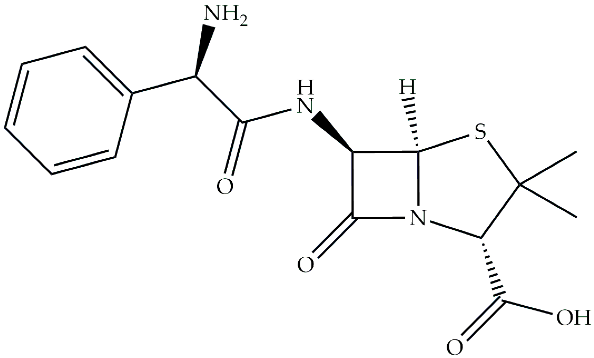
Figure 1. Formula of ampicillin.
Ijms 26 07605 g001
Figure 2. Distribution diagram of ampicillin.
Ijms 26 07605 g002
Figure 3. Distribution diagrams for the systems studied: (a) Mg(II)/ampicillin (1:1 ratio); (b) Mg(II)/ampicillin (1:2 ratio); (c) Ca(II)/ampicillin (1:1 ratio); (d) Ca(II)/ampicillin (1:2 ratio); (e) Sr(II)/ampicillin (1:1 ratio); (f) Sr(II)/ampicillin (1:2 ratio).
Ijms 26 07605 g003
Figure 4. Distribution diagrams for the systems studied: (a) Co(II)/ampicillin (1:1 ratio); (b) Co(II)/ampicillin (1:2 ratio); (c) Ni(II)/ampicillin (1:1 ratio); (d) Ni(II)/ampicillin (1:2 ratio); (e) Cu(II)/ampicillin (1:1 ratio); (f) Cu(II)/ampicillin (1:2 ratio).
Ijms 26 07605 g004
Figure 5. Distribution diagrams for the systems studied: (a) Nd(III)/ampicillin (1:1 ratio); (b) Nd(III)/ampicillin (1:2 ratio); (c) Eu(III)/ampicillin (1:1 ratio); (d) Eu(III)/ampicillin (1:2 ratio); (e) Tb(III)/ampicillin (1:1 ratio); (f) Tb(III)/ampicillin (1:2 ratio).
Ijms 26 07605 g005
Figure 6. UV–Vis absorption spectra of (a) Co(II)/ampicillin (1:1 ratio); (b) Co(II)/ampicillin (1:2 ratio); (c) Ni(II)/ampicillin (1:1 ratio); (d) Ni(II)/ampicillin (1:2 ratio); (e) Cu(II)/ampicillin (1:1 ratio); (f) Cu(II)/ampicillin (1:2 ratio).
Ijms 26 07605 g006
Figure 7. Experimental and simulated EPR spectra of (a) Cu(HAmp); (b) Cu(HAmp)2.
Ijms 26 07605 g007
Figure 8. Emission spectra of the systems: (a) Eu(III)/ampicillin (1:1 ratio); (b) Eu(III)/ampicillin (1:2 ratio); (c) Tb(III)/ampicillin (1:1 ratio); (d) Tb(III)/ampicillin (1:2 ratio).
Ijms 26 07605 g008
Figure 9. IR spectra of (a) Cu(II)/ampicillin (1:1 ratio); (b) Nd(II)/ampicillin (1:1 ratio); (c) Mg(II)/ampicillin (1:2 ratio); (d) Eu(II)/ampicillin (1:2 ratio).
Ijms 26 07605 g009
Figure 10. CD spectra of (a) ampicillin; (b) Co(II)/ampicillin (1:1 ratio); (c) Cu(II)/ampicillin (1:1 ratio); (d) Cu(II)/ampicillin (1:2 ratio).
Ijms 26 07605 g010
Table 1. The overall stability constants (logβ) and the equilibrium constants of formation (logKe) of the complexes formed in the studied systems with ampicillin and s-block elements (standard deviations are given in parentheses).
SpeciesMg(II)Ca(II)Sr(II)
logβlogKelogβlogKelogβlogKe
1:1M(HAmp)9.85(5)2.7510.00(2)2.909.99(1)2.89
M(Amp)3.60(2)3.60--2.60(7)2.60
M(Amp)(OH)----−8.56(5)2.60
1:2M(HAmp)--10.00(2)2.909.99(1)2.89
M(HAmp)(HAmp)20.91(4)6.72----
M(HAmp)(Amp)14.23(5)7.13----
M(Amp)26.96(4)3.36----
Table 2. The overall stability constants (logβ) and the equilibrium constants of formation (logKe) of the complexes formed in the studied systems with ampicillin and d-block elements (standard deviations are given in parentheses).
SpeciesCo(II)Ni(II)Cu(II)
logβlogKelogβlogKelogβlogKe
1:1M(HAmp)9.99(6)2.9010.91(2)3.8210.36(4)3.26
M(Amp)3.12(6)3.123.86(1)3.865.23(5)5.23
M(Amp)(OH)--−6.21(1)3.700.29(2)8.83
M(Amp)(OH)2−14.88(8)12.66--−7.48(3)5.99
M(Amp)(OH)3−25.33(4)3.32--−18.67(4)2.58
1:2M(HAmp)(HAmp)20.49(4)6.3019.99(3)5.8020.54(2)6.35
M(HAmp)(Amp)13.96(4)6.8613.60(3)6.5015.75(4)8.66
M(Amp)26.22(5)3.106.27(2)2.4110.89(2)5.65
M(Amp)2(OH)----4.06(2)6.95
M(Amp)2(OH)2−11.84(3)9.47--−3.85(2)5.85
Table 3. The overall stability constants (logβ) and the equilibrium constants of formation (logKe) of the complexes formed in the studied systems with ampicillin and f-block elements (standard deviations are given in parentheses).
SpeciesNd(III)Eu(III)Tb(III)
logβlogKelogβlogKelogβlogKe
1:1M(HAmp)10.82(2)3.7210.76(4)3.6610.45(2)3.18
M(Amp)3.42(2)3.42----
M(Amp)(OH)3--−21.11(4)20.20−21.06(1)20.24
M2(Amp)--7.47(3)7.47--
1:2M(HAmp)10.82(2)3.7210.76(4)3.6610.45(2)3.35
M(Amp)3.42(2)3.42----
M(HAmp)(Amp)--13.45(5)13.45--
M(Amp)2--6.50(6)6.50--
M(Amp)(OH)3----−21.06(1)20.24
M(Amp)2(OH)3--−16.81(5)4.22--
M(Amp)2(OH)4--−26.77(4)3.81--
Table 4. Spectral parameters of binary complexes formed in systems of d-electron metal ions with ampicillin.
SpeciespHAλmax (nm)ε (dm3·mol−1·cm−1)
1:1Co(II)M(HAmp)5.30.00965179.6
M(Amp)7.90.00845108.4
Ni(II)M(HAmp)5.30.0056
0.0023
670
905
5.6
2.3
M(Amp)8.10.0036
0.0016
675
900
3.6
1.6
Cu(II)M(HAmp)3.60.01877518
M(Amp)4.90.05571055
M(Amp)(OH)6.40.13610130
1:2Co(II)M(HAmp)(HAmp)5.10.01251012
M(HAmp)(Amp)7.00.01151011
M(Amp)28.10.02049020
Ni(II)M(HAmp)(HAmp)5.20.0052
0.0022
700
890
5.2
2.2
M(HAmp)(Amp)6.10.0045
0.0021
710
894
4.5
2.1
M(Amp)28.70.0031
0.0019
644
900
3.1
1.9
Cu(II)M(HAmp)(Amp)4.20.04472544
M(Amp)25.60.13630130
Table 5. Values of Cotton effects for the systems with ampicillin and s-block elements.
Metal/Ampicillin (1:1 Ratio)Metal/Ampicillin (1:2 Ratio)
Mg(II)Ca(II)Sr(II)Mg(II)Ca(II)Sr(II)
pH∆ε (nm)pH∆ε (nm)pH∆ε (nm)pH∆ε (nm)pH∆ε (nm)pH∆ε (nm)
4.313.7 (233)5.012.0 (233)5.012.5 (232)4.525.6 (233)5.026.4 (233)5.025.0 (233)
−12.1 (207)−11.3 (206)−12.2 (207)−23.5 (208)−23.8 (207)−23.1 (207)
64.8 (187)59.5 (187)59.6 (187)126.7 (187)127.0 (187)125.5 (187)
8.811.3 (234) 9.011.8 (233)7.025.1 (234)
−8.9 (206) −7.6 (206)−19.6 (207)
49.7 (188) 46.0 (189)115 (187)
9.024.1 (233)
−15.3 (207)
102.1 (188)
Table 6. Values of Cotton effects for the systems with ampicillin and d-block elements.
Metal/Ampicillin (1:1 Ratio)Metal/Ampicillin (1:2 Ratio)
Co(II)Ni(II)Cu(II)Co(II)Ni(II)Cu(II)
pH∆ε (nm)pH∆ε (nm)pH∆ε (nm)pH∆ε (nm)pH∆ε (nm)pH∆ε (nm)
5.115.2 (233)4.515.7 (233)3.56.5 (242)5.026.8 (233)4.427.0 (233)4.219.2 (235)
−13.0 (207)−15.0 (207)−9.2 (204)−23.7 (207)−21.0 (206)−22.7 (205)
71.4 (187)77.6 (187)42.0 (187)126.1 (187)128.2 (187)102.7 (187)
8.012.3 (233)8.315.5 (233)5.05.1 (242)7.123.8 (233)6.825.7 (232)6.06.9 (232)
−8.7 (207)−7.3 (207)−7.5 (203)−19.3 (206)−19.4 (206)−8.8 (209)
54.4 (188)57.2 (188)27.3 (187)112.4 (188)118.3 (187)44.3 (189)
9.89.4 (233) 6.33.0 (294)8.519.0 (233)8.223.7 (233)7.36.6 (305)
7.0 (232)
−5.0 (207) −2.3 (238)
−3.1 (220)
−11.3 (207)−14.1 (207)−13.3 (210)
48.3 (188) 4.3 (194)88.7 (188)98.7 (188)36.0 (188)
10.58.2 (233) 9.22.6 (301)
2.6 (260)
10.017.9 (233) 9.26.2 (314)
2.2 (234)
−3.4 (207) −6.5 (221)
−7.0 (210)
−5.5 (200)
−7.7 (208) −15.7 (210)
44.0 (189) 84.1 (189) 30.9 (186)
Table 7. Values of Cotton effects for the systems with ampicillin and f-block elements.
Metal/Ampicillin (1:1 Ratio)Metal/Ampicillin (1:2 Ratio)
Nd(III)Eu(III)Tb(III)Nd(III)Eu(III)Tb(III)
pH∆ε (nm)pH∆ε (nm)pH∆ε (nm)pH∆ε (nm)pH∆ε (nm)pH∆ε (nm)
5.010.8 (236)5.013.3 (236)5.015.9 (236)5.025.8 (235)2.423.3 (232)5.028.4 (235)
−9.8 (204)−13.5 (207)−14.0 (206)−24.8 (206)−27.9 (206)−26.0 (206)
57.1 (187)74.3 (187)80.2 (187)131.6 (187)123.3 (187)137.3 (188)
7.710.7 (235)7.513.5 (235)9.213.8 (236)8.122.9 (235)5.026.7 (233)9.224.7 (235)
−8.4 (205)−10.6 (205)−11.1 (206)−17.0 (207)−23.1 (206)−19.3 (206)
48.6 (187)66.4 (187)60.4 (188)104.0 (188)127.1 (187)108.7 (189)
10.011.9 (236) 7.423.9 (235)
−8.8 (206) −19.2 (207)
54.9 (188) 112.9 (188)
9.022.9 (234)
−16.2 (207)
101.4 (188)
10.020.9 (235)
−15.7 (207)
91.2 (189)
Disclaimer/Publisher’s Note: The statements, opinions and data contained in all publications are solely those of the individual author(s) and contributor(s) and not of MDPI and/or the editor(s). MDPI and/or the editor(s) disclaim responsibility for any injury to people or property resulting from any ideas, methods, instructions or products referred to in the content.