Article

Rho/SRF Inhibitor Modulates Mitochondrial Functions

Donald W. Reynolds Department of Geriatrics and Institute on Aging, University of Arkansas for Medical Sciences, Little Rock, AR 72205, USA
*
Authors to whom correspondence should be addressed.
Int. J. Mol. Sci. 2022, 23(19), 11536; https://doi.org/10.3390/ijms231911536
Submission received: 30 August 2022 / Revised: 22 September 2022 / Accepted: 26 September 2022 / Published: 29 September 2022
(This article belongs to the Special Issue Mitochondrial Function and Communication 2.0)

Abstract

:
CCG-1423 is a Rho A pathway inhibitor that has been reported to inhibit Rho/SRF-mediated transcriptional regulation. Serum response factor and its cofactors, which include ternary complex factors and myocardin-related transcription factors, regulate various cellular functions. In this study, we observed that CCG-1423 modulates the mitochondrial functions. The effect of this small molecule drug was determined by measuring mitochondrial function using an XFe96 Analyzer and an Oxygraph 2k (O2k) high-resolution respirometer. CCG-1423 treatment significantly reduced oxidative phosphorylation in a dose-dependent manner. However, CCG-1423 increased the glycolytic rate. We also observed that histone 4 at lysine-16 underwent hyperacetylation with the treatment of this drug. Immunolabeling with F-actin and MitoTracker revealed the alteration in the actin cytoskeleton and mitochondria. Taken together, our findings highlight a critical role of CCG-1423 in inhibiting the transcription of SRF/p49 and PGC-1α, β, resulting in the downregulation of mitochondrial genes, leading to the repression of mitochondrial oxidative phosphorylation and overall ATP reduction. This study provides a better understanding of the effects of CCG-1423 on mitochondria, which may be useful for the assessment of the potential clinical application of CCG-1423 and its derivatives.

1. Introduction

Serum response factor (SRF) is an important transcription factor that plays a crucial role in multiple biological processes in many cells, such as muscle cells (cardiac, skeletal, and smooth), endothelial cells, fibroblasts, hepatocytes, immune cells, and neurons [1,2,3]. SRF regulates differential gene expression through cofactor(s) recruitment, among which the most well-known are myocardin-related transcription factors and the members of the ternary complex factors. Myocardin family transcriptional co-activators include myocardin, also known as MKL (megakaryoblastic leukemia), which plays an important role in stimulating transcriptional activity of SRF. In the nucleus, different cofactors associate with SRF, and this association drives gene expression via the CC(A/T-rich)6GG cis-element, also called the CArG box [4]. This complex regulates actin cytoskeleton and motility [5,6]. SRF is also regulated by Rho GTPases and the Rho/SRF signaling has also been studied in terms of mechanisms including melanoma metastasis and fibrotic pathological pathways [7,8,9].
A small molecule screen of inhibitors of Rho-induced SRF-mediated transcription initially discovered CCG-1423 as an inhibitor of MKL/SRF signaling. CCG-1423 is formally named as N-[2-[4(4-chlorophenyl) amino]-1-methyl-2-oxoethoxy]-3, 5-bis (trifluoromethyl)-benzamide [10]. It has been shown that CCG-1423 binds to the nuclear localization signal region of G-actin-binding sites of the MKL family of proteins [11]. CCG-1423 prevents MKL’s association with importin-α/β, causing inhibition of nuclear import of MKL and MKL/SRF-mediated gene transcription. CCG-1423 can also indirectly reduce the nuclear accumulation of MKL and repress SRF activation. Others have shown that the inhibitory effects of CCG-1423 on cancer cell invasions [10] tend to improve other pathological changes including insulin resistance [12], neointima formation in vascular diseases [13], and fibrogenesis [14].
It is evident that dysfunction in mitochondrial metabolism is the cause of several inherited and acquired diseases. Mitochondria are essential organelles that have several critical functions in the cell, including metabolism and signaling [15]. SRF regulates cell metabolism by maintaining mitochondrial dynamics, fatty acid translocation, and determining the fate of electron transport chain (ETC) complex proteins [16]. Epigenetic modifications are major components affecting the activation of genes downstream of SRF [17]. Chromatin structure and function are greatly influenced by histone protein post-translational modifications, and they usually regulate the DNA transcription factor binding by altering either acetylation or methylation. SRF is involved in mechanisms controlling chromatin structure [18]. The reversible addition of an acetyl group to lysine residues and protein acetylation regulates the transcriptional activity [19,20]. Furthermore, many studies have demonstrated the crucial role of SRF in cellular migration and normal actin cytoskeleton and contractile biology [21,22]. Previously, our group had discovered p49/STRAP, a serum response factor binding protein [23]. The NADH dehydrogenase ubiquinone oxidoreductase subunit AB1 (NDUFAB1), a subunit of complex I, interacts with p49/STRAP [24]. Moreover, p49/STRAP over-expression promoted histone H4 deacetylation, which was accompanied by downregulation of peroxisome proliferator-activated receptor gamma coactivator 1-alpha (PGC 1α) and mitofusin 1 and 2 expression [25]. Mitofusins, which are regulated by PGC-1α, are important mitochondrial fusion proteins [26]. Therefore, p49/STRAP has been demonstrated to have its role in mitochondrial function and dynamics.
We herein investigated the role CCG-1423 on SRF and its cofactor p49/STRAP and whether it repressed the mitochondrial function and biogenesis in mouse skeletal myoblast cells. Our data demonstrate that CCG-1423 deacetylated the SRF protein and caused histone 4 at lysine 16 (H4K16) hyperacetylation. It also changed the actin cytoskeleton conformation. In addition, it reduced the transcriptional activity of PGC-1α, PGC-1β, and affected the other mitochondrial genes involved in biogenesis and function. We have shown the effects of CCG-1423 on the functional aspects of mitochondrial function of oxidative phosphorylation (oxygen consumption rate, OCR), glycolysis (extracellular acidification rate, ECAR), and total ATP production. These findings help to elucidate the role of CCG-1423 in mitochondrial function and might suggest areas for future study in the preclinical evaluation of CCG-1423 as a potential novel therapeutic agent.

2. Results

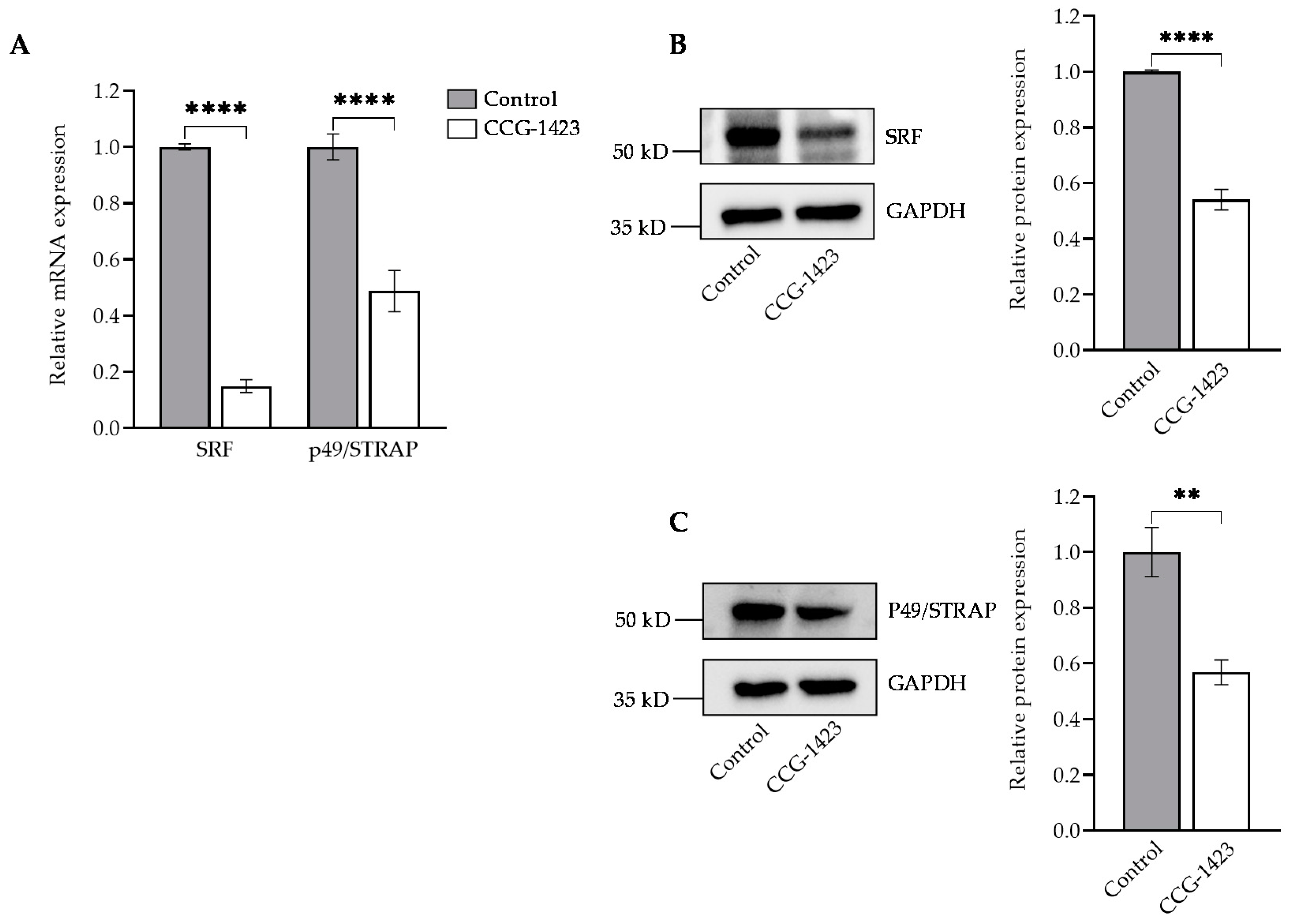
2.1. CCG-1423 Downregulates SRF, p49/STRAP Expression and Does Not Affect the Cell Viability

We first examined whether CCG-1423 affected C2C12 cell viability. The treatment of CCG-1423 at different concentrations for 24 h did not significantly affect the C2C12 cell viability. At higher concentrations, 1 μM and 10 μM of CCG-1423 did have a slight effect on the cell viability (Supplementary Figure S1). The inhibitory effects of CCG-1423 on SRF and p49/STRAP was tested at 10 μM dosage of CCG-1423 for 24 h, resulting in significant reduction of mRNA expression levels and protein levels of SRF and p49/STRAP (Figure 1A,B). Thus, CCG-1423 downregulates both SRF and p49/STRAP.

2.2. CCG-1423 Increases H4 Lysine-16 Acetylation but Decreases the Level of Acetylated-Lysine SRF

We explored a possible regulation of SRF by CCG-1423 in immunoprecipitation experiments using an SRF antibody specifically pulled down SRF compared with vehicle control after 24 h, 10 μM dosage treatment. The strong signal of SRF pulldown confirmed the specificity of the SRF antibody (Figure 2A). The immunoprecipitation evidence for acetylated-lysine/SRF shows SRF is deacetylated on the treatment of CCG-1423 (Figure 2A). We then determined acetylation levels of H4K16 since it plays an important role in transcription and chromatin packaging [27]. We performed immunoprecipitation to specifically pull down the H4. Interestingly, our data showed that pre-treatment with CCG-1423 robustly increased H4K16 acetylation (Figure 2B).

2.3. CCG-1423 Represses the Genes Involved in Mitochondrial Biogenesis, Fusion, and Fission

We next assessed the effect of CCG-1423 on the gene expression of PGC-1α and PGC-1β. The PGC-1α and -1β are the upstream regulators of mitochondrial genes [28,29]. Our data indicate that CCG-1423 reduced the PGC-1α and PGC-1β expression at mRNA levels (Figure 3A) as well as PGC-1α protein levels (Figure 3B). We also determined the effect of CCG-1423 on mitochondrial fusion and fission genes. CCG-1423 significantly reduced the expression level of mitofusin-2 (MFN2) and mitochondrial fission 1 (Fis1) whereas it did not change the mRNA expression level of mitofusin-1 (MFN1) and optic atrophy-1 (Opa1) (Figure 3C). CCG-1423 also altered the expression levels of mitochondrial complex I subunit genes NDUFV1, NDUFV2, NDUFS1, and NDUFAB1 (Figure 3D).

2.4. Actin Assembly and Mitochondrial Morphology

SRF is a crucial transcription regulator of normal actin cytoskeleton and cellular migration [30]. The f-actin phalloidin staining for actin filaments traversing the entire length of the cells (in Figure 4A) were altered by CCG-1423 treatment (in Figure 4B). SRF inhibition decreased the f-actin formation and changed the cell structure. To further investigate the effect of CCG-1423 on mitochondrial morphology and membrane potential, the MitoTracker staining was performed. As shown in Figure 4C (vehicle control) and Figure 4D (treated), CCG-1423 reduced the mitochondrial size and lowered the mitochondrial membrane potential.

2.5. CCG-1423 Differentially Regulates Cellular Bioenergetics

To investigate the two major energy pathways, oxidative phosphorylation, and glycolysis, the Seahorse XFe96 Analyzer was utilized. CCG-1423 repressed the basal respiration paralleled by a significant reduction in the maximal respiratory capacity and spare respiratory capacity in a dose-dependent manner as compared to the vehicle control (Figure 5A). CCG-1423 increased the basal glycolysis and compensatory glycolysis in a dose-dependent trend (Figure 5B). Furthermore, we also determined the total ATP production from OCR and ECAR. As shown in Figure 5C, mitochondrial ATP was reduced with the treatment of different dosages of CCG-1423. The inhibitory effects on OCR reduced the mitochondrial ATP production, possibly forcing the cells to compensate by increasing glycolysis to meet energy demands.
Next, we tested the inhibitory effects of CCG-1423 on OCR with Oxygraph 2k high-resolution respirometer in the intact C2C12 cells. As shown in Figure 6A, the basal respiration, ATP-linked respiration, as well as the maximal capacity of the mitochondrial ETC was significantly repressed with the treatment of CCG-1423. With the substrate-inhibitor-titration, we also assessed the inhibitory effects on the Electron transport complexes I (NADH/ubiquinone oxidoreductase), II (succinate dehydrogenase), III (cytochrome c reductase), and IV (cytochrome c oxidase). Intriguingly, CCG-1423 represses the functional activity of all four complexes of ETC (Figure 6B).

3. Discussion

Recent advances in elucidating the gene transcriptional mechanisms downstream of Rho (i.e., MKL and SRF) have led to the discovery of CCG-1423, a first-generation inhibitor of Rho/MKL1/SRF-mediated gene transcription [10]. Since its initial discovery, a novel small-molecule inhibitor, CCG-1423, has demonstrated significant promise in various preclinical disease models, as an anti-fibrotic agent [14], a possible therapeutic agent in type II diabetes [12], an anti-angiogenic agent [31], and most importantly, in metastatic cancer [32,33,34,35,36].
CCG-1423 has been shown to inhibit the transcriptional signaling by RhoA GTPase family and inhibit proteins and genes, such as β-catenin, TAZ, and p-LATS1, which are involved in promoting proliferation of cancer cell lines. Interestingly, growing evidence suggests a major interest in pursuing the study of mitochondria biology in cancer and targeting this organelle therapeutically [37]. There are multiple ongoing clinical trials exploring the therapeutic effects of Oxidative Phosphorylation (OXPHOS) inhibitors in different tumor types [38]. Many preclinical studies have suggested anti-tumor activity of ETC CI inhibitors, for instance, metformin, the most common anti-diabetic drug [39,40,41]. Previously, our group has also demonstrated the role of metformin against low glucose-induced elevated oxygen consumption in a process that may involve reducing oxidative stress [42].
Although CCG-1423 is a pleiotropic drug, the effects of this small molecule inhibitor on mitochondria function are largely unknown. Our study is focused on exploring whether CCG-1423 could be utilized to target mitochondria metabolism. To address this gap in knowledge, we first defined the effect of CCG-1423 on the cell viability of C2C12 cells. As shown in Supplementary Figure S1, MTS assay demonstrated that incubation with different dosages of the drug molecule from 10 nm to 10 μM for 24 h did not affect the cell viability of C2C12. Our observation is consistent with no effect of the same dosage and for an extended period of 72h of CCG-1423 on cortical neurons [43]. Furthermore, other studies have also implicated the dose dependent inhibitory effects on SRF-mediated transcription by CCG-1423 and it has been shown that high doses of CCG-1423 (5 to 10 μM) are necessary for the reduction of smooth muscle α-actin expression at the protein levels [44]. The precise molecular mechanism of actions of CCG-1423 is not fully understood, but our findings indicate that CCG-1423 may disrupt Rho signaling through functional inhibition of SRF transcriptional activity (Figure 1A). Our data also suggest the inhibitory effects of CCG-1423 on the cofactor p49/STRAP, along with SRF (Figure 1A, B). The attenuation of MKL/SRF signaling is one of the main downstream effects of CCG-1423, but importantly our data suggest CCG-1423 has more than one cellular target. SRF binds to the serum response element in the promoter region of target genes and participates in cell cycle regulation, apoptosis, cell growth, and cell differentiation [45,46,47]. Our data indicates that CCG-1423 repressed the SRF acetylation at lysine (Figure 2). This was an expected outcome since many studies have shown the role of histone acetylation in SRF signaling [48]. Histone acetylation is a critical epigenetic modification that changes chromatin architecture and regulates gene expression by opening or closing the chromatin structure [49]. It has been studied that anisomycin and TNFα induce H4 hyperacetylation via the SAPK/JNK pathway [50]. CCG-1423 inhibition of SRF/p49 mediated transcription induces H4K16 hyperacetylation (Figure 2). Furthermore, histone hyperacetylation has been linked to inducing mitochondrial dysfunction [51,52].
PGC-1α and β are critical regulators of transcriptional control of mitochondrial biogenesis [25], as it directly stimulates transcription of the Mfn1 and Mfn2 genes [53]. Others have shown that PGC-1α stimulates the transcriptional activity of the Fis1 promoter [26]. We showed that CCG-1423 also significantly reduced the expression level of PGC-1α,-1 β, Mfn2, and Fis1 (Figure 3C). Presumably, inhibition of the PGC-1α gene by CCG-1423 negatively regulated the expression of Mfn2 and Fis1. PGC-1α regulates the multiple transcriptional factors involved in mitochondrial biogenesis, such as transcription of the gene coding mitochondrial transcription factor A (TFAM), a gene required for mitochondrial biogenesis [54]. It has been well studied that PGC-1α can also regulate the composition and functions of individual mitochondria [55,56,57]. Thus, CCG-1423 inhibition of PGC-1α could have affected mitochondrial biogenesis and overall function. Mitochondrial oxidative phosphorylation consists of five multienzyme complexes (Complexes I–V) located in the mitochondrial inner membrane [58]. Mitochondrial respiratory complex I is critical to the balance in NAD: NADH levels [59]. CCG-1423 also repressed the gene expression of complex I genes (Figure 3D). Previous studies by our group have also suggested that NDUFAB1, a subunit of complex I, interacts with p49/STRAP [24]. Therefore, CCG-1423 inhibition of p49/STRAP may have indirectly altered the gene expression of NDUFAB1. The actin cytoskeleton mediated differentiation by regulating SRF transcriptional activity has been well studied [60]. Our data in immunolabeling experiments validate the SRF actin regulation since CCG-1423 affects the actin organization in C2C12 cells (Figure 4A,B). Furthermore, it also affects the mitochondrial membrane potential and mitochondrial structure (Figure 4C,D).
In this study, we investigated the OCR in C2C12 cells after the CCG-1423 treatment, which is associated with reducing mitochondrial respiration (Figure 5A). This inhibitory effect illustrates an impaired mitochondrial function that leads to a reduction of the components of ETC machinery and a decrease in oxidative phosphorylation. Energy consumption from metabolic activities in normal cells relies primarily on mitochondrial oxidative phosphorylation, which is efficient and generates more ATP than glycolysis [61]. The inefficient pathway for energy production by aerobic glycolysis in cancer cells was due to a permanent impairment of mitochondrial OXPHOS [62]. Our data in Figure 5B validates the Warburg concept of this compensatory mechanism. On the treatment of CCG-1423, OXPHOS was inhibited but simultaneously glycolysis was increased as compared to the control at different dosages. Furthermore, total ATP production was greatly reduced with the treatment of CCG-1423 (Figure 5C), which validates the MitoTracker immunolabeling data in Figure 4. We also confirmed the inhibitory effects of CCG-1423 on mitochondrial function with the intact C2C12 cells in high-resolution respirometry, O2k-FluoRespirometer (Figure 6A). The inhibitory effects of CCG-1423 on mitochondrial function are not only limited to C2C12 cells, but we have also tested the inhibitory effects of CCG-1423 on the intact H9C2, a rat cardiomyoblast cell line, using O2k-FluoRespirometer (Supplementary Figure S2). Our data also indicated the importance of CCG-1423 at different complex levels of ETC, as it repressed the activity of ETC in complex I-IV (Figure 6B).
In conclusion, CCG-1423 inhibits SRF/p49 mediated transcription and contributes to H4K16 hyperacetylation. Most importantly, it blocked the gene expression of PGC-1α and β and downregulated the mitochondrial genes involved in biogenesis and function. Furthermore, it repressed the mitochondrial oxidative phosphorylation mechanism and reduced total ATP production (Figure 7). Oxidative phosphorylation inhibition has become a potential tool for cancer therapy, and recent therapeutic interventions suggest that some specific tumors might respond to mitochondria inhibitors. Our study supports the effective use of CCG-1423 in repressing mitochondrial genes and function in C2C12 and H9C2 cells representative of vascular and cardiogenic cells. The aging cardiovascular system is vulnerable to damage from oxidative stress and potentially CCG-1423 could be tried to slow down metabolism during periods of high oxidative stress. Although we did not use cancer cell lines in our study, we postulate that the same mechanism of action of CCG-1423 might also prevent cancer cell growth and invasion. However, future studies are required to test the efficacy of this drug as a cardiovascular as well as cancer therapeutic agent.

4. Materials and Methods

4.1. Cell Culture and Cell Proliferation Assay

The mouse skeletal muscle C2C12 cell line was obtained from the American Type Culture Collection (ATCC, CRL-1772), and all the cell culture reagents were obtained from Thermo Fisher Scientific as previously described [63]. The proliferation ability of C2C12 cells was assessed using (3-(4,5-dimethylthiazol-2-yl)-5-(3-carboxymethoxyphenyl)-2-(4-sulfophenyl)-2H-tetrazolium) (MTS reagent) (Abcam, ab197010). C2C12 cells (5 × 10³/well) were seeded in a 96-well microtiter plate with a final volume of 200 μL/well DMEM containing 10% FBS and treated with different concentrations of CCG-1423 (Cayman Chemicals, 10010350) for 24 h. Furthermore, 20 μL of an MTS reagent was added to each well three hours before the end of the incubation period following the manufacturer’s instructions. The absorbance of each well was detected at 490 nm with a microplate reader (BioTek Synergy H1).

4.2. Relative Quantification of Gene Expression

The cells were pre-treated for 24 h with a 10 μM dosage of CCG-1423. The isolation of total RNA, cDNA synthesis, and quantification of gene expression were performed as previously described [64]. The sequence of the qPCR primers is described in Supplementary Table S1.

4.3. Immunoprecipitation and Western Blot Analysis

Cells were washed with cold phosphate-buffered saline (PBS) and lysed with RIPA lysis buffer system (SantaCruz, sc-24948A) for 10 min at 4 °C after 24 h, 10 µM CCG-1423 treatment. The immunoprecipitation and Western blotting were carried out as previously described [24]. Antibodies that were used included SRF (G-20, SC-335), p49/STRAP antibody [23], PGC-1α (Abcam, 106814), Histone H4 (Cell signaling, L64C1), anti-acetyl-Histone H4 (Lys16) (Cell signaling, E2B8W) and anti-acetyl-Lysine (EMD Millipore, 4G12), anti-goat HRP (Santacruz, sc-20200), anti-mouse HRP (Invitrogen, 62-6520), and anti-rabbit AP (Bio-Rad, 64251130). Immunoreactive bands were visualized with ECL, and images were captured on a ChemiDoc MP Imaging System (Bio-Rad) and analyzed using ImageJ software (National Institutes of Health, Bethesda, MD, USA), and the relative level was obtained by comparison with the GAPDH or total protein level.

4.4. MitoTracker and Immunofluorescence Staining

C2C12 cells were seeded with 1.0 × 10⁵ cells per well in a 35 mm MatTek microscopy glass dish (Fisher Scientific, PDCFOS30) and treated with 10 μM of CCG-1423 for 24 h, whereas 0.5% DMSO treatment was used as control. As described previously [24], cells were fixed with 3.7% formaldehyde (Fisher Scientific, SF100-4) for 10 min at room temperature and permeabilized with PBS containing 0.2% Triton X-100 for 5 min. Cells were washed with PBS and blocked with 3% BSA for 1 h at room temperature. Cells were then incubated with ActinGreen (ThermoFisher, Waltham, MA, USA, R37110) using rhodamine-phalloidin (1:200) for 20 min. Samples were then counterstained for 5 min with DAPI (1:1000) to label the nuclei (ThermoFisher, Waltham, MA, USA, D1306). Cells were stained with MitoTracker™ Red CMXRos (Invitrogen, Waltham, MA, USA, M7512) for 30 min following the manufacturer’s instructions. Fluorescent images were captured with a Zeiss LSM 880 confocal microscope with ZEN blue 3.2 software (Carl Zeiss Microscopy, White Plains, NY, USA). The collected images were analyzed, and fluorescence intensity was quantified using ImageJ software.

4.5. Measurement of Mitochondrial Oxygen Consumption, Glycolytic and ATP Rate

C2C12 cells were seeded at 12,000 cells/well in XFe96 Well plates (Seahorse Bioscience, Billerica, MA, USA). The cells were treated with different concentrations of CCG-1423 and 0.5% DMSO as control, for 24 h. Then, the cells were subjected to extracellular flux analysis using the XF Cell Mito Stress Test Kit (Agilent, 103015-100), XF Glycolytic Rate Assay Kit (Agilent, 103344-100), and XF Real-Time ATP Rate Assay (Agilent, 103592-100). The measurement was performed as previously described [64,65].

4.6. High-Resolution Respirometry

The mitochondrial respiration parameters in the intact C2C12 cells were measured using an Oxygraph 2k (O2k) high-resolution respirometer and processed with DatLab 6.2 software (Oroboros Instruments GmbH, Innsbruck, Austria). Cells were treated with a 10 μM dose of CCG-1423 for 24 h and 0.5% DMSO treatment was used as control. Aliquots of 1 × 10⁶ treated cells were analyzed. Briefly, cells were collected and suspended in 2 mL of Mir05 buffer [66]. Air calibration of a polarographic oxygen sensor was performed routinely before each experiment. According to the O2K manufacturer’s protocol, 5 nM of oligomycin (Sigma, O4876) was added to both chambers. Oligomycin is an inhibitor of ATP synthase and is used to induce a LEAK respiration state of respiration. Then, 0.5–3 µM of CCCP (Sigma, C2759), an uncoupler of mitochondrial oxidative phosphorylation, was added until maximal mitochondrial ET capacity was achieved. Furthermore, 2.5 μM of antimycin A (Sigma, A8674) was added to inhibit the ATP synthase and reduce the OCR. The activity of mitochondrial respiratory complexes was measured according to the substrate-inhibitor-titration protocols adapted as described in [67,68]. First, C2C12 cells (2 × 10⁶) were incubated for 15 min at 4 °C with digitonin (Sigma, D5628) (8 μM/million cells), prepared in MiRO5 buffer to the permeabilize cells. Non-phosphorylating respiration was induced by adding the complex I-linked substrate 2 mM malate (Sigma, M1000) and 10 mM glutamate (Sigma, G1626), followed by 2.5 mM ADP (Sigma, A5285) to achieve state 3 respiration. Subsequently, 10µM of cytochrome C (Sigma, C7752) was added to check the flux control efficiency. Furthermore, 0.5 µM Rotenone (Sigma, R8875) was added to inhibit complex I. Then, 10 mM succinate (Sigma, S2378) was added to evaluate OXPHOS-capacity of complex II and III-linked activity, followed by 1 mM malonic acid (Sigma, M1296) to inhibit complex II respiration and 2.5 µM antimycin A to inhibit complex III. Furthermore, 0.5 mM tetramethyl-p-phenylenediamine, TMPD (Sigma, T3134), and 2 mM ascorbate (Sigma, A7631) to stabilize the TMPD was added to initiate the Complex IV respiration and inhibited using 100 mM Azide (Sigma, 438456). Data analysis was performed with DatLab 6.2 software (Innsbruck, Austria) and cellular respiration of each mitochondrial complex was expressed as oxygen flux (pmol/s*Million Cells).

4.7. Statistical Analysis

Statistical analysis was performed with GraphPad Prism 9.1.1 Software Inc (Dotmatics, Boston, MA, USA). Results are represented by mean values ± SD of at least three independent experiments, as indicated by n. For simple comparisons, a two-tailed Student’s t-test was used. For multiple group comparison, one way ANOVA was used. p-values inferior to 0.05 were considered statistically significant (* p < 0.05; ** p <0.01; *** p < 0.001, **** p < 0.0001).

Supplementary Materials

The following supporting information can be downloaded at: https://www.mdpi.com/article/10.3390/ijms231911536/s1.

Author Contributions

Conceived and designed the experiments: X.Z. and J.Y.W.; Performed the experiments: P.P., B.N., F.S.A., A.V., J.C., G.K. and Y.C.; Analyzed the data: P.P., X.Z. and G.A.; Wrote the paper: P.P., X.Z. and J.Y.W.; Statistical analysis: P.P., X.Z. and G.A. All authors have read and agreed to the published version of the manuscript.

Funding

This study was supported in part by the Claude D. Pepper Older American Independence Center grant (P30AG28718) from National Institute on Aging (NIA).

Institutional Review Board Statement

Not applicable.

Informed Consent Statement

Not applicable.

Data Availability Statement

The raw data are available without reservation upon reasonable request.

Acknowledgments

The authors thank Laura Gocio and Amanda Pangle for assistance. The authors are also thankful to the UAMS digital microscopy core facility for the use of their equipment and expertise.

Conflicts of Interest

The authors declare no conflict of interest in regard to this manuscript.

References

  1. McDonald, O.G.; Wamhoff, B.R.; Hoofnagle, M.H.; Owens, G.K. Control of SRF binding to CArG box chromatin regulates smooth muscle gene expression in vivo. J. Clin. Investig. 2006, 116, 36–48. [Google Scholar] [CrossRef] [PubMed]
  2. Miano, J.M. Serum response factor: Toggling between disparate programs of gene expression. J. Mol. Cell Cardiol. 2003, 35, 577–593. [Google Scholar] [CrossRef]
  3. Xie, L. MKL1/2 and ELK4 co-regulate distinct serum response factor (SRF) transcription programs in macrophages. BMC Genom. 2014, 15, 301. [Google Scholar] [CrossRef]
  4. Esnault, C.; Stewart, A.; Gualdrini, F.; East, P.; Horswell, S.; Matthews, N.; Treisman, R. Rho-actin signaling to the MRTF coactivators dominates the immediate transcriptional response to serum in fibroblasts. Genes Dev. 2014, 28, 943–958. [Google Scholar] [CrossRef]
  5. Miano, J.M.; Long, X.; Fujiwara, K. Serum response factor: Master regulator of the actin cytoskeleton and contractile apparatus. Am. J. Physiol. Cell Physiol. 2007, 292, C70–C81. [Google Scholar] [CrossRef]
  6. Olson, E.N.; Nordheim, A. Linking actin dynamics and gene transcription to drive cellular motile functions. Nat. Rev. Mol. Cell Biol. 2010, 11, 353–365. [Google Scholar] [CrossRef] [PubMed]
  7. Miralles, F.; Posern, G.; Zaromytidou, A.-I.; Treisman, R. Actin dynamics control SRF activity by regulation of its coactivator MAL. Cell 2003, 113, 329–342. [Google Scholar] [CrossRef]
  8. Tsou, P.-S.; Haak, A.J.; Khanna, D.; Neubig, R.R. Cellular mechanisms of tissue fibrosis. 8. Current and future drug targets in fibrosis: Focus on Rho GTPase-regulated gene transcription. Am. J. Physiol. Cell Physiol. 2014, 307, C2–C13. [Google Scholar] [CrossRef]
  9. Sakai, N.; Chun, J.; Duffield, J.S.; Wada, T.; Luster, A.D.; Tager, A.M. LPA1-induced cytoskeleton reorganization drives fibrosis through CTGF-dependent fibroblast proliferation. FASEB J. 2013, 27, 1830–1846. [Google Scholar] [CrossRef]
  10. Evelyn, C.R.; Wade, S.M.; Wang, Q.; Wu, M.; Iñiguez-Lluhí, J.A.; Merajver, S.D.; Neubig, R.R. CCG-1423: A small-molecule inhibitor of RhoA transcriptional signaling. Mol. Cancer Ther. 2007, 6, 2249–2260. [Google Scholar] [CrossRef] [Green Version]
  11. Hayashi, K.; Watanabe, B.; Nakagawa, Y.; Minami, S.; Morita, T. RPEL proteins are the molecular targets for CCG-1423, an inhibitor of Rho signaling. PLoS ONE 2014, 9, e89016. [Google Scholar] [CrossRef]
  12. Jin, W.; Goldfine, A.B.; Boes, T.; Henry, R.R.; Ciaraldi, T.P.; Kim, E.-Y.; Emecan, M.; Fitzpatrick, C.; Sen, A.; Shah, A.; et al. Increased SRF transcriptional activity in human and mouse skeletal muscle is a signature of insulin resistance. J. Clin. Investig. 2011, 121, 918–929. [Google Scholar] [CrossRef] [PubMed]
  13. Minami, T.; Kuwahara, K.; Nakagawa, Y.; Takaoka, M.; Kinoshita, H.; Nakao, K.; Kuwabara, Y.; Yamada, Y.; Yamada, C.; Shibata, J.; et al. Reciprocal expression of MRTF-A and myocardin is crucial for pathological vascular remodelling in mice. EMBO J. 2012, 31, 4428–4440. [Google Scholar] [CrossRef] [PubMed]
  14. Johnson, L.A.; Rodansky, E.S.; Haak, A.J.; Larsen, S.D.; Neubig, R.R.; Higgins, P.D.R. Novel Rho/MRTF/SRF inhibitors block matrix-stiffness and TGF-β-induced fibrogenesis in human colonic myofibroblasts. Inflamm. Bowel Dis. 2014, 20, 154–165. [Google Scholar] [CrossRef] [PubMed]
  15. Karnkowska, A.; Vacek, V.; Zubáčová, Z.; Treitli, S.C.; Petrželková, R.; Eme, L.; Novák, L.; Žárský, V.; Barlow, L.D.; Herman, E.K.; et al. A Eukaryote without a mitochondrial organelle. Curr. Biol. 2016, 26, 1274–1284. [Google Scholar] [CrossRef]
  16. Guo, Y.; Jardin, B.D.; Zhou, P.; Sethi, I.; Akerberg, B.N.; Toepfer, C.N.; Ai, Y.; Li, Y.; Ma, Q.; Guatimosim, S.; et al. Hierarchical and Stage-Specific Regulation of Murine Cardiomyocyte Maturation by Serum Response Factor. Nat. Commun. 2018, 9, 3837. [Google Scholar] [CrossRef]
  17. Rugowska, A.; Starosta, A.; Konieczny, P. Epigenetic modifications in muscle regeneration and progression of duchenne muscular dystrophy. Clin. Epigenet. 2021, 13, 13. [Google Scholar] [CrossRef]
  18. Mack, C.P. Signaling mechanisms that regulate smooth muscle cell differentiation. Arterioscler. Thromb. Vasc. Biol. 2011, 31, 1495–1505. [Google Scholar] [CrossRef]
  19. Arif, M.; Selvi, B.R.; Kundu, T.K. Lysine acetylation: The tale of a modification from transcription regulation to metabolism. Chembiochem 2010, 11, 1501–1504. [Google Scholar] [CrossRef]
  20. Millar, C.B.; Kurdistani, S.K.; Grunstein, M. Acetylation of yeast histone H4 lysine 16: A switch for protein interactions in heterochromatin and euchromatin. Cold Spring Harb. Symp. Quant. Biol. 2004, 69, 193–200. [Google Scholar] [CrossRef] [Green Version]
  21. Schratt, G.; Philippar, U.; Berger, J.; Schwarz, H.; Heidenreich, O.; Nordheim, A. Serum response factor is crucial for actin cytoskeletal organization and focal adhesion assembly in embryonic stem cells. J. Cell Biol. 2002, 156, 737–750. [Google Scholar] [CrossRef] [PubMed]
  22. Ellis, P.D.; Martin, K.M.; Rickman, C.; Metcalfe, J.C.; Kemp, P.R. Increased actin polymerization reduces the inhibition of serum response factor activity by Yin Yang 1. Biochem. J. 2002, 364, 547–554. [Google Scholar] [CrossRef] [PubMed]
  23. Zhang, X.; Azhar, G.; Zhong, Y.; Wei, J.Y. Identification of a novel serum response factor cofactor in cardiac gene regulation. J. Biol. Chem. 2004, 279, 55626–55632. [Google Scholar] [CrossRef]
  24. Zhang, X.; Azhar, G.; Helms, S.; Zhong, Y.; Wei, J.Y. Identification of a subunit of NADH-dehydrogenase as a P49/STRAP-binding protein. BMC Cell Biol. 2008, 9, 8. [Google Scholar] [CrossRef] [PubMed]
  25. Zhang, X.; Williams, E.D.; Azhar, G.; Rogers, S.C.; Wei, J.Y. Does P49/STRAP, a SRF-binding protein (SRFBP1), modulate cardiac mitochondrial function in aging? Exp. Gerontol. 2016, 82, 150–159. [Google Scholar] [CrossRef]
  26. Martin, O.J.; Lai, L.; Soundarapandian, M.M.; Leone, T.C.; Zorzano, A.; Keller, M.P.; Attie, A.D.; Muoio, D.M.; Kelly, D.P. A role for peroxisome proliferator-activated receptor γ coactivator-1 in the control of mitochondrial dynamics during postnatal cardiac growth. Circ Res. 2014, 114, 626–636. [Google Scholar] [CrossRef]
  27. Shia, W.-J.; Pattenden, S.G.; Workman, J.L. Histone H4 Lysine 16 Acetylation Breaks the Genome’s Silence. Genome Biol. 2006, 7, 217. [Google Scholar] [CrossRef]
  28. Esterbauer, H.; Oberkofler, H.; Krempler, F.; Patsch, W. Human peroxisome proliferator activated receptor gamma coactivator 1 (PPARGC1) gene: CDNA sequence, genomic organization, chromosomal localization, and tissue expression. Genomics 1999, 62, 98–102. [Google Scholar] [CrossRef]
  29. Finck, B.N.; Kelly, D.P. PGC-1 Coactivators: Inducible regulators of energy metabolism in health and disease. J. Clin. Investig. 2006, 116, 615–622. [Google Scholar] [CrossRef]
  30. Pipes, G.C.T.; Creemers, E.E.; Olson, E.N. The myocardin family of transcriptional coactivators: Versatile regulators of cell growth, migration, and myogenesis. Genes Dev. 2006, 20, 1545–1556. [Google Scholar] [CrossRef] [Green Version]
  31. Gau, D.; Veon, W.; Capasso, T.L.; Bottcher, R.; Shroff, S.; Roman, B.L.; Roy, P. Pharmacological intervention of MKL/SRF signaling by CCG-1423 impedes endothelial cell migration and angiogenesis. Angiogenesis 2017, 20, 663–672. [Google Scholar] [CrossRef] [PubMed]
  32. Lisabeth, E.M.; Kahl, D.; Gopallawa, I.; Haynes, S.E.; Misek, S.A.; Campbell, P.L.; Dexheimer, T.S.; Khanna, D.; Fox, D.A.; Jin, X.; et al. identification of pirin as a molecular target of the CCG-1423/CCG-203971 series of antifibrotic and antimetastatic compounds. ACS Pharmacol. Transl. Sci. 2019, 2, 92–100. [Google Scholar] [CrossRef] [PubMed]
  33. Ma, X.; Yu, H. Global burden of cancer. Yale J. Biol. Med. 2006, 79, 85–94. [Google Scholar] [PubMed]
  34. Haga, R.B.; Ridley, A.J. Rho GTPases: Regulation and roles in cancer cell biology. Small GTPases 2016, 7, 207–221. [Google Scholar] [CrossRef]
  35. Nakajima, M.; Hayashi, K.; Egi, Y.; Katayama, K.; Amano, Y.; Uehata, M.; Ohtsuki, M.; Fujii, A.; Oshita, K.; Kataoka, H.; et al. Effect of Wf-536, a NOVEL ROCK inhibitor, against metastasis of B16 melanoma. Cancer Chemother. Pharmacol. 2003, 52, 319–324. [Google Scholar] [CrossRef]
  36. Wei, L.; Surma, M.; Shi, S.; Lambert-Cheatham, N.; Shi, J. Novel insights into the roles of Rho kinase in cancer. Arch. Immunol. Ther. Exp. 2016, 64, 259–278. [Google Scholar] [CrossRef]
  37. Zong, W.-X.; Rabinowitz, J.D.; White, E. Mitochondria and cancer. Mol. Cell 2016, 61, 667–676. [Google Scholar] [CrossRef]
  38. Dalton, K.M.; Lochmann, T.L.; Floros, K.V.; Calbert, M.L.; Kurupi, R.; Stein, G.T.; McClanaghan, J.; Murchie, E.; Egan, R.K.; Greninger, P.; et al. Catastrophic ATP loss underlies a metabolic combination therapy tailored for MYCN-amplified neuroblastoma. Proc. Natl. Acad. Sci. USA 2021, 118, e2009620118. [Google Scholar] [CrossRef]
  39. Wheaton, W.W.; Weinberg, S.E.; Hamanaka, R.B.; Soberanes, S.; Sullivan, L.B.; Anso, E.; Glasauer, A.; Dufour, E.; Mutlu, G.M.; Budigner, G.S.; et al. Metformin inhibits mitochondrial complex I of cancer cells to reduce tumorigenesis. eLife 2014, 3, e02242. [Google Scholar] [CrossRef]
  40. Roesch, A.; Vultur, A.; Bogeski, I.; Wang, H.; Zimmermann, K.M.; Speicher, D.; Körbel, C.; Laschke, M.W.; Gimotty, P.A.; Philipp, S.E.; et al. Overcoming intrinsic multidrug resistance in melanoma by blocking the mitochondrial respiratory chain of slow-cycling JARID1Bhigh cells. Cancer Cell 2013, 23, 811–825. [Google Scholar] [CrossRef] [Green Version]
  41. Shackelford, D.B.; Abt, E.; Gerken, L.; Vasquez, D.S.; Seki, A.; Leblanc, M.; Wei, L.; Fishbein, M.C.; Czernin, J.; Mischel, P.S.; et al. LKB1 inactivation dictates therapeutic response of non-small cell lung cancer to the metabolism drug phenformin. Cancer Cell 2013, 23, 143–158. [Google Scholar] [CrossRef] [PubMed]
  42. Williams, E.D.; Rogers, S.C.; Zhang, X.; Azhar, G.; Wei, J.Y. Elevated oxygen consumption rate in response to acute low-glucose stress: Metformin restores rate to normal level. Exp. Gerontol. 2015, 70, 157–162. [Google Scholar] [CrossRef] [PubMed]
  43. Kikuchi, K.; Shiota, J.; Yamada, T.; Ishikawa, M.; Ihara, D.; Fukuchi, M.; Tsuda, M.; Tabuchi, A. Rho signaling inhibitor, CCG-1423, inhibits axonal elongation and dendritic complexity of rat cortical neurons. Biochem. Biophys. Res. Commun. 2017, 492, 474–479. [Google Scholar] [CrossRef] [PubMed]
  44. Watanabe, B.; Minami, S.; Ishida, H.; Yoshioka, R.; Nakagawa, Y.; Morita, T.; Hayashi, K. Stereospecific inhibitory effects of CCG-1423 on the cellular events mediated by myocardin-related transcription factor A. PLoS ONE 2015, 10, e0136242. [Google Scholar] [CrossRef]
  45. Gineitis, D.; Treisman, R. Differential usage of signal transduction pathways defines two types of serum response factor target gene. J. Biol. Chem. 2001, 276, 24531–24539. [Google Scholar] [CrossRef]
  46. Holst, B.; Holliday, N.D.; Bach, A.; Elling, C.E.; Cox, H.M.; Schwartz, T.W. Common structural basis for constitutive activity of the ghrelin receptor family. J. Biol. Chem. 2004, 279, 53806–53817. [Google Scholar] [CrossRef]
  47. Whitmarsh, A.J.; Shore, P.; Sharrocks, A.D.; Davis, R.J. Integration of MAP kinase signal transduction pathways at the serum response element. Science 1995, 269, 403–407. [Google Scholar] [CrossRef]
  48. Connelly, J.T.; Mishra, A.; Gautrot, J.E.; Watt, F.M. Shape-induced terminal differentiation of human epidermal stem cells requires P38 and is regulated by histone acetylation. PLoS ONE 2011, 6, e27259. [Google Scholar] [CrossRef]
  49. Gräff, J.; Tsai, L.-H. Histone acetylation: Molecular mnemonics on the chromatin. Nat. Rev. Neurosci. 2013, 14, 97–111. [Google Scholar] [CrossRef]
  50. Alberts, A.S.; Geneste, O.; Treisman, R. Activation of SRF-regulated chromosomal templates by Rho-family GTPases requires a signal that also induces H4 hyperacetylation. Cell 1998, 92, 475–487. [Google Scholar] [CrossRef] [Green Version]
  51. Huang, M.; Lou, D.; Charli, A.; Kong, D.; Jin, H.; Zenitsky, G.; Anantharam, V.; Kanthasamy, A.; Wang, Z.; Kanthasamy, A.G. Mitochondrial dysfunction–induced H3K27 hyperacetylation perturbs enhancers in Parkinson’s disease. JCI Insight 2021, 6, e138088. [Google Scholar] [CrossRef] [PubMed]
  52. Kopinski, P.K.; Janssen, K.A.; Schaefer, P.M.; Trefely, S.; Perry, C.E.; Potluri, P.; Tintos-Hernandez, J.A.; Singh, L.N.; Karch, K.R.; Campbell, S.L.; et al. Regulation of nuclear epigenome by mitochondrial DNA heteroplasmy. Proc. Natl. Acad. Sci. USA 2019, 116, 16028–16035. [Google Scholar] [CrossRef] [PubMed]
  53. Scarpulla, R.C. Metabolic control of mitochondrial biogenesis through the PGC-1 family regulatory network. Biochim. Biophys. Acta (BBA) Mol. Cell Res. 2011, 1813, 1269–1278. [Google Scholar] [CrossRef]
  54. Ekstrand, M.I.; Falkenberg, M.; Rantanen, A.; Park, C.B.; Gaspari, M.; Hultenby, K.; Rustin, P.; Gustafsson, C.M.; Larsson, N.-G. Mitochondrial transcription factor A regulates MtDNA copy number in mammals. Hum. Mol. Genet. 2004, 13, 935–944. [Google Scholar] [CrossRef] [PubMed]
  55. Austin, S.; St-Pierre, J. PGC1α and mitochondrial metabolism—Emerging concepts and relevance in ageing and neurodegenerative disorders. J. Cell Sci. 2012, 125, 4963–4971. [Google Scholar] [CrossRef]
  56. Wu, Z.; Puigserver, P.; Andersson, U.; Zhang, C.; Adelmant, G.; Mootha, V.; Troy, A.; Cinti, S.; Lowell, B.; Scarpulla, R.C.; et al. Mechanisms controlling mitochondrial biogenesis and respiration through the thermogenic coactivator PGC-1. Cell 1999, 98, 115–124. [Google Scholar] [CrossRef]
  57. Chen, L.; Qin, Y.; Liu, B.; Gao, M.; Li, A.; Li, X.; Gong, G. PGC-1α-Mediated Mitochondrial Quality Control: Molecular Mechanisms and Implications for Heart Failure. Front. Cell Dev. Biol. 2022, 10, 871357. [Google Scholar] [CrossRef]
  58. Hatefi, Y. The mitochondrial electron transport and oxidative phosphorylation system. Annu. Rev. Biochem. 1985, 54, 1015–1069. [Google Scholar] [CrossRef]
  59. Santidrian, A.F.; Matsuno-Yagi, A.; Ritland, M.; Seo, B.B.; LeBoeuf, S.E.; Gay, L.J.; Yagi, T.; Felding-Habermann, B. Mitochondrial complex I activity and NAD+/NADH balance regulate breast cancer progression. J. Clin. Investing. 2013, 123, 1068–1081. [Google Scholar] [CrossRef]
  60. Connelly, J.T.; Gautrot, J.E.; Trappmann, B.; Tan, D.W.-M.; Donati, G.; Huck, W.T.S.; Watt, F.M. Actin and serum response factor transduce physical cues from the microenvironment to regulate epidermal stem cell fate decisions. Nat. Cell Biol. 2010, 12, 711–718. [Google Scholar] [CrossRef]
  61. Koppenol, W.H.; Bounds, P.L.; Dang, C.V. Otto Warburg’s Contributions to Current Concepts of Cancer Metabolism. Nat. Rev. Cancer 2011, 11, 325–337. [Google Scholar] [CrossRef] [PubMed]
  62. Zheng, J. Energy metabolism of cancer: Glycolysis versus oxidative phosphorylation (review). Oncol. Lett. 2012, 4, 1151–1157. [Google Scholar] [CrossRef] [PubMed]
  63. Rogers, S.C.; Zhang, X.; Azhar, G.; Luo, S.; Wei, J.Y. Exposure to high or low glucose levels accelerates the appearance of markers of endothelial cell senescence and induces dysregulation of nitric oxide synthase. J. Gerontol. Ser. A 2013, 68, 1469–1481. [Google Scholar] [CrossRef] [PubMed]
  64. Zhang, X.; Ameer, F.S.; Azhar, G.; Wei, J.Y. Alternative splicing increases sirtuin gene family diversity and modulates their subcellular localization and function. IJMS 2021, 22, 473. [Google Scholar] [CrossRef] [PubMed]
  65. Zhang, X.; Azhar, G.; Wei, J.Y. The expression of MicroRNA and MicroRNA clusters in the aging heart. PLoS ONE 2012, 7, e34688. [Google Scholar] [CrossRef]
  66. Gnaiger, E. Mitochondrial pathways and respiratory control. In An Introduction to OXPHOS Analysis, 5th ed.; Bioenergetics Communications: Axams, Austria, 2020. [Google Scholar] [CrossRef]
  67. Tobacyk, J.; Kc, G.; MacMillan-Crow, L.A. Overexpression of MnSOD protects against cold storage-induced mitochondrial injury but not against OMA1-Dependent OPA1 proteolytic processing in rat renal proximal tubular cells. Antioxidants 2021, 10, 1272. [Google Scholar] [CrossRef] [PubMed]
  68. Pesta, D.; Gnaiger, E. High-resolution respirometry: OXPHOS protocols for human cells and permeabilized fibers from small biopsies of human muscle. In Mitochondrial Bioenergetics; Palmeira, C.M., Moreno, A.J., Eds.; Methods in Molecular Biology; Humana Press: Totowa, NJ, USA, 2012; Volume 810, pp. 25–58. ISBN 978-1-61779-381-3. [Google Scholar]
Figure 1. CCG-1423 represses SRF and p49/STRAP. (AC) C2C12 cells were treated with a 10 µM dosage for 24 h. (A) The mRNA expression of SRF and p49/STRAP was evaluated in qPCR. DMSO was used as control. Immunoblotting was used to evaluate the protein levels of (B) SRF and (C) p49/STRAP. GAPDH was used as the total protein loading control and relative protein expression was quantified in (B,C). ** p < 0.01, **** p < 0.0001 (n = 3).
Ijms 23 11536 g001
Figure 2. Immunoprecipitation of SRF acetylated lysine and H4K16 protein. (A) CCG-1423 represses the SRF acetylated-lysine (Ac-SRF), representative western blot of acetylated-lysine SRF and SRF in lysates from CCG-1423 treated and non-treated C2C12 cells. GAPDH was used as a loading control. (B) H4K16 was hyperacetylated on the CCG-1423 treatment, representative western blot image of H4K16 and total histone H4 protein was used as a loading control (n = 3).
Ijms 23 11536 g002
Figure 3. Repression of mitochondrial genes involved in biogenesis and function. (A) RT-qPCR analysis demonstrated the inhibition of PGC-1α and -1β gene expression following the treatment of CCG-1423. (B) Protein levels of PGC-1α were analyzed by Western blot and GAPDH was used as a loading control. Densitometric analysis shows inhibition of PGC-1α. (C) MFN2 and Fis1 gene expression levels reduced whereas MFN1 and Opa1 were unaffected. (D) Downregulation of mitochondrial genes involved in complex-I. * p < 0.05, ** p < 0.01, *** p < 0.001, **** p < 0.0001, ns: p > 0.05 (n = 3).
Ijms 23 11536 g003
Figure 4. Immunolabeling of Actin and MitoTracker. C2C12 cells at 24 h post-treatment, cells incubated with phalloidin (green) and MitoTracker (red) conjugated dyes. DAPI (blue) was used for nuclear counterstaining. (A,C) Vehicle (DMSO) (B,D) CCG-1423 treated cells, at 10 μM dosage. 63× oil objective is used; scale bars indicate 10 μm. (E) MitoTracker stained cells are randomly selected microscopic fields in four individual repetitions where the fluorescence intensity was quantified using ImageJ. **** p < 0.0001 (n = 4).
Ijms 23 11536 g004
Figure 5. Mitochondria functional analysis of oxygen consumption rate, extracellular acidification rate, and total ATP production. (A) OCR and basal respiration, maximum respiration, and spare respiratory capacity in CCG-1423 treated cells with different dosages. OCR was repressed in a dose dependent manner upon the treatment of CCG-1423 in C2C12 cells. (B) CCG-1423 upregulated the ECAR in a dose dependent manner. (C) Total ATP production was decreased in both mechanisms of oxidative phosphorylation (mitochondrial-ATP) and glycolysis (glycolysis-ATP). * p < 0.05, *** p < 0.001, **** p < 0.0001, ns: p > 0.05 (n = 4).
Ijms 23 11536 g005
Figure 6. High-resolution respiratory analysis of oxidative phosphorylation. (A) OCR and basal respiration, leak respiration, ATP-linked respiration, Maximum ET capacity was analyzed using an Oroboros O2K instrument. Intact C2C12 cells were pre-treated with CCG-1423 with 10 µM for 24 h treatment. OCR and other functional aspects indicated above in the figure were repressed. (B) C2C12 cells were permeabilized and were used to determine OCR levels at complexes of ETC. OCR at complex I–IV of ETC were repressed on the treatment of CCG-1423. ** p < 0.01, *** p < 0.001, **** p < 0.0001 (n = 3).
Ijms 23 11536 g006
Figure 7. Schematic demonstrating the effects of CCG-1423 on mitochondria function. CCG-1423 inhibits SRF, p49/STRAP and PGC1-α, β, and causes H4K16 hyperacetylation, which modulates the functional activity of oxidative phosphorylation, glycolysis, and total ATP. Arrows indicate: = increase, ↓ = decrease, and Ijms 23 11536 i001 = inhibition.
Ijms 23 11536 g007
Publisher’s Note: MDPI stays neutral with regard to jurisdictional claims in published maps and institutional affiliations.

