


Iron(III) Complexes with Substituted Salicylaldehydes: Synthesis, Interaction with DNA and Serum Albumins, and Antioxidant Activity


Abstract
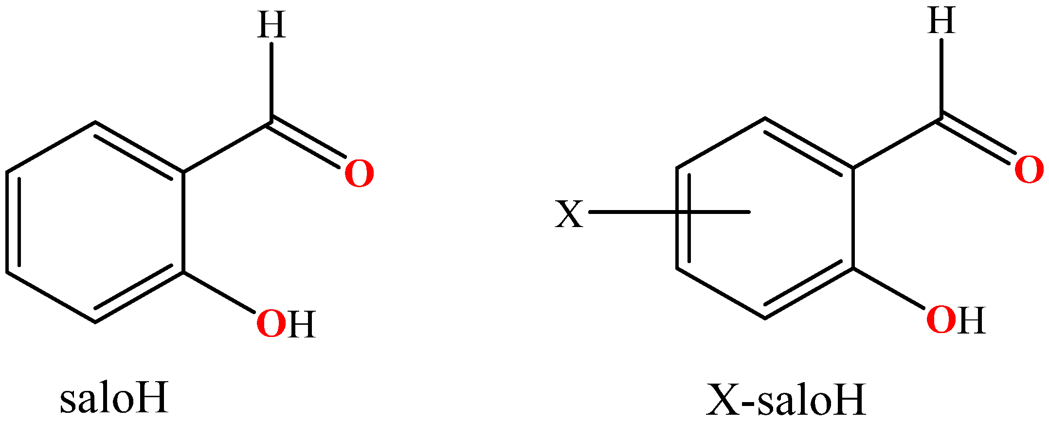
1. Introduction
2. Results
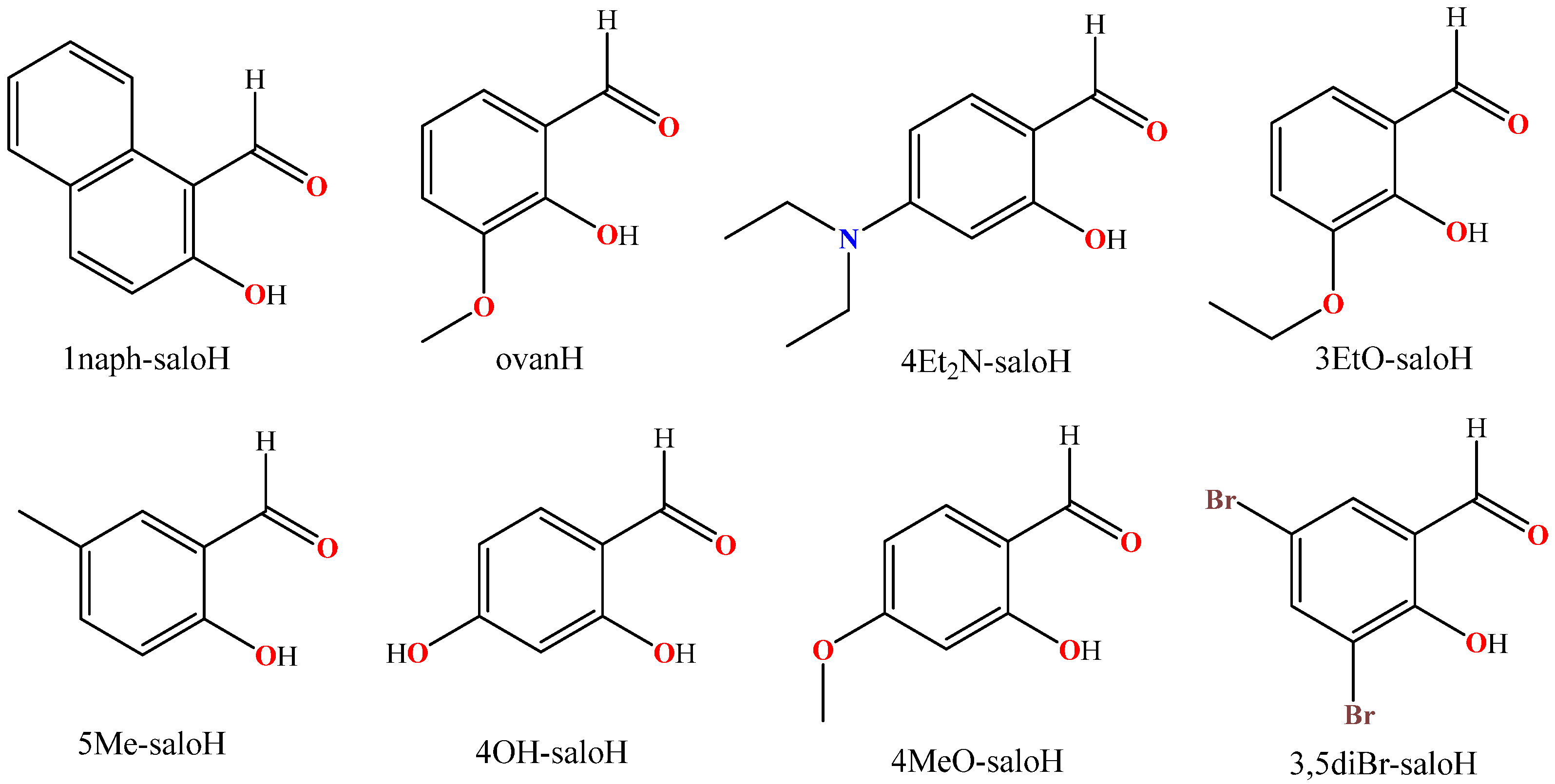
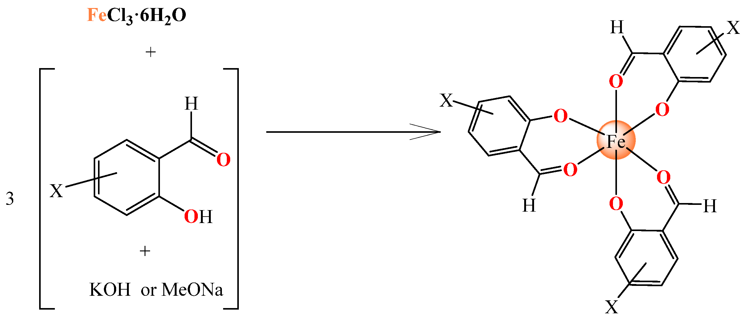
2.1. Synthesis and Characterization
2.2. Structures the Complexes
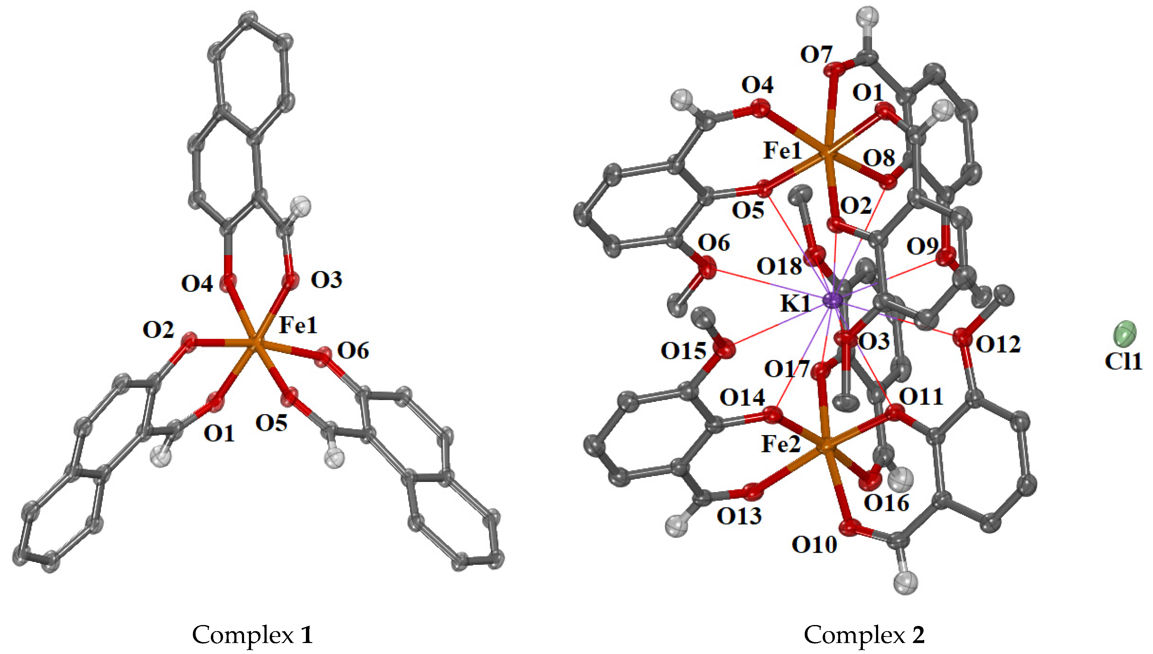
2.2.1. Crystal Structures of Complexes 1 and 2
2.2.2. Proposed Structures for Complexes 3–8
2.3. Interaction of the Complexes with CT DNA
2.3.1. Interaction of the Complexes with CT DNA Studied with UV-Vis Spectroscopy
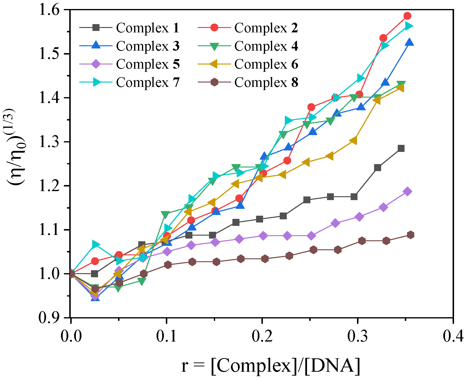
2.3.2. CT DNA Viscosity Measurements
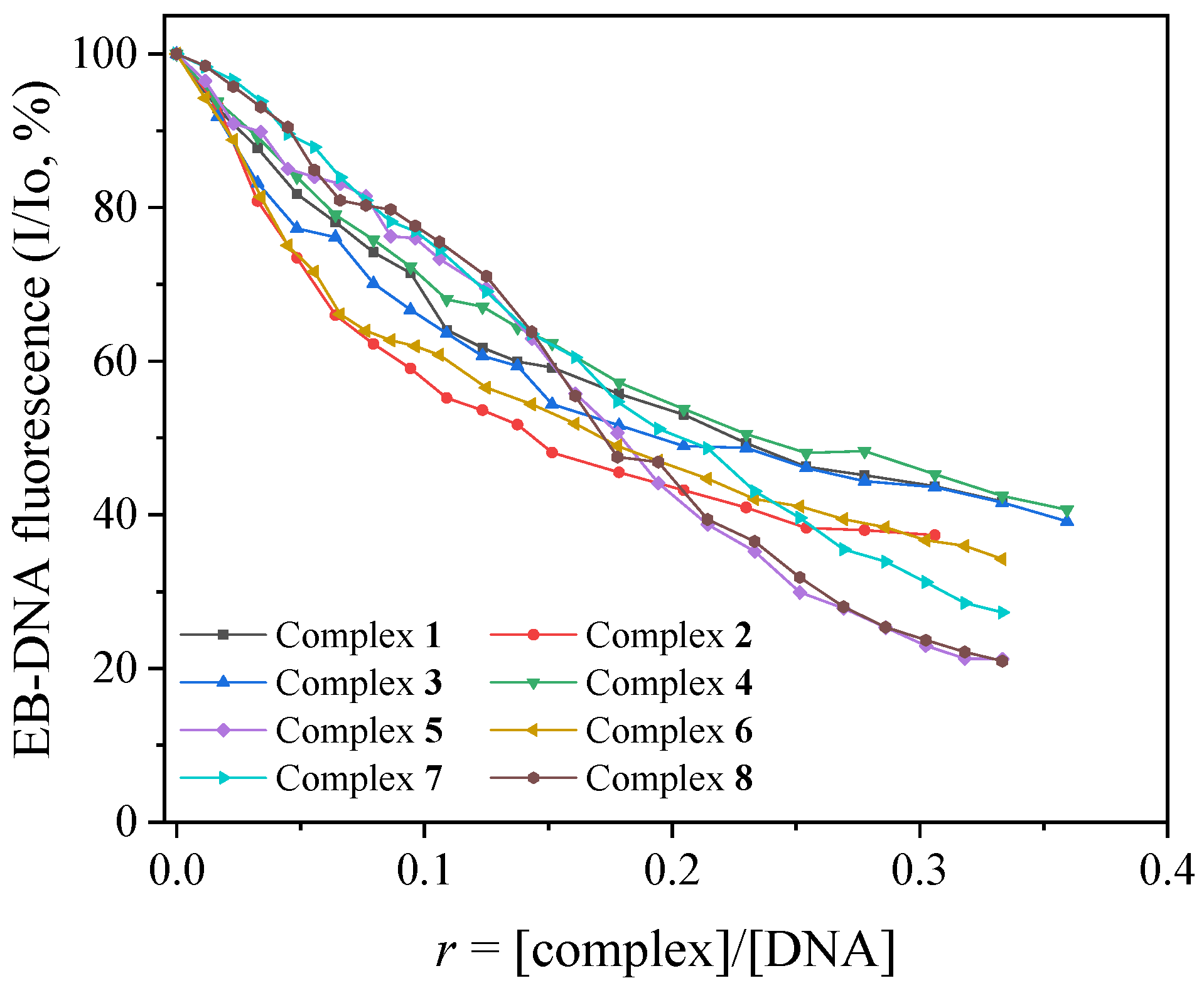
2.3.3. Competitive Studies with EB
2.3.4. Thermodynamic Parameters for the Interaction of Complexes with CT DNA
2.4. (Photo)Cleavage of pBR322 Plasmid DNA
2.5. Interaction of the Compounds with Albumins
2.6. Antioxidant Activity of the Complexes
3. Materials and Methods
3.1. Materials-Instrumentation-Physical Measurements
3.2. Synthesis of the Complexes
3.3. Single-Crystal X-Ray Crystallography
3.4. Study of the Biological Profile of the Complexes
4. Conclusions
Supplementary Materials
Author Contributions
Funding
Institutional Review Board Statement
Informed Consent Statement
Data Availability Statement
Conflicts of Interest
Abbreviations
| 1naph-saloH | 2-hydroxy-1-naphthaldehyde |
| 3,5diBr-saloH | 3,5-dibromo-salicylaldehyde |
| 3EtO-saloH | 3-ethoxy-salicylaldehyde |
| 4Et2N-saloH | 4-diethylamino-salicylaldehyde |
| 4MeO-saloH | 4-methoxy-salicylaldehyde |
| 4OH-saloH | 4-hydroxy-salicylaldehyde |
| 5Me-saloH | 5-methyl-salicylaldehyde |
| ABTS | 2,2′-azinobis-(3-ethylbenzothiazoline-6-sulfonic acid) |
| BHT | Butylated hydroxytoluene |
| BSA | Bovine serum albumin |
| CT | Calf thymus |
| DPPH | 1,1-diphenyl-picrylhydrazyl |
| EB | Ethidium bromide |
| HSA | Human serum albumin |
| K | SA-binding constant |
| Kb | DNA-binding constant |
| Kq | Quenching constant |
| KSV | Stern-Volmer constant |
| NDGA | Nordihydroguaiaretic acid |
| ovan | 3-methoxy-salicylaldehyde, o-vanillin |
| pDNA | pBR322 plasmid DNA |
| SA | Serum albumin |
| saloH | Salicylaldehyde |
| trolox | 6-hydroxy-2,5,7,8-tetramethylchroman-2-carboxylic acid |
| X-saloH | Substituted salicylaldehyde |
References
- Klimova, B.; Storek, M.; Valis, M.; Kuca, K. Global View on Rare Diseases: A Mini Review. Curr. Med. Chem. 2017, 24, 3153–3158. [Google Scholar] [CrossRef] [PubMed]
- Fermaglich, L.J.; Miller, K.L. A comprehensive study of the rare diseases and conditions targeted by orphan drug designations and approvals over the forty years of the Orphan Drug Act. Orphanet J. Rare Dis. 2023, 18, 163. [Google Scholar] [CrossRef]
- Abrams, M.J.; Murrer, B.A. Metal Compounds in Therapy and Diagnosis. Science 1993, 261, 725–730. [Google Scholar] [CrossRef]
- Karges, J.; Stokes, R.W.; Cohen, S.M. Metal complexes for therapeutic applications. Trends Chem. 2021, 3, 523–534. [Google Scholar] [CrossRef]
- Abbaspour, N.; Hurrell, R.; Kelishadi, R. Review on iron and its importance for human health. J. Res. Med. Sci. 2014, 19, 164. [Google Scholar]
- de Morais, T.R.; Gambero, A. Iron chelators in obesity therapy-Old drugs from a new perspective? Eur. J. Pharmacol. 2019, 861, 172614. [Google Scholar] [CrossRef] [PubMed]
- Sukhbaatar, N.; Weichhart, T. Iron Regulation: Macrophages in Control. Pharmaceuticals 2018, 11, 137. [Google Scholar] [CrossRef]
- Rout, G.R.; Sahoo, S. Role of iron in plant growth and metabolism. Rev. Agric. Sci. 2015, 3, 1–24. [Google Scholar] [CrossRef]
- Zhang, H.; Zhabyeyev, P.; Wang, S.; Oudit, G.Y. Role of iron metabolism in heart failure: From iron deficiency to iron overload. Biochim. Biophys. Acta (BBA)-Mol. Basis Dis. 2019, 1865, 1925–1937. [Google Scholar] [CrossRef]
- Lieu, P.T.; Heiskala, M.; Peterson, P.A.; Yang, Y. The roles of iron in health and disease. Mol. Asp. Med. 2001, 22, 1–87. [Google Scholar] [CrossRef]
- Ying, J.-F.; Lu, Z.-B.; Fu, L.-Q.; Tong, Y.; Wang, Z.; Li, W.-F.; Mou, X.-Z. The role of iron homeostasis and iron-mediated ROS in cancer. Am. J. Cancer Res. 2021, 11, 1895. [Google Scholar] [PubMed] [PubMed Central]
- Kuang, Y.; Wang, Q. Iron and lung cancer. Cancer Lett. 2019, 464, 56–61. [Google Scholar] [CrossRef] [PubMed]
- Vessieres, A. Iron Compounds as Anticancer Agents. In Metal-Based Anticancer Agents; Casini, A., Vessieres, A., Meier-Menches, S.M., Eds.; Royal Society of Chemistry: London, UK, 2019; pp. 62–90. [Google Scholar] [CrossRef]
- Arias, L.S.; Pessan, J.P.; Vieira, A.P.M.; De Lima, T.M.T.; Delbem, A.C.B.; Monteiro, D.R. Iron Oxide Nanoparticles for Biomedical Applications: A Perspective on Synthesis, Drugs, Antimicrobial Activity, and Toxicity. Antibiotics 2018, 7, 46. [Google Scholar] [CrossRef]
- Lin, J.F.; Wu, C.C.; Liao, Y.J.; Jakfar, S.; Tang, Z.B.; Chen, J.K.; Lin, F.H. In Vitro and In Vivo Evaluations of Mesoporous Iron Particles for Iron Bioavailability. Int. J. Mol. Sci. 2019, 20, 5291. [Google Scholar] [CrossRef]
- Wani, W.A.; Baig, U.; Shreaz, S.; Shiekh, R.A.; Iqbal, P.F.; Jameel, E.; Ahmad, A.; Mohd-Setapar, S.H.; Mushtaque, M.; Hun, L.T. Recent advances in iron complexes as potential anticancer agents. New J. Chem. 2016, 40, 1063–1090. [Google Scholar] [CrossRef]
- Naureen, B.; Miana, G.A.; Shahid, K.; Asghar, M.; Tanveer, S.; Sarwar, A. Iron (III) and zinc (II) monodentate Schiff base metal complexes: Synthesis, characterisation and biological activities. J. Mol. Struct. 2021, 1231, 129946. [Google Scholar] [CrossRef]
- Dimitrakopoulou, A.; Dendrinou-Samara, C.; Pantazaki, A.A.; Raptopoulou, C.; Terzis, A.; Samaras, E.; Kessissoglou, D.P. Interaction of Fe(III) with herbicide-carboxylato ligands. Di-, tri- and tetra-nuclear compounds: Structure, antimicrobial study and DNA interaction. Inorganica Chim. Acta 2007, 360, 546–556. [Google Scholar] [CrossRef]
- Dong, Y.R.; Cheng, S.J.; Qi, G.H.; Yang, Z.P.; Yin, S.Y.; Chen, G.T. Antimicrobial and antioxidant activities of Flammulina velutipes polysacchrides and polysacchride-iron(III) complex. Carbohydr. Polym. 2017, 161, 26–32. [Google Scholar] [CrossRef]
- Adjimani, J.P.; Asare, P. Antioxidant and free radical scavenging activity of iron chelators. Toxicol. Rep. 2015, 2, 721–728. [Google Scholar] [CrossRef]
- Dimiza, F.; Barmpa, A.; Chronakis, A.; Hatzidimitriou, A.G.; Sanakis, Y.; Papadopoulos, A.N.; Psomas, G. Iron(III) Complexes with Non-Steroidal Anti-Inflammatory Drugs: Structure, Antioxidant and Anticholinergic Activity, and Interaction with Biomolecules. Int. J. Mol. Sci. 2023, 24, 6391. [Google Scholar] [CrossRef]
- Dimiza, F.; Hatzidimitriou, A.G.; Sanakis, Y.; Papadopoulos, A.N.; Psomas, G. Trinuclear and tetranuclear iron(III) complexes with fenamates: Structure and biological profile. J. Inorg. Biochem. 2021, 218, 111410. [Google Scholar] [CrossRef]
- Michalski, C.; Mohagheghi, H.; Nimtz, M.; Pasteels, J.; Ober, D. Salicyl Alcohol Oxidase of the Chemical Defense Secretion of Two Chrysomelid Leaf Beetles. J. Biol. Chem. 2008, 283, 19219–19228. [Google Scholar] [CrossRef]
- Pelttari, E.; Lehtinen, M.; Elo, H. Substituted salicylaldehydes as potential antimicrobial drugs: Minimal inhibitory and microbicidal concentrations. Z. Fur Naturforschung-Sect. C J. Biosci. 2011, 66, 571–580. [Google Scholar] [CrossRef] [PubMed]
- Pelttari, E.; Karhumaki, E.; Langshaw, J.; Perakyla, H.; Elo, H. Antimicrobial properties of substituted salicylaldehydes and related compounds. Z. Fur Naturforschung-Sect. C J. Biosci. 2007, 62, 487–497. [Google Scholar] [CrossRef]
- Bountagkidou, O.G.; Ordoudi, S.A.; Tsimidou, M.Z. Structure-antioxidant activity relationship study of natural hydroxybenzaldehydes using in vitro assays. Food Res. Int. 2010, 43, 2014–2019. [Google Scholar] [CrossRef]
- Ntanatsidis, S.; Perontsis, S.; Konstantopoulou, S.; Kalogiannis, S.; Hatzidimitriou, A.G.; Papadopoulos, A.N.; Psomas, G. Manganese(II) complexes of substituted salicylaldehydes and α-diimines: Synthesis, characterization and biological activity. J. Inorg. Biochem. 2022, 227, 111693. [Google Scholar] [CrossRef] [PubMed]
- Psarras, G.I.; Zianna, A.; Hatzidimitriou, A.G.; Psomas, G. Coordination Compounds of Nickel(II) with 3,5-Dibromo-Salicylaldehyde: Structure and Interaction with Biomolecules. Inorganics 2024, 12, 138. [Google Scholar] [CrossRef]
- Papadopoulos, Z.; Doulopoulou, E.; Zianna, A.; Hatzidimitriou, A.G.; Psomas, G. Copper(II) Complexes of 5-Fluoro-Salicylaldehyde: Synthesis, Characterization, Antioxidant Properties, Interaction with DNA and Serum Albumins. Molecules 2022, 27, 8929. [Google Scholar] [CrossRef]
- Zianna, A.; Geromichalos, G.; Fiotaki, A.M.; Hatzidimitriou, A.G.; Kalogiannis, S.; Psomas, G. Palladium(II) Complexes of Substituted Salicylaldehydes: Synthesis, Characterization and Investigation of Their Biological Profile. Pharmaceuticals 2022, 15, 886. [Google Scholar] [CrossRef]
- Ristovic, M.S.; Zianna, A.; Psomas, G.; Hatzidimitriou, A.G.; Coutouli-Argyropoulou, E.; Lalia-Kantouri, M. Interaction of dinuclear cadmium(II) 5-Cl-salicylaldehyde complexes with calf-thymus DNA. Mater. Sci. Eng. C 2016, 61, 579–590. [Google Scholar] [CrossRef]
- Gkisiou, C.; Malis, G.; Hatzidimitriou, A.G.; Psomas, G. Erbium(III) coordination compounds with substituted salicylaldehydes: Characterization and biological profile. J. Inorg. Biochem. 2023, 242, 112161. [Google Scholar] [CrossRef]
- Zianna, A.; Vradi, E.; Hatzidimitriou, A.G.; Kalogiannis, S.; Psomas, G. Zinc(II) complexes of 3-bromo-5-chloro-salicylaldehyde: Characterization and biological activity. Dalton Trans. 2022, 51, 17629–17641. [Google Scholar] [CrossRef] [PubMed]
- Gkritzali, M.; Georgila, M.; Hatzidimitriou, A.G.; Kalogiannis, S.; Psomas, G. Neutral and cationic nickel(II) complexes with substituted salicylaldehydes: Characterization, antibacterial activity, and interaction with biomacromolecules. J. Inorg. Biochem. 2023, 247, 112339. [Google Scholar] [CrossRef]
- Selakovic, S.; Rodic, M.V.; Novakovic, I.; Matic, I.Z.; Stanojkovic, T.; Pirkovic, A.; Zivkovic, L.; Spremo-Potparevic, B.; Milcic, M.; Medakovic, V.; et al. Cu(II) complexes with a salicylaldehyde derivative and α-diimines as co-ligands: Synthesis, characterization, biological activity. Experimental and theoretical approach. Dalton Trans. 2024, 53, 2770–2788. [Google Scholar] [CrossRef] [PubMed]
- Zianna, A.; Geromichalos, G.D.; Hatzidimitriou, A.G.; Coutouli-Argyropoulou, E.; Lalia-Kantouri, M.; Psomas, G. Palladium(II) complexes with salicylaldehyde ligands: Synthesis, characterization, structure, in vitro and in silico study of the interaction with calf-thymus DNA and albumins. J. Inorg. Biochem. 2019, 194, 85–96. [Google Scholar] [CrossRef]
- Zianna, A.; Psomas, G.; Hatzidimitriou, A.; Lalia-Kantouri, M. Copper(II) complexes of salicylaldehydes and 2-hydroxyphenones: Synthesis, structure, thermal decomposition study and interaction with calf-thymus DNA and albumins. RSC Adv. 2015, 5, 37495–37511. [Google Scholar] [CrossRef]
- Zianna, A.; Geromichalos, G.; Psoma, E.; Kalogiannis, S.; Hatzidimitriou, A.G.; Psomas, G. Structure and in vitro and in silico biological activity of zinc(II) complexes with 3,5-dichloro-salicylaldehyde. J. Inorg. Biochem. 2022, 229, 111727. [Google Scholar] [CrossRef]
- Zianna, A.; Geromichalou, E.; Geromichalos, G.; Fiotaki, A.M.; Hatzidimitriou, A.G.; Kalogiannis, S.; Psomas, G. Zinc(II) complexes of 3,5-dibromo-salicylaldehyde and α-diimines: Synthesis, characterization and in vitro and in silico biological profile. J. Inorg. Biochem. 2022, 226, 111659. [Google Scholar] [CrossRef] [PubMed]
- Vitomirov, T.; Dimiza, F.; Matic, I.Z.; Stanojkovic, T.; Pirkovic, A.; Zivkovic, L.; Spremo-Potparevic, B.; Novakovic, I.; Andelkovic, K.; Milcic, M.; et al. Copper(II) complexes with 4-(diethylamino)salicylaldehyde and α-diimines: Anticancer, antioxidant, antigenotoxic effects and interaction with DNA and albumins. J. Inorg. Biochem. 2022, 235, 111942. [Google Scholar] [CrossRef]
- Lalia-Kantouri, M.; Gdaniec, M.; Choli-Papadopoulou, T.; Badounas, A.; Papadopoulos, C.D.; Czapik, A.; Geromichalos, G.D.; Sahpazidou, D.; Tsitouroudi, F. Effect of cobalt(II) complexes with dipyridylamine and salicylaldehydes on cultured tumor and non-tumor cells: Synthesis, crystal structure investigations and biological activity. J. Inorg. Biochem. 2012, 117, 25–34. [Google Scholar] [CrossRef]
- Lalia-Kantouri, M.; Papadopoulos, C.D.; Hatzidimitriou, A.G.; Bakas, T.; Pachini, S. A Trinuclear Iron(III) Complex Containing the Semi-Cubane [Fe3(μ3-O)]7+ Core: Structural, Spectroscopic, Magnetic and Electrochemical Study. Z. Anorg. Allg. Chem. 2010, 636, 531–538. [Google Scholar] [CrossRef]
- Anastasiadou, D.; Zianna, A.; Gdaniec, M.; Sigalas, M.P.; Coutouli-Argyropoulou, E.; Czapik, A.; Lalia-Kantouri, M. Unusual coordination mode of 3-methoxysalicylaldehyde in mononuclear zinc(II) complexes with nitrogenous bases: Synthesis, structural characterization and theoretical studies. Polyhedron 2015, 87, 275–285. [Google Scholar] [CrossRef]
- Shool, M.T.; Rudbari, H.A.; Gil-Anton, T.; Cuevas-Vicario, J.V.; García, B.; Busto, N.; Moini, N.; Blacque, O. The effect of halogenation of salicylaldehyde on the antiproliferative activities of {Δ/Λ-[Ru(bpy)2(X,Y-sal)]BF4} complexes. Dalton Trans. 2022, 51, 7658–7672. [Google Scholar] [CrossRef]
- Mandal, A.; Jaman, M.A.; Chakraborty, I.; Chowdhury, S. Salicylaldehyde derived ligands unlocking stable oxorhenium(V) compounds: An insightful role of chloroform dimer and catalysis. J. Mol. Struct. 2025, 1340, 142552. [Google Scholar] [CrossRef]
- Dimitrijevic, T.; Novakovic, I.; Radanovic, D.; Novakovic, S.B.; Rodic, M.V.; Andjelkovic, K.; Sumar-Ristovic, M. Synthesis, spectral and structural characterization and biological activity of Cu(II) complexes with 4-(diethylamino)salicylaldehyde and α-diimines. J. Coord. Chem. 2020, 73, 702–716. [Google Scholar] [CrossRef]
- Kuwar, A.; Tayade, K.; Keshav, K.; Sahoo, S.K.; Mayank; Singh, N. Cu2+-driven metallo-supramolecular self-assembly and its application in sensing of hydroxyl ion. Supramol. Chem. 2018, 30, 52–60. [Google Scholar] [CrossRef]
- Zhu, X.W. Synthesis, Crystal Structures, and Catalytic Properties of Dioxomolybdenum(VI) Complexes Derived from 4-Chloro-2-{[4-Diethylamino-2-Hydroxybenzylidene]amino}phenol. Russ. J. Coord. Chem. 2019, 45, 532–538. [Google Scholar] [CrossRef]
- Abedin, N.; Alshehri, A.H.A.; Almughrbi, A.M.A.; Moore, O.; Alyza, S.; Rusbridge, E.K.; Masood, N.; Egbowon, B.F.; Hargreaves, A.J.; Dafhnis-Calas, F.; et al. Expanding the family of tetrahalide iron complexes: Synthesis, structure and biological applications. Polyhedron 2020, 190, 114755. [Google Scholar] [CrossRef]
- Patel, K.N.; Patel, N.H.; Patel, K.M.; Patel, M.N.; Kothari, I.L. Synthesis, Characterization and Antimicrobial Activities of Some Transition Metal Complexes Containing Two Bidentate (O-O) Monobasic Hydroxy Aldehydes and 2,2′-Bipyridylamine. Synth. React. Inorg. Met.-Org. Chem. 2000, 30, 829–841. [Google Scholar] [CrossRef]
- Geary, W.J. The use of conductivity measurements in organic solvents for the characterisation of coordination compounds. Coord. Chem. Rev. 1971, 7, 81–122. [Google Scholar] [CrossRef]
- Chun, H.; Verani, C.N.; Chaudhuri, P.; Bothe, E.; Bill, E.; Weyhermuller, T.; Wieghardt, K. Molecular and electronic structure of octahedral o-aminophenolato and o-iminobenzosemiquinonato complexes of V(V), Cr(III), Fe(III), and Co(III). Experimental determination of oxidation levels of ligands and metal ions. Inorg. Chem. 2001, 40, 4157–4166. [Google Scholar] [CrossRef] [PubMed]
- Lalia-Kantouri, M.; Dimitriadis, T.; Papadopoulos, C.D.; Gdaniec, M.; Czapik, A.; Hatzidimitriou, A.G. Synthesis and structural characterization of iron(III) complexes with 2-hydroxyphenones. Z. Anorg. Allg. Chem. 2009, 635, 2185–2190. [Google Scholar] [CrossRef]
- Hamlaoui, M.; Hamlaoui, I.; Damous, M.; Belhocine, Y.; Sbei, N.; Ali, F.A.M.; Alghamdi, M.A.; Talab, S.; Rahali, S.; Merazig, H. Synthesis of Two Novel Copper (II) Complexes as Potential Inhibitors of HIV-1 Protease Enzyme: Experimental and Theoretical Investigations. Crystals 2022, 12, 1066. [Google Scholar] [CrossRef]
- Shaikh, N.A.; Rathod, S.N.; Bhat, S.S.; Naveen, S.; Shaikh, S.; Mahesha; Revankar, V.K.; Lokanath, N.K. Synthesis, Structural Characterization and Hirshfeld Surface Analysis of Mixed Ligand Copper(II) Complex. Chem. Data Collect. 2020, 28, 100374. [Google Scholar] [CrossRef]
- Asgharpour, Z.; Farzaneh, F.; Abbasi, A. Synthesis, characterization and immobilization of a new cobalt(II) complex on modified magnetic nanoparticles as catalyst for epoxidation of alkenes and oxidation of activated alkanes. RSC Adv. 2016, 6, 95729–95739. [Google Scholar] [CrossRef]
- Hazra, S.; Majumdar, D.; Frontera, A.; Roy, S.; Gassoumi, B.; Ghalla, H.; Dalai, S. On the Significant Importance of Hg⋯Cl Spodium Bonding/σ/π-Hole/Noncovalent Interactions and Nanoelectronic/Conductivity Applications in Mercury Complexes: Insights from DFT Spectrum. Cryst. Growth Des. 2024, 24, 7246–7261. [Google Scholar] [CrossRef]
- Wang, Y.Z.; Wang, P.; Li, C.; Su, Y.Q. Synthesis, Crystal Structure and Sulfoxidation of an Iron(III) Complex Derived From 3-Methoxysalicylaldehyde, Synthesis and Reactivity in Inorganic. Met.-Org. Nano-Met. Chem. 2016, 46, 868–871. [Google Scholar] [CrossRef]
- Nesterova, O.V.; Vassilyeva, O.Y.; Skelton, B.W.; Bienko, A.; Pombeiro, A.J.L.; Nesterov, D.S. A novel o-vanillin Fe(III) complex catalytically active in C-H oxidation: Exploring the magnetic exchange interactions and spectroscopic properties with different DFT functionals. Dalton Trans. 2021, 50, 14782–14796. [Google Scholar] [CrossRef]
- Vassilyeva, O.Y.; Buvaylo, E.A.; Kokozay, V.N.; Skelton, B.W.; Sobolev, A.N.; Bienko, A.; Ozarowski, A. Ferro- vs. antiferromagnetic exchange between two Ni(II) ions in a series of Schiff base heterometallic complexes: What makes the difference? Dalton Trans. 2021, 50, 2841–2853. [Google Scholar] [CrossRef]
- Costes, J.P.; Dahan, F.; Vendier, L.; Shova, S.; Lorusso, G.; Evangelisti, M. NiII-LnIII complexes with o-vanillin as the main ligand: Syntheses, structures, magnetic and magnetocaloric properties. Dalton Trans. 2018, 47, 1106–1116. [Google Scholar] [CrossRef]
- Vassilyeva, O.Y.; Nesterova, O.V.; Bieńko, A.; Komarnicka, U.K.; Buvaylo, E.A.; Vasylieva, S.M.; Skelton, B.W.; Nesterov, D.S. Heterometallic CuCd and Cu2Zn complexes with o-vanillin and its Schiff-base derivative: Slow magnetic relaxation and catalytic activity associated with CuII centres. Dalton Trans. 2025, 54, 6117–6132. [Google Scholar] [CrossRef] [PubMed]
- Mondal, A.; Mondal, M.; Das, R.; Ghosh, M.; Bhowmik, A.; Biswas, B.; Banerjee, P. A homobimetallic nickel(II) complex for discriminative chromogenic recognition of aqueous cyanide and silver(I) from medicinal products: Role of end-on thiocyanate bridging. Inorganica Chim. Acta 2024, 573, 122322. [Google Scholar] [CrossRef]
- Zhang, S.H.; Li, N.; Ge, C.M.; Feng, C.; Ma, L.F. Structures and magnetism of {Ni2Na2}, {Ni4} and {Ni6IINiIII} 2-hydroxy-3-alkoxy-benzaldehyde clusters. Dalton Trans. 2011, 40, 3000–3007. [Google Scholar] [CrossRef] [PubMed]
- Costes, J.P.; Vendier, L.; Wernsdorfer, W. Structural and magnetic studies of original tetranuclear CoII-LnIII complexes (LnIII = Gd, Tb, Y). Dalton Trans. 2011, 40, 1700–1706. [Google Scholar] [CrossRef]
- Biswas, R.; Drew, M.G.B.; Ghosh, A. Synthesis and crystal structure of a novel octa-aqua bridged star-shaped Ni4K complex. Inorg. Chem. Commun. 2012, 24, 1–3. [Google Scholar] [CrossRef]
- Arion, V.B.; Kravtsov, V.C.; Gradinaru, J.I.; Simonov, Y.A.; Gerbeleu, N.V.; Lipkowski, J.; Wignacourt, J.P.; Vezin, H.; Mentre, O. Potassium-controlled synthesis of heterotopic macrocycles based on isothiosemicarbazide. Inorganica Chim. Acta 2002, 328, 123–133. [Google Scholar] [CrossRef]
- Diop, M.; Sarr, M.; Diop, A.; Thiam, I.E.; Barry, A.H.; Gaye, M.; Alvarez, N.; Ellena, J. Nickel(II)-complex ligand as host for Potassium ion guest. Spectroscopic characterization and X-ray structure determination. IOSR J. Appl. Chem. 2019, 12, 18–24. Available online: https://www.iosrjournals.org/iosr-jac/papers/vol12-issue1/series-1/E1201011824.pdf (accessed on 22 January 2019).
- Jeffery, J.C.; Jelliss, P.A.; Rudd, G.E.A.; Sakanishi, S.; Stone, F.G.A.; Whitehead, J. Some di- and tri-metal complexes derived from the synthon [Ru(CO)2(THF)(η5-7,8-C2B9H11)]. J. Organomet. Chem. 1999, 582, 90–99. [Google Scholar] [CrossRef]
- Wang, Q.; Manzano, R.A.; Tinnermann, H.; Sung, S.; Leforestier, B.; Krämer, T.; Young, R.D. Access to and Reactivity of Fe0, Fe−I, FeI, and FeII PCcarbeneP Pincer Complexes. Angew. Chem. Int. Ed. 2021, 60, 18168–18177. [Google Scholar] [CrossRef]
- Reinfandt, N.; Michenfelder, N.; Schoo, C.; Yadav, R.; Reichl, S.; Konchenko, S.N.; Unterreiner, A.N.; Scheer, M.; Roesky, P.W. d/f-Polypnictides Derived by Non-Classical Ln2+ Compounds: Synthesis, Small Molecule Activation and Optical Properties. Chemistry 2021, 27, 7862–7871. [Google Scholar] [CrossRef]
- Zeglis, B.M.; Pierre, V.C.; Barton, J.K.; Pierre, V.C. Metallo-intercalators and metallo-insertors. Chem. Commun. 2007, 44, 4565–4579. [Google Scholar] [CrossRef] [PubMed]
- Pages, B.J.; Ang, D.L.; Wright, E.P.; Aldrich-Wright, J.R. Metal complex interactions with DNA. Dalton Trans. 2015, 44, 3505–3526. [Google Scholar] [CrossRef]
- Pyle, A.M.; Rehmann, J.P.; Meshoyrer, R.; Kumar, C.V.; Turro, N.J.; Barton, J.K. Mixed-ligand complexes of ruthenium(II): Factors governing binding to DNA. J. Am. Chem. Soc. 2002, 111, 3051–3058. [Google Scholar] [CrossRef]
- Wolfe, A.; Shimer, G.H.; Meehan, T. Polycyclic Aromatic Hydrocarbons Physically Intercalate into Duplex Regions of Denatured DNA. Biochemistry 1987, 26, 6392–6396. [Google Scholar] [CrossRef] [PubMed]
- Dimitrakopoulou, A.; Dendrinou-Samara, C.; Pantazaki, A.A.; Alexiou, M.; Nordlander, E.; Kessissoglou, D.P. Synthesis, structure and interactions with DNA of novel tetranuclear, [Mn4(II/II/II/IV)] mixed valence complexes. J. Inorg. Biochem. 2008, 102, 618–628. [Google Scholar] [CrossRef]
- Pizarro, A.M.; Sadler, P.J. Unusual DNA binding modes for metal anticancer complexes. Biochimie 2009, 91, 1198–1211. [Google Scholar] [CrossRef]
- Lakowicz, J.R. Principles of Fluorescence Spectroscopy; Springer: Berlin/Heidelberg, Germany, 2006; Available online: https://link.springer.com/book/10.1007/978-0-387-46312-4 (accessed on 12 February 2023).
- Heller, D.P.; Greenstock, C.L. Fluorescence lifetime analysis of DNA intercalated ethidium bromide and quenching by free dye. Biophys. Chem. 1994, 50, 305–312. [Google Scholar] [CrossRef]
- Ross, P.D.; Subramanian, S. Thermodynamics of Protein Association Reactions: Forces Contributing to Stability. Biochemistry 1981, 20, 3096–3102. [Google Scholar] [CrossRef]
- Rodrigues, B.M.; Victória, H.F.V.; Leite, G.; Krambrock, K.; Chaves, O.A.; de Oliveira, D.F.; de Q, R.; de Boni, L.; Costa, L.A.S.; Iglesias, B.A. Photophysical, photobiological, and biomolecule-binding properties of new tri-cationic meso-tri(2-thienyl)corroles with Pt(II) and Pd(II) polypyridyl derivatives. J. Inorg. Biochem. 2023, 242, 112149. [Google Scholar] [CrossRef]
- Sakthikumar, K.; Krause, R.W.M.; Isamura, B.K.; Raja, J.D.; Athimoolam, S. Spectro-electrochemical, fluorometric and biothermodynamic evaluation of pharmacologically active morpholine scaffold single crystal ligand and its metal(II) complexes: A comparative study on in-vitro and in-silico screening towards DNA/BSA/SARS-CoV-19. J. Inorg. Biochem. 2022, 236, 111953. [Google Scholar] [CrossRef]
- Sakthikumar, K.; Solomon, R.V.; Raja, J.D. Spectro-electrochemical assessments of DNA/BSA interactions, cytotoxicity, radical scavenging and pharmacological implications of biosensitive and biologically active morpholine-based metal(II) complexes: A combined experimental and computational investigation. RSC Adv. 2019, 9, 14220–14241. [Google Scholar] [CrossRef] [PubMed]
- Kashanian, S.; Askari, S.; Ahmadi, F.; Omidfar, K.; Ghobadi, S.; Tarighat, F.A. In Vitro Study of DNA Interaction with Clodinafop-Propargyl Herbicide. DNA Cell Biol. 2008, 27, 581–586. [Google Scholar] [CrossRef]
- Papastergiou, A.; Perontsis, S.; Gritzapis, P.; Koumbis, A.E.; Koffa, M.; Psomas, G.; Fylaktakidou, K.C. Evaluation of O-alkyl and aryl sulfonyl aromatic and heteroaromatic amidoximes as novel potent DNA photo-cleavers. Photochem. Photobiol. Sci. 2016, 15, 351–360. [Google Scholar] [CrossRef]
- Tarushi, A.; Lafazanis, K.; Kljun, J.; Turel, I.; Pantazaki, A.A.; Psomas, G.; Kessissoglou, D.P. First- and second-generation quinolone antibacterial drugs interacting with zinc(II): Structure and biological perspectives. J. Inorg. Biochem. 2013, 121, 53–65. [Google Scholar] [CrossRef] [PubMed]
- Tarushi, A.; Kljun, J.; Turel, I.; Pantazaki, A.A.; Psomas, G.; Kessissoglou, D.P. Zinc(II) complexes with the quinolone antibacterial drug flumequine: Structure, DNA- and albumin-binding. New J. Chem. 2013, 37, 342–355. [Google Scholar] [CrossRef]
- Stella, L.; Capodilupo, A.L.; Bietti, M. A reassessment of the association between azulene and [60]fullerene. Possible pitfalls in the determination of binding constants through fluorescence spectroscopy. Chem. Commun. 2008, 4744–4746. [Google Scholar] [CrossRef]
- Laitinen, O.H.; Hytönen, V.P.; Nordlund, H.R.; Kulomaa, M.S. Genetically engineered avidins and streptavidins. Cell. Mol. Life Sci. 2006, 63, 2992–3017. [Google Scholar] [CrossRef]
- Lazou, M.; Tarushi, A.; Gritzapis, P.; Psomas, G. Transition metal complexes with a novel guanine-based (E)-2-(2-(pyridin-2-ylmethylene)hydrazinyl)quinazolin-4(3H)-one: Synthesis, characterization, interaction with DNA and albumins and antioxidant activity. J. Inorg. Biochem. 2020, 206, 111019. [Google Scholar] [CrossRef]
- Cini, R.; Giorgi, G.; Cinquantini, A.; Rossi, C.; Sabat, M. Metal Complexes of the Antiinflammatory Drug Piroxicam. Inorg. Chem. 1990, 29, 5197–5200. [Google Scholar] [CrossRef]
- Marchi, R.C.; Campos, I.A.S.; Santana, V.T.; Carlos, R.M. Chemical implications and considerations on techniques used to assess the in vitro antioxidant activity of coordination compounds. Coord. Chem. Rev. 2022, 451, 2142752. [Google Scholar] [CrossRef]
- Ali, B.M.; Boothapandi, M.; Nasar, A.S.S. Nitric oxide, DPPH and hydrogen peroxide radical scavenging activity of TEMPO terminated polyurethane dendrimers: Data supporting antioxidant activity of radical dendrimers. Data Brief 2020, 28, 104972. [Google Scholar] [CrossRef] [PubMed]
- Dairi, S.; Carbonneau, M.A.; Galeano-Diaz, T.; Remini, H.; Dahmoune, F.; Aoun, O.; Belbahi, A.; Lauret, C.; Cristol, J.P.; Madani, K. Antioxidant effects of extra virgin olive oil enriched by myrtle phenolic extracts on iron-mediated lipid peroxidation under intestinal conditions model. Food Chem. 2017, 237, 297–304. [Google Scholar] [CrossRef]
- Kontogiorgis, C.; Hadjipavlou-Litina, D. Biological evaluation of several coumarin derivatives designed as possible anti-inflammatory/antioxidant agents. J. Enzym. Inhib. Med. Chem. 2003, 18, 63–69. [Google Scholar] [CrossRef] [PubMed]
- Wettasinghe, M.; Shahidi, F. Scavenging of reactive-oxygen species and DPPH free radicals by extracts of borage and evening primrose meals. Food Chem. 2000, 70, 17–26. [Google Scholar] [CrossRef]
- Marmur, J. A procedure for the isolation of deoxyribonucleic acid from micro-organisms. J. Mol. Biol. 1961, 3, 208–218. [Google Scholar] [CrossRef]
- Reichmann, M.E.; Rice, S.A.; Thomas, C.A.; Doty, P. A Further Examination of the Molecular Weight and Size of Desoxypentose Nucleic Acid. J. Am. Chem. Soc. 1954, 76, 3047–3053. [Google Scholar] [CrossRef]
- Bruker Analytical X-ray Systems, Inc. Apex2, Version 2 User Manual, M86-E01078; Bruker Analytical X-ray Systems, Inc.: Madison, WI, USA, 2006. [Google Scholar]
- Siemens Industrial Automation, Inc. SADABS: Area-Detector Absorption Correction; Siemens Industrial Automation, Inc.: Plano, TX, USA, 1996. [Google Scholar]
- Palatinus, L.; Chapuis, G. SUPERFLIP-a computer program for the solution of crystal structures by charge flipping in arbitrary dimensions. J. Appl. Crystallogr. 2007, 40, 786–790. [Google Scholar] [CrossRef]
- Betteridge, P.W.; Carruthers, J.R.; Cooper, R.I.; Prout, K.; Watkin, D.J. CRYSTALS version 12: Software for guided crystal structure analysis. J. Appl. Crystallogr. 2003, 36, 1487. [Google Scholar] [CrossRef]
- Wang, Y.-Q.; Zhang, H.-M.; Zhang, G.-C.; Tao, W.-H.; Tang, S.-H. Interaction of the flavonoid hesperidin with bovine serum albumin: A fluorescence quenching study. J. Lumin. 2007, 126, 211–218. [Google Scholar] [CrossRef]
- Ruch, R.J.; Cheng, S.J.; Klaunig, J.E. Prevention of cytotoxicity and inhibition of intercellular communication by antioxidant catechins isolated from Chinese green tea. Carcinogenesis 1989, 10, 1003–1008. [Google Scholar] [CrossRef]
- de Meulenaer, J.; Tompa, H. The absorption correction in crystal structure analysis. Acta Crystallogr. 1965, 19, 1014–1018. [Google Scholar] [CrossRef]


| Bond | Length (Å) | Bond | Length (Å) |
|---|---|---|---|
| Complex 1 | |||
| Fe–Oaldehyde | Fe–Ophenolato | ||
| Fe1–O1 | 1.976(4) | Fe1–O2 | 2.024(3) |
| Fe1–O3 | 1.994(4) | Fe1–O4 | 1.965(3) |
| Fe1–O5 | 2.042(3) | Fe1–O6 | 1.967(3) |
| Complex 2 | |||
| Fe–Oaldehyde | Fe–Ophenolato | ||
| Fe1–O1 | 2.0673(18) | Fe1–O2 | 1.9344(16) |
| Fe1–O4 | 2.0656(18) | Fe1–O5 | 1.9357(16) |
| Fe1–O7 | 2.0755(17) | Fe1–O8 | 1.9384(17) |
| Fe2–O10 | 2.049(2) | Fe2–O11 | 1.9346(19) |
| Fe2–O13 | 2.070(2) | Fe2–O14 | 1.933(2) |
| Fe2–O16 | 2.080(2) | Fe2–O17 | 1.942(2) |
| K⋯Omethoxy | K⋯Ophenolato | ||
| K1⋯O3 | 3.101(2) | K1⋯O2 | 2.9761(18) |
| K1⋯O6 | 3.078 (2) | K1⋯O5 | 2.9155(17) |
| K1⋯O9 | 3.054(2) | K1⋯O8 | 2.8843(17) |
| K1⋯O12 | 3.097(2) | K1⋯O11 | 2.8776(19) |
| K1⋯O15 | 3.136(2) | K1⋯O14 | 2.8616(19) |
| K1⋯O18 | 3.227(2) | K1⋯O17 | 2.998(2) |
| Compound | λmax (nm) (ΔA/Aο (%)) a, Δλ (nm) b) | Kb (M−1) |
|---|---|---|
| [Fe(1naph-salo)3], 1 | 315(−23 a, −9 b); 356(<− c, +7); 408(>+ d, +1) | 1.62(±0.01) × 107 |
| {K[Fe(ovan)3]2}Cl, 2 | 320(−14, +13); 354(−10, +1); 410(>+, +1) | 1.65(±0.13) × 106 |
| [Fe(5Me-salo)3], 3 | 333(−35, +5); 409(>+, +3) | 9.12(±0.95) × 105 |
| [Fe(3EtO-salo)3], 4 | 340(−24, 0); 379(>+, −5) | 8.89(±0.14) × 104 |
| [Fe(4OH-salo)3], 5 | 278(<−, −4); 315(>+, +15) | 6.64(±1.06) × 105 |
| [Fe(4MeO-salo)3], 6 | 282(−17, −9); 316(<–, +2); 386(>+, −2.5) | 6.17(±1.13) × 105 |
| [Fe(4Et2N-salo)3], 7 | 349(+12, −1); 407(<−, 0) | 5.62(±1.09) × 105 |
| [Fe(3,5diBr-salo)3], 8 | 319(−30, +3); 426(>+, 0) | 1.05(±0.21) × 106 |
| 1naph-saloH | 315(−22, −9); 356(−62, +9); 409(>+, +2) | 1.97(±0.10) × 106 |
| ovanH [37] | 340(−28, +3); 400(sh) e (+>, 0) | 9.84(±0.16) × 104 |
| 5Me-saloH [37] | 335(−10, 0) | 1.17(±0.11) × 106 |
| 3EtO-saloH | 267(−25, 0); 340(−33, +4); 403(>+, +4) | 1.16(±0.13) × 106 |
| 4OH-saloH | 282(−13, −1); 315(+24, +21) | 2.38(±0.17) × 105 |
| 4MeO-saloH [27] | 315(−44, +1) | 9.25(±0.12) × 105 |
| 4Et2N-saloH [30] | 349(−1, 0) | 5.06(±0.14) × 105 |
| 3,5diBr-saloH [39] | 337(<−, elim f); 427(>+, 0) | 3.71(±0.14) × 105 |
| Compound | ΔΙ/Ιο (%) | Ksv (M−1) | Kq (M−1 s−1) |
|---|---|---|---|
| [Fe(1naph-salo)3], 1 | 58.3 | 7.11(±0.15) × 104 | 3.09(±0.07) × 1012 |
| {K[Fe(ovan)3]2}Cl, 2 | 62.6 | 1.01(±0.03) × 105 | 4.38(±0.14) × 1012 |
| [Fe(5Me-salo)3], 3 | 60.9 | 6.99(±0.22) × 104 | 3.04(±0.10) × 1012 |
| [Fe(3EtO-salo)3], 4 | 59.3 | 1.35(±0.02) × 105 | 5.88(±0.08) × 1012 |
| [Fe(4OH-salo)3], 5 | 78.8 | 8.58(±0.35) × 104 | 3.73(±0.15) × 1012 |
| [Fe(4MeO-salo)3], 6 | 65.8 | 9.17(±0.13) × 104 | 3.99(±0.06) × 1012 |
| [Fe(4Et2N-salo)3], 7 | 72.7 | 1.17(±0.06) × 105 | 5.08(±0.25) × 1012 |
| [Fe(3,5diBr-salo)3], 8 | 79.1 | 3.88(±0.14) × 104 | 1.69(±0.62) × 1012 |
| Compound | Kq(BSA) (M−1 s−1) | K(BSA) (M−1) | K(BSA,ibuprofen) (M−1) | K(BSA,warfarin) (M−1) |
| [Fe(1naph-salo)3], 1 | 8.94(±0.28) × 1012 | 4.42(±0.26) × 105 | 8.57(±0.24) × 104 | 8.73(±0.42) × 104 |
| {K[Fe(ovan)3]2}Cl, 2 | 9.28(±0.38) × 1012 | 6.07(±0.34) × 104 | 1.15(±0.03) × 105 | 2.57(±0.16) × 104 |
| [Fe(5Me-salo)3], 3 | 2.78(±0.05) × 1012 | 3.39(±0.25) × 104 | 5.99(±0.20) × 104 | 5.38(±0.22) × 104 |
| [Fe(3EtO-salo)3], 4 | 5.41(±0.17) × 1012 | 1.15(±0.04) × 105 | 7.10(±0.16) × 104 | 3.75(±0.19) × 104 |
| [Fe(4OH-salo)3], 5 | 1.81(±0.10) × 1013 | 8.63(±0.50) × 104 | 5.20(±0.23) × 104 | 1.47(±0.11) × 105 |
| [Fe(4MeO-salo)3], 6 | 6.03(±0.24) × 1012 | 7.02(±0.54) × 103 | 2.15(±0.11) × 104 | 2.85(±0.23) × 104 |
| [Fe(4Et2N-salo)3], 7 | 1.66(±0.05) × 1013 | 8.61(±0.44) × 104 | 9.82(±0.32) × 104 | 2.48(±0.10) × 104 |
| [Fe(3,5diBr-salo)3], 8 | 2.54(±0.09) × 1013 | 1.21(±0.04) × 106 | 1.31(±0.10) × 106 | 1.33(±0.08) × 106 |
| Compound | Kq(HSA) (M−1 s−1) | K(HSA) (M−1) | K(HSA,ibuprofen) (M−1) | K(HSA,warfarin) (M−1) |
| [Fe(1naph-salo)3], 1 | 4.22(±0.14) × 1012 | 9.99(±0.23) × 105 | 7.13(±0.23) × 104 | 1.44(±0.05) × 105 |
| {K[Fe(ovan)3]2}Cl, 2 | 8.23(±0.49) × 1012 | 3.67(±0.25) × 104 | 5.11(±0.13) × 104 | 2.17(±0.06) × 105 |
| [Fe(5Me-salo)3], 3 | 1.22(±0.06) × 1012 | 2.17(±0.12) × 105 | 4.56(±0.24) × 104 | 1.98(±0.07) × 105 |
| [Fe(3EtO-salo)3], 4 | 2.47(±0.12) × 1012 | 1.45(±0.07) × 105 | 5.11(±0.20) × 104 | 2.13(±0.07) × 105 |
| [Fe(4OH-salo)3], 5 | 2.08(±0.13) × 1013 | 8.03(±0.23) × 104 | 5.46(±0.29) × 104 | 1.83(±0.06) × 105 |
| [Fe(4MeO-salo)3], 6 | 4.23(±0.15) × 1012 | 8.48(±0.55) × 104 | 1.82(±0.02) × 104 | 2.01(±0.05) × 105 |
| [Fe(4Et2N-salo)3], 7 | 1.09(±0.03) × 1013 | 1.52(±0.07) × 105 | 5.20(±0.05) × 104 | 3.29(±0.10) × 105 |
| [Fe(3,5diBr-salo)3], 8 | 2.01(±0.05) × 1013 | 1.99(±0.09) × 106 | 1.24(±0.05) × 106 | 1.71(±0.14) × 106 |
| Compound | DPPH% (30 min/60 min) | ABTS% | %H2O2 |
|---|---|---|---|
| [Fe(1naph-salo)3], 1 | 8.60 ± 0.62/6.46 ± 0.89 | 17.78 ± 0.53 | 68.69 ± 0.05 |
| {K[Fe(ovan)3]2}Cl, 2 | 33.85 ± 0.17/49.03 ± 0.71 | 85.69 ± 0.67 | 54.30 ± 0.48 |
| [Fe(5Me-salo)3], 3 | 11.91 ± 0.11/11.18 ± 0.67 | 31.29 ± 0.62 | 44.04 ± 0.36 |
| [Fe(3EtO-salo)3], 4 | 23.70 ± 0.42/29.40 ± 0.71 | 78.62 ± 0.71 | 71.10 ± 0.06 |
| [Fe(4OH-salo)3], 5 | 0.86 ± 0.34/2.62 ± 0.45 | 84.72 ± 0.76 | 72.28 ± 0.19 |
| [Fe(4MeO-salo)3], 6 | 4.83 ± 0.93/5.43 ± 0.27 | 11.05 ± 0.59 | 47.76 ± 0.61 |
| [Fe(4Et2N-salo)3], 7 | Not active | 57.07 ± 0.67 | 49.46 ± 0.51 |
| [Fe(3,5diBr-salo)3], 8 | Not active | 60.39 ± 0.53 | 85.86 ± 0.89 |
| BHT | 70.23 ± 0.95/88.60 ± 0.27 | Not tested | Not tested |
| NDGA | 93.51 ± 0.12/93.54 ± 0.11 | Not tested | Not tested |
| Trolox | Not tested | 89.25 ± 0.11 | Not tested |
| L-ascorbic acid | Not tested | Not tested | 60.51 ± 0.54 |
Disclaimer/Publisher’s Note: The statements, opinions and data contained in all publications are solely those of the individual author(s) and contributor(s) and not of MDPI and/or the editor(s). MDPI and/or the editor(s) disclaim responsibility for any injury to people or property resulting from any ideas, methods, instructions or products referred to in the content. |
© 2025 by the authors. Licensee MDPI, Basel, Switzerland. This article is an open access article distributed under the terms and conditions of the Creative Commons Attribution (CC BY) license (https://creativecommons.org/licenses/by/4.0/).
Share and Cite
Papadopoulos, Z.; Hatzidimitriou, A.G.; Psomas, G. Iron(III) Complexes with Substituted Salicylaldehydes: Synthesis, Interaction with DNA and Serum Albumins, and Antioxidant Activity. Molecules 2025, 30, 2383. https://doi.org/10.3390/molecules30112383
Papadopoulos Z, Hatzidimitriou AG, Psomas G. Iron(III) Complexes with Substituted Salicylaldehydes: Synthesis, Interaction with DNA and Serum Albumins, and Antioxidant Activity. Molecules. 2025; 30(11):2383. https://doi.org/10.3390/molecules30112383
Chicago/Turabian StylePapadopoulos, Zisis, Antonios G. Hatzidimitriou, and George Psomas. 2025. "Iron(III) Complexes with Substituted Salicylaldehydes: Synthesis, Interaction with DNA and Serum Albumins, and Antioxidant Activity" Molecules 30, no. 11: 2383. https://doi.org/10.3390/molecules30112383
APA StylePapadopoulos, Z., Hatzidimitriou, A. G., & Psomas, G. (2025). Iron(III) Complexes with Substituted Salicylaldehydes: Synthesis, Interaction with DNA and Serum Albumins, and Antioxidant Activity. Molecules, 30(11), 2383. https://doi.org/10.3390/molecules30112383