Article

Psilocin, the Psychoactive Metabolite of Psilocybin, Modulates Select Neuroimmune Functions of Microglial Cells in a 5-HT2 Receptor-Dependent Manner

Laboratory of Cellular and Molecular Pharmacology, Department of Biology, University of British Columbia Okanagan Campus, Kelowna, BC V1V 1V7, Canada
*
Author to whom correspondence should be addressed.
Molecules 2024, 29(21), 5084; https://doi.org/10.3390/molecules29215084
Submission received: 29 August 2024 / Revised: 16 October 2024 / Accepted: 17 October 2024 / Published: 28 October 2024
(This article belongs to the Section Medicinal Chemistry)

Abstract

:
Neuroinflammation that is caused by microglia, the main immune cells of the brain, contributes to neurodegenerative diseases. Psychedelics, including psilocybin and lysergic acid diethylamide (LSD), possess certain anti-inflammatory properties and, therefore, should be considered as drug candidates for treating neuroinflammatory pathologies. When ingested, psilocybin is rapidly dephosphorylated to yield psilocin, which crosses the blood–brain barrier and exerts psychotropic activity by interacting with the 5-hydroxytryptamine 2A receptors (5-HT2ARs) on neurons. Since microglia express all three 5-HT2R isoforms, we hypothesized that, by interacting with these receptors, psilocin beneficially modulates select neuroimmune functions of microglia. We used microglia-like cell lines to demonstrate that psilocin, at non-toxic concentrations, did not affect the secretion of tumor necrosis factor (TNF) by immune-stimulated microglial cells, but significantly inhibited their phagocytic activity, the release of reactive oxygen species (ROS), and nitric oxide (NO) production. The inhibitory activity of psilocin on the latter two functions was similar to that of two selective 5-HT2R agonists, namely, 25I-NBOH and Ro60-0175. The role of this subfamily of receptors was further demonstrated by the application of 5-HT2R antagonists cyproheptadine and risperidone. Psilocin should be considered a novel drug candidate that might be effective in treating neuroimmune disorders, such as neurodegenerative diseases, where reactive microglia are significant contributors.

1. Introduction

While the aggregation of amyloid β and the hyperphosphorylation of tau play well-established roles in the pathophysiology of Alzheimer’s disease (AD), the involvement of persistent neuroinflammation in the development and progression of this and other neurodegenerative disorders is increasingly accepted [1]. Neuronal death and neuroinflammatory responses in AD are accompanied by the chronic overactivation of glial cells, particularly microglia [2,3]. When confronted with abnormal protein aggregates, microglia facilitate the innate neuroimmune response. In part, this involves the clearance of harmful particles by phagocytosis and the release of cytokines, such as the pro-inflammatory tumor necrosis factor (TNF), and cytotoxins, including reactive oxygen species (ROS) and nitric oxide (NO) [4]. While these functions are essential for mounting an effective acute immune response, neurodegeneration, once triggered, leads to the chronic adverse reactivity of microglia. This causes the sustained secretion of pro-inflammatory mediators and excess phagoptosis, contributing to neuronal death [5,6]; therefore, anti-inflammatory therapies aimed at ameliorating reactive microglia represent feasible options in the search for effective treatments for these disorders [7,8].
Anti-inflammatory drugs are a class of medication aimed at suppressing or modulating inflammatory responses. The long-term use of conventional non-steroidal anti-inflammatory drugs (NSAIDs) has previously been associated with a decreased risk of AD in epidemiological studies [9]; however, conflicting results have been observed in clinical trials [10]. Additionally, several other commonly used inflammation-reducing therapies, such as corticosteroids, have proven to lack protective effects in AD treatment [11]; therefore, novel molecular targets and drug candidates that can be used to effectively suppress neuroinflammation in a broad range of neurodegenerative diseases need to be identified. Anti-inflammatory drugs often exert their effects by reducing the activity of enzymes and transcription factors, such as cyclooxygenase (COX)-2, nicotinamide adenine dinucleotide phosphate (NADPH)-dependent oxidase (NOX), inducible NO synthase (iNOS), and nuclear factor (NF)-κB. Such inhibitory activities diminish the production of inflammation-inducing and -propagating prostaglandins, ROS, NO, and cytokines [12,13,14,15]. Psychedelics, including psilocybin, mescaline, and lysergic acid diethylamide (LSD), have recently been shown to possess anti-inflammatory properties [16]. Since this heterogenous group of compounds remains relatively unexplored in the context of neuroinflammation and neurodegenerative diseases, they may possess untapped potential as drug candidates for the treatment of these disorders [17].
Depression is often associated with alterations in 5-hydroxytryptamine (5-HT, serotonin) neurotransmission. Most first-line antidepressants modulate specific aspects of 5-HT signaling, such as its reuptake, but there are treatment-resistant cases that do not respond to such therapies [18]. Notably, increasing evidence indicates that neuroinflammation also contributes to the pathogenesis of depression [19]. Treatment-resistant depression cases in particular are associated with increased TNF concentrations [20]. In addition, higher-than-normal plasma concentrations of both ROS and NO have been detected in individuals experiencing depressive symptoms [21,22]. Recently, antidepressant drugs, such as selective serotonin reuptake inhibitors (SSRIs), have been shown to reduce neuroinflammation [23]. Therefore, drugs with antidepressant activity that are also confirmed to cross the blood–brain barrier should be considered for use to suppress neuroinflammation in neurodegenerative diseases [24,25].
Psilocin (4-hydroxy-N,N-dimethyltryptamine) is the bioactive metabolite of the naturally occurring psychedelic alkaloid psilocybin, which was first isolated from mushrooms of the Psilocybe genus [26]. Since their discovery, methods of synthetic production for both psilocin and psilocybin have been developed. When ingested, psilocybin undergoes rapid dephosphorylation by alkaline phosphatase in the small intestine, kidneys, and plasma to yield psilocin, which freely crosses the blood–brain barrier and exerts psychotropic activity [27]. The distinctive psychedelic effects induced by psilocin result from altered serotonin signaling, primarily due to the activation of the 5-HT2A receptor (5-HT2AR) isoform on frontal cortex pyramidal neurons [28,29]. Likely due to this stimulatory activity of its main metabolite, psilocybin has emerged as a promising novel antidepressant, as shown in multiple human clinical trials, significantly alleviating symptoms in various forms of depression, including in treatment-resistant cases [30,31,32]. Recently, it has been proposed that the clinically meaningful antidepressant activity of psilocin could be due in part to its anti-inflammatory effects, which are also mediated by 5-HTRs expressed by neurons and glial cells [16,33]. Therefore, we hypothesized that the anti-neuroinflammatory activity of psilocin is mediated by its activation of 5-HT2R isoforms expressed by microglia, thereby downregulating their phagocytic activity and reducing their secretion of cytotoxins.
To test this hypothesis, in addition to psilocin, we employed two established ligands of 5-HT2Rs, namely, 25I-NBOH and Ro60-0175. All three compounds exhibit unique binding patterns with regard to 5-HTRs [34,35]. Psilocin displays a high level of affinity for the 5-HT2AR but is also capable of binding to the 5-HT2BR, 5-HT2CR, and 5-HT1AR isoforms, albeit with a lower level of affinity [36,37]. 25I-NBOH demonstrates a high level of affinity for the 5-HT2AR and, in contrast to psilocin, is highly selective for this receptor isoform in relation to other members of the 5-HT2R subfamily [38]. Conversely, Ro60-0175 has a higher level of affinity towards the 5-HT2CR isoform than 5-HT2A [39]. In this study, we found that psilocin significantly inhibited phagocytic activity and the production of ROS and NO by immune-activated microglial cells. We also demonstrated that the inhibitory activity of psilocin on ROS and NO production by microglial cells was similar to that of the two established 5-HT2R agonists, 25I-NBOH and Ro60-0175. Additionally, the 5-HT2R antagonists cyproheptadine and risperidone were employed to provide further evidence that the identified modulatory effects of psilocin were 5-HT2R-dependent.

2. Results

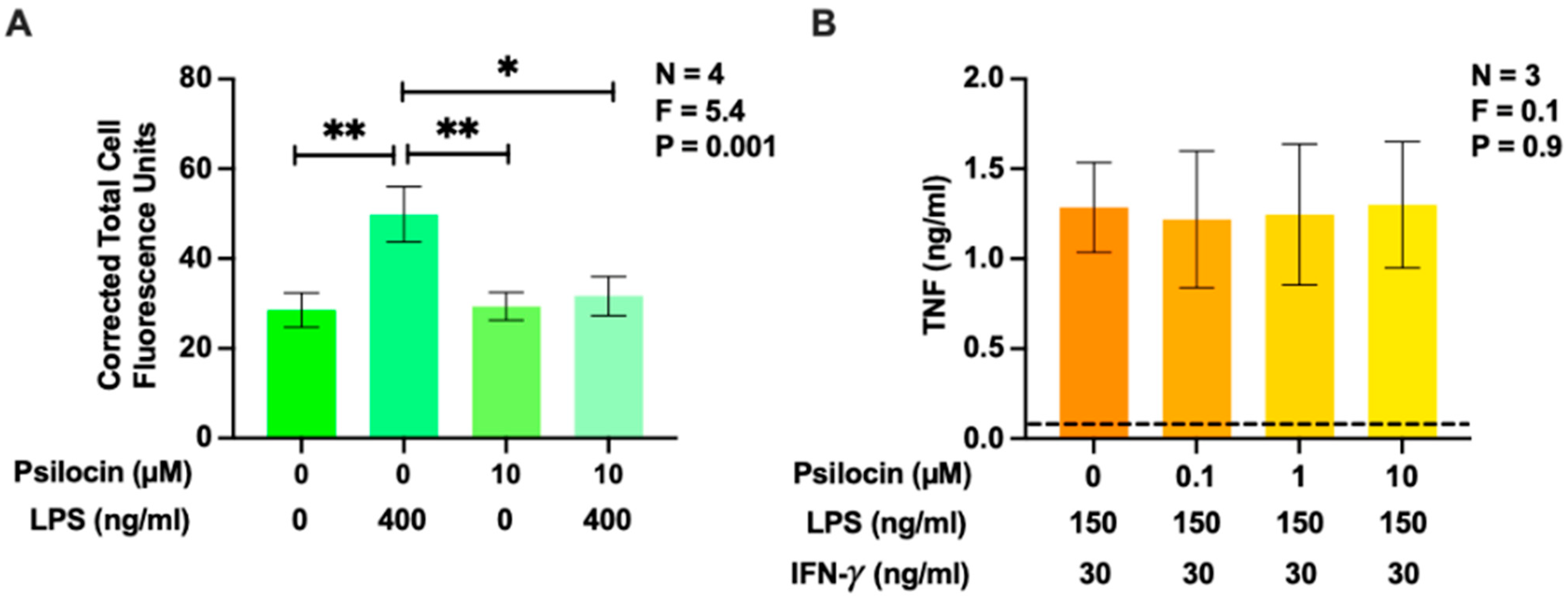
2.1. Effects of Psilocin on Phagocytosis of Latex Beads and Secretion of Tumor Necrosis Factor by BV-2 Murine Microglia

The phagocytic activity of primary murine microglia has been shown to decrease in response to treatment with the 5-HT2R agonists 5-HT and 2,5-dimethoxy-4-iodoamphetamine (DOI) [40]. In a more recent investigation, Kozlowska et al. [41] showed that psilocin downregulates the phagocytosis of viable neurons by primary murine CD11b+ microglia. Additionally, psilocin has been demonstrated to inhibit TNF secretion by RAW 264.7 murine macrophages [42] and exposure to psilocybin reduces TNF levels in mouse brains and human sera [33,43]; however, the opposite effect has been reported on serum TNF levels in rats [44]. Therefore, we first investigated whether psilocin inhibited the phagocytic activity and TNF secretion of immune-stimulated BV-2 murine microglia. The bacterial endotoxin lipopolysaccharide (LPS) and the combination of LPS plus interferon (IFN)-γ were employed as known inducers of phagocytic activity and cytokine secretion in multiple cell types, including BV-2 cells [45,46]. Psilocin was used at up to the maximum non-toxic concentration (10 µM), which was determined in preliminary experiments. The phagocytic activity of BV-2 cells was nearly doubled after their incubation with LPS for 24 h (Figure 1A). On its own, psilocin did not affect this function but completely inhibited the stimulatory effect of LPS. This effect was not caused by the cytotoxicity of psilocin since, at the 0.01 to 10 μM range, it did not inhibit TNF secretion by BV-2 cells (Figure 1B). The lack of toxic effects of psilocin towards immune-stimulated BV-2 cells at this concentration range was also confirmed using the 3-(4,5-dimethylthiazol-2-yl)-2,5-diphenyl tetrazolium bromide (MTT) assay (Figure A1).

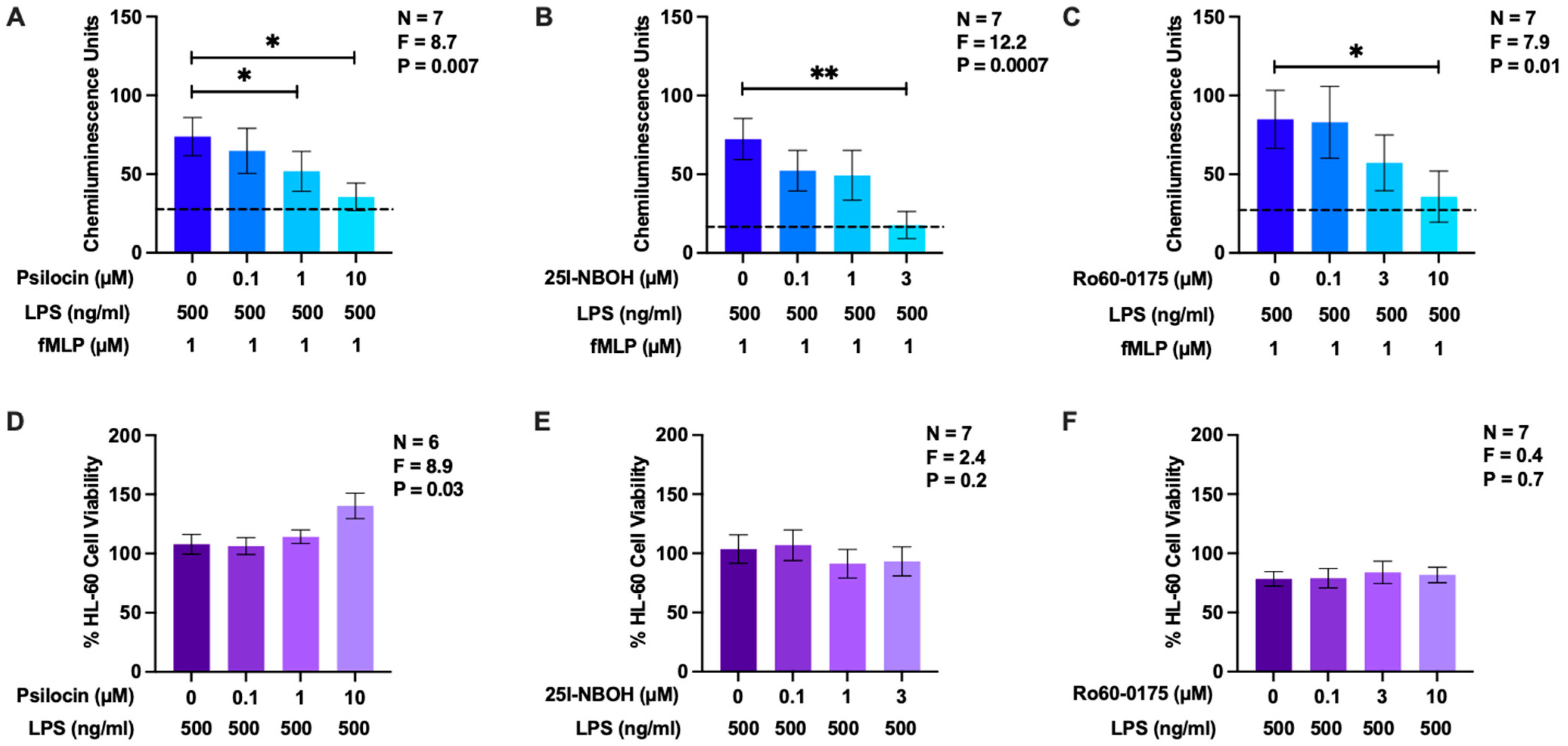
2.2. Effects of Psilocin, 25I-NBOH, and Ro60-0175 on Reactive Oxygen Species Generation by HL-60 Human Microglia-Like Cells

Nkadimeng et al. [47] report that the use of extracts from psilocybin-containing mushrooms decreases intracellular ROS levels in cardiomyocytes. To the best of our knowledge, the effects of psilocin or psilocybin on the generation of ROS by primary microglia or microglial model cells have not been determined. To investigate the effects of psilocin on ROS production in microglia-like cells, we used differentiated human HL-60 myelomonocytes, which respond similarly to microglia with regard to the respiratory burst when they are primed with LPS and subsequently stimulated with the bacterial peptide N-formyl-Met-Leu-Phe (fMLP) [48,49,50]. While psilocin has a high affinity for 5-HT2Rs, it also binds to other 5-HTR isoforms. Therefore, in addition to psilocin, we studied the effects of two other 5-HT2R ligands on the respiratory burst response: the specific 5-HT2AR agonists 25I-NBOH and Ro60-0175. The latter is more selective towards 5-HT2CR than 5-HT2AR. We hypothesized that psilocin would display a level of biological activity comparable to that of these established specific agonists when applied to microglial 5-HT2Rs. All three drugs were used at or below the maximum non-toxic concentrations determined in the preliminary experiments.
In comparison to the unprimed cells, LPS priming upregulated ROS production by HL-60 cells in response to fMLP stimulation (Figure 2A–C). Psilocin (1 and 10 µM), 25I-NBOH (3 µM), and Ro60-0175 (10 µM) significantly decreased the production of ROS by LPS-primed and fMLP-stimulated HL-60 cells. 25I-NBOH was the most effective drug, inhibiting approximately 75% of the maximal CHL signal (Figure 2B), followed by Ro60-0175 (Figure 2C) and psilocin (Figure 2A), which caused 58% and 52% reductions, respectively. At the concentrations tested, psilocin, 25I-NBOH, and Ro60-0175 did not significantly decrease the viability of HL-60 human cells (Figure 2D–F). This confirmed that the observed inhibitory effects of these drugs on ROS production were not due to the induction of HL-60 cell death.

2.3. Effects of Psilocin, 25I-NBOH, and Ro60-0175 on Nitric Oxide Production by BV-2 Murine Microglia Cells

Due to the established relationship between oxidative and nitrosative stress, we also investigated the effects of psilocin, 25I-NBOH, and Ro60-0175 at non-toxic concentrations on NO production by immune-stimulated BV-2 murine microglia cells. Unstimulated BV-2 microglia do not produce detectable levels of NO; therefore, the combination of LPS plus IFN-γ was used to induce NO secretion by BV-2 cells [45]. Figure 3A–C illustrates that BV-2 microglia, stimulated for 24 h with LPS plus IFN-γ, secreted detectable levels of NO. Psilocin, 25I-NBOH, and Ro60-0175 administration in a concentration-dependent manner decreased NO production by stimulated BV-2 cells. The most effective drug was 25I-NBOH, inhibiting maximal NO production by approximately 36% (Figure 3B). At the concentrations studied, psilocin, 25I-NBOH, or Ro60-0175 did not significantly affect the viability of BV-2 cells (Figure 3D–F); therefore, the observed inhibitory effects of these drugs on NO production were not caused by decreased BV-2 cell viability.

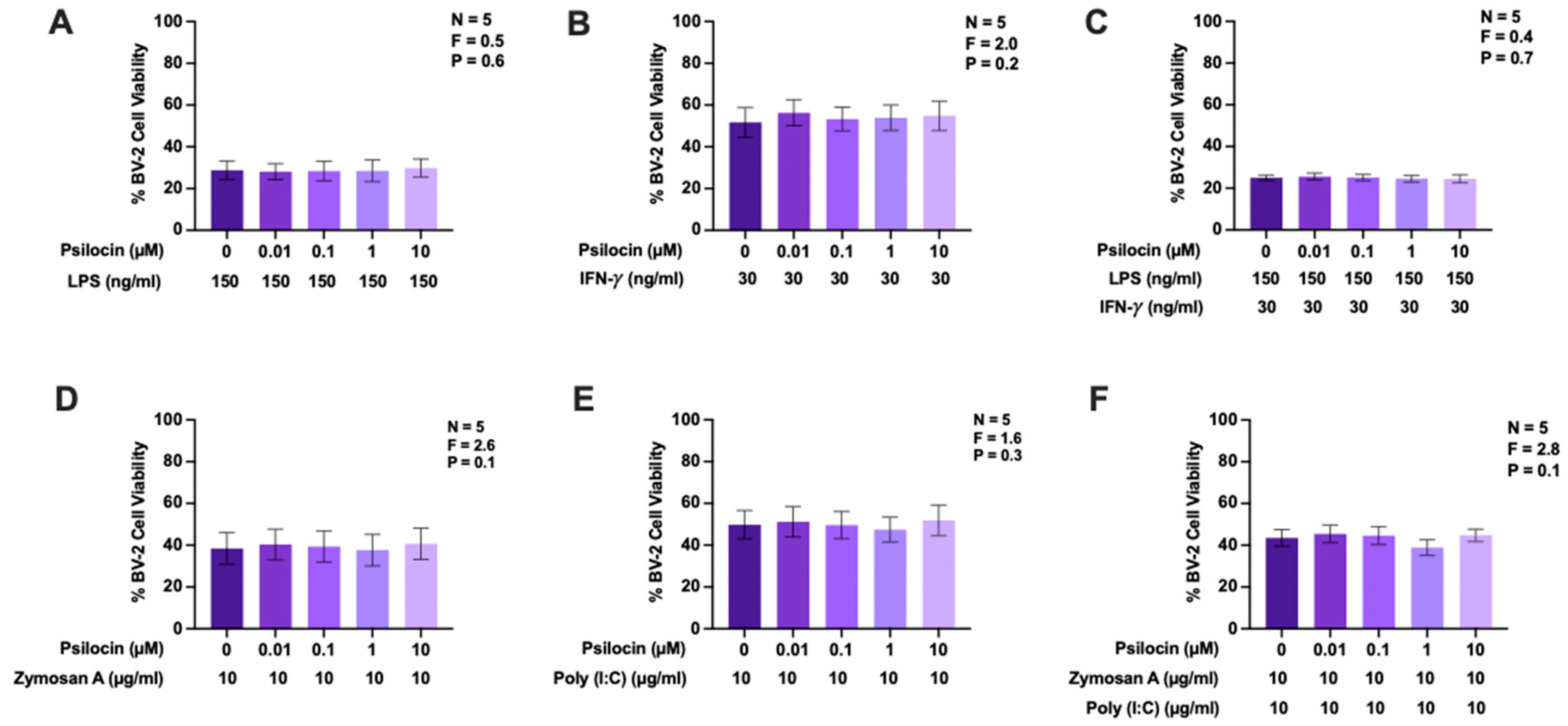
2.4. Effects of Psilocin on Nitric Oxide Production by BV-2 Murine Microglia Exposed to Diverse Stimuli

To further elucidate the inhibitory effects of psilocin on NO production by BV-2 microglia, four different stimuli relating to inflammatory signaling were utilized. LPS, IFN-γ, zymosan A, and polyinosinic:polycytidylic acid (poly (I:C)) were selected based on their demonstrated ability to induce NO secretion by BV-2 cells as well as the diversity of signaling pathways activated and immune responses initiated by these stimuli [45]. Figure 4 shows that exposure to the selected stimuli or two combinations, namely, LPS plus IFN-γ and zymosan A plus poly (I:C), upregulated NO production by BV-2 cells. The inhibitory activities of psilocin towards the generation of NO by BV-2 murine microglia were similar (11–22% reduction) with all stimuli and with all the combinations used in this study. At the concentrations studied, psilocin did not significantly affect the viability of BV-2 cells under any of these stimulatory conditions (Figure A1). Therefore, the observed inhibitory effect of psilocin on NO production was not caused by decreased BV-2 cell viability.

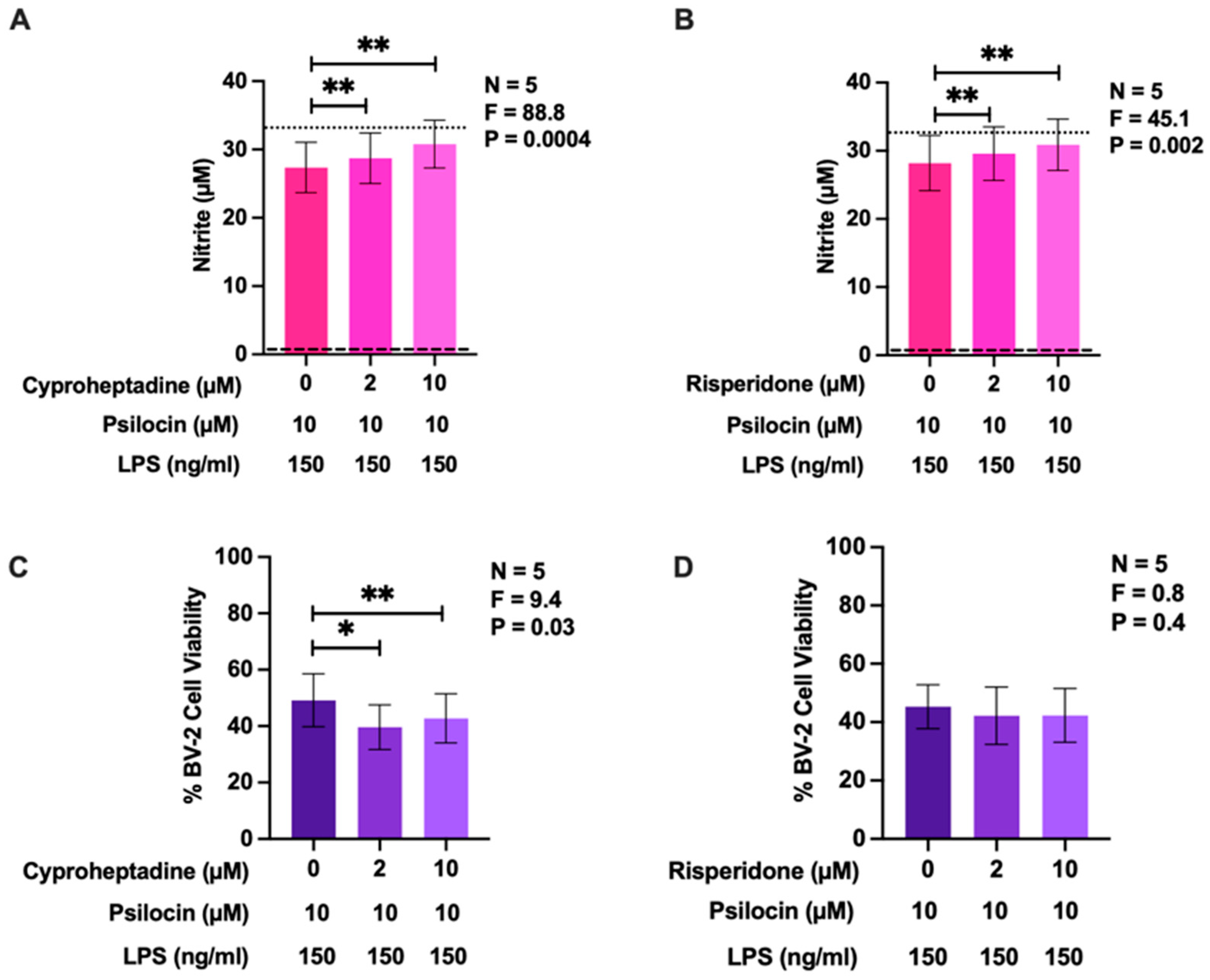
2.5. Effects of 5-HT2 Receptor Antagonists on the Inhibitory Activity of Psilocin

To confirm that the observed cellular effects of psilocin were mediated by 5-HT2R isoforms, we employed two antagonists of this 5-HTR subtype, cyproheptadine and risperidone, at non-toxic concentrations determined in preliminary experiments. Similar to Figure 3A, Figure 5 shows that psilocin (10 µM) inhibited LPS-induced NO secretion by BV-2 microglia. In this experiment, cells treated with psilocin and LPS exhibited approximately 80% of the maximum level of NO production compared to cells stimulated with LPS in the absence of psilocin or antagonists. At the two concentrations used (2 and 10 µM), both cyproheptadine (Figure 5A) and risperidone (Figure 5B) significantly suppressed the inhibitory effect of psilocin on NO generation by LPS-stimulated BV-2 cells. At these concentrations, risperidone was not toxic (Figure 5D) but cyproheptadine lowered the viability of BV-2 microglia when subsequently treated with both psilocin and LPS by up to 7% (Figure 5C). However, this small cytotoxic effect of cyproheptadine cannot account for the observed increase in NO production, as reduced cell viability typically results in decreased NO levels.

3. Materials and Methods

3.1. Reagents

The following reagents were obtained from Sigma Aldrich (Oakville, ON, Canada): MTT (M2128), bisbenzimide Hoechst 33258 trihydrochloride (B1155), carboxylate-modified polystyrene fluorescent yellow-green latex beads (L4655), LPS (from Escherichia coli O55:B5; L6529), luminol sodium salt (A4685), N-(1-naphthyl)-ethylenediamine dihydrochloride (222488), fMLP (F3506), poly (I:C) (P0913), psilocin (P-098), and zymosan A (from Saccharomyces cerevisiae; Z4250). The 5-HT2R agonists (S)-6-chloro-5-fluoro-1H-indole-2-propanamine (Ro60-0175; 29520) and 2-((2-(4-iodo-2,5-dimethoxyphenyl)ethylamino)methyl)phenol (25I-NBOH; 14909) were purchased from Cayman Chemicals (Ann Arbor, MI, USA). The 5-HT2R antagonists cyproheptadine hydrochloride sesquihydrate (C3218) and risperidone (R0087) were obtained from the Tokyo Chemical Institute (TCI; Tokyo, Japan). Recombinant murine IFN-γ (315-05) and ELISA development kits, used to detect murine TNF (900-T54) levels, were purchased from PeproTech (Embrun, ON, Canada). All cell culture media and other reagents were obtained from ThermoFisher Scientific (Ottawa, ON, Canada).

3.2. Cell Culture

The HL-60 human myelomonocytic cell line was purchased from the American Type Culture Collection (ATCC, Manassas, VA, USA) and the BV-2 murine microglia cell line was a generous donation from Dr. G. Garden (University of Washington, Seattle, WA, USA). All cells were maintained in Dulbecco’s modified Eagle medium/Ham’s F-12 nutrient mixture (DMEM/F-12), supplemented with 10% heat-inactivated bovine calf serum (CBS), penicillin (100 U/mL), streptomycin (100 µg/mL), and amphotericin B (500 ng/mL). HL-60 cells were used following differentiation with dimethyl sulfoxide (DMSO), as outlined in Section 2.5, while BV-2 cells were used undifferentiated. The media used in cell culture experiments contained 2%, 5%, or 10% heat-inactivated CBS, as well as the aforementioned antibiotics and antimycotic agents. Cells were cultured at 37 °C in a humid atmosphere comprising 5% CO2 and 95% O2.

3.3. Measurement of Phagocytic Activity

BV-2 murine microglia cells were utilized as microglial models to display phagocytic activity and were induced to secrete inflammatory cytokines and cytotoxins. Immune stimulation also induces the expression of iNOS in BV-2 microglia, leading to the production of NO [45,48,51,52]. Fluorescent latex beads were used to assess the phagocytic activity of BV-2 microglia as previously described [48]. The experimental conditions of this assay and all other assays performed in this study are summarized in Table 1. Cells were seeded in four-chambered glass-bottom Petri dishes at a density of 0.05 million per mL in 500 μL of DMEM/F-12 containing 5% CBS. They were then allowed to adhere for 24 h. Psilocin or its vehicle solution (20% v/v acetonitrile in deionized water) was administered on its own or 20 min prior to the addition of LPS or its vehicle solution (phosphate-buffered saline, known as PBS). After a 24 h incubation period, we added 2 µL of fluorescent latex beads (1 µm diameter; 0.1 million particles/µL) to the wells for 1 h. Following the removal of excess beads, the cells were fixed with 500 μL of cold 70% ethanol for 5 min and washed with warm PBS. After the cell nuclei were stained by adding 5 µL of bisbenzimide solution (2 μg/mL), cells were imaged at 40X magnification with a Zeiss AxioObserver.Z1 inverted widefield fluorescence microscope using ZEN 2.0 software. Measurements of corrected total cell fluorescence intensities were performed by an investigator, who was blinded to the experimental conditions, using the NIH ImageJ (version 1.53, FIJI build).

3.4. Measurement of Nitric Oxide and Tumor Necrosis Factor

BV-2 murine microglia were seeded in 24-well plates at a volume of 1 mL and a density of 0.2 million cells per mL in DMEM/F-12 containing 5% CBS. After a 24 h incubation period, the following drugs (or their vehicle solutions) were added as described in the results section: psilocin (dissolved in 20% v/v acetonitrile), the 5-HT2R agonists 25I-NBOH (DMSO) and Ro60-0175 (DMSO), and the 5-HT2R antagonists cyproheptadine (DMSO) and risperidone (DMSO). Following a 20 min incubation period, LPS, IFN-γ, LPS plus IFN-γ, zymosan A, poly (I:C), zymosan A plus poly (I:C), or their vehicle solutions (PBS or deionized water for poly (I:C) only) were added to the wells. Following a 24 h incubation period, the concentration of nitrite, a stable metabolite of NO, in cell-free supernatants was measured by adding the Griess reagent (1% sulfanilamide, 2.5% orthophosphoric acid, and 0.1% N-(1-naphthyl)-ethylenediamine dihydrochloride in deionized water) in a 1:1 ratio with the well volume. A calibration curve was created using standard nitrite concentrations in a cell culture medium. Optical density at 570 nm was measured immediately using a microplate reader. The collected cell-free supernatants were also used to measure concentrations of secreted TNF using ELISAs according to the instructions provided by the manufacturer (PeproTech).

3.5. Measurement of Reactive Oxygen Species

DMSO-differentiated HL-60 human myelomonocytic cells were used to model the microglial respiratory burst since they upregulate the expression of the NOX subunits, required for ROS production during this cellular response [53]. The chemiluminescence (CHL) assay was performed as described previously, albeit with minor modifications [48,54]. HL-60 cells were seeded in 6 cm cell culture plates in DMEM/F-12 containing 10% CBS and 1.3% DMSO at a volume of 20 mL and a density of 0.2 million cells per ml. After a six-day incubation period, chosen to allow for differentiation, cells were resuspended in fresh medium at a density of one million cells per ml. One mL aliquots of the culture were then added to 24-well plates and exposed to psilocin, 25I-NBOH, Ro60-0175, or their vehicle solutions. LPS was used as a priming agent and applied 20 min after the addition of the drugs. After a 24 h incubation period, the contents of each well were individually resuspended in phenol red-free DMEM/F-12 containing 2% CBS, and then seeded into a 96-well plate at a volume of 85 µL and a concentration of 0.5 million cells per ml. Using the injectors of a FLUOstar Omega microplate reader (BMG Labtech; Ortenberg, Germany), 10 µL of luminol sodium salt (10 mg/mL in PBS) and 5 µL of fMLP (20 µM in PBS) solutions were sequentially added into each well. This step was followed by CHL intensity measurement for a 25 min period. The ROS production induced by fMLP was considered to be luminol-dependent CHL [55].

3.6. Measurement of Cell Viability

Viable cells converted MTT into insoluble purple formazan crystals [56]. The cell cultures used in this study were exposed to MTT (0.5 mg/mL) for 1 h. This was followed by the addition of a solubilizing solution (20% w/v sodium lauryl sulfate and 50% v/v N,N-dimethylformamide in water) in a 1:1 ratio with well volume. The crystals were dissolved by using an orbital plate shaker for 3 h. Optical density was measured at 570 nm using a microplate reader. The data were normalized using values obtained from cells incubated in fresh growth medium only.

3.7. Statistical Analyses
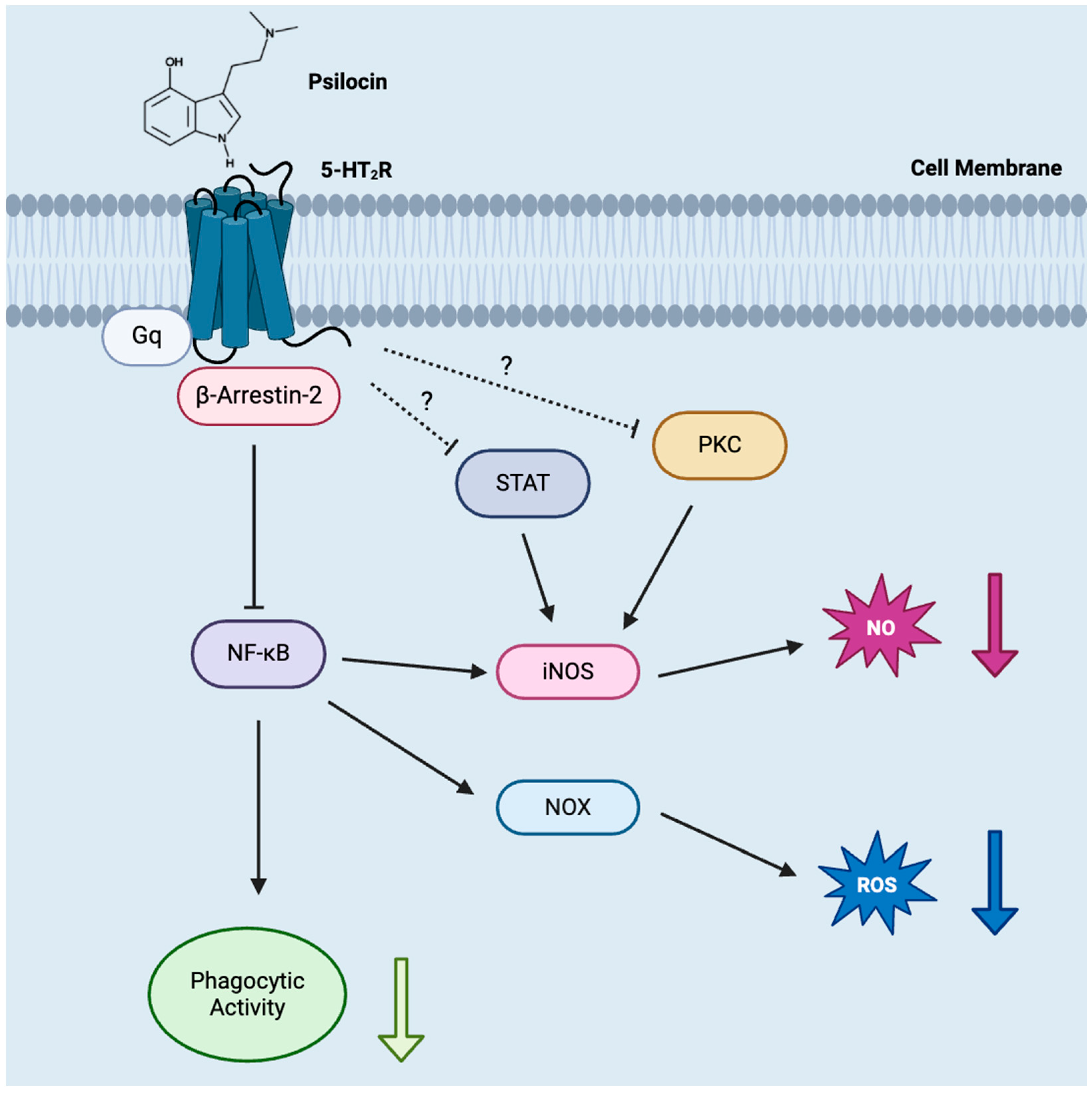
Statistical analyses and graphing were completed using Prism GraphPad software (version 10.1.0, GraphPad Software Inc., La Jolla, CA, USA). Randomized block design one-way analysis of variance (ANOVA), followed by Dunnett’s multiple comparison post hoc test, was used in most cases. One-way ANOVA was applied to phagocytosis experimental data. This was followed by Tukey’s multiple comparisons test. Data were collected from independent experiments, performed on separate days, and are shown as means ± standard error of the mean (SEM). Significance was established at p < 0.05.

4. Discussion

While the use of psilocybin-containing mushrooms for their consciousness-altering properties spans centuries and many cultures, the non-hallucinogenic effects of this substance, as well as its main metabolite psilocin, have only recently been identified and are not yet well understood [57]. 5-HT2R-mediated antidepressant and anti-neuroinflammatory activities are two of the significant non-psychotropic properties reported for both psilocybin and psilocin with relevance in the context of affective disorders and neurodegenerative diseases [16,58]. Since microglia play a central role in neuroimmune responses associated with these pathologies, we tested the hypothesis that psilocin modulates select neuroimmune functions of microglia in a manner that could be beneficial for the treatment of the disorders mentioned above. Additionally, we hypothesized that 5-HT2Rs are responsible for the observed beneficial effects of psilocin. To test these hypotheses, we used BV-2 murine microglia and HL-60 human myelomonocytic cells as microglia models, since several functional responses to immune stimulation in both these types of cell mimic those of their primary counterparts. Importantly, both BV-2 [59] and HL-60 cells [60,61] have already been reported to express all three 5-HT2R isoforms (A, B, and C).
First, by demonstrating the inhibitory effect of psilocin on phagocytosis of latex beads by BV-2 murine microglia (see Figure 6), we confirmed the validity of a previous study [40] and preliminary data by Kozlowska et al. [41] showing the downregulation of the phagocytic activity of murine microglia by 5-HT2R agonists and psilocin, respectively. Notably, psilocin did not affect the phagocytic activity of unstimulated BV-2 microglia and only reduced the LPS-induced uptake of latex particles. Such a selective activity towards immune-stimulated cells could be used to modulate the phagocytic activity of microglia under pathological conditions. Many researchers agree that the phagocytosis of cell fragments, myelin debris, and abnormal protein aggregates is a beneficial activity of microglia that should not be interfered with or even facilitated under specific pathological conditions; however, the suppression of the exacerbated phagocytic activity of reactive microglia, leading to cellular damage and overactive synaptic pruning, could be advantageous in the treatment of other conditions such as AD, neurotrauma, and ischemic stroke [62,63,64,65,66].
The observed lack of the effect of psilocin in the 0.1 to 10 µM range on TNF secretion by BV-2 microglia supports the mounting evidence that the modulation of microglial TNF expression by this alkaloid is cell type-dependent. For example, no effect on TNF secretion by immune-stimulated human THP-1 microglia-like cells and human T-cells was reported after their exposure to 0.3–30 µM psilocin, which is similar to the concentration range used in our experiments [67]. However, an inhibitory effect of psilocin on TNF production was observed when using LPS-stimulated RAW 264.7 murine macrophages [42]. Even though elevated TNF is implicated in the development of neuroinflammation, particularly in the context of neurodegenerative diseases and depression [19], psilocin may not be effective at suppressing the production of this cytokine by microglia.
Our study revealed the previously unknown inhibitory effect of psilocin on the respiratory burst activity of human microglia-like cells, resulting in the reduced production of ROS. This activity was a characteristic of not only psilocin but also two specific 5-HT2R agonists, which implicated this subfamily of 5-HTRs in the protective effect exhibited by psilocin. ROS play multifaceted roles in the CNS, and at high concentrations, such as those associated with increased microglial respiratory burst activity, they are linked to oxidative damage and the cytotoxicity observed in a multitude of neurological disorders [68].
Similar to their effects on ROS production, psilocin and the two 5-HT2R agonists had small yet significant inhibitory effects on NO release by BV-2 microglia. Our observations implicating the 5-HT2R subfamily in these suppressive actions of psilocin align well with other studies reporting that the activation of 5-HT2AR [69] and 5-HT2CR [70] isoforms is associated with lowered iNOS activity, leading to decreased NO levels. To the best of our knowledge, reduced NO production by microglia in the presence of psilocin has not been reported before, but it is noteworthy that extracts from Psilocybe natalensis, a psilocybin-containing mushroom, have been shown to reduce NO release from LPS-stimulated RAW 264.7 murine macrophages. Nitric oxide modulates multiple CNS signaling pathways under physiological conditions. However, when present at high concentrations, it is also capable of reacting with ROS to form peroxynitrite, a significant contributor to oxidative damage in various neuropathologies, including neurodegenerative diseases and neurotrauma [71,72]. Thus, the observed inhibitory effects of psilocin on ROS and NO production by microglial cells could explain its reported protective activity in certain human neuropathologies and makes it a suitable candidate for further preclinical studies aimed at identifying drugs which are effective at suppressing microglia-driven neuroinflammatory diseases.
Most previous studies investigating the effects of psilocin or psilocybin on microglial functions used LPS to induce a reactive state in this cell type. The Toll-like receptor (TLR)4-dependent signaling pathways and functional outcomes triggered by LPS exposure are well characterized in microglia and their documented involvement in various neuropathologies makes this endotoxin an appropriate immune stimulant for the in vitro and in vivo modeling of neuroinflammatory responses [73]. Since our experiments demonstrated the suppressive activity of psilocin on three different LPS-induced inflammatory functions of microglial cells, we next aimed to investigate whether psilocin was effective under other stimulatory conditions known to engage alternative signaling pathways in microglia [45]. We discovered that psilocin had a similar inhibitory effect on NO production triggered by LPS, IFN-γ, zymosan A, poly (I:C), and two different combinations of these stimulants. LPS is typically used to simulate bacterial infection [74], zymosan A mimics fungal pathogens and interacts with microglial TLR2 [75], poly (I:C) models viral particles and binds to TLR3 [76], and IFN-γ is a potent endogenous pro-inflammatory cytokine that activates IFN-γ receptors (IFNGR) and is known to be upregulated in several human neuropathologies [77,78]. The activation of these different receptors induces distinct downstream mechanisms [45]. The fact that psilocin inhibited NO release induced by these diverse stimuli could indicate that, by interacting with 5-HT2Rs, it modulated multiple signaling pathways regulating the inflammatory response of microglia. The ability to modify microglial responses to diverse pro-inflammatory stimuli makes psilocin a good drug candidate for the treatment of neuroinflammatory conditions that are triggered by diverse causative agents and conditions.
Finally, we obtained direct evidence that the inhibitory effect of psilocin on one of the pro-inflammatory responses of microglia, namely NO production, was mediated by 5-HT2Rs. We were not interested in the contributions of the individual isoforms of 5-HT2Rs; therefore, we selected two structurally distinct antagonists, cyproheptadine and risperidone, which are known to bind with different affinities to all three (A, B, and C) isoforms of this receptor subfamily [79,80,81]. Both these drugs effectively blocked the inhibitory effect of psilocin on BV-2 microglia, which indicated that this alkaloid exerted its activity by binding and activating at least one of the 5-HT2R isoforms. Detailed molecular studies will be needed to pinpoint the exact molecular targets when applying psilocin to microglia, but 5-HT2AR and 5-HT2CR isoforms are the likely candidate receptors due to their more prominent expression across various brain regions compared to 5-HT2BR [82] Notably, the A and C isoforms of 5-HT2R expressed by neurons are directly linked to the psychotropic effects of psilocybin and psilocin [28,36]; therefore, designing and testing novel derivatives of psilocin that possess anti-neuroinflammatory properties but do display hallucinogenic activity is an avenue of research that can be pursued in the future [83].
Downstream signaling pathways responsible for the suppressive actions of psilocin observed in this study also require further investigation (see Figure 6). It is known that 5-HT2AR is a G protein-coupled receptor (GPCR) and that psychedelics, after binding to this receptor, activate both Gq and β-arrestin-2 transducers [83]. Coupling through β-arrestin-2, in turn, has been shown to negatively regulate NF-κB activity [84]. It has also been reported that the expression of NOX, a primary producer of ROS in macrophages, depends on NF-κB activity [85] and that the expression and activity of iNOS in macrophages are highly upregulated by NF-κB [86]; therefore, this transcription factor may mediate the inhibitory effects of psilocin on ROS and NO production by microglia. Similarly, due to the previously reported dependence of the phagocytic activity of murine macrophages on NF-κB activity [87], it could also be responsible for the reduced phagocytosis by BV-2 microglia, which was observed in this study. In this regard, the previously reported inhibitory effect of 10–15 µM psilocybin on NF-κB activity in LPS-stimulated human THP-1 microglia-like cells is notable [88]. In addition to NF-κB, protein kinase C (PKC) and signal transducer and activator of transcription (STAT) have been linked to the inhibitory effect of 5-HT2R agonists on iNOS expression [69,89,90], but the contribution of these pathways needs to be confirmed regarding psilocin-induced inhibitory effects in microglia.
Herein, we demonstrate that the psychotropic alkaloid psilocin inhibits three different neuroimmune functions of microglia model cells without affecting their viability or having an effect on TNF secretion. The roles of 5-HT2Rs, as mediators of the cellular effects of psilocin, are indicated by the observations that (1) two other agonists of this receptor subfamily display effects that are comparable to those of psilocin in two of the cellular assays employed, and (2) the inhibitory effect of psilocin on BV-2 microglia NO release is blocked by two different antagonists of 5-HT2Rs. Our study has significant limitations since only in vitro models of neuroinflammatory activation of microglia were employed, which involved using immortal cell lines as microglial model cells. There are several important experimental steps that are required for the validation of psilocin as a candidate anti-neuroinflammatory drug that could potentially prevent or slow down neurodegenerative diseases that are characterized by the adverse activation of microglia. They include additional in vitro studies using primary human microglia or human induced pluripotent stem cell (iPSC)-derived microglia. Human iPSC-derived brain organoids, which contain all major brain cell types as well as humanized animal models of neurodegenerative diseases, could be used subsequently to study the anti-neuroinflammatory and neuroprotective effects of psilocin before it advances to human clinical trials. Nevertheless, to the best of our knowledge, this is the first study demonstrating the 5-HT2R-mediated inhibition of select neuroimmune functions of microglia by psilocin. While the inhibitory effect on the phagocytic activity of microglia could have adverse outcomes under certain neuropathological conditions, the lowering of excess ROS and NO production by microglia likely induces protective effects that could be beneficial in the treatment of neuroimmune disorders such as stroke, neurotrauma, and neurodegenerative diseases [72]. We conclude that psilocin holds merit not only as a novel antidepressant, but also as a potential therapeutic agent that can be used to mitigate neuroinflammation in various neuropathologies where reactive microglial play a prominent role [91]. This justifies further preclinical research to characterize the precise molecular mechanism of action of psilocin and also studies that aim to create non-psychotropic derivatives of this alkaloid that still possess its anti-neuroinflammatory activity.

Author Contributions

K.R.W.: conceptualization, methodology, formal analysis, data curation, writing—original draft, and writing—review and editing. N.A.H.B.: methodology, formal analysis, data curation, writing—original draft, and writing—review and editing. I.R.: methodology, formal analysis, data curation, and writing—review and editing. B.K.G.: methodology, formal analysis, data curation, and writing—review and editing. I.A.L.: methodology, formal analysis, data curation, and writing—review and editing. A.K.: funding acquisition, project administration, conceptualization, methodology, writing—original draft, and writing—review and editing. All authors have read and agreed to the published version of the manuscript.

Funding

This work was supported by grants from the Jack Brown and Family Alzheimer’s Disease Research Foundation, the Natural Sciences and Engineering Research Council of Canada, and the University of British Columbia Okanagan Campus.

Institutional Review Board Statement

Not applicable.

Informed Consent Statement

Not applicable.

Data Availability Statement

The data presented in this study are available on request from the authors.

Acknowledgments

The authors would like to thank Z. Mohammad and E. Creed for their assistance with the experiments and formatting of the figures as well as all members of the Laboratory of Cellular and Molecular Pharmacology for the helpful discussions and comments on the manuscript. Figure 6 in this manuscript was created with BioRender.com.

Conflicts of Interest

The authors declare no conflicts of interest.

Abbreviations

5-HT, 5-hydroxytryptamine, serotonin; 5-HTR, 5-hydroxytryptamine receptor; AD, Alzheimer’s disease; ANOVA, analysis of variance; CBS, calf bovine serum; CHL, chemiluminescence; CNS, central nervous system; COX, cyclooxygenase; DMEM/F-12, Dulbecco’s modified Eagle medium/Ham’s F-12 nutrient mixture; DMSO, dimethyl sulfoxide; ELISA, enzyme-linked immunosorbent assay; fMLP, N-formyl-Met-Leu-Phe; IFN, interferon; iNOS, inducible nitric oxide synthase; iPSC, induced pluripotent stem cell; LPS, lipopolysaccharide; LSD, lysergic acid diethylamide; MTT, 3-(4,5-dimethylthiazol-2-yl)-2,5-diphenyl tetrazolium bromide; NADPH, nicotinamide adenine dinucleotide phosphate; NF-κB, nuclear factor-κB; NO, nitric oxide; NOX, nicotinamide adenine dinucleotide phosphate-dependent oxidase; NSAID, non-steroidal anti-inflammatory drug; PBS, phosphate-buffered saline; poly (I:C), polyinosinic:polycytidylic acid; PKC, protein kinase C; ROS, reactive oxygen species; SEM, standard error of the mean; SSRI, selective serotonin reuptake inhibitor; STAT, signal transducer and activator of transcription; TNF, tumor necrosis factor; TLR, Toll-like receptor.

Appendix A

Figure A1. Effects of psilocin on viability of BV-2 murine microglia exposed to diverse stimuli. Varying concentrations of psilocin were administered 20 min prior to the addition of four different stimuli or their combinations as displayed in the figure panels (AF). Following a 24 h incubation period, viability of cells was assessed by the MTT assay. Data from five independent experiments completed on different days are shown as means ± SEM. p and F values displayed in the figures were calculated using randomized block one-way ANOVA.
Molecules 29 05084 g0a1

References

  1. Busche, M.A.; Hyman, B.T. Synergy between amyloid-β and tau in Alzheimer’s disease. Nat. Neurosci. 2020, 23, 1183–1193. [Google Scholar] [CrossRef] [PubMed]
  2. Heneka, M.T.; Carson, M.J.; El Khoury, J.; Landreth, G.E.; Brosseron, F.; Feinstein, D.L.; Jacobs, A.H.; Wyss-Coray, T.; Vitorica, J.; Ransohoff, R.M.; et al. Neuroinflammation in Alzheimer’s disease. Lancet Neurol. 2015, 14, 388–405. [Google Scholar] [CrossRef] [PubMed]
  3. DeTure, M.A.; Dickson, D.W. The neuropathological diagnosis of Alzheimer’s disease. Mol. Neurodegener. 2019, 14, 32. [Google Scholar] [CrossRef] [PubMed]
  4. Rodriguez-Gomez, J.A.; Kavanagh, E.; Engskog-Vlachos, P.; Engskog, M.K.R.; Herrera, A.J.; Espinosa-Oliva, A.M.; Joseph, B.; Hajji, N.; Venero, J.L.; Burguillos, M.A. Microglia: Agents of the CNS pro-inflammatory response. Cells 2020, 9, 1717. [Google Scholar] [CrossRef] [PubMed]
  5. Butler, C.A.; Popescu, A.S.; Kitchener, E.J.A.; Allendorf, D.H.; Puigdellívol, M.; Brown, G.C. Microglial phagocytosis of neurons in neurodegeneration, and its regulation. J. Neurochem. 2021, 158, 621–639. [Google Scholar] [CrossRef]
  6. Ganguly, U.; Kaur, U.; Chakrabarti, S.S.; Sharma, P.; Agrawal, B.K.; Saso, L.; Chakrabarti, S. Oxidative stress, neuroinflammation, and NADPH oxidase: Implications in the pathogenesis and treatment of Alzheimer’s disease. Oxid. Med. Cell. Longev. 2021, 2021, 7086512. [Google Scholar] [CrossRef]
  7. Muzio, L.; Viotti, A.; Martino, G. Microglia in neuroinflammation and neurodegeneration: From understanding to therapy. Front. Neurosci. 2021, 15, 742065. [Google Scholar] [CrossRef]
  8. Ozben, T.; Ozben, S. Neuro-inflammation and anti-inflammatory treatment options for Alzheimer’s disease. Clin. Biochem. 2019, 72, 87–89. [Google Scholar] [CrossRef]
  9. Zhang, C.; Wang, Y.; Wang, D.; Zhang, J.; Zhang, F. NSAID exposure and risk of Alzheimer’s disease: An updated meta-analysis from cohort studies. Front. Aging Neurosci. 2018, 10, 83. [Google Scholar] [CrossRef]
  10. Pasqualetti, P.; Bonomini, C.; Dal Forno, G.; Paulon, L.; Sinforiani, E.; Marra, C.; Zanetti, O.; Maria Rossini, P. A randomized controlled study on effects of ibuprofen on cognitive progression of Alzheimer’s disease. Aging Clin. Exp. Res. 2009, 21, 102–110. [Google Scholar] [CrossRef]
  11. Wang, J.; Tan, L.; Wang, H.-F.; Tan, C.-C.; Meng, X.-F.; Wang, C.; Tang, S.-W.; Yu, J.-T. Anti-inflammatory drugs and risk of Alzheimer’s disease: An updated systematic review and meta-analysis. J. Alzheimer’s Dis. 2015, 44, 385–396. [Google Scholar] [CrossRef] [PubMed]
  12. Singh, A.; Yau, Y.F.; Leung, K.S.; El-Nezami, H.; Lee, J.C.-Y. Interaction of polyphenols as antioxidant and anti-inflammatory compounds in brain–liver–gut axis. Antioxidants 2020, 9, 669. [Google Scholar] [CrossRef] [PubMed]
  13. Koppula, S.; Kumar, H.; Kim, I.S.; Choi, D.-K. Reactive oxygen species and inhibitors of inflammatory enzymes, NADPH oxidase, and iNOS in experimental models of Parkinson’s disease. Mediat. Inflamm. 2012, 2012, 823902. [Google Scholar] [CrossRef] [PubMed]
  14. Yagami, T.; Koma, H.; Yamamoto, Y. Pathophysiological roles of cyclooxygenases and prostaglandins in the central nervous system. Mol. Neurobiol. 2016, 53, 4754–4771. [Google Scholar] [CrossRef] [PubMed]
  15. Crinelli, R.; Antonelli, A.; Bianchi, M.; Gentilini, L.; Scaramucci, S.; Magnani, M. Selective inhibition of NF-KB activation and TNF-α production in macrophages by red blood cell-mediated delivery of dexamethasone. Blood Cells Mol. Dis. 2000, 26, 211–222. [Google Scholar] [CrossRef]
  16. Flanagan, T.W.; Nichols, C.D. Psychedelics as anti-inflammatory agents. Int. Rev. Psychiatry 2018, 30, 363–375. [Google Scholar] [CrossRef]
  17. Kozlowska, U.; Nichols, C.; Wiatr, K.; Figiel, M. From Psychiatry to neurology: Psychedelics as prospective sherapeutics for neurodegenerative disorders. J. Neurochem. 2022, 162, 89–108. [Google Scholar] [CrossRef]
  18. Galecki, P.; Mossakowska-Wojcik, J.; Talarowska, M. The anti-inflammatory mechanism of antidepressants—SSRIs, SNRIs. Prog. Neuropsychopharmacol. Biol. Psychiatry 2018, 80, 291–294. [Google Scholar] [CrossRef]
  19. Troubat, R.; Barone, P.; Leman, S.; Desmidt, T.; Cressant, A.; Atanasova, B.; Brizard, B.; El Hage, W.; Surget, A.; Belzung, C.; et al. Neuroinflammation and depression: A review. Eur. J. Neurosci. 2021, 53, 151–171. [Google Scholar] [CrossRef]
  20. Strawbridge, R.; Arnone, D.; Danese, A.; Papadopoulos, A.; Herane Vives, A.; Cleare, A.J. Inflammation and clinical response to treatment in depression: A meta-analysis. Eur. Neuropsychopharmacol. 2015, 25, 1532–1543. [Google Scholar] [CrossRef]
  21. Kim, Y.-K.; Paik, J.-W.; Lee, S.-W.; Yoon, D.; Han, C.; Lee, B.-H. Increased plasma nitric oxide level associated with suicide attempt in depressive patients. Prog. Neuropsychopharmacol. Biol. Psychiatry 2006, 30, 1091–1096. [Google Scholar] [CrossRef] [PubMed]
  22. Rybka, J.; Kedziora-Kornatowska, K.; Banas-Lezanska, P.; Majsterek, I.; Carvalho, L.A.; Cattaneo, A.; Anacker, C.; Kedziora, J. Interplay between the pro-oxidant and antioxidant systems and proinflammatory cytokine levels, in relation to iron metabolism and the erythron in depression. Free Radic. Biol. Med. 2013, 63, 187–194. [Google Scholar] [CrossRef] [PubMed]
  23. Patel, S.; Keating, B.A.; Dale, R.C. Anti-inflammatory properties of commonly used psychiatric drugs. Front. Neurosci. 2023, 16, 1039379. [Google Scholar] [CrossRef] [PubMed]
  24. Gutierrez, I.L.; Gonzalez-Prieto, M.; Caso, J.R.; Garcia-Bueno, B.; Leza, J.C.; Madrigal, J.L.M. Reboxetine treatment reduces neuroinflammation and neurodegeneration in the 5xFAD mouse model of Alzheimer’s disease: Role of CCL2. Mol. Neurobiol. 2019, 56, 8628–8642. [Google Scholar] [CrossRef]
  25. Nykamp, M.J.; Zorumski, C.F.; Reiersen, A.M.; Nicol, G.E.; Cirrito, J.; Lenze, E.J. Opportunities for drug repurposing of serotonin reuptake inhibitors: Potential uses in inflammation, infection, cancer, neuroprotection, and Alzheimer’s disease prevention. Pharmacopsychiatry 2022, 55, 24–29. [Google Scholar] [CrossRef]
  26. Strauss, D.; Ghosh, S.; Murray, Z.; Gryzenhout, M. An overview on the taxonomy, phylogenetics and ecology of the psychedelic genera Psilocybe, Panaeolus, Pluteus and Gymnopilus. Front. For. Glob. Chang. 2022, 5, 813998. [Google Scholar] [CrossRef]
  27. Dinis-Oliveira, R.J. Metabolism of psilocybin and psilocin: Clinical and forensic toxicological relevance. Drug Metab. Rev. 2017, 49, 84–91. [Google Scholar] [CrossRef]
  28. Madsen, M.K.; Fisher, P.M.; Burmester, D.; Dyssegaard, A.; Stenbæk, D.S.; Kristiansen, S.; Johansen, S.S.; Lehel, S.; Linnet, K.; Svarer, C.; et al. Psychedelic effects of psilocybin correlate with serotonin 2A receptor occupancy and plasma psilocin levels. Neuropsychopharmacology 2019, 44, 1328–1334. [Google Scholar] [CrossRef]
  29. Gonzalez-Maeso, J.; Weisstaub, N.V.; Zhou, M.; Chan, P.; Ivic, L.; Ang, R.; Lira, A.; Bradley-Moore, M.; Ge, Y.; Zhou, Q.; et al. Hallucinogens recruit specific cortical 5-HT2A receptor-mediated signaling pathways to affect behavior. Neuron 2007, 53, 439–452. [Google Scholar] [CrossRef]
  30. Carhart-Harris, R.; Giribaldi, B.; Watts, R.; Baker-Jones, M.; Murphy-Beiner, A.; Murphy, R.; Martell, J.; Blemings, A.; Erritzoe, D.; Nutt, D.J. Trial of psilocybin versus escitalopram for depression. N. Engl. J. Med. 2021, 384, 1402–1411. [Google Scholar] [CrossRef]
  31. Goodwin, G.M.; Aaronson, S.T.; Alvarez, O.; Arden, P.C.; Baker, A.; Bennett, J.C.; Bird, C.; Blom, R.E.; Brennan, C.; Brusch, D.; et al. Single-dose psilocybin for a treatment-resistant episode of major depression. N. Engl. J. Med. 2022, 387, 1637–1648. [Google Scholar] [CrossRef] [PubMed]
  32. Vargas, A.S.; Luis, A.; Barroso, M.; Gallardo, E.; Pereira, L. Psilocybin as a new approach to treat depression and anxiety in the context of life-threatening diseases—A systematic review and meta-analysis of clinical trials. Biomedicines 2020, 8, 331. [Google Scholar] [CrossRef] [PubMed]
  33. Zanikov, T.; Gerasymchuk, M.; Ghasemi Gojani, E.; Robinson, G.I.; Asghari, S.; Groves, A.; Haselhorst, L.; Nandakumar, S.; Stahl, C.; Cameron, M.; et al. The effect of combined treatment of psilocybin and eugenol on lipopolysaccharide-induced brain inflammation in mice. Molecules 2023, 28, 2624. [Google Scholar] [CrossRef] [PubMed]
  34. Bos, M.; Jenck, F.; Martin, J.R.; Moreau, J.-L.; Sleight, A.J.; Wichmann, J.; Widmer, U. Novel agonists of 5HT2C receptors. Synthesis and biological evaluation of substituted 2-(Indol-1-Yl)-1-methylethylamines and 2-(indeno[1,2-b]pyrrol-1-Yl)-1-methylethylamines: Improved therapeutics for obsessive compulsive disorder. J. Med. Chem. 1997, 40, 2762–2769. [Google Scholar] [CrossRef]
  35. Braden, M.R.; Parrish, J.C.; Naylor, J.C.; Nichols, D.E. Molecular interaction of serotonin 5-HT2A receptor residues Phe339(6.51) and Phe340(6.52) with superpotent N-benzyl phenethylamine agonists. Mol. Pharmacol. 2006, 70, 1956–1964. [Google Scholar] [CrossRef]
  36. Rickli, A.; Moning, O.D.; Hoener, M.C.; Liechti, M.E. Receptor interaction profiles of novel psychoactive tryptamines compared with classic hallucinogens. Eur. Neuropsychopharmacol. 2016, 26, 1327–1337. [Google Scholar] [CrossRef]
  37. Blair, J.B.; Kurrasch-Orbaugh, D.; Marona-Lewicka, D.; Cumbay, M.G.; Watts, V.J.; Barker, E.L.; Nichols, D.E. Effect of ring fluorination on the pharmacology of hallucinogenic tryptamines. J. Med. Chem. 2000, 43, 4701–4710. [Google Scholar] [CrossRef]
  38. Arantes, L.C.; Junior, E.F.; de Souza, L.F.; Cardoso, A.C.; Alcantara, T.L.F.; Liao, L.M.; Machado, Y.; Lordeiro, R.A.; Neto, J.C.; Andrade, A.F.B. 25I-NBOH: A new potent serotonin 5-HT2A receptor agonist identified in blotter paper seizures in Brazil. Forensic Toxicol. 2017, 35, 408–414. [Google Scholar] [CrossRef]
  39. Martin, J.R.; Bos, M.; Jenck, F.; Moreau, J.; Mutel, V.; Sleight, A.J.; Wichmann, J.; Andrews, J.S.; Berendsen, H.H.; Broekkamp, C.L.; et al. 5-HT2C receptor agonists: Pharmacological characteristics and therapeutic potential. J. Pharmacol. Exp. Ther. 1998, 286, 913–924. [Google Scholar]
  40. Krabbe, G.; Matyash, V.; Pannasch, U.; Mamer, L.; Boddeke, H.W.G.M.; Kettenmann, H. Activation of serotonin receptors promotes microglial injury-induced motility but attenuates phagocytic activity. Brain Behav. Immun. 2012, 26, 419–428. [Google Scholar] [CrossRef]
  41. Kozlowska, U.; Klimczak, A.; Wiatr, K.; Figiel, M. The DMT and psilocin treatment changes CD11b+ activated microglia immunological phenotype. BioRxiv 2021. [Google Scholar] [CrossRef]
  42. Laabi, S.; LeMmon, C.; Vogel, C.; Chacon, M.; Jimenez, V.M. Deciphering psilocybin: Cytotoxicity, anti-inflammatory effects, and mechanistic insights. Int. Immunopharmacol. 2024, 130, 111753. [Google Scholar] [CrossRef] [PubMed]
  43. Mason, N.L.; Szabo, A.; Kuypers, K.P.C.; Mallaroni, P.A.; de la Torre Fornell, R.; Reckweg, J.T.; Tse, D.H.Y.; Hutten, N.R.P.W.; Feilding, A.; Ramaekers, J.G. Psilocybin induces acute and persisting alterations in immune status in healthy volunteers: An experimental, placebo-controlled study. Brain Behav. Immun. 2023, 114, 299–310. [Google Scholar] [CrossRef] [PubMed]
  44. Bove, G.M.; Mokler, D.J. Effects of a single dose of psilocybin on cytokines, chemokines and leptin in rat serum. J. Psychedelic Stud. 2023, 6, 171–175. [Google Scholar] [CrossRef]
  45. Bernath, A.K.; Murray, T.E.; Yang, S.; Gibon, J.; Klegeris, A. Microglia secrete distinct sets of neurotoxins in a stimulus-dependent manner. Brain Res. 2023, 1807, 148315. [Google Scholar] [CrossRef]
  46. Wenzel, T.J.; Gates, E.J.; Ranger, A.L.; Klegeris, A. Short-chain fatty acids (SCFAs) alone or in combination regulate select immune functions of microglia-like cells. Mol. Cell. Neurosci. 2020, 105, 103493. [Google Scholar] [CrossRef]
  47. Nkadimeng, S.M.; Steinmann, C.M.L.; Eloff, J.N. Effects and safety of Psilocybe Cubensis and Panaeolus Cyanescens magic mushroom extracts on endothelin-1-induced hypertrophy and cell injury in cardiomyocytes. Sci. Rep. 2020, 10, 22314. [Google Scholar] [CrossRef]
  48. Greuel, B.K.; Da Silva, D.E.; Robert-Gostlin, V.N.; Klegeris, A. Natural compounds oridonin and shikonin exhibit potentially beneficial regulatory effects on select functions of microglia. Brain Sci. 2024, 14, 328. [Google Scholar] [CrossRef]
  49. Hughes, J.E.; Stewart, J.; Barclay, G.R.; Govan, J.R. Priming of neutrophil respiratory burst activity by lipopolysaccharide from Burkholderia Cepacia. Infect. Immun. 1997, 65, 4281–4287. [Google Scholar] [CrossRef]
  50. Levy, R.; Rotrosen, D.; Nagauker, O.; Leto, T.L.; Malech, H.L. Induction of the respiratory burst in HL-60 cells. Correlation of Function and Protein Expression. J. Immunol. 1990, 145, 2595–2601. [Google Scholar] [CrossRef]
  51. Li, C.; Wu, Y.; Huang, M.-Y.; Song, X.-J. Characterization of inflammatory signals in BV-2 microglia in response to Wnt3a. Biomedicines 2023, 11, 1121. [Google Scholar] [CrossRef] [PubMed]
  52. Zhou, Y.; Wu, Z.; Cao, X.; Ding, L.; Wen, Z.; Bian, J.-S. HNO suppresses LPS-induced inflammation in BV-2 microglial cells via inhibition of NF-ΚB and P38 MAPK pathways. Pharmacol. Res. 2016, 111, 885–895. [Google Scholar] [CrossRef] [PubMed]
  53. Dakik, H.; El Dor, M.; Leclerc, J.; Kouzi, F.; Nehme, A.; Deynoux, M.; Debeissat, C.; Khamis, G.; Ducrocq, E.; Ibrik, A.; et al. Characterization of NADPH oxidase expression and activity in acute myeloid leukemia cell lines: A correlation with the differentiation status. Antioxidants 2021, 10, 498. [Google Scholar] [CrossRef]
  54. Gouveia, A.; Bajwa, E.; Klegeris, A. Extracellular cytochrome c as an intercellular signaling molecule regulating microglial functions. Biochim. Biophys. Acta—Gen. Subj. 2017, 1861, 2274–2281. [Google Scholar] [CrossRef] [PubMed]
  55. Jin, Y.; Dixon, B.; Jones, L.; Gorbet, M. The differential reactive oxygen species production of tear neutrophils in response to various stimuli in vitro. Int. J. Mol. Sci. 2021, 22, 12899. [Google Scholar] [CrossRef]
  56. Kumar, P.; Nagarajan, A.; Uchil, P.D. Analysis of cell viability by the MTT assay. Cold Spring Harb. Protoc. 2018, 2018, pdb.prot095505. [Google Scholar] [CrossRef]
  57. Nichols, D.E. Psilocybin: From ancient magic to modern medicine. J. Antibiot. 2020, 73, 679–686. [Google Scholar] [CrossRef]
  58. Watford, T.; Masood, N. Psilocybin, an effective treatment for major depressive disorder in adults—A systematic review. Clin. Psychopharmacol. Neurosci. 2024, 22, 2–12. [Google Scholar] [CrossRef]
  59. Glebov, K.; Lochner, M.; Jabs, R.; Lau, T.; Merkel, O.; Schloss, P.; Steinhauser, C.; Walter, J. Serotonin stimulates secretion of exosomes from microglia cells. Glia 2015, 63, 626–634. [Google Scholar] [CrossRef]
  60. Zhang, Y.; Chen, K.; Sloan, S.A.; Bennett, M.L.; Scholze, A.R.; O’Keeffe, S.; Phatnani, H.P.; Guarnieri, P.; Caneda, C.; Ruderisch, N.; et al. An RNA-sequencing transcriptome and splicing database of glia, neurons, and vascular vells of the cerebral cortex. J. Neurosci. 2014, 34, 11929–11947. [Google Scholar] [CrossRef]
  61. Chen, M.-L.; Wu, S.; Tsai, T.-C.; Wang, L.-K.; Tsai, F.-M. Regulation of neutrophil phagocytosis of Escherichia Coli by antipsychotic drugs. Int. Immunopharmacol. 2014, 23, 550–557. [Google Scholar] [CrossRef] [PubMed]
  62. Rajendran, L.; Paolicelli, R.C. Microglia-mediated synapse loss in Alzheimer’s disease. J. Neurosci. 2018, 38, 2911–2919. [Google Scholar] [CrossRef] [PubMed]
  63. Brucato, F.H.; Benjamin, D.E. Synaptic pruning in Alzheimer’s disease: Role of the complement system. Glob. J. Med. Res. 2020, 20, 1–20. [Google Scholar] [CrossRef] [PubMed]
  64. Fu, R.; Shen, Q.; Xu, P.; Luo, J.J.; Tang, Y. Phagocytosis of microglia in the central nervous system diseases. Mol. Neurobiol. 2014, 49, 1422–1434. [Google Scholar] [CrossRef] [PubMed]
  65. Simoncicova, E.; Goncalves de Andrade, E.; Vecchiarelli, H.A.; Awogbindin, I.O.; Delage, C.I.; Tremblay, M.-E. Present and future of microglial pharmacology. Trends Pharmacol. Sci. 2022, 43, 669–685. [Google Scholar] [CrossRef]
  66. Brown, G.C. Cell death by phagocytosis. Nat. Rev. Immunol. 2024, 24, 91–102. [Google Scholar] [CrossRef]
  67. Rudin, D.; Areesanan, A.; Liechti, M.E.; Grundemann, C. Classic psychedelics do not affect T cell and monocyte immune responses. Front. Psychiatry 2023, 14, 1042440. [Google Scholar] [CrossRef]
  68. Buccellato, F.R.; D’Anca, M.; Fenoglio, C.; Scarpini, E.; Galimberti, D. Role of oxidative damage in Alzheimer’s disease and neurodegeneration: From pathogenic mechanisms to biomarker discovery. Antioxidants 2021, 10, 1353. [Google Scholar] [CrossRef]
  69. Yu, B.; Becnel, J.; Zerfaoui, M.; Rohatgi, R.; Boulares, A.H.; Nichols, C.D. Serotonin 5-hydroxytryptamine2A receptor activation suppresses tumor necrosis factor-α-induced inflammation with extraordinary potency. J. Pharmacol. Exp. Ther. 2008, 327, 316–323. [Google Scholar] [CrossRef]
  70. Maura, G.; Marcoli, M.; Pepicelli, O.; Rosu, C.; Viola, C.; Raiteri, M. Serotonin inhibition of the NMDA receptor/nitric oxide/cyclic GMP pathway in human neocortex slices: Involvement of 5-HT 2C and 5-HT 1A receptors. Br. J. Pharmacol. 2000, 130, 1853–1858. [Google Scholar] [CrossRef]
  71. Aghili-Mehrizi, S.; Williams, E.; Yan, S.; Willman, M.; Willman, J.; Lucke-Wold, B. Secondary mechanisms of neurotrauma: A closer look at the evidence. Diseases 2022, 10, 30. [Google Scholar] [CrossRef] [PubMed]
  72. Olufunmilayo, E.O.; Gerke-Duncan, M.B.; Holsinger, R.M.D. Oxidative stress and antioxidants in neurodegenerative disorders. Antioxidants 2023, 12, 517. [Google Scholar] [CrossRef] [PubMed]
  73. Leitner, G.R.; Wenzel, T.J.; Marshall, N.; Gates, E.J.; Klegeris, A. Targeting toll-like receptor 4 to modulate neuroinflammation in central nervous system disorders. Expert Opin. Ther. Targets 2019, 23, 865–882. [Google Scholar] [CrossRef] [PubMed]
  74. Faure, E.; Equils, O.; Sieling, P.A.; Thomas, L.; Zhang, F.X.; Kirschning, C.J.; Polentarutti, N.; Muzio, M.; Arditi, M. Bacterial lipopolysaccharide activates NF-ΚB through toll-like receptor 4 (TLR-4) in cultured human dermal endothelial cells. J. Biol. Chem. 2000, 275, 11058–11063. [Google Scholar] [CrossRef] [PubMed]
  75. Sato, M.; Sano, H.; Iwaki, D.; Kudo, K.; Konishi, M.; Takahashi, H.; Takahashi, T.; Imaizumi, H.; Asai, Y.; Kuroki, Y. Direct binding of toll-like receptor 2 to zymosan, and zymosan-induced NF-κB zctivation and TNF-α secretion are down-regulated by lung collectin surfactant protein A. J. Immunol. 2003, 171, 417–425. [Google Scholar] [CrossRef]
  76. Matsumoto, M.; Seya, T. TLR3: Interferon induction by double-stranded RNA including Poly(I:C). Adv. Drug Deliv. Rev. 2008, 60, 805–812. [Google Scholar] [CrossRef]
  77. Larkin, J.; Johnson, H.M.; Subramaniam, P.S. Differential nuclear localization of the IFNGR-1 and IFNGR-2 subunits of the IFN-γ receptor complex following activation by IFN-γ. J. Interferon Cytokine Res. 2000, 20, 565–576. [Google Scholar] [CrossRef]
  78. Belardelli, F. Role of interferons and other cytokines in the regulation of the immune response. APMIS 1995, 103, 161–179. [Google Scholar] [CrossRef]
  79. Kapur, S.; Zipursky, R.; Jones, C.; Wilson, A.; DaSilva, J.; Houle, S. Cyproheptadine: A potent in vivo serotonin antagonist. Am. J. Psychiatry 1997, 154, 884. [Google Scholar] [CrossRef]
  80. Schotte, A.; Janssen, P.F.M.; Gommeren, W.; Luyten, W.H.M.L.; Van Gompel, P.; Lesage, A.S.; De Loore, K.; Leysen, J.E. Risperidone compared with new and reference antipsychotic drugs: In vitro and in vivo receptor binding. Psychopharmacology 1996, 124, 57–73. [Google Scholar] [CrossRef]
  81. Shah, U.H.; Gaitonde, S.A.; Moreno, J.L.; Glennon, R.A.; Dukat, M.; González-Maeso, J. Revised pharmacophore model for 5-HT2A receptor antagonists derived from the atypical antipsychotic agent risperidone. ACS Chem. Neurosci. 2019, 10, 2318–2331. [Google Scholar] [CrossRef] [PubMed]
  82. Barnes, N.M.; Sharp, T. A review of central 5-HT receptors and their function. Neuropharmacology 1999, 38, 1083–1152. [Google Scholar] [CrossRef] [PubMed]
  83. Wallach, J.; Cao, A.B.; Calkins, M.M.; Heim, A.J.; Lanham, J.K.; Bonniwell, E.M.; Hennessey, J.J.; Bock, H.A.; Anderson, E.I.; Sherwood, A.M.; et al. Identification of 5-HT2A receptor signaling pathways associated with psychedelic potential. Nat. Commun. 2023, 14, 8221. [Google Scholar] [CrossRef] [PubMed]
  84. Cheshmehkani, A.; Senatorov, I.S.; Dhuguru, J.; Ghoneim, O.; Moniri, N.H. Free-fatty acid receptor-4 (FFA4) modulates ROS generation and COX-2 expression via the C-terminal β-arrestin phosphosensor in RAW 264.7 macrophages. Biochem. Pharmacol. 2017, 146, 139–150. [Google Scholar] [CrossRef] [PubMed]
  85. Anrather, J.; Racchumi, G.; Iadecola, C. NF-ΚB regulates phagocytic NADPH oxidase by inducing the expression of Gp91. J. Biol. Chem. 2006, 281, 5657–5667. [Google Scholar] [CrossRef]
  86. Kolyada, A.Y.; Savikovsky, N.; Madias, N.E. Transcriptional regulation of the human iNOS gene in vascular-smooth-muscle cells and macrophages: Evidence for tissue specificity. Biochem. Biophys. Res. Commun. 1996, 220, 600–605. [Google Scholar] [CrossRef]
  87. Zhu, F.; Yue, W.; Wang, Y. The nuclear factor kappa B (NF-κB) activation is required for phagocytosis of Staphylococcus Aureus by RAW 264.7 cells. Exp. Cell Res. 2014, 327, 256–263. [Google Scholar] [CrossRef]
  88. Gojani, E.; Wang, B.; Li, D.; Kovalchuk, O.; Kovalchuk, I. The effects of psilocybin on lipopolysaccharide-induced inflammation in THP-1 human macrophages. Psychoactives 2024, 3, 48–64. [Google Scholar] [CrossRef]
  89. Stempelj, M.; Kedinger, M.; Augenlicht, L.; Klampfer, L. Essential role of the JAK/STAT1 signaling pathway in the expression of inducible nitric-oxide synthase in intestinal epithelial cells and its regulation by butyrate. J. Biol. Chem. 2007, 282, 9797–9804. [Google Scholar] [CrossRef]
  90. Hecker, M.; Preib, C.; Klemm, P.; Busse, R. Inhibition by antioxidants of nitric oxide synthase expression in murine macrophages: Role of nuclear factor kB and interferon regulatory factor 1. Br. J. Pharmacol. 1996, 118, 2178–2184. [Google Scholar] [CrossRef]
  91. Butzlaff, M.; Ponimaskin, E. The role of serotonin receptors in Alzheimer’s disease. Opera Med. Physiol. 2016, 2, 77–86. [Google Scholar]
Figure 1. The effect of psilocin on the phagocytosis of latex beads and the secretion of TNF by BV-2 murine microglia. Varying concentrations of psilocin, shown on the abscissa, were administered 20 min prior to the addition of LPS (A) and LPS plus IFN-γ (B). After a 24 h incubation period, cells were exposed to fluorescent latex beads for 1 h and corrected total cell fluorescence values, taken from 189 to 192 randomly selected cells per experimental condition, were measured in a blinded manner (A). In a separate experiment, TNF concentration in cell-free supernatants was quantified after 24 h of exposure to stimuli. The dashed line represents the detection limit of the enzyme-linked immunosorbent assay (ELISA) (B). Data from 3 to 4 experiments, completed on different days, are shown as means ± SEM. The p and F values displayed in the figures were calculated using one-way (A) or randomized block one-way (B) ANOVA. * p < 0.05 and ** p < 0.01, as determined according to Tukey’s post hoc test.
Molecules 29 05084 g001
Figure 2. The effects of psilocin, 25I-NBOH, and Ro60-0175 on ROS generation by HL-60 human microglia-like cells and their viability. Varying concentrations of psilocin, 25I-NBOH, or Ro60-0175, shown on the abscissa, were added to differentiated HL-60 cells 20 min prior to their priming with LPS for 24 h. The respiratory burst response was induced by fMLP, and CHL was measured to quantify ROS production (AC). The viability of HL-60 cells in separate wells was measured by the MTT assay (DF). The dashed line represents the mean baseline CHL signal from unprimed but fMLP-stimulated cells. Data from 6 to 7 experiments completed on different days are shown as means ± SEM. p and F values displayed in the figures were calculated using randomized block one-way ANOVA. * p < 0.05 and ** p < 0.01 according to Dunnett’s post hoc test.
Molecules 29 05084 g002
Figure 3. The effects of psilocin, 25I-NBOH, and Ro60-0175 on NO production and the viability of BV-2 murine microglia. Varying concentrations of psilocin, 25I-NBOH, or Ro60-0175, shown on the abscissa, were added to BV-2 cells 20 min prior to stimulation with LPS plus IFN-γ. Following a 24 h incubation period, nitrite in cell-free supernatants was quantified by the Griess assay (AC) and cell viability was measured by the MTT assay (DF). The dashed line represents the detection limit of the Griess assay (AC). Data from eight independent experiments completed on different days are shown as means ± SEM. The p and F values displayed in the figures were calculated using randomized block one-way ANOVA. * p < 0.05, determined according to Dunnett’s post hoc test.
Molecules 29 05084 g003
Figure 4. The effect of psilocin on NO production, induced by diverse stimuli, by BV-2 murine microglia. Varying concentrations of psilocin were administered 20 min prior to the addition of four different stimuli and their combinations, as displayed in the figure panels (AF). Following a 24 h incubation period, the level of nitrite in cell-free supernatants was quantified by the Griess assay. The dashed line represents the detection limit of the Griess assay. The corresponding cell viability data are presented in Figure A1. Data from five independent experiments completed on different days are shown as means ± SEM. p and F values displayed in the figures were calculated using randomized block one-way ANOVA. * p < 0.05, determined according to Dunnett’s post hoc test.
Molecules 29 05084 g004
Figure 5. Effects of 5-HT2R antagonists cyproheptadine and risperidone on the inhibitory activity of psilocin on NO production by BV-2 murine microglia. Psilocin (10 µM) was administered 20 min after cyproheptadine or risperidone and 20 min prior to stimulation with LPS. Following a 24 h incubation period, the presence of nitrite in cell-free supernatant was quantified by the Griess assay (A,B) and cell viability was assessed by the MTT assay (C,D). The dashed line represents the detection limit of the Griess assay, while the dotted line represents NO production by LPS-stimulated BV-2 cells in the absence of psilocin or 5-HT2R antagonists (A,B). Data from five experiments completed on different days are shown as means ± SEM. p and F values displayed in the figures were calculated using randomized block one-way ANOVA. * p < 0.05 and ** p < 0.01, determined according to Dunnett’s post hoc test.
Molecules 29 05084 g005
Figure 6. Intracellular signaling pathways activated by the binding of psilocin to microglial 5-HT2R.
Molecules 29 05084 g006
Table 1. Experimental conditions used to measure the activation parameters of microglia-like cells.
Parameter Studied and the Cell Type UsedAssayDrugs Added 20 min Before Stimulants
(Solvent Used in Controls)
Concentrations of Drugs Stimulants (Solvent Used in Controls)Concentrations of Stimulants Duration of Exposure to Stimulants
Phagocytic activity of BV-2 murine microgliaEngulfment of fluorescent latex beads Psilocin (20% v/v acetonitrile)10 µMLPS (PBS)400 ng/mL24 h
TNF secretion by BV-2 murine microgliaELISAPsilocin (20% v/v acetonitrile)0.1–10 µMLPS (PBS)150 ng/mL24 h
LPS + IFN (PBS)150 ng/mL +
30 ng/mL
Production of NO by BV-2 murine microgliaGriess assayPsilocin (20% v/v acetonitrile)0.01–10 µMLPS (PBS)150 ng/mL24 h
IFN (PBS)30 ng/mL
25I-NBOH (DMSO)0.1–3 µMLPS + IFN (PBS)150 ng/mL +
30 ng/mL
Zymosan A (PBS)10 µg/mL
Ro60-0175 (DMSO)0.1–10 µMPoly (I:C)
(deionized H2O)
10 µg/mL
Zymosan (PBS) +
Poly (I:C)
(deionized H2O)
10 µg/mL +
10 µg/mL
Generation of ROS by DMSO-differentiated human HL-60 cellsLuminol-dependent chemiluminescencePsilocin (20% v/v acetonitrile)0.01–10 µMPriming: LPS (PBS)
Stimulation: fMLP
(PBS)
500 ng/mL
1 µM
24 h
25 min
25I-NBOH (DMSO)0.1–3 µM
Ro60-0175 (DMSO)0.1–10 µM
Viability of all cell typesMTT assayPsilocin (20% v/v acetonitrile)

25I-NBOH (DMSO)

Ro60-0175 (DMSO)
Correspond to the functional assays listed aboveCorrespond to the functional assays listed aboveCorrespond to the functional assays listed above24 h
Disclaimer/Publisher’s Note: The statements, opinions and data contained in all publications are solely those of the individual author(s) and contributor(s) and not of MDPI and/or the editor(s). MDPI and/or the editor(s) disclaim responsibility for any injury to people or property resulting from any ideas, methods, instructions or products referred to in the content.

