
Screening of Big Pharma’s Library against Various in-house Biological Targets


Abstract
:1. Introduction
2. Results and Discussion
3. Materials and Methods
3.1. Inhibition of Mur Enzymes and DdlB
3.2. Inhibition of hBChE and mAChE
3.3. Inhibition of hMAO-A and hMAO-B
4. Conclusions
Supplementary Materials
Author Contributions
Funding
Data Availability Statement
Acknowledgments
Conflicts of Interest
Sample Availability
References
- Macarron, R. Critical Review of the Role of HTS in Drug Discovery. Drug Discov. Today 2006, 11, 277–279. [Google Scholar] [CrossRef] [PubMed]
- Macarron, R.; Banks, M.N.; Bojanic, D.; Burns, D.J.; Cirovic, D.A.; Garyantes, T.; Green, D.V.S.; Hertzberg, R.P.; Janzen, W.P.; Paslay, J.W.; et al. Impact of High-Throughput Screening in Biomedical Research. Nat. Rev. Drug Discov. 2011, 10, 188–195. [Google Scholar] [CrossRef] [PubMed]
- Karawajczyk, A.; Giordanetto, F.; Benningshof, J.; Hamza, D.; Kalliokoski, T.; Pouwer, K.; Morgentin, R.; Nelson, A.; Müller, G.; Piechot, A.; et al. Expansion of Chemical Space for Collaborative Lead Generation and Drug Discovery: The European Lead Factory Perspective. Drug Discov. Today 2015, 20, 1310–1316. [Google Scholar] [CrossRef] [PubMed] [Green Version]
- Volochnyuk, D.M.; Ryabukhin, S.V.; Moroz, Y.S.; Savych, O.; Chuprina, A.; Horvath, D.; Zabolotna, Y.; Varnek, A.; Judd, D.B. Evolution of Commercially Available Compounds for HTS. Drug Discov. Today 2019, 24, 390–402. [Google Scholar] [CrossRef]
- Follmann, M.; Briem, H.; Steinmeyer, A.; Hillisch, A.; Schmitt, M.H.; Haning, H.; Meier, H. An Approach towards Enhancement of a Screening Library: The Next Generation Library Initiative (NGLI) at Bayer—Against All Odds? Drug Discov. Today 2019, 24, 668–672. [Google Scholar] [CrossRef]
- da Silva Rocha, S.F.L.; Olanda, C.G.; Fokoue, H.H.; Sant’Anna, C.M.R. Virtual Screening Techniques in Drug Discovery: Review and Recent Applications. Curr. Top. Med. Chem. 2019, 19, 1751–1767. [Google Scholar] [CrossRef]
- Li, Q. Application of Fragment-Based Drug Discovery to Versatile Targets. Front. Mol. Biosci. 2020, 7, 180. [Google Scholar] [CrossRef]
- Goodnow, R.A.; Dumelin, C.E.; Keefe, A.D. DNA-Encoded Chemistry: Enabling the Deeper Sampling of Chemical Space. Nat. Rev. Drug Discov. 2017, 16, 131–147. [Google Scholar] [CrossRef]
- Brown, D.G.; Boström, J. Where Do Recent Small Molecule Clinical Development Candidates Come From? J. Med. Chem. 2018, 61, 9442–9468. [Google Scholar] [CrossRef]
- Boss, C.; Hazemann, J.; Kimmerlin, T.; Von Korff, M.; Lüthi, U.; Peter, O.; Sander, T.; Siegrist, R. The Screening Compound Collection: A Key Asset for Drug Discovery. CHIMIA 2017, 71, 667. [Google Scholar] [CrossRef]
- Proj, M.; Knez, D.; Sosič, I.; Gobec, S. Redox Active or Thiol Reactive? Optimization of Rapid Screens to Identify Less Evident Nuisance Compounds. Drug Discov. Today 2022, 27, 1733–1742. [Google Scholar] [CrossRef] [PubMed]
- Pushpakom, S.; Iorio, F.; Eyers, P.A.; Escott, K.J.; Hopper, S.; Wells, A.; Doig, A.; Guilliams, T.; Latimer, J.; McNamee, C.; et al. Drug Repurposing: Progress, Challenges and Recommendations. Nat. Rev. Drug Discov. 2019, 18, 41–58. [Google Scholar] [CrossRef] [PubMed]
- Biopharma Mini Library—Research. Available online: https://www.merckgroup.com/en/research/open-innovation/biopharma-open-innovation-portal/biopharma-mini-library.html (accessed on 19 April 2022).
- Barreteau, H.; Kovač, A.; Boniface, A.; Sova, M.; Gobec, S.; Blanot, D. Cytoplasmic Steps of Peptidoglycan Biosynthesis. FEMS Microbiol. Rev. 2008, 32, 168–207. [Google Scholar] [CrossRef] [PubMed] [Green Version]
- Hrast, M.; Sosič, I.; Šink, R.; Gobec, S. Inhibitors of the Peptidoglycan Biosynthesis Enzymes MurA-F. Bioorganic Chem. 2014, 55, 2–15. [Google Scholar] [CrossRef] [PubMed]
- Hampel, H.; Mesulam, M.-M.; Cuello, A.C.; Farlow, M.R.; Giacobini, E.; Grossberg, G.T.; Khachaturian, A.S.; Vergallo, A.; Cavedo, E.; Snyder, P.J.; et al. The Cholinergic System in the Pathophysiology and Treatment of Alzheimer’s Disease. Brain 2018, 141, 1917–1933. [Google Scholar] [CrossRef]
- Haake, A.; Nguyen, K.; Friedman, L.; Chakkamparambil, B.; Grossberg, G.T. An Update on the Utility and Safety of Cholinesterase Inhibitors for the Treatment of Alzheimer’s Disease. Expert Opin. Drug Saf. 2020, 19, 147–157. [Google Scholar] [CrossRef]
- Youdim, M.B.H. Monoamine Oxidase Inhibitors, and Iron Chelators in Depressive Illness and Neurodegenerative Diseases. J. Neural Transm. 2018, 125, 1719–1733. [Google Scholar] [CrossRef]
- Riederer, P.; Müller, T. Monoamine Oxidase-B Inhibitors in the Treatment of Parkinson’s Disease: Clinical–Pharmacological Aspects. J. Neural Transm. 2018, 125, 1751–1757. [Google Scholar] [CrossRef]
- Mialet-Perez, J.; Parini, A. Cardiac Monoamine Oxidases: At the Heart of Mitochondrial Dysfunction. Cell Death Dis. 2020, 11, 54 . [Google Scholar] [CrossRef]
- Iacovino, L.G.; Manzella, N.; Resta, J.; Vanoni, M.A.; Rotilio, L.; Pisani, L.; Edmondson, D.E.; Parini, A.; Mattevi, A.; Mialet-Perez, J.; et al. Rational Redesign of Monoamine Oxidase A into a Dehydrogenase to Probe ROS in Cardiac Aging. ACS Chem. Biol. 2020, 15, 1795–1800. [Google Scholar] [CrossRef]
- Park, J.-H.; Ju, Y.H.; Choi, J.W.; Song, H.J.; Jang, B.K.; Woo, J.; Chun, H.; Kim, H.J.; Shin, S.J.; Yarishkin, O.; et al. Newly Developed Reversible MAO-B Inhibitor Circumvents the Shortcomings of Irreversible Inhibitors in Alzheimer’s Disease. Sci. Adv. 2019, 5, eaav0316. [Google Scholar] [CrossRef] [PubMed] [Green Version]
- Wang, X.; Li, B.; Kim, Y.J.; Wang, Y.-C.; Li, Z.; Yu, J.; Zeng, S.; Ma, X.; Choi, I.Y.; Di Biase, S.; et al. Targeting Monoamine Oxidase A for T Cell–Based Cancer Immunotherapy. Sci. Immunol. 2021, 6, eabh2383. [Google Scholar] [CrossRef] [PubMed]
- Gross, M.E.; Agus, D.B.; Dorff, T.B.; Pinski, J.K.; Quinn, D.I.; Castellanos, O.; Gilmore, P.; Shih, J.C. Phase 2 Trial of Monoamine Oxidase Inhibitor Phenelzine in Biochemical Recurrent Prostate Cancer. Prostate Cancer Prostatic Dis. 2021, 24, 61–68. [Google Scholar] [CrossRef] [PubMed]
- Gobert, R.P.; van den Eijnden, M.; Szyndralewiez, C.; Jorand-Lebrun, C.; Swinnen, D.; Chen, L.; Gillieron, C.; Pixley, F.; Juillard, P.; Gerber, P.; et al. GLEPP1/Protein-Tyrosine Phosphatase φ Inhibitors Block Chemotaxis in Vitro and in Vivo and Improve Murine Ulcerative Colitis. J. Biol. Chem. 2009, 284, 11385–11395. [Google Scholar] [CrossRef] [Green Version]
- Copeland, R.A. Evaluation of Enzyme Inhibitors in Drug Discovery: A Guide for Medicinal Chemists and Pharmacologists, 2nd ed.; Wiley, John Wiley & Sons: Hoboken, NJ, USA, 2013. [Google Scholar]
- Seelig, A. The Role of Size and Charge for Blood–Brain Barrier Permeation of Drugs and Fatty Acids. J. Mol. Neurosci. 2007, 33, 32–41. [Google Scholar] [CrossRef] [Green Version]
- Mederski, W.W.; Dorsch, D.; Bokel, H.H.; Beier, N.; Lues, I.; Schelling, P. Non-peptide angiotensin II receptor antagonists: Synthesis and biological activity of a series of novel 4,5-dihydro-4-oxo-3H-imidazo[4,5-c]pyridine derivatives. J. Med. Chem. 1994, 27, 1632–1645. [Google Scholar] [CrossRef]
- Mederski, W.W.; Dorsch, D.; Osswald, M.; Beier, N.; Lues, I.; Minck, K.-O.; Schelling, P.; Ladstetter, B.J. 4,5-Dihydro-4-oxo-3H-imidazo[4,5-c]pyridines: Potent arylacetic acid-derived AT1 antagonists with improved affinity for the AT2 receptor. Bioorg. Med. Chem. Lett. 1995, 5, 2665–2670. [Google Scholar] [CrossRef]
- Mederski, W.W.; Dorsch, D.; Osswald, M.; Schwartz, H.; Beier, N.; Christadler, M.; Minck, K.O.; Schelling, P.; Schmitges, C.J. Novel 4,5-dihydro-4-oxo-3H-imidazo[4,5-c]pyridines. Potent angiotensin II receptor antagonists with high affinity for both the AT1 and AT2 subtypes. Eur. J. Med. Chem. 1997, 32, 479–491. [Google Scholar] [CrossRef]
- Stieber, F.; Jonczyk, A.; Hoelzemann, G.; Buchstaller, H.-P.; Burgdorf, L.T.; Rautenberg, W.; Greiner, H. Heterocyclic Substituted Bisarylurea Derivates as Kinase Inhibitors. WO2006040056A1, 20 April 2006. [Google Scholar]
- Heinrich, T.; Zenke, F.; Krier, M.; Schiemann, K. Novel Heterocyclic Compounds for Use as Metap-2 Inhibitors. WO2010083870, 29 July 2010. [Google Scholar]
- Boettcher, H.; Barnickel, G.; Hausberg, H.H.; Haase, A.F.; Seyfried, C.A.; Eiermann, V. Synthesis and Dopaminergic Activity of Some 3-(1,2,3,6-Tetrahydro-1-Pyridylalkyl)Indoles. A Novel Conformational Model to Explain Structure-Activity Relationships. J. Med. Chem. 1992, 35, 4020–4026. [Google Scholar] [CrossRef]
- Meden, A.; Knez, D.; Malikowska-Racia, N.; Brazzolotto, X.; Nachon, F.; Svete, J.; Sałat, K.; Grošelj, U.; Gobec, S. Structure-Activity Relationship Study of Tryptophan-Based Butyrylcholinesterase Inhibitors. Eur. J. Med. Chem. 2020, 208, 112766. [Google Scholar] [CrossRef]
- Meden, A.; Knez, D.; Brazzolotto, X.; Nachon, F.; Dias, J.; Svete, J.; Stojan, J.; Grošelj, U.; Gobec, S. From Tryptophan-Based Amides to Tertiary Amines: Optimization of a Butyrylcholinesterase Inhibitor Series. Eur. J. Med. Chem. 2022, 234, 114248. [Google Scholar] [CrossRef]
- Lanzetta, P.A.; Alvarez, L.J.; Reinach, P.S.; Candia, O.A. An Improved Assay for Nanomole Amounts of Inorganic Phosphate. Anal. Biochem. 1979, 100, 95–97. [Google Scholar] [CrossRef]
- Zhou, M.; Diwu, Z.; Panchuk-Voloshina, N.; Haugland, R.P. A Stable Nonfluorescent Derivative of Resorufin for the Fluorometric Determination of Trace Hydrogen Peroxide: Applications in Detecting the Activity of Phagocyte NADPH Oxidase and Other Oxidases. Anal. Biochem. 1997, 253, 162–168. [Google Scholar] [CrossRef] [PubMed]
- Von der Eltz, H.; Guder, H.J.; Muhlegger, K. New Hydrolase Substrates. U.S. Patent 4900822A, 13 February 1990. [Google Scholar]
- Seyfried, C.A.; Greiner, H.E.; Haase, A.F. Biochemical and functional studies on EMD 49,980: A potent, selectively presynaptic D-2 dopamine agonist with actions on serotonin systems. Eur. J. Pharmacol. 1989, 24, 31–41. [Google Scholar] [CrossRef]
- Klimke, A.; Klieser, E. Antipsychotic efficacy of the dopaminergic autoreceptor agonist EMD 49980 (Roxindol). Results of an open clinical study. Pharmacopsychiatry 1991, 24, 107–112. [Google Scholar] [CrossRef]
- Kasper, S.; Fuger, J.; Zinner, H.J.; Bäuml, J.; Möller, H.J. Early clinical results with the neuroleptic roxindole (EMD 49,980) in the treatment of schizophrenia-an open study. Eur. Neuropsychopharmacol. 1992, 2, 91–95. [Google Scholar] [CrossRef]
- Gründer, G.; Wetzel, H.; Hammes, E.; Benkert, O. Roxindole, a dopamine autoreceptor agonist, in the treatment of major depression. Psychopharmacology 1993, 111, 123–126. [Google Scholar] [CrossRef]
- Bravi, D.; Davis, T.L.; Mouradian, M.M.; Chase, T.N. Treatment of Parkinson's disease with the partial dopamine agonist EMD 49980. Mov. Disord. 1993, 8, 195–197. [Google Scholar] [CrossRef]
- Seyfried, C.A.; Adam, G. Studies on the interaction of roxindole with brain monoamine oxidases and dopaminergic neurones in vitro and in vivo. Pharmacol. Toxicol. 1994, 74, 314–320. [Google Scholar] [CrossRef]
- Seyfried, C.A.; Bartoszyk, G.D. Sensitivity of dopamine D2 receptors following long-term treatment with roxindole. Eur. J. Pharmacol. 1994, 12, 67–72. [Google Scholar] [CrossRef]
- Wiedemann, K.; Kellner, M. Endocrine characterization of the new dopamine autoreceptor agonist roxindole. Exp. Clin. Endocrinol. 1994, 102, 284–288. [Google Scholar] [CrossRef] [PubMed]
- Bartoszyk, G.D.; Harting, J.; Minck, K.O. Roxindole: Psychopharmacological profile of a dopamine D2 autoreceptor agonist. J. Pharmacol. Exp. Ther. 1996, 276, 41–48. [Google Scholar] [PubMed]
- Kleven, M.S.; Koek, W. Differential effects of direct and indirect dopamine agonists on eye blink rate in cynomolgus monkeys. J. Pharmacol. Exp. Ther. 1996, 279, 1211–1219. [Google Scholar] [PubMed]
- Bartoszyk, G.D. Anxiolytic effects of dopamine receptor ligands: I. Involvement of dopamine autoreceptors. Life Sci. 1998, 62, 649–663. [Google Scholar] [CrossRef]

| Compound | MurA | mAChE | hBChE | |||
|---|---|---|---|---|---|---|
| RA a (%) | IC50 (µM) | RA b (%) | IC50 (nM) | RA b (%) | IC50 (nM) | |
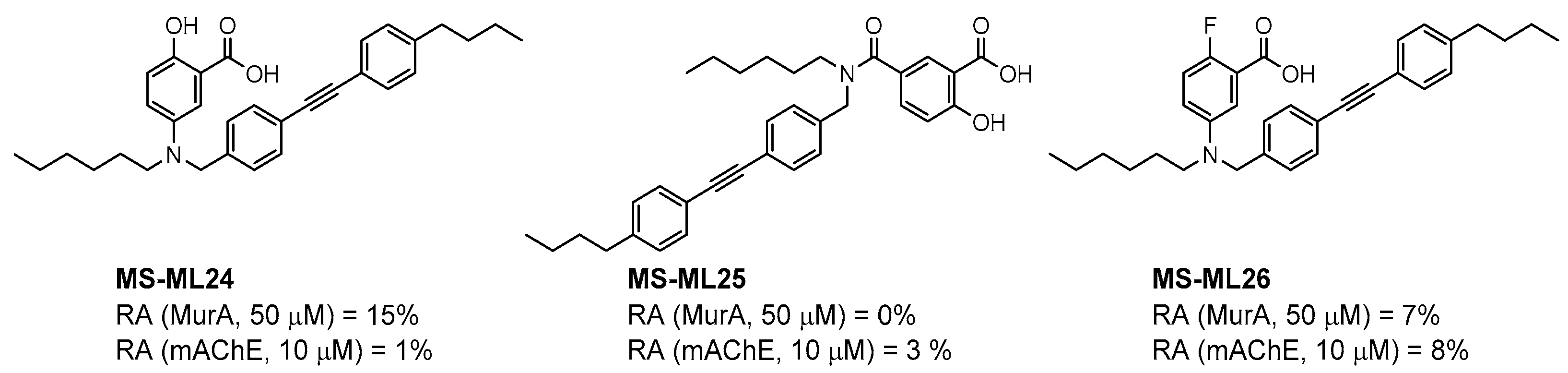
| MS-ML24 | 0 | 7 ± 3 | 1 | 294 ± 25 | 9 | 949 ± 46 |
| MS-ML25 | 0 | 6 ± 2 | 3 | 485 ± 79 | 55 | / |
| MS-ML26 | 0 | 7 ± 4 | 8 | 275 ± 60 | 57 | / |
| 1 | 0 | No clear dose-dependency | 2 | 1200 ± 50 | 100 | / |
| 2 | 64 | / | 99 | / | 100 | / |
| 3 | 58 | / | 100 | / | 100 | / |
| 4 | 14 | 102 ± 15 | 100 | / | 100 | / |
| 5 | 0 | 7 ± 3 | 2 | 1100 ± 100 | 16 | 2400 ± 200 |
| 6 | 0 | 27 ± 7 | 13 | 4800 ± 1300 | 95 | / |
| 7 | 100 | / | 19 | 682 ± 86 | 45 | 19,700 ± 10,000 c |
| 8 | 0 | 14 ± 5 | 22 | 368 ± 52 | 71 | / |
| 9 | 10 | 91 ± 13 | 52 | 1800 ± 450 | 80 | / |
| Fosfomycin | 0 | 0.24 ± 0.02 | / | / | / | / |
| Tacrine | 0 | 106 ± 10 | 0 | 12 ± 3 | ||
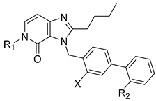
| Compound | R1 | R2 | X | MurC | |
|---|---|---|---|---|---|
| RA a (%) | IC50 (µM) | ||||
| MS-ML03 |  |  | H | 40 | 85 ± 15 |
| MS-ML05 |  |  | F | 32 | 101 ± 23 |
| 10 |  |  | H | 30 | 92 ± 11 |
| 11 |  |  | H | 20 | 77 ± 12 |
| 12 |  |  | H | 27 | 84 ±15 |
| 13 |  |  | H | 34 | 109 ± 26 |
| 14 |  |  | H | 26 | 90 ± 13 |
| 15 |  |  | F | 59 | / |
| 16 |  |  | F | 53 | / |
| 17 |  |  | H | 75 | / |
| 18 |  | −CN | H | 58 | / |
| 19 | 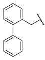 |  | H | 15 | 75 ± 9 |
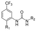
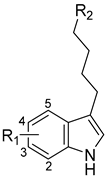
| Compound | R1 | R2 | hMAO-B | hMAO-A | |
|---|---|---|---|---|---|
| RA a (%) | IC50 (nM) | RA a (%) | |||
| MS-ML80 |  | 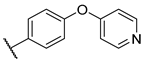 | 10 | 405 ± 76 | 78 |
| 20 |  | 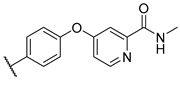 | 99 | / | 60 |
| 21 |  |  | 88 | / | 92 |
| 22 |  |  | 6 | 710 ± 100 | 111 |
| 23 |  |  | 24 | 3000 ± 800 | 100 |
| 24 | –F |  | 100 | / | 100 |
| 25 |  |  | 86 | / | 79 |
| 26 |  | 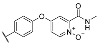 | 98 | / | 89 |
| 27 |  |  | 6 | 465 ± 42 | 117 |
| 28 |  |  | 15 | 2600 ± 100 | 94 |
| 29 |  | 89 | / | 86 | |
| Triazaindolizine | |||||
| MS-ML31 | 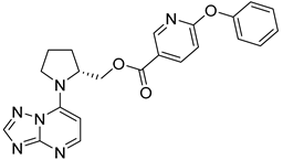 | 17 | 912 ± 122 | 98 | |
| Safinamide | 0 | 29 ± 2 | / | ||
| Harmaline | / | / | 0 b | ||
| Compound | R1 * | R2 | hBChE | mAChE | ||
|---|---|---|---|---|---|---|
| RA a (%) | IC50 (nM) | RA a (%) | IC50 (µM) | |||
| MS-ML10 | –H | 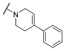 | 0 | 35.6 ± 0.3 | 64 | / |
| MS-ML11 | –OH (5) | 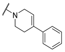 | 0 | 78.8 ± 10.3 | 82 | / |
| 30 | –H |  | 6 | 400 ± 57 | 76 | / |
| 31 |  (5) |  | 0 | 59.9 ± 5.5 | 44 | 14 ± 4 |
| 32 |  (4) |  | 49 | 14500 ± 2200 | 71 | / |
| 33 | –OH (6) |  | 3 | 267 ± 1 | 61 | / |
| 34 | –H |  | 7 | 493 ± 88 | 31 | 7 ± 1 |
| 35 | –CN (5) |  | 31 | 2900 ± 100 | 59 | / |
| 36 |  (5) |  | 97 | / | 78 | / |
| 37 |  (5) |  | 23 | 2300 ± 100 | 88 | / |
| 38 |  (5) |  | 4 | 319 ± 51 | 100 | / |
| 39 | –F (5) |  | 0 | 70 ± 5 | 47% | 15 ± 5 |
Publisher’s Note: MDPI stays neutral with regard to jurisdictional claims in published maps and institutional affiliations. |
© 2022 by the authors. Licensee MDPI, Basel, Switzerland. This article is an open access article distributed under the terms and conditions of the Creative Commons Attribution (CC BY) license (https://creativecommons.org/licenses/by/4.0/).
Share and Cite
Knez, D.; Gobec, S.; Hrast, M. Screening of Big Pharma’s Library against Various in-house Biological Targets. Molecules 2022, 27, 4484. https://doi.org/10.3390/molecules27144484
Knez D, Gobec S, Hrast M. Screening of Big Pharma’s Library against Various in-house Biological Targets. Molecules. 2022; 27(14):4484. https://doi.org/10.3390/molecules27144484
Chicago/Turabian StyleKnez, Damijan, Stanislav Gobec, and Martina Hrast. 2022. "Screening of Big Pharma’s Library against Various in-house Biological Targets" Molecules 27, no. 14: 4484. https://doi.org/10.3390/molecules27144484
APA StyleKnez, D., Gobec, S., & Hrast, M. (2022). Screening of Big Pharma’s Library against Various in-house Biological Targets. Molecules, 27(14), 4484. https://doi.org/10.3390/molecules27144484