
Design and Development of D‒α‒Tocopheryl Polyethylene Glycol Succinate‒block‒Poly(ε-Caprolactone) (TPGS−b−PCL) Nanocarriers for Solubilization and Controlled Release of Paclitaxel

 ,
,  ,
,  ,
,
Abstract
1. Introduction
2. Materials and Methods
2.1. Materials
2.2. Methods
2.2.1. Synthesis of TPGS-b-PCL Copolymers
2.2.2. Characterization of TPGS-b-PCL Copolymers
Molecular Weight and Polydispersity of TPGS-b-PCL Copolymers
Fourier Transform Infrared (FTIR) Spectroscopy
X-ray Diffraction (XRD)
Differential Scanning Calorimetry (DSC)
2.2.3. Preparation of Drug-Free and PAX-Loaded TPGS-b-PCL Micelles
2.2.4. Characterization of TPGS-b-PCL Micelles
Size, Polydispersity, and CMC
Morphology
Encapsulation Efficiency and Drug Loading
2.2.5. In Vitro Drug Release
2.2.6. Data Analysis
3. Results and Discussion
3.1. Synthesis and Characterization of TPGS-b-PCL Copolymers
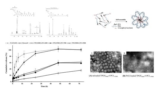
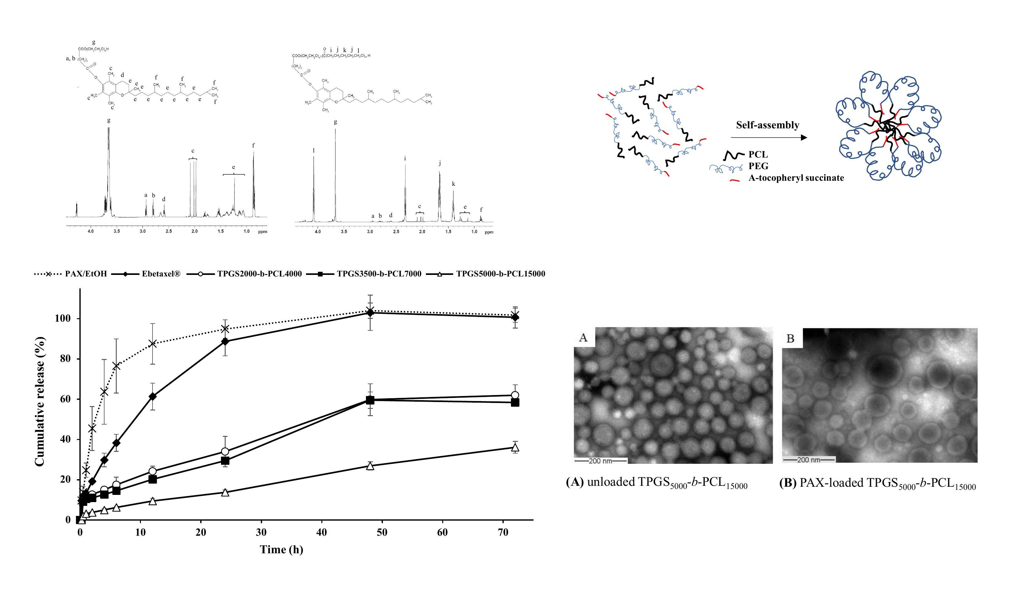
3.1.1. 1H NMR
3.1.2. GPC
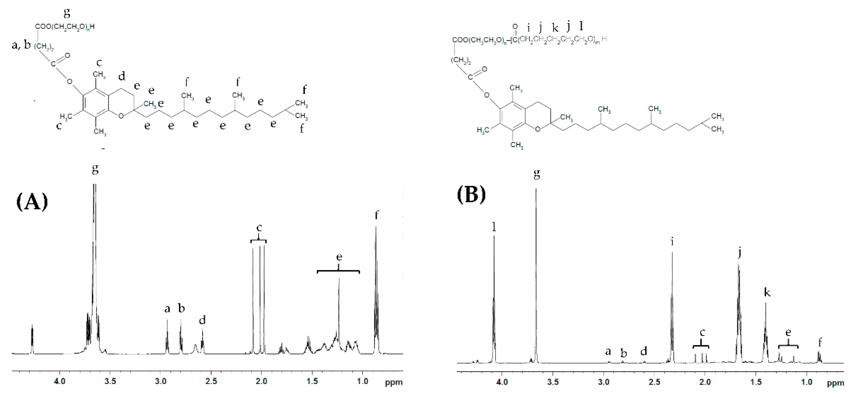
3.1.3. FTIR
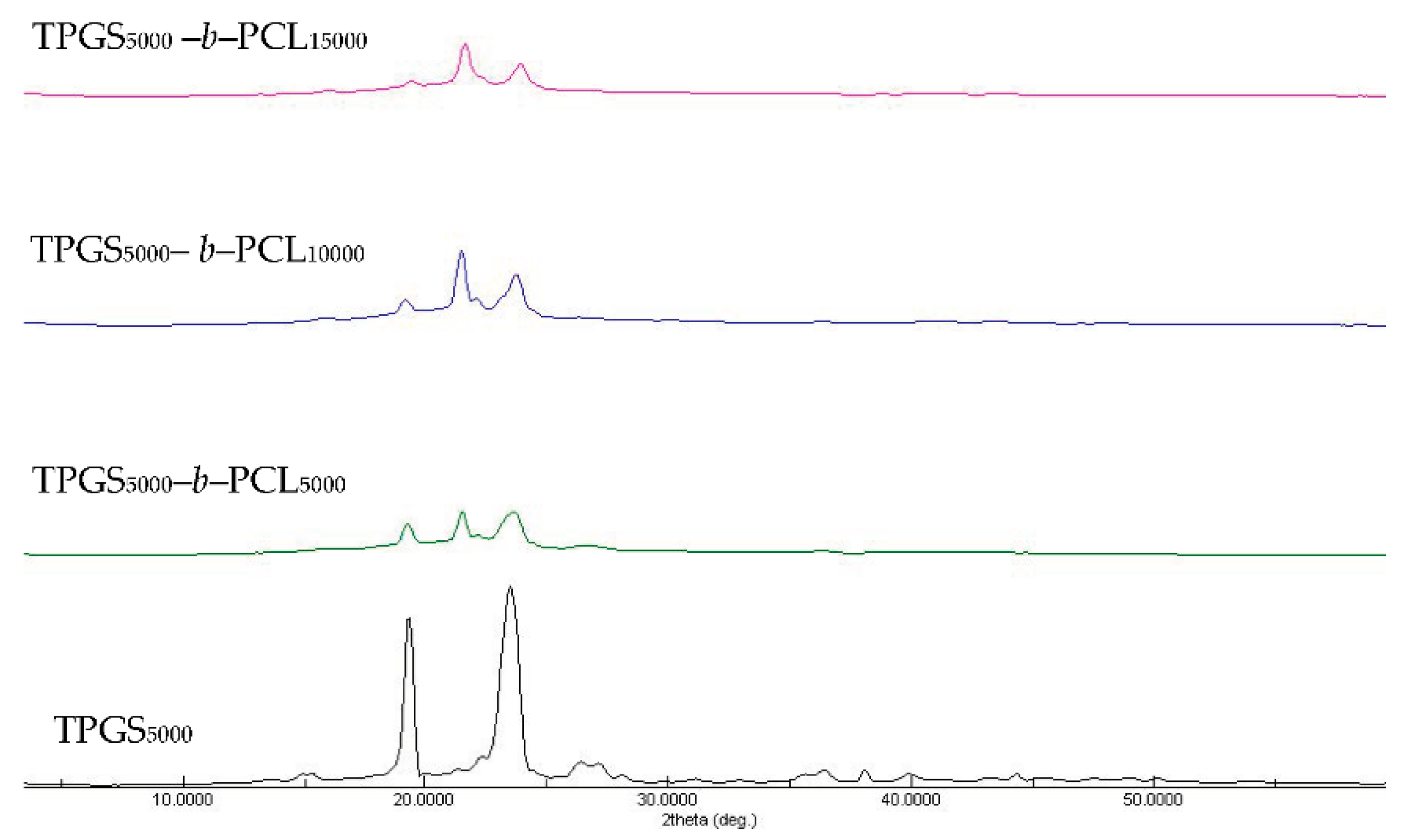
3.1.4. XRD
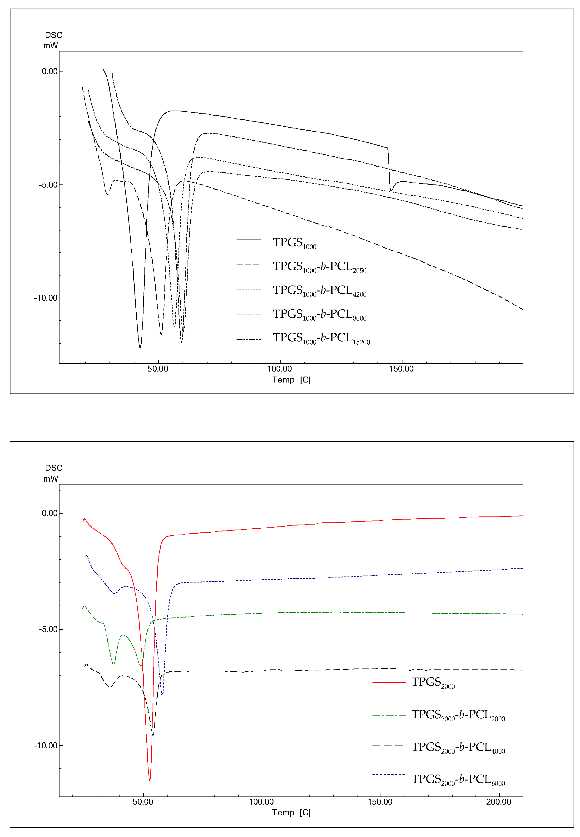
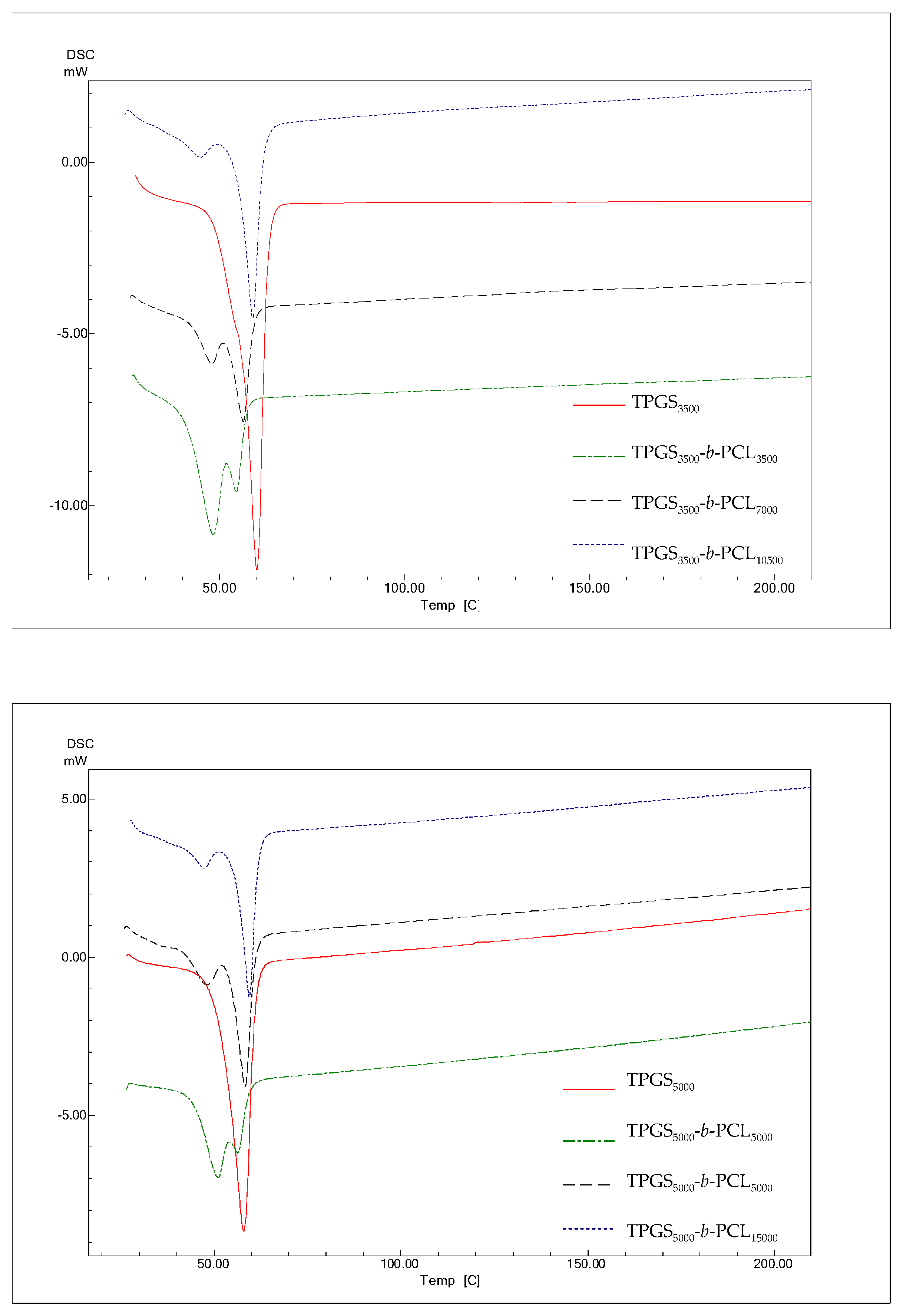
3.1.5. DSC
3.2. Preparation and Characterization of Drug-Free and PAX-Loaded TPGS-PCL Nanocarriers
3.2.1. Size, Polydispersity, and CMC
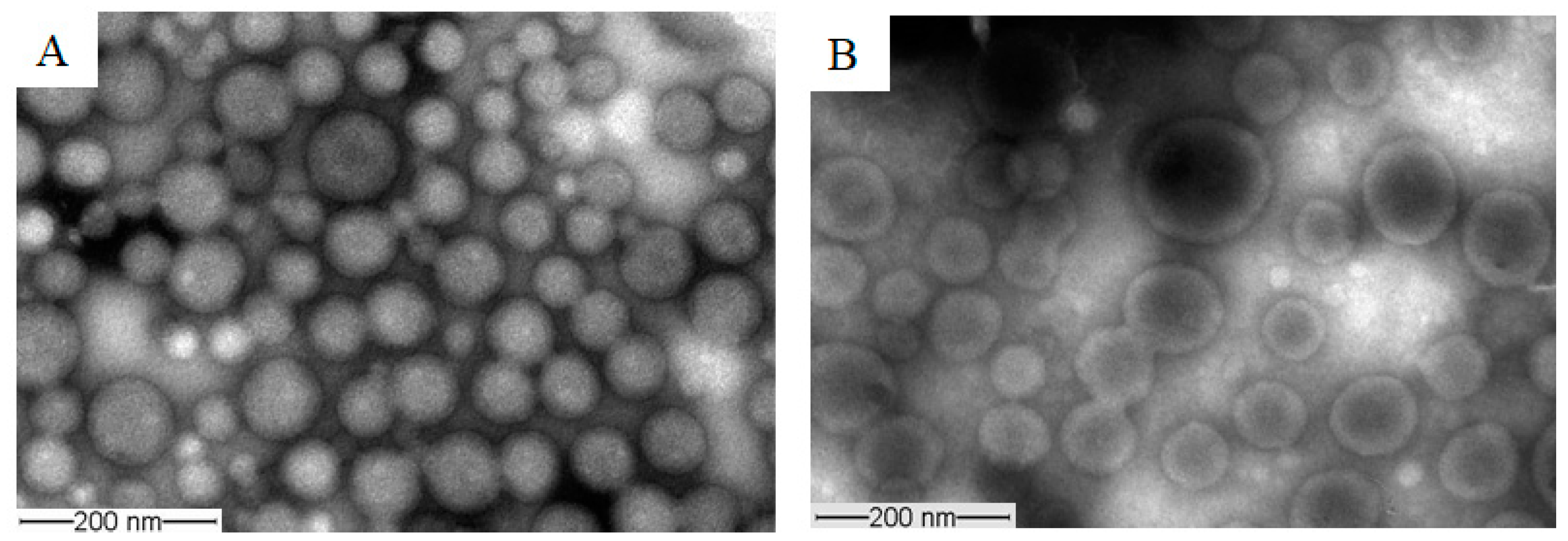
3.2.2. Morphology
3.2.3. Encapsulation Efficiency and Drug Loading
3.3. In Vitro Release of PAX from the Nanocarriers
4. Conclusions
Supplementary Materials
Author Contributions
Funding
Institutional Review Board Statement
Informed Consent Statement
Data Availability Statement
Conflicts of Interest
References
- Zhang, Z.; Tan, S.; Feng, S.S. Vitamin E TPGS as a molecular biomaterial for drug delivery. Biomaterials 2012, 33, 4889–4906. [Google Scholar] [CrossRef]
- Collnot, E.M.; Baldes, C.; Wempe, M.F.; Hyatt, J.; Navarro, L.; Edgar, K.J.; Schaefer, U.F.; Lehr, C.M. Influence of vitamin E TPGS poly(ethylene glycol) chain length on apical efflux transporters in Caco-2 cell monolayers. J. Control. Release 2006, 111, 35–40. [Google Scholar] [CrossRef]
- Collnot, E.M.; Baldes, C.; Schaefer, U.F.; Edgar, K.J.; Wempe, M.F.; Lehr, C.M. Vitamin E TPGS P-glycoprotein inhibition mechanism: Influence on conformational flexibility, intracellular ATP levels, and role of time and site of access. Mol. Pharm. 2010, 7, 642–651. [Google Scholar] [CrossRef]
- Duhem, N.; Danhier, F.; Préat, V. Vitamin E-based nanomedicines for anti-cancer drug delivery. J. Control. Release 2014, 182, 33–44. [Google Scholar] [CrossRef]
- Muthu, M.S.; Kulkarni, S.A.; Liu, Y.; Feng, S.-S. Development of docetaxel-loaded vitamin E TPGS micelles: Formulation optimization, effects on brain cancer cells and biodistribution in rats. Nanomedicine 2012, 7, 353–364. [Google Scholar] [CrossRef]
- Woodruff, M.A.; Hutmacher, D.W. The return of a forgotten polymer—Polycaprolactone in the 21st century. Prog. Polym. Sci. 2010, 35, 1217–1256. [Google Scholar] [CrossRef]
- Dash, T.K.; Konkimalla, V.B. Poly-epsilon-caprolactone based formulations for drug delivery and tissue engineering: A review. J. Control. Release 2012, 158, 15–33. [Google Scholar] [CrossRef] [PubMed]
- Sinha, V.R.; Bansal, K.; Kaushik, R.; Kumria, R.; Trehan, A. Poly-epsilon-caprolactone microspheres and nanospheres: An overview. Int. J. Pharm. 2004, 278, 1–23. [Google Scholar] [CrossRef] [PubMed]
- Glavas, L.; Olsén, P.; Odelius, K.; Albertsson, A.-C. Achieving Micelle Control through Core Crystallinity. Biomacromolecules 2013, 14, 4150–4156. [Google Scholar] [CrossRef]
- Aliabadi, H.M.; Mahmud, A.; Sharifabadi, A.D.; Lavasanifar, A. Micelles of methoxy poly(ethylene oxide)-b-poly(epsilon-caprolactone) as vehicles for the solubilization and controlled delivery of cyclosporine A. J. Control. Release 2005, 104, 301–311. [Google Scholar] [CrossRef]
- Binkhathlan, Z.; Hamdy, D.A.; Brocks, D.R.; Lavasanifar, A. Development of a polymeric micellar formulation for valspodar and assessment of its pharmacokinetics in rat. Eur. J. Pharm. Biopharm. 2010, 75, 90–95. [Google Scholar] [CrossRef]
- Carrillo-Castillo, T.D.; Castro-Carmona, J.S.; Luna-Velasco, A.; Zaragoza-Contreras, E.A. pH-responsive polymer micelles for methotrexate delivery at tumor microenvironments. e-Polymers 2020, 20, 624–635. [Google Scholar] [CrossRef]
- Sze, L.P.; Li, H.Y.; Lai, K.L.A.; Chow, S.F.; Li, Q.; KennethTo, K.W.; Lam, T.N.T.; Lee, W.Y.T. Oral delivery of paclitaxel by polymeric micelles: A comparison of different block length on uptake, permeability and oral bioavailability. Colloids Surf. B Biointerfaces 2019, 184, 110554. [Google Scholar] [CrossRef]
- Cai, S.; Vijayan, K.; Cheng, D.; Lima, E.M.; Discher, D.E. Micelles of Different Morphologies—Advantages of Worm-like Filomicelles of PEO-PCL in Paclitaxel Delivery. Pharm. Res. 2007, 24, 2099–2109. [Google Scholar] [CrossRef] [PubMed]
- Zhang, Z.; Mei, L.; Feng, S.S. Paclitaxel drug delivery systems. Expert Opin. Drug Deliv. 2013, 10, 325–340. [Google Scholar] [CrossRef]
- Sarisozen, C.; Vural, I.; Levchenko, T.; Hincal, A.A.; Torchilin, V.P. PEG-PE-based micelles co-loaded with paclitaxel and cyclosporine A or loaded with paclitaxel and targeted by anticancer antibody overcome drug resistance in cancer cells. Drug Deliv. 2012, 19, 169–176. [Google Scholar] [CrossRef]
- Dorr, R.T. Pharmacology and toxicology of Cremophor EL diluent. Ann. Pharmacother. 1994, 28, S11–S14. [Google Scholar] [CrossRef]
- Gelderblom, H.; Verweij, J.; Nooter, K.; Sparreboom, A. Cremophor EL: The drawbacks and advantages of vehicle selection for drug formulation. Eur. J. Cancer 2001, 37, 1590–1598. [Google Scholar] [CrossRef]
- Weiss, R.B.; Donehower, R.C.; Wiernik, P.H.; Ohnuma, T.; Gralla, R.J.; Trump, D.L.; Baker, J.R., Jr.; Van Echo, D.A.; Von Hoff, D.D.; Leyland-Jones, B. Hypersensitivity reactions from taxol. J. Clin. Oncol. 1990, 8, 1263–1268. [Google Scholar] [CrossRef] [PubMed]
- Sofias, A.M.; Dunne, M.; Storm, G.; Allen, C. The battle of “nano” paclitaxel. Adv. Drug Deliv. Rev. 2017, 122, 20–30. [Google Scholar] [CrossRef] [PubMed]
- Binkhathlan, Z.; Lavasanifar, A. Chapter 15—Effects of block copolymer micelles on the pharmacokinetics of encapsulated drugs. In Nanoarchitectonics in Biomedicine; Grumezescu, A.M., Ed.; William Andrew Publishing: Norwich, NY, USA, 2019; pp. 507–546. [Google Scholar]
- Bernabeu, E.; Helguera, G.; Legaspi, M.J.; Gonzalez, L.; Hocht, C.; Taira, C.; Chiappetta, D.A. Paclitaxel-loaded PCL-TPGS nanoparticles: In vitro and in vivo performance compared with Abraxane(R). Colloids Surf. B Biointerfaces 2014, 113, 43–50. [Google Scholar] [CrossRef]
- Bernabeu, E.; Gonzalez, L.; Legaspi, M.J.; Moretton, M.A.; Chiappetta, D.A. Paclitaxel-Loaded TPGS-b-PCL Nanoparticles: In Vitro Cytotoxicity and Cellular Uptake in MCF-7 and MDA-MB-231 Cells versus mPEG-b-PCL Nanoparticles and Abraxane(R). J. Nanosci. Nanotechnol. 2016, 16, 160–170. [Google Scholar] [CrossRef] [PubMed]
- Gill, K.K.; Kaddoumi, A.; Nazzal, S. Mixed micelles of PEG(2000)-DSPE and vitamin-E TPGS for concurrent delivery of paclitaxel and parthenolide: Enhanced chemosenstization and antitumor efficacy against non-small cell lung cancer (NSCLC) cell lines. Eur. J. Pharm. Sci. 2012, 46, 64–71. [Google Scholar] [CrossRef] [PubMed]
- Zhang, Z.; Lee, S.H.; Gan, C.W.; Feng, S.S. In vitro and in vivo investigation on PLA-TPGS nanoparticles for controlled and sustained small molecule chemotherapy. Pharm. Res. 2008, 25, 1925–1935. [Google Scholar] [CrossRef] [PubMed]
- Binkhathlan, Z.; Elhasi, S.; Brocks, D.R.; Lavasanifar, A. Characterization of the self assembly of methoxy poly(ethylene oxide)-block-poly(alpha-benzyl carboxylate-epsilon-caprolactone) for the solubilization and in vivo delivery of valspodar. Curr. Drug Deliv. 2012, 9, 164–171. [Google Scholar] [CrossRef]
- Zhang, Z.; Feng, S.S. Nanoparticles of poly(lactide)/vitamin E TPGS copolymer for cancer chemotherapy: Synthesis, formulation, characterization and in vitro drug release. Biomaterials 2006, 27, 262–270. [Google Scholar] [CrossRef]
- Topel, Ö.; Çakır, B.A.; Budama, L.; Hoda, N. Determination of critical micelle concentration of polybutadiene-block-poly(ethyleneoxide) diblock copolymer by fluorescence spectroscopy and dynamic light scattering. J. Mol. Liq. 2013, 177, 40–43. [Google Scholar] [CrossRef]
- Bilensoy, E.; Gurkaynak, O.; Ertan, M.; Sen, M.; Hincal, A.A. Development of nonsurfactant cyclodextrin nanoparticles loaded with anticancer drug paclitaxel. J. Pharm. Sci. 2008, 97, 1519–1529. [Google Scholar] [CrossRef]
- Chu, B.; Qu, Y.; Huang, Y.; Zhang, L.; Chen, X.; Long, C.; He, Y.; Ou, C.; Qian, Z. PEG-derivatized octacosanol as micellar carrier for paclitaxel delivery. Int. J. Pharm. 2016, 500, 345–359. [Google Scholar] [CrossRef]
- Wei, Y.; Xu, S.; Wang, F.; Zou, A.; Zhang, S.; Xiong, Y.; Cao, S.; Zhang, Q.; Wang, Y.; Jiang, X. A Novel Combined Micellar System of Lapatinib and Paclitaxel with Enhanced Antineoplastic Effect Against Human Epidermal Growth Factor Receptor-2 Positive Breast Tumor In Vitro. J. Pharm. Sci. 2015, 104, 165–177. [Google Scholar] [CrossRef]
- Wang, Y.; Wang, C.; Gong, C.; Wang, Y.; Guo, G.; Luo, F.; Qian, Z. Polysorbate 80 coated poly (ɛ-caprolactone)–poly (ethylene glycol)–poly (ɛ-caprolactone) micelles for paclitaxel delivery. Int. J. Pharm. 2012, 434, 1–8. [Google Scholar] [CrossRef]
- Moore, J.W. Mathematical Comparison of Dissolution Profiles. Pharm. Technol. 1996, 20, 64–75. [Google Scholar]
- Costa, P.; Sousa Lobo, J.M. Modeling and comparison of dissolution profiles. Eur. J. Pharm. Sci. 2001, 13, 123–133. [Google Scholar] [CrossRef]
- Suksiriworapong, J.; Phoca, K.; Ngamsom, S.; Sripha, K.; Moongkarndi, P.; Junyaprasert, V.B. Comparison of poly(ε-caprolactone) chain lengths of poly(ε-caprolactone)-co-d-α-tocopheryl-poly(ethylene glycol) 1000 succinate nanoparticles for enhancement of quercetin delivery to SKBR3 breast cancer cells. Eur. J. Pharm. Biopharm. 2016, 101, 15–24. [Google Scholar] [CrossRef] [PubMed]
- Zhang, H.; Liu, G.; Zeng, X.; Wu, Y.; Yang, C.; Mei, L.; Wang, Z.; Huang, L. Fabrication of genistein-loaded biodegradable TPGS-b-PCL nanoparticles for improved therapeutic effects in cervical cancer cells. Int. J. Nanomed. 2015, 10, 2461–2473. [Google Scholar] [CrossRef]
- Sun, J.; Chen, X.; He, C.; Jing, X. Morphology and Structure of Single Crystals of Poly(ethylene glycol)−Poly(ε-caprolactone) Diblock Copolymers. Macromolecules 2006, 39, 3717–3719. [Google Scholar] [CrossRef]
- Shin, I.G.; Kim, S.Y.; Lee, Y.M.; Cho, C.S.; Sung, Y.K. Methoxy poly(ethylene glycol)/epsilon-caprolactone amphiphilic block copolymeric micelle containing indomethacin. I. Preparation and characterization. J. Control. Release 1998, 51, 1–11. [Google Scholar] [CrossRef]
- Koulouktsi, C.; Nanaki, S.; Barmpalexis, P.; Kostoglou, M.; Bikiaris, D. Preparation and characterization of Alendronate depot microspheres based on novel poly(-ε-caprolactone)/Vitamin E TPGS copolymers. Int. J. Pharm. X 2019, 1, 100014. [Google Scholar] [CrossRef]
- Yin, G.; Chen, G.; Zhou, Z.; Li, Q. Modification of PEG-b-PCL block copolymer with high melting temperature by the enhancement of POSS crystal and ordered phase structure. RSC Adv. 2015, 5, 33356–33363. [Google Scholar] [CrossRef]
- Vaishya, R.D.; Gokulgandhi, M.; Patel, S.; Minocha, M.; Mitra, A.K. Novel dexamethasone-loaded nanomicelles for the intermediate and posterior segment uveitis. AAPS PharmSciTech 2014, 15, 1238–1251. [Google Scholar] [CrossRef]
- Sun, J.; He, C.; Zhuang, X.; Jing, X.; Chen, X. The crystallization behavior of poly(ethylene glycol)-poly(ε-caprolactone) diblock copolymers with asymmetric block compositions. J. Polym. Res. 2011, 18, 2161–2168. [Google Scholar] [CrossRef]
- Gajra, B.; Dalwadi, C.; Patel, R. Formulation and optimization of itraconazole polymeric lipid hybrid nanoparticles (Lipomer) using box behnken design. DARU J. Pharm. Sci. 2015, 23, 3. [Google Scholar] [CrossRef] [PubMed]
- Dezhu, M.; Xiaolie, L.; Ruiyun, Z.; Nishi, T. Miscibility and spherulites in blends of poly(ϵ-caprolactone) with ethylene terephthalate-caprolactone copolyester. Polymer 1996, 37, 1575–1581. [Google Scholar] [CrossRef]
- Majumdar, R.; Alexander, K.S.; Riga, A.T. Physical characterization of polyethylene glycols by thermal analytical technique and the effect of humidity and molecular weight. Die Pharm. Int. J. Pharm. Sci. 2010, 65, 343–347. [Google Scholar] [CrossRef]
- Pielichowski, K.; Flejtuch, K. Differential scanning calorimetry studies on poly(ethylene glycol) with different molecular weights for thermal energy storage materials. Polym. Adv. Technol. 2002, 13, 690–696. [Google Scholar] [CrossRef]
- Nanaki, S.G.; Pantopoulos, K.; Bikiaris, D.N. Synthesis of biocompatible poly(varepsilon-caprolactone)- block-poly(propylene adipate) copolymers appropriate for drug nanoencapsulation in the form of core-shell nanoparticles. Int. J. Nanomed. 2011, 6, 2981–2995. [Google Scholar] [CrossRef][Green Version]
- Bogdanov, B.; Vidts, A.; Van Den Buicke, A.; Verbeeck, R.; Schacht, E. Synthesis and thermal properties of poly(ethylene glycol)-poly(ϵ-caprolactone) copolymers. Polymer 1998, 39, 1631–1636. [Google Scholar] [CrossRef]
- He, C.; Sun, J.; Ma, J.; Chen, X.; Jing, X. Composition Dependence of the Crystallization Behavior and Morphology of the Poly(ethylene oxide)-poly(ε-caprolactone) Diblock Copolymer. Biomacromolecules 2006, 7, 3482–3489. [Google Scholar] [CrossRef]
- Li, L.; Meng, F.; Zhong, Z.; Byelov, D.; Jeu, W.H.d.; Feijen, J. Morphology of a highly asymmetric double crystallizable poly(ε-caprolactone-b-ethylene oxide) block copolymer. J. Chem. Phys. 2007, 126, 024904. [Google Scholar] [CrossRef]
- Sharma, R.; Murali, R.; Murthy, C.N. Clouding and Aggregation Behavior of PPO-PEO-PPO Triblock Copolymer (Pluronic®25R4) in Surfactant Additives Environment. Tenside Surfactants Deterg. 2012, 49, 136–144. [Google Scholar] [CrossRef]
- Mahmud, A.; Xiong, X.-B.; Lavasanifar, A. Novel Self-Associating Poly(ethylene oxide)-block-poly(ε-caprolactone) Block Copolymers with Functional Side Groups on the Polyester Block for Drug Delivery. Macromolecules 2006, 39, 9419–9428. [Google Scholar] [CrossRef]
- Atanase, L.I.; Winninger, J.; Delaite, C.; Riess, G. Micellization and demicellization of amphiphilic poly(vinyl acetate)-graft-poly(N-vinyl-pyrrolidone) graft copolymers in the presence of sodium dodecyl sulfate. Colloids Surf. A Physicochem. Eng. Asp. 2014, 461, 287–294. [Google Scholar] [CrossRef]
- Fairley, N.; Hoang, B.; Allen, C. Morphological Control of Poly(ethylene glycol)-block-poly(ε-caprolactone) Copolymer Aggregates in Aqueous Solution. Biomacromolecules 2008, 9, 2283–2291. [Google Scholar] [CrossRef] [PubMed]
- Qi, W.; Ghoroghchian, P.P.; Li, G.; Hammer, D.A.; Therien, M.J. Aqueous self-assembly of poly(ethylene oxide)-block-poly(ε-caprolactone) (PEO-b-PCL) copolymers: Disparate diblock copolymer compositions give rise to nano- and meso-scale bilayered vesicles. Nanoscale 2013, 5, 10908–10915. [Google Scholar] [CrossRef] [PubMed]
- Ghezzi, M.; Pescina, S.; Padula, C.; Santi, P.; Del Favero, E.; Cantù, L.; Nicoli, S. Polymeric micelles in drug delivery: An insight of the techniques for their characterization and assessment in biorelevant conditions. J. Control. Release 2021, 332, 312–336. [Google Scholar] [CrossRef]
- Aliabadi, H.M.; Elhasi, S.; Mahmud, A.; Gulamhusein, R.; Mahdipoor, P.; Lavasanifar, A. Encapsulation of hydrophobic drugs in polymeric micelles through co-solvent evaporation: The effect of solvent composition on micellar properties and drug loading. Int. J. Pharm. 2007, 329, 158–165. [Google Scholar] [CrossRef]
- Ali, R.; Farah, A.; Binkhathlan, Z. Development and characterization of methoxy poly(ethylene oxide)-block-poly(ε-caprolactone) (PEO-b-PCL) micelles as vehicles for the solubilization and delivery of tacrolimus. Saudi Pharm. J. 2017, 25, 258–265. [Google Scholar] [CrossRef]
- Soleymani Abyaneh, H.; Vakili, M.R.; Zhang, F.; Choi, P.; Lavasanifar, A. Rational design of block copolymer micelles to control burst drug release at a nanoscale dimension. Acta Biomater. 2015, 24, 127–139. [Google Scholar] [CrossRef]
- Shahin, M.; Lavasanifar, A. Novel self-associating poly(ethylene oxide)-b-poly(epsilon-caprolactone) based drug conjugates and nano-containers for paclitaxel delivery. Int. J. Pharm. 2010, 389, 213–222. [Google Scholar] [CrossRef] [PubMed]
- Aliabadi, H.M.; Lavasanifar, A. Polymeric micelles for drug delivery. Expert Opin. Drug Deliv. 2006, 3, 139–162. [Google Scholar] [CrossRef] [PubMed]
- Simonutti, R.; Bertani, D.; Marotta, R.; Ferrario, S.; Manzone, D.; Mauri, M.; Gregori, M.; Orlando, A.; Masserini, M. Morphogenic effect of common solvent in the self-assembly behavior of amphiphilic PEO-b-PLA. Polymer 2021, 218, 123511. [Google Scholar] [CrossRef]
- Binkhathlan, Z.; Ali, R.; Qamar, W.; Al-Lawati, H.; Lavasanifar, A. Pharmacokinetic and Tissue Distribution of Orally Administered Cyclosporine A-Loaded poly(ethylene oxide)-block-Poly(ε-caprolactone) Micelles versus Sandimmune® in Rats. Pharm. Res. 2021, 38, 51–65. [Google Scholar] [CrossRef] [PubMed]


| Block Copolymer a | Theoretical Molecular Weight (g/mol) | Mn (g/mol) b | Mn (g/mol) c | Đ d | Diameter e (nm) | Polydispersity e | CAC f (µM) |
|---|---|---|---|---|---|---|---|
| TPGS1000 | 1513 | - | - | - | - | - | 132.2 g |
| TPGS1000-b-PCL2050 § | 3560 | 3560 | 5330 | 1.06 | - | - | - |
| TPGS1000-b-PCL4200 | 5500 | 5700 | 9000 | 1.29 | - | - | - |
| TPGS1000-b-PCL8000 | 9500 | 9500 | 15,200 | 1.27 | - | - | - |
| TPGS1000-b-PCL15200 | 17,500 | 16,700 | 23,100 | 1.17 | - | - | - |
| TPGS1000-b-PCL20800 | 21,500 | 22,300 | 35,100 | 1.10 | - | - | - |
| TPGS1000-b-PCL25100 | 26,600 | 26,600 | 38,950 | 1.11 | - | - | - |
| TPGS1000-b-PCL30400 | 31,500 | 31,900 | 43,250 | 1.14 | - | - | - |
| TPGS2000 | 2513 | - | - | - | - | - | 186.2 ± 16.2 |
| TPGS2000-b-PCL2000 | 4500 | 4550 | 5950 | 1.25 | 70.5 ± 6.2 | 0.54 ± 0.30 | 41.00 ± 3.06 * |
| TPGS2000-b-PCL4000 | 6500 | 6400 | 8350 | 1.55 | 86.5 ± 3.9 | 0.45 ± 0.04 | 25.16 ± 3.10 * |
| TPGS2000-b-PCL6000 | 8500 | 8800 | 10,500 | 1.71 | 170.1 ± 9.4 | 0.21 ± 0.02 | 18.06 ± 0.20 *# |
| TPGS3500 | 4013 | - | - | - | - | - | 165.7 ± 15.0 |
| TPGS3500-b-PCL3500 | 7500 | 7100 | 4600 | 1.81 | 89.2 ± 4.1 | 0.35 ± 0.06 | 24.00 ± 1.81 * |
| TPGS3500-b-PCL7000 | 11,000 | 10,300 | 6400 | 1.62 | 61.3 ± 1.7 | 0.27 ± 0.02 | 15.88 ± 2.41 * |
| TPGS3500-b-PCL10500 | 14,500 | 13,900 | 10,300 | 1.62 | 81.7 ± 3.6 | 0.23 ± 0.03 | 8.38 ± 0.88 * |
| TPGS5000 | 5513 | - | - | - | - | - | 123.6 ± 6.8 |
| TPGS5000-b-PCL5000 | 10,500 | 10,300 | 4600 | 1.90 | 81.1 ± 2.8 | 0.27 ± 0.06 | 12.30 ± 0.82 * |
| TPGS5000-b-PCL10000 | 15,500 | 15,600 | 8100 | 1.53 | 75.0 ± 4.6 | 0.19 ± 0.05 | 7.29 ± 0.89 * |
| TPGS5000-b-PCL15000 | 20,500 | 20,550 | 12,100 | 1.70 | 77.0 ± 4.6 | 0.21 ± 0.03 | 5.44 ± 0.36 * |
| Block Copolymer | Drug: Polymer Ratio (w/w) | Drug Loading (% w/w) a | Encapsulation Efficiency (%) a | Diameter (nm) b | Polydispersity b |
|---|---|---|---|---|---|
| TPGS2000-b-PCL2000 | 1:10 | 0.66 ± 0.01 | 6.70 ± 0.13 | 195.1 ± 31.8 * | 0.52 ± 0.18 |
| 1:20 | 0.35 ± 0.01 | 7.10 ± 0.08 | 131.1 ± 69.7 | 0.46 ± 0.13 | |
| 1:30 | 0.32 ± 0.02 | 8.04 ± 0.39 | 155.1 ± 31.3 | 0.53 ± 0.10 | |
| TPGS2000-b-PCL4000 | 1:10 | 0.85 ± 0.01 | 8.60 ± 0.08 | 93.5 ± 11.4 | 0.50 ± 0.13 |
| 1:20 | 0.60 ± 0.02 | 11.98 ± 0.44 | 77.2 ± 11.4 | 0.39 ± 0.06 | |
| 1:30 | 0.57 ± 0.01 | 17.24 ± 0.14 | 77.6 ± 6.5 | 0.40 ± 0.06 | |
| TPGS2000-b-PCL6000 | 1:10 | 0.75 ± 0.05 | 7.54 ± 0.54 | 209.4 ± 5.1 * | 0.25 ± 0.03 |
| 1:20 | 0.52 ± 0.02 | 10.54 ± 0.45 | 242.7 ± 7.5 | 0.43 ± 0.10 | |
| 1:30 | 0.27 ± 0.01 | 8.04 ± 0.39 | 199.2 ± 45.3 | 0.46 ± 0.10 | |
| TPGS3500-b-PCL3500 | 1:10 | 1.00 ± 0.05 | 10.07 ± 0.62 | 81.9 ± 7.8 | 0.33 ± 0.03 |
| 1:20 | 0.63 ± 0.03 | 12.67 ± 0.54 | 85.1 ± 6.8 | 0.34 ± 0.11 | |
| 1:30 | 0.47 ± 0.02 | 13.24 ± 0.36 | 74.4 ± 1.3 | 0.30 ± 0.03 | |
| TPGS3500-b-PCL7000 | 1:10 | 1.05 ± 0.03 | 10.59 ± 0.26 | 74.8 ± 9.5 | 0.33 ± 0.11 |
| 1:20 | 0.63 ± 0.02 | 12.58 ± 0.47 | 60.6 ± 1.0 | 0.31 ± 0.03 | |
| 1:30 | 0.60 ± 0.02 | 17.28 ± 0.53 | 67.2 ± 2.0 | 0.21 ± 0.01 | |
| TPGS3500-b-PCL10500 | 1:10 | 0.83 ± 0.02 | 8.42 ± 0.19 | 91.4 ± 4.1 * | 0.32 ± 0.01 |
| 1:20 | 0.58 ± 0.01 | 11.68 ± 0.19 | 80.1 ± 0.7 | 0.24 ± 0.02 | |
| 1:30 | 0.57 ± 0.03 | 17.10 ± 1.00 | 94.0 ± 1.1 | 0.25 ± 0.01 | |
| TPGS5000-b-PCL5000 | 1:10 | 0.85 ± 0.05 | 8.55 ± 0.51 | 83.7 ± 3.0 | 0.31 ± 0.01 |
| 1:20 | 0.59 ± 0.04 | 11.58 ± 0.51 | 92.7 ± 1.0 | 0.21 ± 0.01 | |
| 1:30 | 0.51 ± 0.02 | 14.32 ± 0.46 | 96.1 ± 1.3 | 0.16 ± 0.03 | |
| TPGS5000-b-PCL10000 | 1:10 | 0.84 ± 0.01 | 8.45 ± 0.06 | 76.6 ± 0.6 | 0.21 ± 0.04 |
| 1:20 | 0.63 ± 0.03 | 12.76 ± 0.57 | 79.7 ± 3.8 | 0.20 ± 0.02 | |
| 1:30 | 0.52 ± 0.02 | 15.72 ± 0.60 | 79.1 ± 2.3 | 0.19 ± 0.03 | |
| TPGS5000-b-PCL15000 | 1:10 | 0.80 ± 0.02 | 8.06 ± 0.19 | 76.4 ± 2.9 | 0.22 ± 0.02 |
| 1:20 | 0.62 ± 0.02 | 12.56 ± 0.49 | 85.0 ± 0.7 | 0.16 ± 0.02 | |
| 1:30 | 0.59 ± 0.03 | 17.68 ± 0.30 | 93.2 ± 1.0 | 0.18 ± 0.05 |
| Solution/Formulation | Difference Factor (f1) | Similarity Factor (f2) |
|---|---|---|
| PAX/EtOH vs. Ebetaxel® | 23.72 | 32.75 |
| Ebetaxel® vs. TPGS2000-b-PCL4000 | 48.45 | 24.28 |
| Ebetaxel® vs. TPGS3500-b-PCL7000 | 52.62 | 22.66 |
| Ebetaxel® vs. TPGS5000-b-PCL15000 | 77.64 | 14.81 |
| TPGS2000-b-PCL4000 vs. TPGS3500-b-PCL7000 * | 8.09 | 75.75 |
| TPGS2000-b-PCL4000 vs. TPGS5000-b-PCL15000 | 56.62 | 36.38 |
| TPGS3500-b-PCL7000 vs. TPGS5000-b-PCL15000 | 52.80 | 38.93 |
Publisher’s Note: MDPI stays neutral with regard to jurisdictional claims in published maps and institutional affiliations. |
© 2021 by the authors. Licensee MDPI, Basel, Switzerland. This article is an open access article distributed under the terms and conditions of the Creative Commons Attribution (CC BY) license (https://creativecommons.org/licenses/by/4.0/).
Share and Cite
Yusuf, O.; Ali, R.; Alomrani, A.H.; Alshamsan, A.; Alshememry, A.K.; Almalik, A.M.; Lavasanifar, A.; Binkhathlan, Z. Design and Development of D‒α‒Tocopheryl Polyethylene Glycol Succinate‒block‒Poly(ε-Caprolactone) (TPGS−b−PCL) Nanocarriers for Solubilization and Controlled Release of Paclitaxel. Molecules 2021, 26, 2690. https://doi.org/10.3390/molecules26092690
Yusuf O, Ali R, Alomrani AH, Alshamsan A, Alshememry AK, Almalik AM, Lavasanifar A, Binkhathlan Z. Design and Development of D‒α‒Tocopheryl Polyethylene Glycol Succinate‒block‒Poly(ε-Caprolactone) (TPGS−b−PCL) Nanocarriers for Solubilization and Controlled Release of Paclitaxel. Molecules. 2021; 26(9):2690. https://doi.org/10.3390/molecules26092690
Chicago/Turabian StyleYusuf, Osman, Raisuddin Ali, Abdullah H. Alomrani, Aws Alshamsan, Abdullah K. Alshememry, Abdulaziz M. Almalik, Afsaneh Lavasanifar, and Ziyad Binkhathlan. 2021. "Design and Development of D‒α‒Tocopheryl Polyethylene Glycol Succinate‒block‒Poly(ε-Caprolactone) (TPGS−b−PCL) Nanocarriers for Solubilization and Controlled Release of Paclitaxel" Molecules 26, no. 9: 2690. https://doi.org/10.3390/molecules26092690
APA StyleYusuf, O., Ali, R., Alomrani, A. H., Alshamsan, A., Alshememry, A. K., Almalik, A. M., Lavasanifar, A., & Binkhathlan, Z. (2021). Design and Development of D‒α‒Tocopheryl Polyethylene Glycol Succinate‒block‒Poly(ε-Caprolactone) (TPGS−b−PCL) Nanocarriers for Solubilization and Controlled Release of Paclitaxel. Molecules, 26(9), 2690. https://doi.org/10.3390/molecules26092690