
Isoflavonoid-Antibiotic Thin Films Fabricated by MAPLE with Improved Resistance to Microbial Colonization

 , ,
, ,  ,
,  ,
,  ,
,  and
and 
Abstract
1. Introduction
2. Results and Discussions
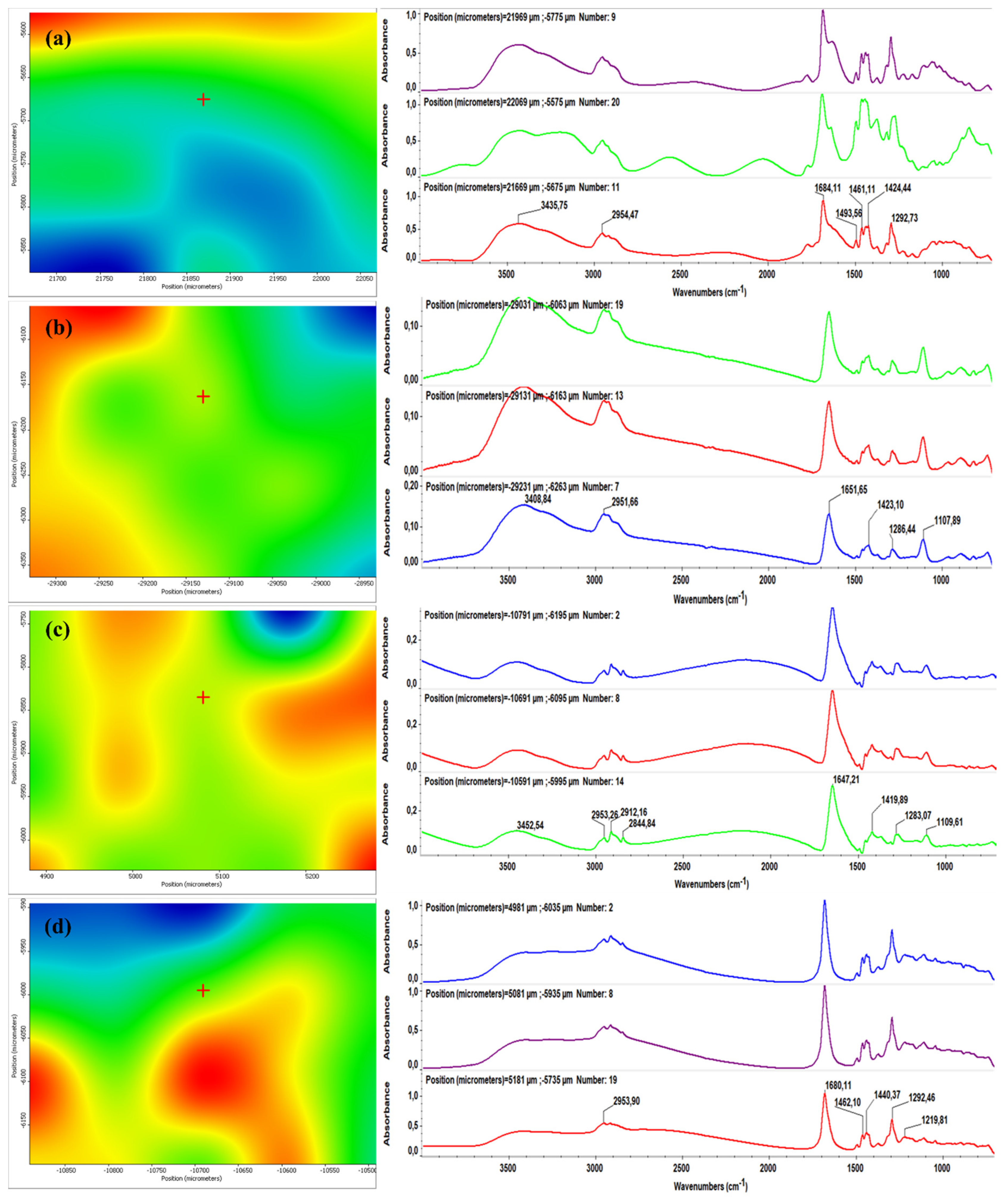
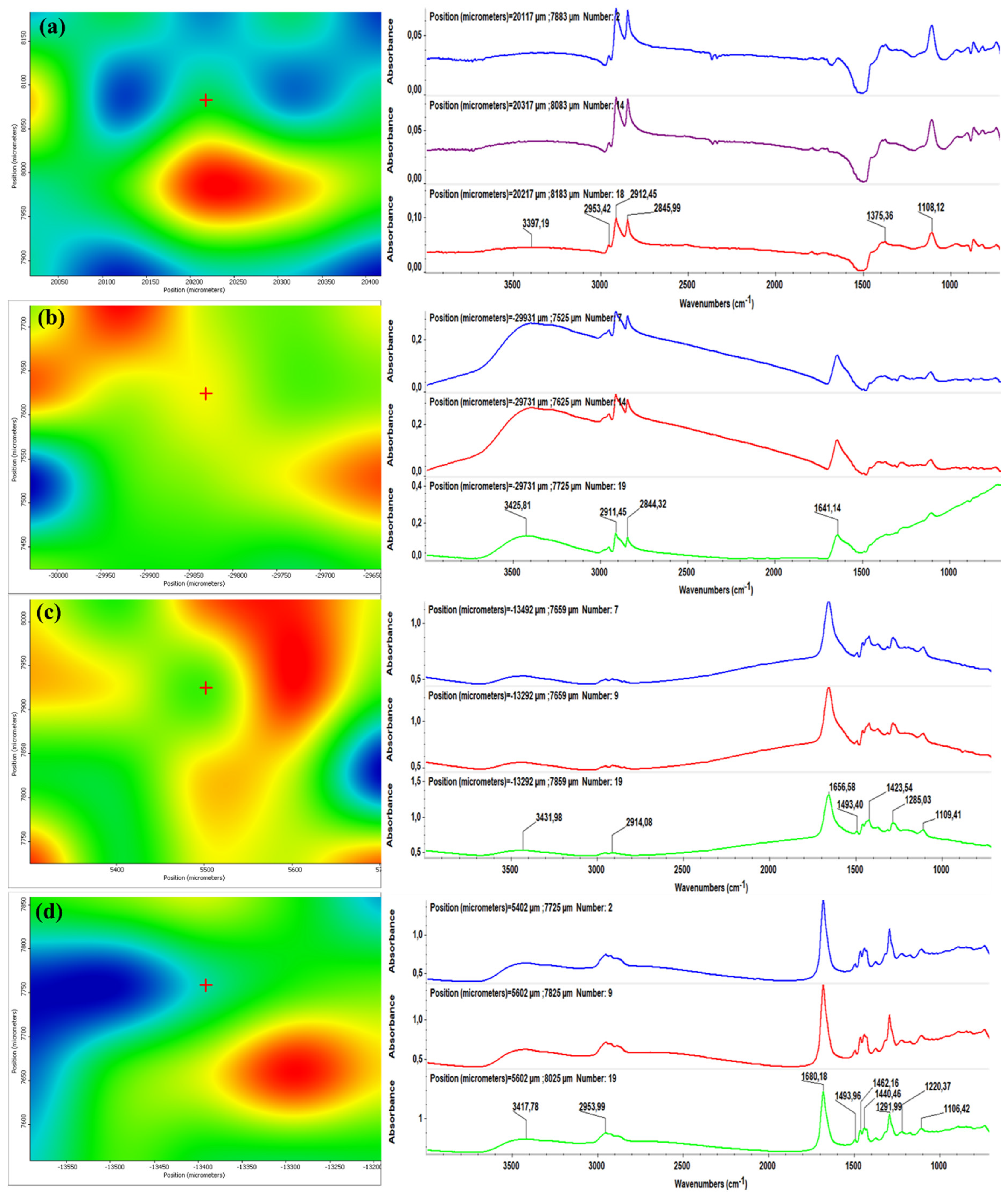
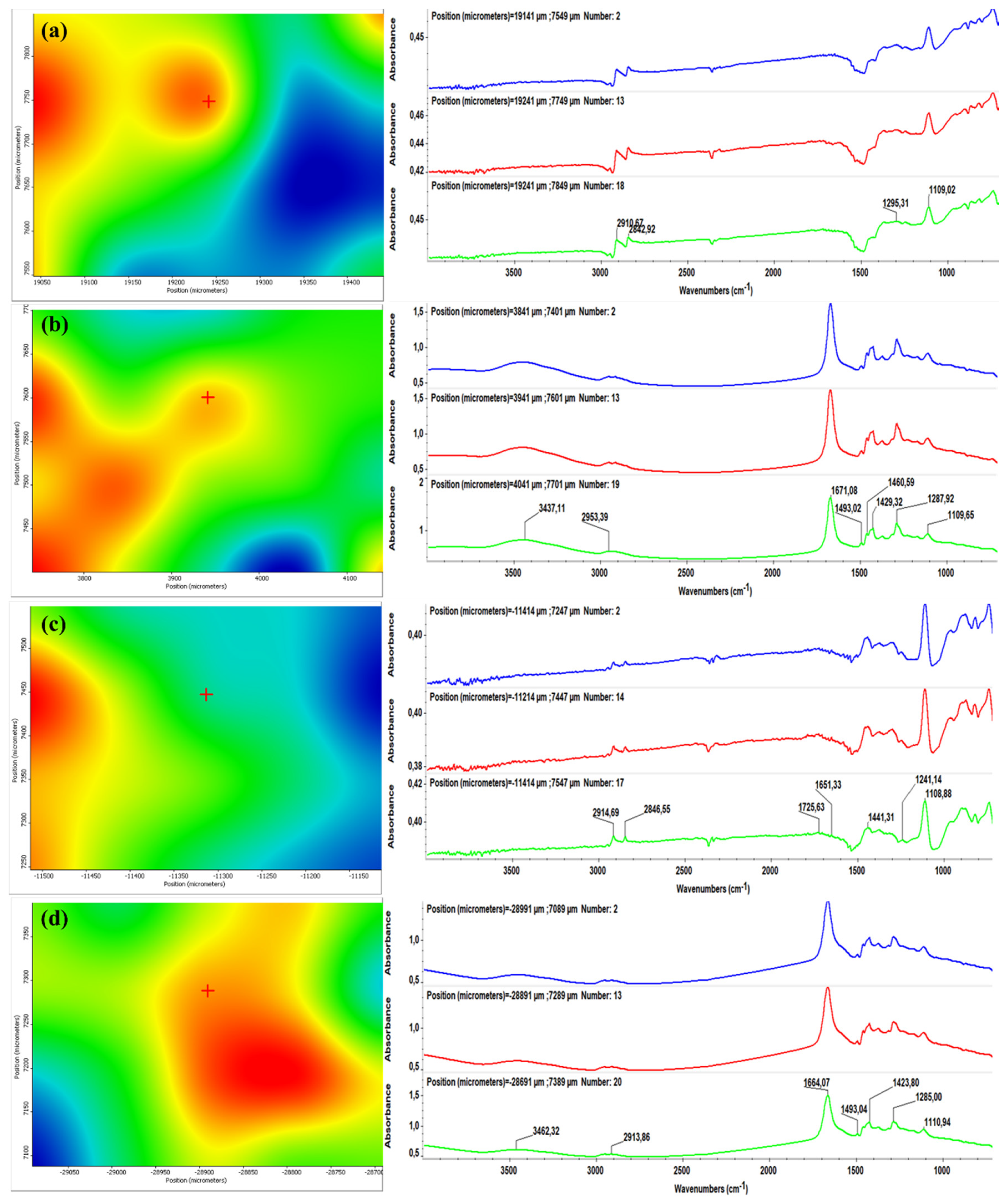
2.1. FT-IR and IRM
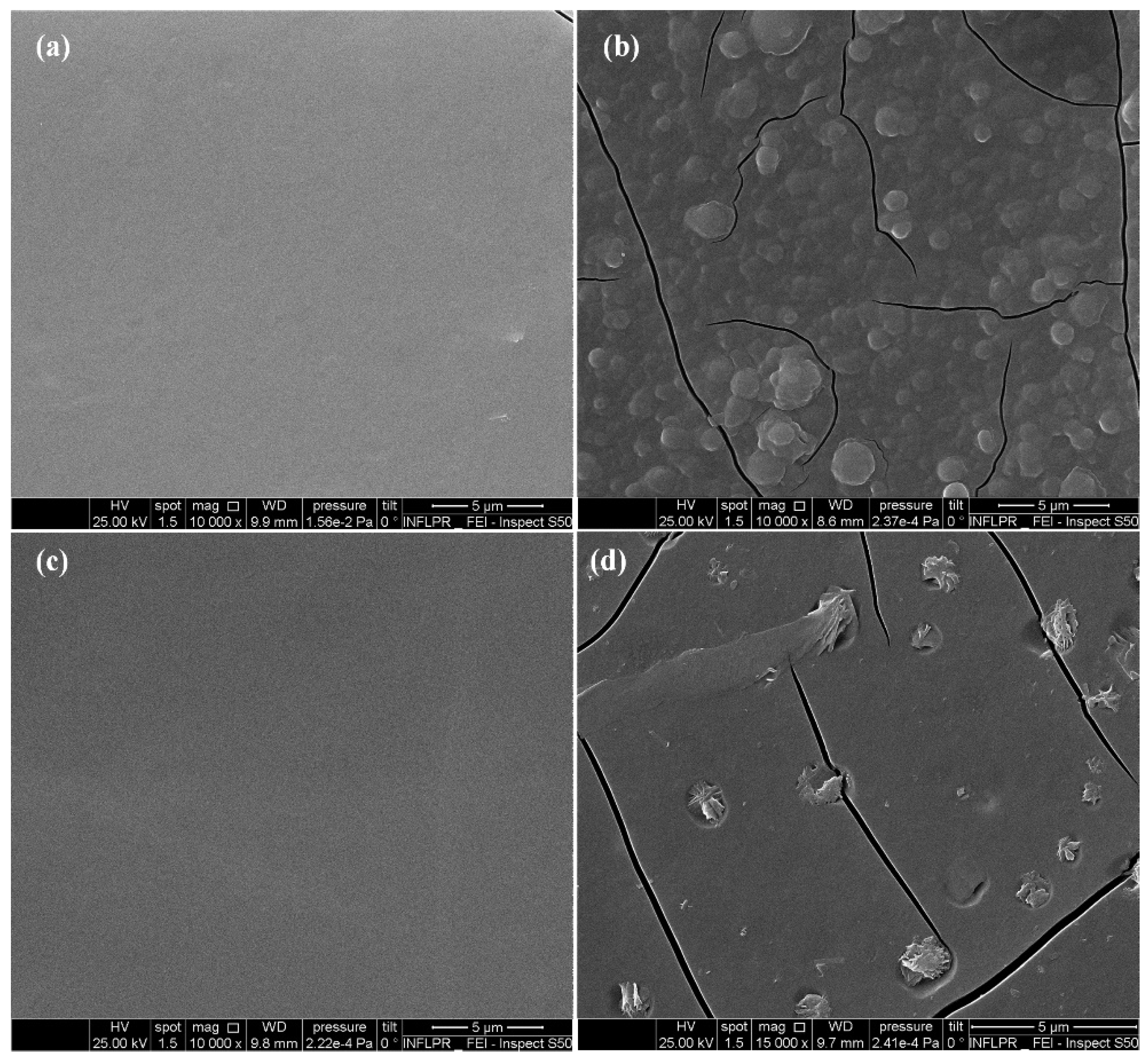
2.2. SEM
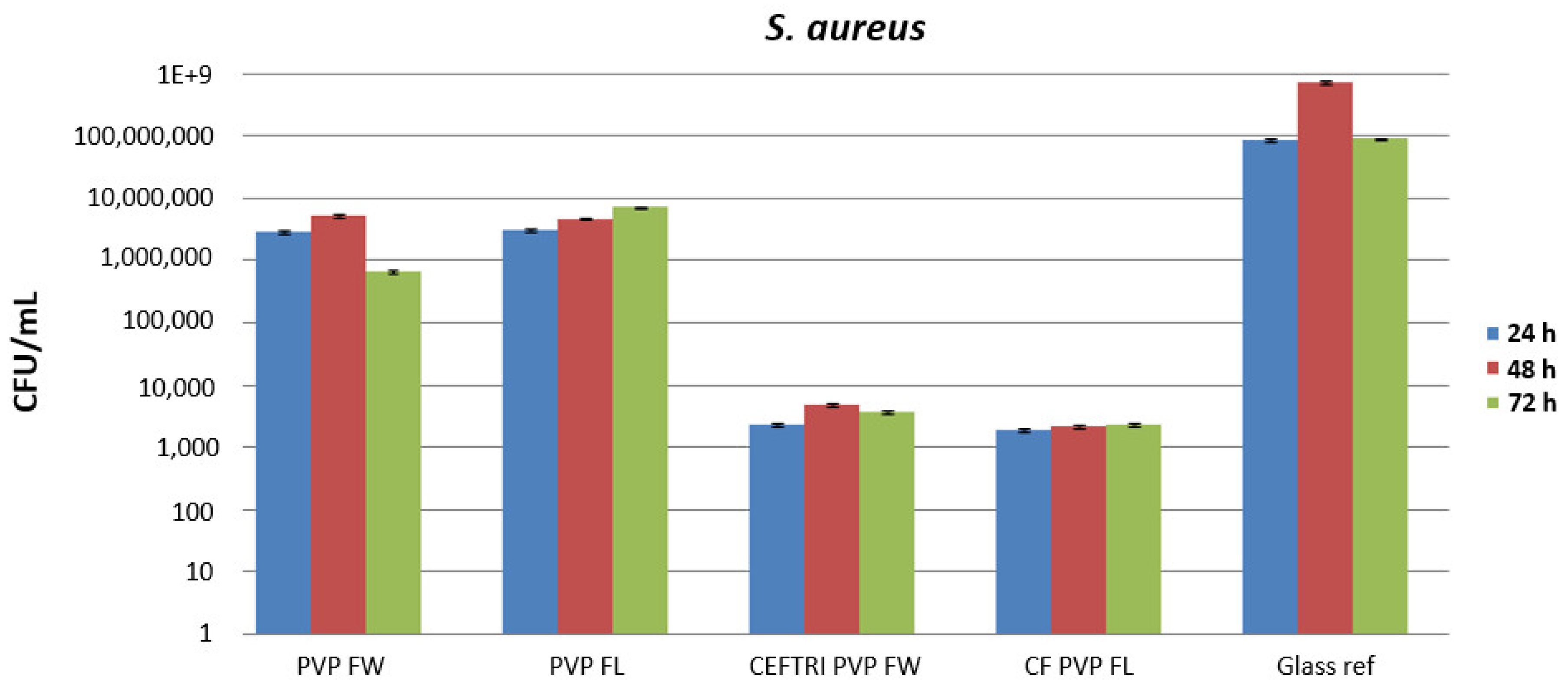
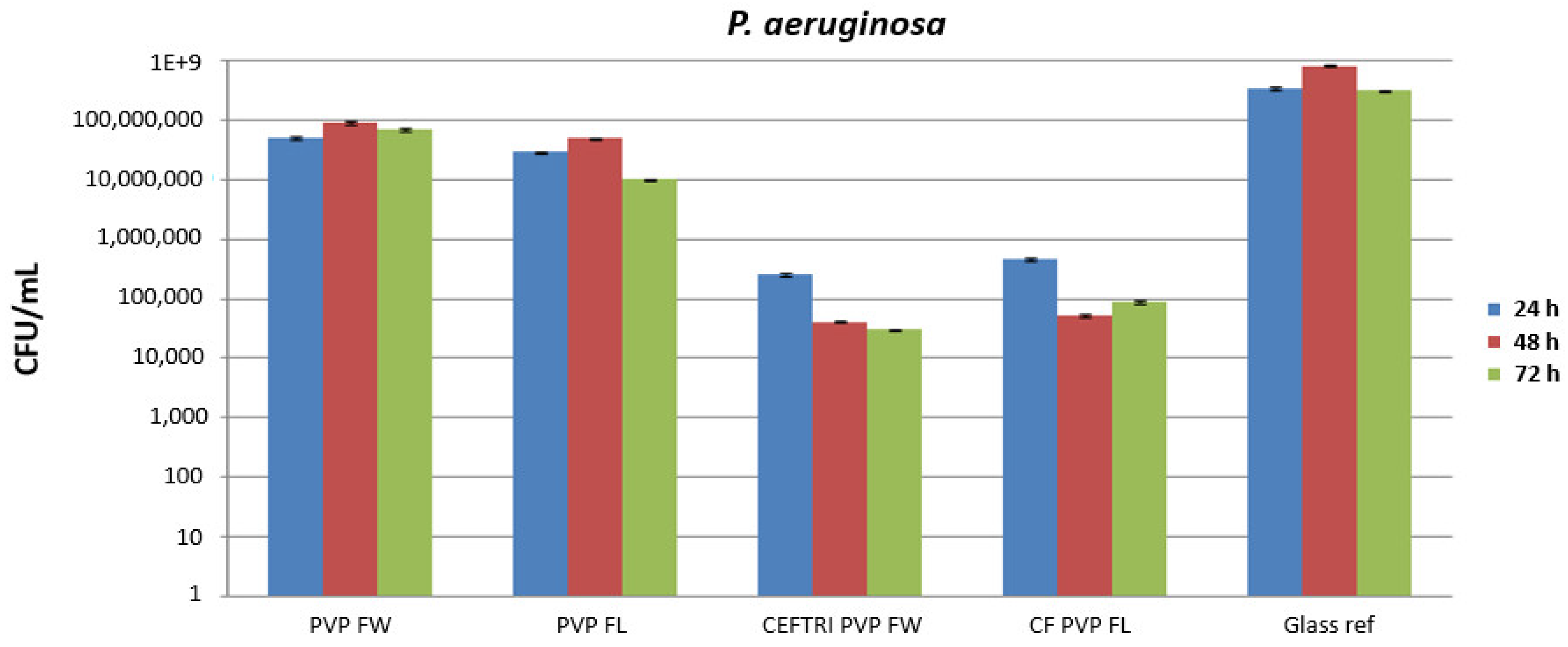
2.3. Antimicrobial and Antibiofilm Evaluation
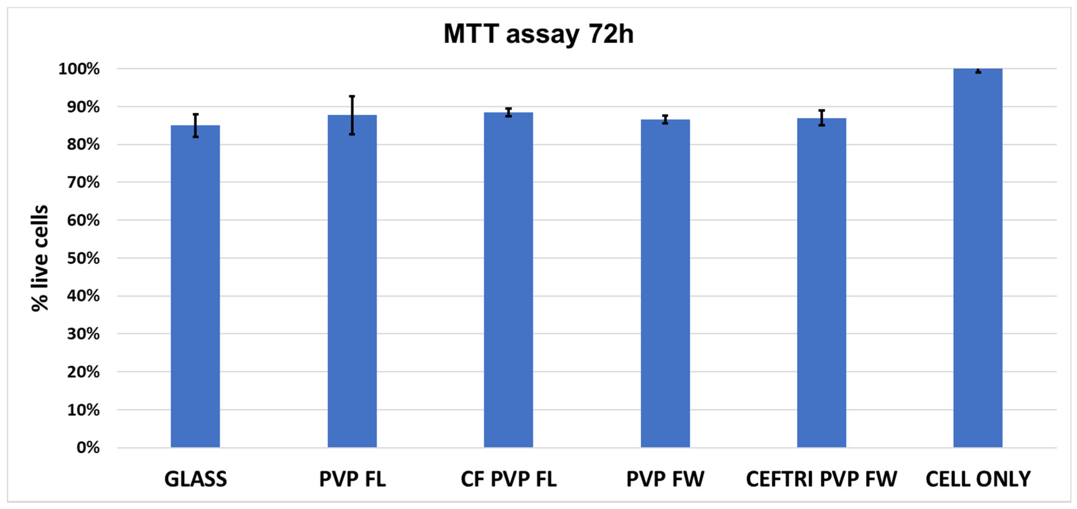
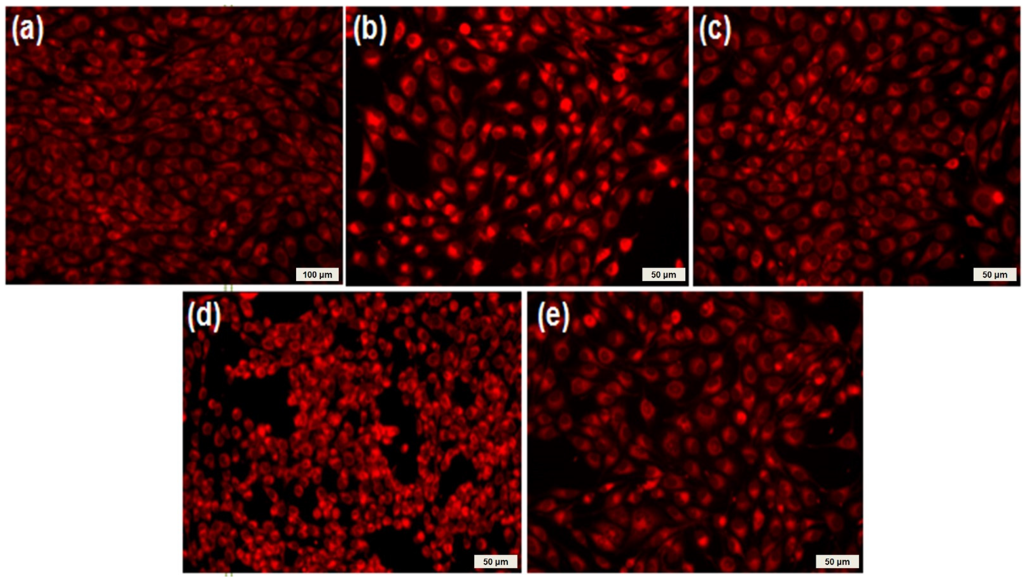
2.4. Biocompatibility Evaluation
3. Materials and Methods
3.1. Materials
3.2. Preparation of Mixes for Drop-Cast and MAPLE Depositions
- i.
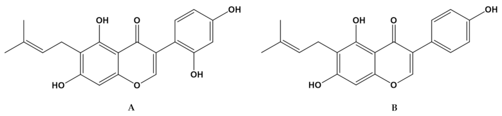
- Solution A: Cefuroxime:PVP (2% in DMSO):Luteone, 4:1:1 wt.% (CF/PVP/FL symbol).
- ii.
- Solution B: PVP (2% in DMSO):Luteone, 1:1 w.t% (PVP/FL symbol).
- iii.
- Solution C: Ceftriaxone:PVP (2% in DMSO):Wighteone, 4:1:1 wt.% (CEFTRI/PVP/FW symbol).
- iv.
- Solution D: PVP (2% in DMSO):Wighteone, 1:1 wt.% (PVP/FW symbol).
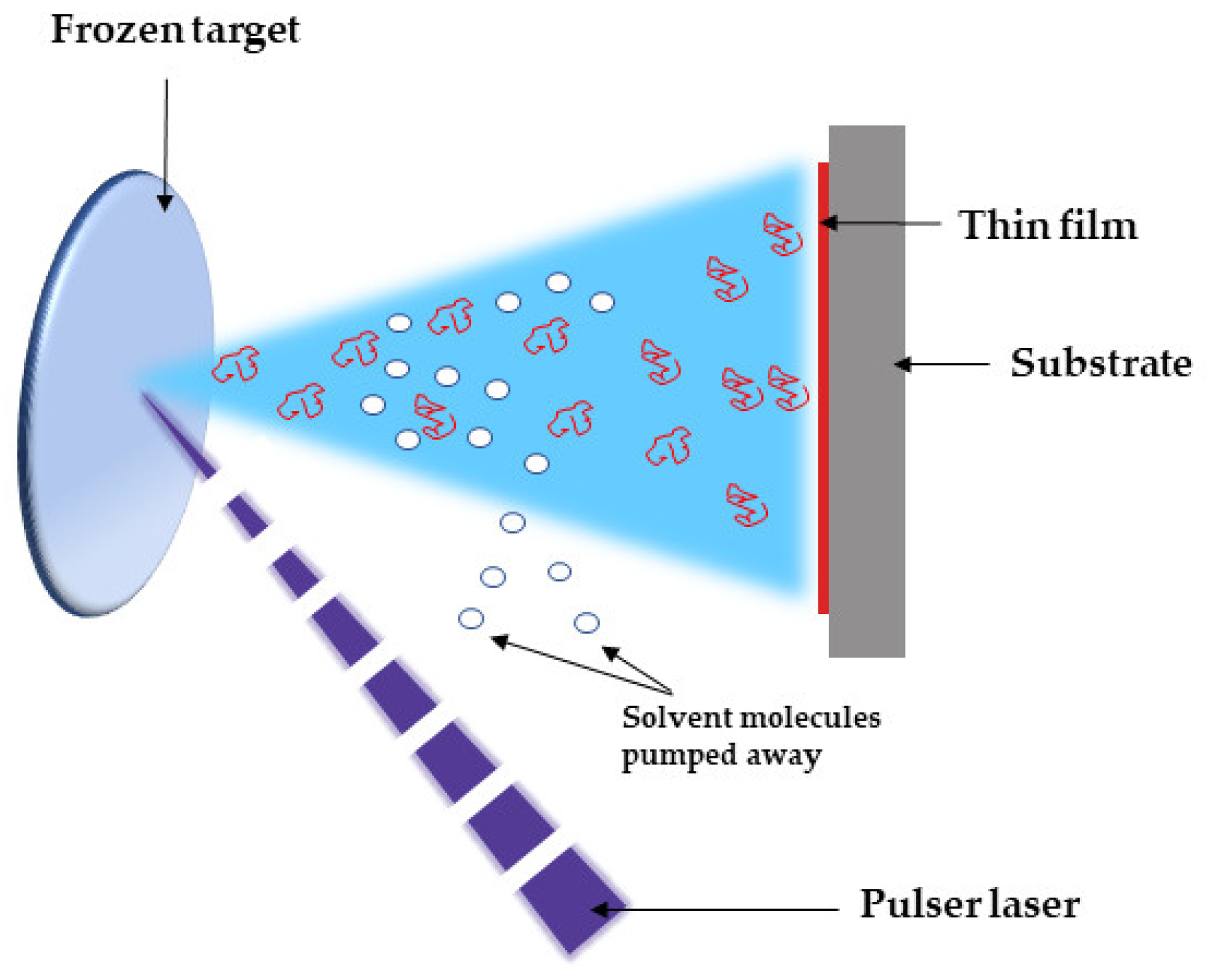
3.3. MAPLE Experimental Conditions
3.4. Characterization Methods
3.4.1. Fourier-Transform Infrared Spectroscopy (FT-IR) and Infrared Microscopy (IRM)
3.4.2. Scanning Electron Microscopy (SEM)
3.4.3. Antimicrobial Evaluation
3.4.4. Biocompatibility Evaluation
3.4.5. Statistical Analysis
4. Conclusions
Author Contributions
Funding
Institutional Review Board Statement
Informed Consent Statement
Data Availability Statement
Conflicts of Interest
Sample Availability
References
- Bloom, D.E.; Cadarette, D. Infectious disease threats in the twenty-first century: Strengthening the global response. Front. Immunol. 2019, 10, 549. [Google Scholar] [CrossRef]
- Podgoreanu, P.; Negrea, S.M.; Buia, R.; Delcaru, C.; Trusca, S.B.; Lazar, V.; Chifiriuc, M.C. Alternative strategies for fighting multidrug resistant bacterial infections. Biointerface Res. Appl. Chem. 2019, 9, 3834–3841. [Google Scholar] [CrossRef]
- Lucien, M.A.B.; Canarie, M.F.; Kilgore, P.E.; Jean-Denis, G.; Fénélon, N.; Pierre, M.; Cerpa, M.; Joseph, G.A.; Maki, G.; Zervos, M.J.; et al. Antibiotics and antimicrobial resistance in the COVID-19 era: Perspective from resource-limited settings. Int. J. Infect. Dis. 2021, 104, 250–254. [Google Scholar] [CrossRef]
- Sehmi, S.K.; Lourenco, C.; Alkhuder, K.; Pike, S.D.; Noimark, S.; Williams, C.K.; Shaffer, M.S.P.; Parkin, I.P.; MacRobert, A.J.; Allan, E. Antibacterial surfaces with activity against antimicrobial resistant bacterial pathogens and endospores. ACS Infect. Dis. 2020, 6, 939–946. [Google Scholar] [CrossRef]
- Darwesh, O.M.; Barakat, K.M.; Mattar, M.Z.; Sabae, S.Z.; Hassan, S.H. Production of antimicrobial blue green pigment Pyocyanin by marine Pseudomonas aeruginosa. Biointerface Res. Appl. Chem. 2019, 9, 4334–4339. [Google Scholar] [CrossRef]
- Lee, M.H.; Lee, G.A.; Lee, S.H.; Park, Y.-H. A systematic review on the causes of the transmission and control measures of outbreaks in long-term care facilities: Back to basics of infection control. PLoS ONE. 2020, 15, e0229911. [Google Scholar] [CrossRef]
- Friedman, N.D.; Temkin, E.; Carmeli, Y. The negative impact of antibiotic resistance. Clin. Microbiol. Infect. 2016, 22, 416–422. [Google Scholar] [CrossRef]
- Sizentsov, A.; Mindolina, Y.; Barysheva, E.; Ponomareva, P.; Kunavina, E.; Levenets, T.; Dudko, A.; Kvan, O. Effectiveness of combined use of antibiotics, essential metals and probiotic bacterial strain complexes against multidrug resistant pathogens. Biointerface Res. Appl. Chem. 2020, 10, 4830–4836. [Google Scholar] [CrossRef]
- Abdelghany, A.M.; Ayaad, D.M.; Mahmoud, S.M. Antibacterial and Energy Gap Correlation of PVA/SA Biofilms Doped With Selenium Nanoparticles. Biointerface Res. Appl. Chem. 2020, 10, 6280–6288. [Google Scholar] [CrossRef]
- Srinivasan, R.; Santhakumari, S.; Poonguzhali, P.; Geetha, M.; Dyavaiah, M.; Xiangmin, L. Bacterial Biofilm Inhibition: A Focused Review on Recent Therapeutic Strategies for Combating the Biofilm Mediated Infections. Front. Microbiol. 2021, 12, 676458. [Google Scholar] [CrossRef]
- Hooshdar, P.; Kermanshahi, R.K.; Ghadam, P.; Khosravi-Darani, K. A Review on Production of Exopolysaccharide and Biofilm in Probiotics Like Lactobacilli and Methods of Analysis. Biointerface Res. Appl. Chem. 2020, 10, 6058–6075. [Google Scholar] [CrossRef]
- Petersen, P.J.; Labthavikul, P.; Jones, C.H.; Bradford, P.A. In vitro antibacterial activities of tigecycline in combination with other antimicrobial agents determined by chequerboard and time-kill kinetic analysis. J. Antimicrob. Chemother. 2006, 57, 573–576. [Google Scholar] [CrossRef]
- Leekha, S.; Terrell, C.L.; Edson., R.S. General principles of antimicrobial therapy. Mayo Clin. Proc. 2011, 86, 156–167. [Google Scholar] [CrossRef] [PubMed]
- Gradelski, E.; Valera, L.; Bonner, D.; Fung-Tomc, J. Synergistic activities of gatifloxacin in combination with other antimicrobial agents against Pseudomonas aeruginosa and related species. Antimicrob. Agents Chemother. 2001, 45, 3220–3222. [Google Scholar] [CrossRef]
- Abreu, A.C.; McBain, A.J.; Simoes, M. Plants as sources of new antimicrobials and resistance-modifying agents. Nat. Prod. Rep. 2012, 29, 1007–1021. [Google Scholar] [CrossRef]
- Ventola, C.L. The antibiotic resistance crisis, part 1: Causes and threats. P&T 2015, 40, 277–283. [Google Scholar]
- Cushnie, T.P.T.; Lamb, A.J. Recent advances in understanding the antibacterial properties of flavonoids. Int. J. Antimicrob. Agents. 2011, 38, 99–107. [Google Scholar] [CrossRef]
- Amini, M.; Safaie, N.; Salmani, M.J.; Shams-Bakhs, M. Antifungal activity of three medicinal plant essential oils against some phytopathogenic fungi. Trakia J. Sci. 2012, 10, 1–8. [Google Scholar]
- Ahad, B.; Shahri, W.; Rasool, H.; Reshi, Z.A.; Rasool, S.; Hussain, T. Medicinal Plants and Herbal Drugs: An Overview. In Medicinal and Aromatic Plants. Healthcare and Industrial Applications.; Aftab, T., Hakeem, K.R., Eds.; Springer Nature: Cham, Switzerland, 2021. [Google Scholar] [CrossRef]
- Beecher, G.R. Overview of dietary flavonoids: Nomenclature, occurrence and intake. J. Nutr. 2003, 133, 3248S–3254S. [Google Scholar] [CrossRef]
- Abotaleb, M.; Samuel, S.M.; Varghese, E.; Varghese, S.; Kubatka, P.; Liskova, A.; Büsselberg, D. Flavonoids in Cancer and Apoptosis. Cancers 2019, 11, 28. [Google Scholar] [CrossRef]
- Hatano, T.; Shintani, Y.; Aga, Y.; Shiota, S.; Tsuchiya, T.; Yoshida, T. Phenolic constituents of licorice. VIII. Structures of glicophenone and glicoisoflavanone, and effects of licorice phenolics on methicillin-resistant Staphylococcus aureus. Chem. Pharm. Bull. 2000, 48, 1286–1292. [Google Scholar] [CrossRef]
- Santra, H.K.; Banerjee, D. Natural products as fungicide and their role in crop protection. In Natural bioactive products in sustainable agriculture; Singh, J., Yadav, A., Eds.; Springer: Singapore, 2020. [Google Scholar] [CrossRef]
- Juma, B.F.; Majinda, R.R.T. Three new compounds from Erythrina lysistemon and their antimicrobial, radical scavenging activities and their brine shrimp lethality. 11th NAPRECA Symposium Book of Proceedings, Antananarivo, Madagascar, 9–12 August 2005; pp. 97–109. [Google Scholar]
- Cao, Z.W.; Zeng, Q.; Pei, H.J.; Ren, L.D.; Bai, H.Z.; Na, R.N. HSP90 expression and its association with wighteone metabolite response in HER2-positive breast cancer cells. Oncol. Lett. 2016, 11, 3719–3722. [Google Scholar] [CrossRef][Green Version]
- Ingham, J.L.; Tahara, S.; Harborne, J.B. Fungitoxic isoflavones from Lupinus albus and other Lupinus species. Z Naturforsch. C. 1983, 38, 194–200. [Google Scholar] [CrossRef]
- Friend, J. Plant phenolics, lignification and plant disease. In Progress in Phytochemistry, 1st ed.; Reinhold, L., Harborne, J.B., Swain, T., Eds.; Pergamon: Oxford, UK, 1981; Volume 7, pp. 197–261. [Google Scholar]
- Shanmuganathan, R.; MubarakAli, D.; Prabakar, D.; Muthukumar, H.; Thajuddin, N.; Kumar, S.S.; Pugazhendhi, A. An enhancement of antimicrobial efficacy of biogenic and ceftriaxone-conjugated silver nanoparticles: Green approach. Environ. Sci. Pollut. Res. 2018, 25, 10362–10370. [Google Scholar] [CrossRef] [PubMed]
- Amin, M.U.; Khurram, M.; Khattak, B.; Khan, J. Antibiotic additive and synergistic action of rutin, morin and quercetin against methicillin resistant Staphylococcus aureus. BMC Complement. Altern. Med. 2015, 15, 59. [Google Scholar] [CrossRef] [PubMed]
- Mun, S.-H.; Kang, O.-H.; Joung, D.-K.; Kim, S.-B.; Seo, Y.-S.; Choi, J.-G.; Lee, Y.-S.; Cha, S.-W.; Ahn, Y.-S.; Han, S.H.; et al. Combination Therapy of Sophoraflavanone B against MRSA: In Vitro Synergy Testing. Evid. Based Complement. Alternat. Med. 2013, 823794. [Google Scholar]
- Sadgrove, N.J.; Jones, G.L. From petri dish to patient: Bioavailability estimation and mechanism of action for antimicrobial and immunomodulatory natural products. Front. Microbiol. 2019, 10, 2470. [Google Scholar] [CrossRef]
- Rana, A.C.; Gulliya, B. Chemistry and Pharmacology of Flavonoids- A Review. Indian J. Pharm. Educ. 2019, 53, 8–20. [Google Scholar] [CrossRef]
- Caesar, L.K.; Cech, N.B. Synergy and antagonism in natural product extracts: When 1 + 1 does not equal 2. Nat. Prod. Rep. 2019, 36, 869–888. [Google Scholar] [CrossRef]
- Aboody, M.S.A.; Mickymaray, S. Anti-fungal efficacy and mechanisms of flavonoids. Antibiotics 2020, 9, 45. [Google Scholar] [CrossRef]
- Ahmad, R.; Srivastava, S.; Ghosh, S.; Khare, S.K. Phytochemical delivery through nanocarriers: A review. Colloids Surf. B Biointerfaces 2021, 197, 111389. [Google Scholar] [CrossRef]
- Khan, F.; Tabassum, N.; Kim, Y.M. A strategy to control colonization of pathogens: Embedding of lactic acid bacteria on the surface of urinary catheter. Appl. Microbiol. Biotechnol. 2020, 104, 9053–9066. [Google Scholar] [CrossRef]
- Hogan, S.; Stevens, N.T.; Humphreys, H.; O’Gara, J.P.; O’Neill, E. Current and future approaches to the prevention and treatment of staphylococcal medical device-related infections. Curr. Pharm. Des. 2015, 21, 100–113. [Google Scholar] [CrossRef]
- Francolini, I.; Donelli, G.; Crisante, F.; Taresco, V.; Piozzi, A. Antimicrobial Polymers for Anti-biofilm Medical Devices: State-of-Art and Perspectives. In Biofilm-based Healthcare-associated Infections. Advances in Experimental Medicine and Biology; Donelli, G., Ed.; Springer: Cham, Switzerland, 2015; Volume 831, pp. 93–117. [Google Scholar] [CrossRef]
- Cordero, H.C.P.; Abarca, R.Q.; Vargas, K.D.; Troncoso, I.P. Polymeric materials with antifouling, biocidal, antiviral and antimicrobial properties; Elaboration method and its uses. U.S. Patent 9,481,800 B2, 1 November 2016. [Google Scholar]
- Makabenta, J.M.V.; Nabawy, A.; Li, C.H.; Schmidt-Malan, S.; Patel, R.; Rotello, V.M. Nanomaterial-based therapeutics for antibiotic-resistant bacterial infections. Nat. Rev. Microbiol. 2021, 19, 23–36. [Google Scholar] [CrossRef]
- Landis, R.F.; Li, C.H.; Gupta, A.; Lee, Y.W.; Yazdani, M.; Ngernyuang, N.; Altinbasak, I.; Mansoor, S.; Khichi, M.A.; Sanyal, A.; et al. Biodegradable nanocomposite antimicrobials for the eradication of multidrug-resistant bacterial biofilms without accumulated resistance. J. Am. Chem. Soc. 2018, 140, 6176–6182. [Google Scholar] [CrossRef]
- McGill, R.A.; Chrisey, D.B. Method of Producing a Film Coating by Matrix Assisted Pulsed Laser Deposition. U.S. Patent Application No. 6,025,036, 15 February 2000. [Google Scholar]
- Chrisey, D.B.; McGill, R.A.; Horwitz, J.S.; Pique, A.; Ringeisen, B.R.; Bubb, D.M.; Wu, P.K. Novel Laser-Based Deposition of Active Protein Thin Films. Chem. Rev. 2003, 103, 553–576. [Google Scholar] [CrossRef]
- Cristescu, R.; Patz, T.; Narayan, R.J.; Menegazzo, N.; Mizaikoff, B.; Mihaiescu, D.E.; Messersmith, P.B.; Stamatin, I.; Mihailescu, I.N.; Chrisey, D.B. Processing of mussel adhesive protein analog thin films by matrix assisted pulsed laser evaporation. Appl. Surf. Sci. 2005, 247, 217–224. [Google Scholar] [CrossRef]
- Doraiswamy, A.; Narayan, R.J.; Cristescu, R.; Mihailescu, I.N.; Chrisey, D.B. Laser processing of natural mussel adhesive protein thin films. Mater. Sci. Eng. C 2007, 27, 409–413. [Google Scholar] [CrossRef]
- Cristescu, R.; Popescu, C.; Popescu, A.C.; Grigorescu, S.; Duta, L.; Mihailescu, I.N.; Caraene, G.; Albulescu, R.; Albulescu, L.; Andronie, A.; et al. Functionalized polyvinyl alcohol derivatives thin films for controlled drug release and targeting systems: MAPLE deposition and morphological, chemical and in vitro characterization. Appl. Surf. Sci. 2009, 255, 5600–5604. [Google Scholar] [CrossRef]
- Riggs, B.C.; Dias, A.D.; Schiele, N.R.; Cristescu, R.; Huang, Y.; Corr, D.T.; Chrisey, D.B. Matrix-assisted pulsed laser methods for biofabrication. MRS Bull. 2011, 36, 1043–1050. [Google Scholar] [CrossRef]
- Cristescu, R.; Popescu, C.; Socol, G.; Iordache, I.; Mihailescu, I.N.; Mihaiescu, D.E.; Grumezescu, A.M.; Balan, A.; Stamatin, I.; Chifiriuc, C.; et al. Magnetic core/shell nanoparticle thin films deposited by MAPLE: Investigation by chemical, morphological and in vitro biological assays. Appl. Surf. Sci. 2012, 258, 9250–9255. [Google Scholar] [CrossRef]
- Limban, C.; Missir, A.V.; Grumezescu, A.M.; Oprea, A.E.; Grumezescu, V.; Vasile, B.S.; Socol, G.; Trusca, R.; Caproiu, M.T.; Chifiriuc, M.C.; et al. Bioevaluation of novel anti-biofilm coatings based on PVP/Fe3O4 nanostructures and 2-((4-Ethylphenoxy)methyl)-N- (arylcarbamothioyl)benzamides. Molecules 2014, 19, 12011–12030. [Google Scholar] [CrossRef]
- Cristescu, R.; Surdu, A.V.; Grumezescu, A.M.; Oprea, A.E.; Trusca, R.; Vasile, O.; Dorcioman, G.; Visan, A.; Socol, G.; Mihailescu, I.N.; et al. Microbial colonization of biopolymeric thin films containing natural compounds and antibiotics fabricated by MAPLE. Appl. Surf. Sci. 2015, 336, 234–239. [Google Scholar] [CrossRef]
- Negut, I.; Visan, A.I.; Popescu, C.; Cristescu, R.; Ficai, A.; Grumezescu, A.M.; Chifiriuc, M.C.; Boehm, R.D.; Yamaleyeva, D.; Taylor, M.; et al. Successful release of voriconazole and flavonoids from MAPLE deposited bioactive surfaces. Appl. Sci. 2019, 9, 786. [Google Scholar] [CrossRef]
- Cristescu, R.; Visan, A.; Socol, G.; Surdu, A.V.; Oprea, A.E.; Grumezescu, A.M.; Chifiriuc, M.C.; Boehm, R.D.; Yamaleyeva, D.; Taylor, M.; et al. Antimicrobial activity of biopolymeric thin films containing flavonoid natural compounds and silver nanoparticles fabricated by MAPLE: A comparative study. Appl. Surf. Sci. 2016, 374, 290–296. [Google Scholar] [CrossRef]
- Abian, O.; Ortega-Alarcon, D.; Jimenez-Alesanco, A.; Ceballos-Laita, L.; Vega, S.; Reyburn, H.T.; Rizzuti, B.; Velazquez-Campoy, A. Structural stability of SARS-CoV-2 3CLpro and identification of quercetin as an inhibitor by experimental screening. Int. J. Biol. Macromol. 2020, 164, 1693–1703. [Google Scholar] [CrossRef] [PubMed]
- Cristescu, R.; Narayan, R.J.; Chrisey, D.B. Novel antimicrobial surfaces to defeat COVID-19 transmission. MRS Adv. 2020, 5, 2839–2851. [Google Scholar] [CrossRef]
- Hadi, A.G.; Kadhom, M.; Hairunisa, N.; Yousif, E.; Mohammed, S.A. A Review on COVID-19: Origin, Spread, Symptoms, Treatment, and Prevention. Biointerface Res. Appl. Chem. 2020, 10, 7234–7242. [Google Scholar] [CrossRef]
- Rahma, A.; Munir, M.M.; Khairurrijal, P.A.; Suendo, V.; Rachmawati, H. Intermolecular Interactions and the Release Pattern of Electrospun Curcumin-Polyvinyl(pyrrolidone). Fiber. Biol. Pharm. Bull. 2016, 39, 163–173. [Google Scholar] [CrossRef]
- Marimuthu, S.; Nantheeswaran, S.; Shanmugarathinam, A.; Purachikody, A. Formulation and characterization of cefuroxime axetil Floating microspheres. Int. J. Pharm. 2012, 2, 526–533. [Google Scholar] [CrossRef]
- Chandra, D.; Kohli, G.; Prasad, K.; Bisht, G.; Punetha, V.D.; Khetwal, K.; Devrani, M.K.; Pandey, H. Phytochemical and Ethnomedicinal Uses of Family Violaceae. Curr. Res. Chem. 2015, 7, 44–52. [Google Scholar] [CrossRef]
- Trifunschi, S.; Munteanu, M.F.; Agotici., V.; Pintea (Ardelean), S.; Gligor, R. Determination of Flavonoid and Polyphenol Compounds in Viscum Album and Allium Sativum Extracts. Int. Cur. Pharm. J. 2015, 4, 382–385. [Google Scholar] [CrossRef]
- Noh, C.H.C.; Azmin, N.F.M.; Amid, A. Principal Component Analysis Application on Flavonoids Characterization. ASTESJ 2017, 2, 435–440. [Google Scholar] [CrossRef]
- Lane, G.A.; Newman, R.H. Isoflavones from Lupinus angustifolius root. Phytochemistry 1987, 26, 295–300. [Google Scholar] [CrossRef]
- Kinoshita, T.; Ichinose, K.; Takahashi, C.; Ho, F.C.; Wu, J.B.; Sankawa, U. Chemical studies on Sophora tomentosa: The isolation of a new class of isoflavonoid. Chem. Pharm. Bull. 1990, 38, 2756–2759. [Google Scholar] [CrossRef]
- Savoia, D. Plant-derived antimicrobial compounds: Alternatives to antibiotics. Future Microbiol. 2012, 7, 979–990. [Google Scholar] [CrossRef]
- Górniak, I.; Bartoszewski, R.; Króliczewski, J. Comprehensive review of antimicrobial activities of plant flavonoids. Phytochem. Rev. 2019, 18, 241–272. [Google Scholar] [CrossRef]
- Deepika, M.S.; Thangam, R.; Vijayakumar, T.S.; Sasirekha, R.; Vimala, R.T.V.; Sivasubramanian, S.; Arun, S.; Babu, M.D.; Thirumurugan, R. Antibacterial synergy between rutin and florfenicol enhances therapeutic spectrum against drug resistant Aeromonas hydrophila. Microb. Pathog. 2019, 135, 103612. [Google Scholar] [CrossRef] [PubMed]
- Farhadi, F.; Khameneh, B.; Iranshahi, M.; Iranshahy, M. Antibacterial activity of flavonoids and their structure–activity relationship: An update review. Phytother. Res. 2019, 33, 13–40. [Google Scholar] [CrossRef] [PubMed]
- Kayisli, U.A.; Aksu, C.A.H.; Berkkanoglu, M.; Arici, A. Estrogenicity of isoflavones on human endometrial stromal and glandular cells. J. Clin. Endocrinol. Metab. 2002, 87, 5539–5544. [Google Scholar] [CrossRef]
- Smolińska, E.; Moskot, M.; Jakóbkiewicz-Banecka, J.; Węgrzyn, G.; Banecki, B.; Szczerkowska-Dobosz, A.; Purzycka-Bohdan, D.; Gabig-Cimińska, M. Molecular action of isoflavone genistein in the human epithelial cell line HaCaT. PLoS ONE 2018, 13, e0192297. [Google Scholar] [CrossRef] [PubMed]
- Lungu, M.; Gavriliu, S.; Enescu, E.; Ion, I.; Bratulescu, A.; Mihaescu, G.; Maruţescu, L.; Chifiriuc, M.C. Silver–titanium dioxide nanocomposites as effective antimicrobial and antibiofilm agents. J. Nanopart. Res. 2014, 16, 2203. [Google Scholar] [CrossRef]
- Grumezescu, V.; Socol, G.; Grumezescu, A.M.; Holban, A.M.; Ficai, A.; Trusca, R.; Bleotu, C.; Balaure, P.C.; Cristescu, R.; Chifiriuc, M.C. Functionalized antibiofilm thin coatings based on PLA-PVA microspheres loaded with usnic acid natural compounds fabricated by MAPLE. Appl. Surf. Sci. 2014, 302, 262–267. [Google Scholar] [CrossRef]
- Grumezescu, V.; Holban, A.M.; Iordache, F.; Socol, G.; Mogosanu, G.D.; Grumezescu, A.M.; Ficai, A.; Vasile, B.S.; Trusca, R.; Chifiriuc, M.C.; et al. MAPLE fabricated magnetite@eugenol and (3-hidroxybutyric acid-co-3-hidroxyvaleric acid)-polyvinyl alcohol microspheres coated surfaces with anti-microbial properties. Appl. Surf. Sci. 2014, 306, 16–22. [Google Scholar] [CrossRef]
- Curutiu, C.; Chifiriuc, M.C.; Iordache, F.; Bleotu, C.; Lazar, V.; Mogoanta, C.A.; Popescu, C.R.; Grigore, R.; Bertesteanu, S.V. Fluorescence analysis of apoptosis induced by Pseudomonas aeruginosa in endothelial cells. Rom. J. Morphol. Embryol. 2014, 55, 313–317. [Google Scholar]


Publisher’s Note: MDPI stays neutral with regard to jurisdictional claims in published maps and institutional affiliations. |
© 2021 by the authors. Licensee MDPI, Basel, Switzerland. This article is an open access article distributed under the terms and conditions of the Creative Commons Attribution (CC BY) license (https://creativecommons.org/licenses/by/4.0/).
Share and Cite
Grumezescu, V.; Negut, I.; Cristescu, R.; Grumezescu, A.M.; Holban, A.M.; Iordache, F.; Chifiriuc, M.C.; Narayan, R.J.; Chrisey, D.B. Isoflavonoid-Antibiotic Thin Films Fabricated by MAPLE with Improved Resistance to Microbial Colonization. Molecules 2021, 26, 3634. https://doi.org/10.3390/molecules26123634
Grumezescu V, Negut I, Cristescu R, Grumezescu AM, Holban AM, Iordache F, Chifiriuc MC, Narayan RJ, Chrisey DB. Isoflavonoid-Antibiotic Thin Films Fabricated by MAPLE with Improved Resistance to Microbial Colonization. Molecules. 2021; 26(12):3634. https://doi.org/10.3390/molecules26123634
Chicago/Turabian StyleGrumezescu, Valentina, Irina Negut, Rodica Cristescu, Alexandru Mihai Grumezescu, Alina Maria Holban, Florin Iordache, Mariana Carmen Chifiriuc, Roger J. Narayan, and Douglas B. Chrisey. 2021. "Isoflavonoid-Antibiotic Thin Films Fabricated by MAPLE with Improved Resistance to Microbial Colonization" Molecules 26, no. 12: 3634. https://doi.org/10.3390/molecules26123634
APA StyleGrumezescu, V., Negut, I., Cristescu, R., Grumezescu, A. M., Holban, A. M., Iordache, F., Chifiriuc, M. C., Narayan, R. J., & Chrisey, D. B. (2021). Isoflavonoid-Antibiotic Thin Films Fabricated by MAPLE with Improved Resistance to Microbial Colonization. Molecules, 26(12), 3634. https://doi.org/10.3390/molecules26123634