
Appraisal of Comparative Therapeutic Potential of Undoped and Nitrogen-Doped Titanium Dioxide Nanoparticles


 ,
,  ,
,
Abstract
1. Introduction
2. Results and Discussion
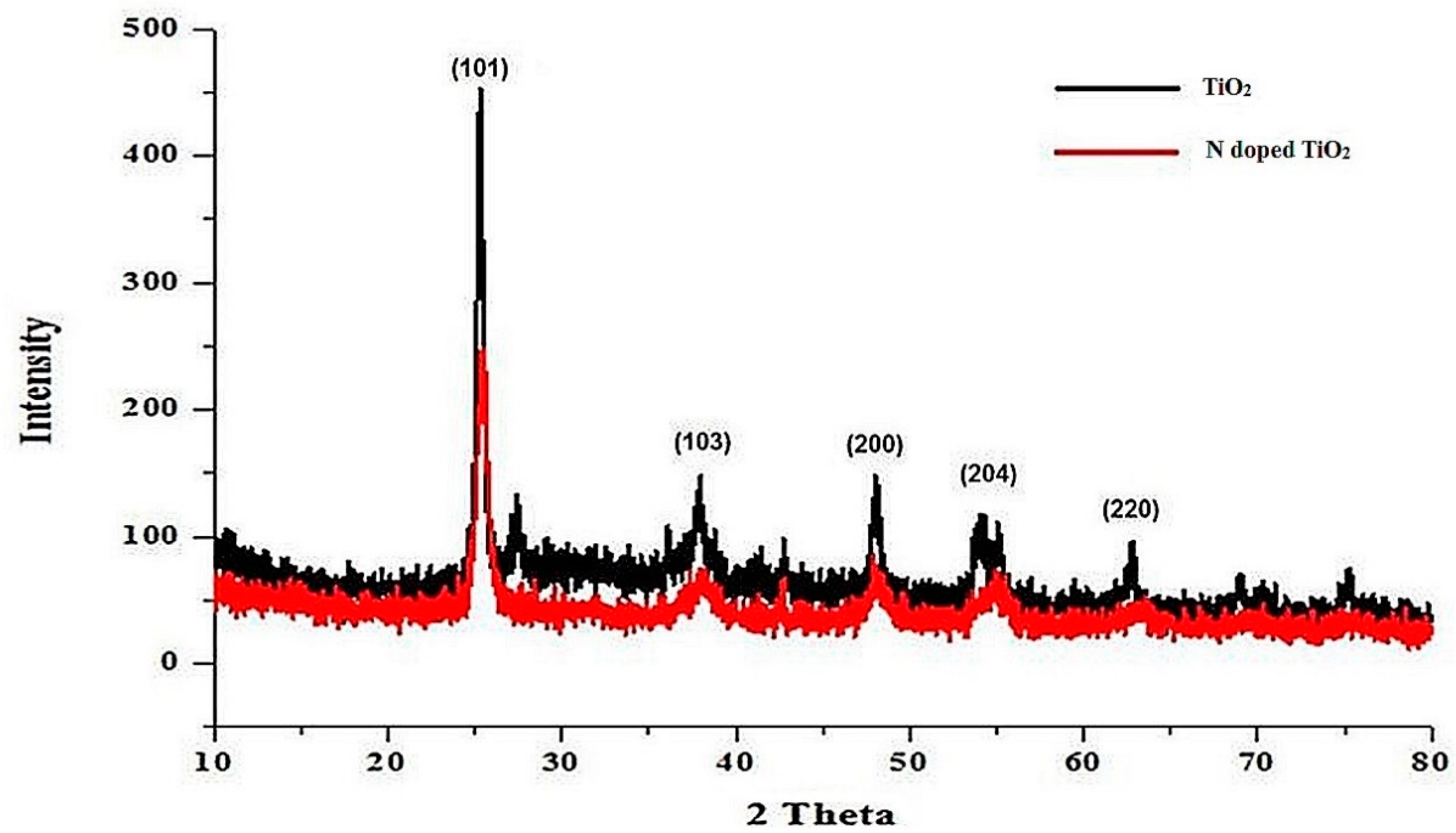
2.1. X-Ray Diffraction (XRD)
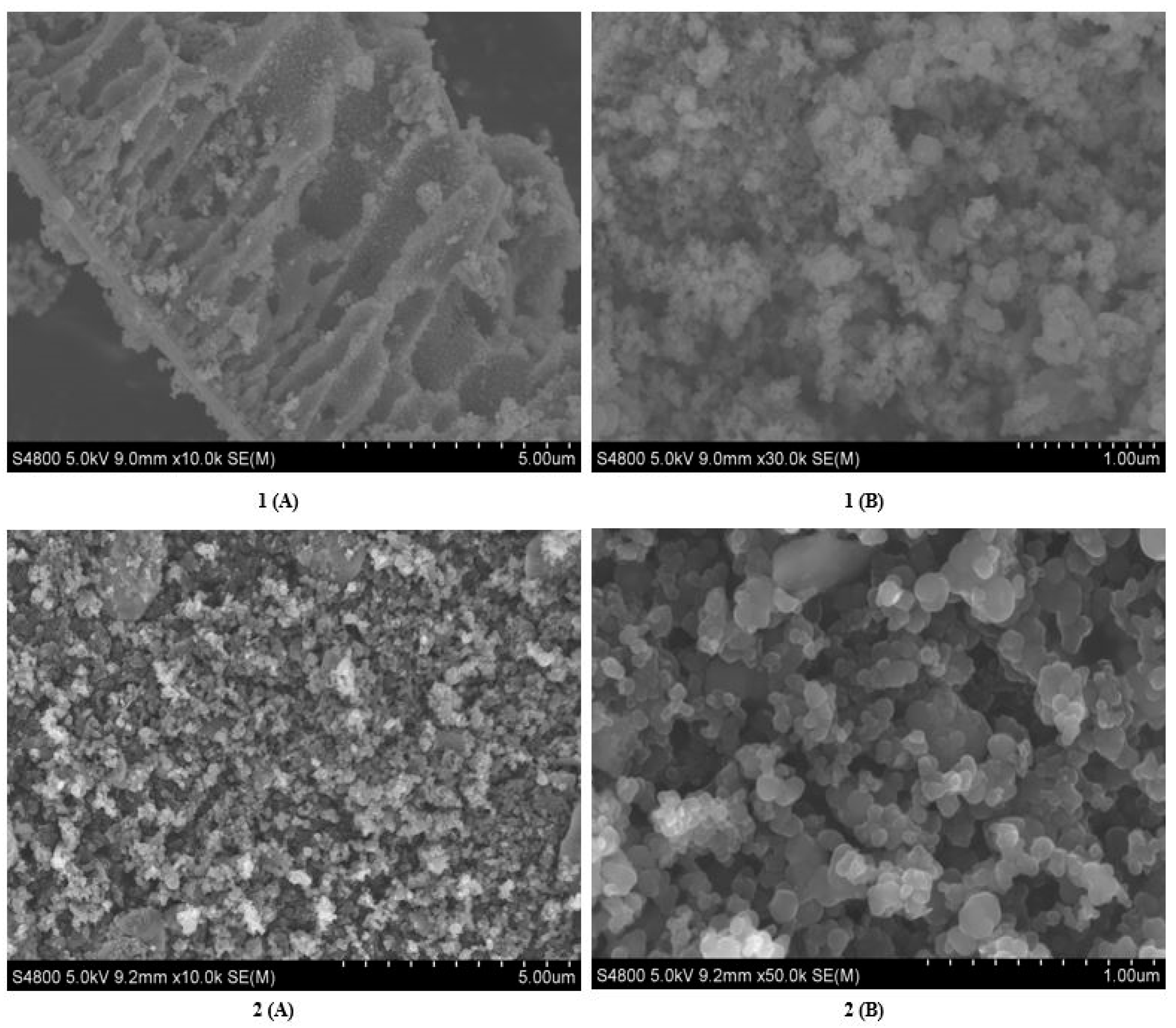
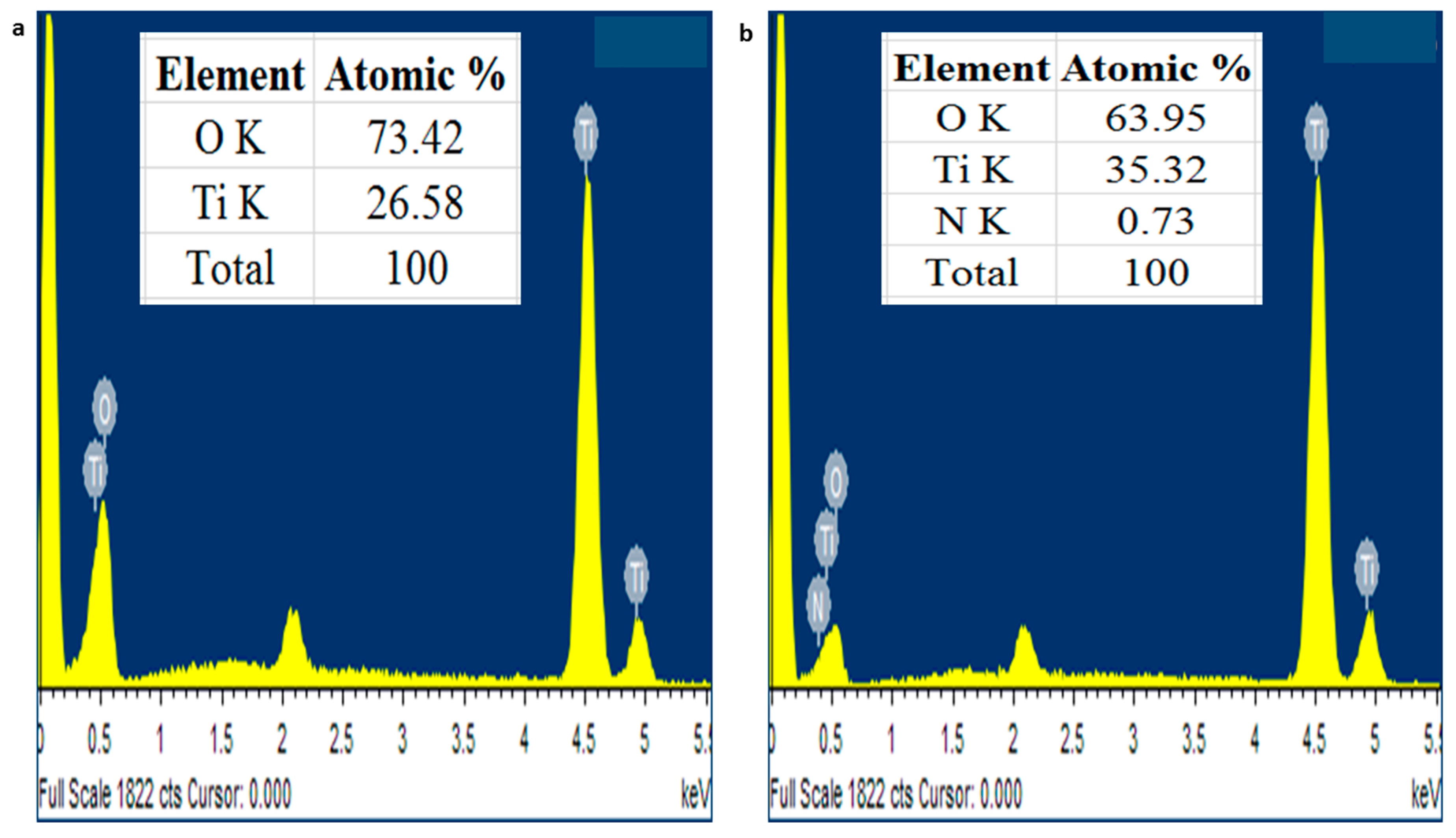
2.2. Scanning Electron Microscopy (SEM) and Energy Dispersive X-Ray (EDX)
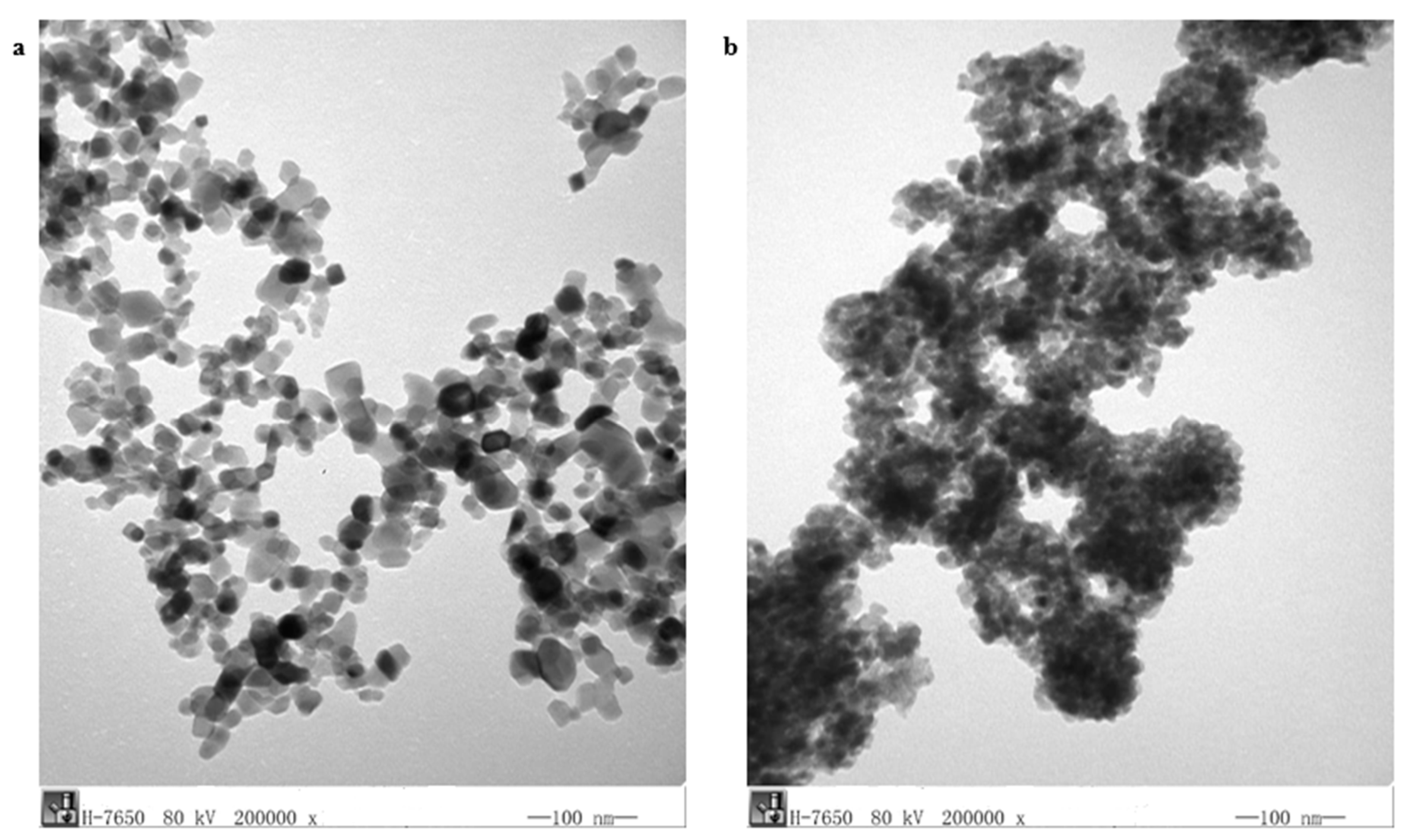
2.3. Transmission Electron Microscopy (TEM)
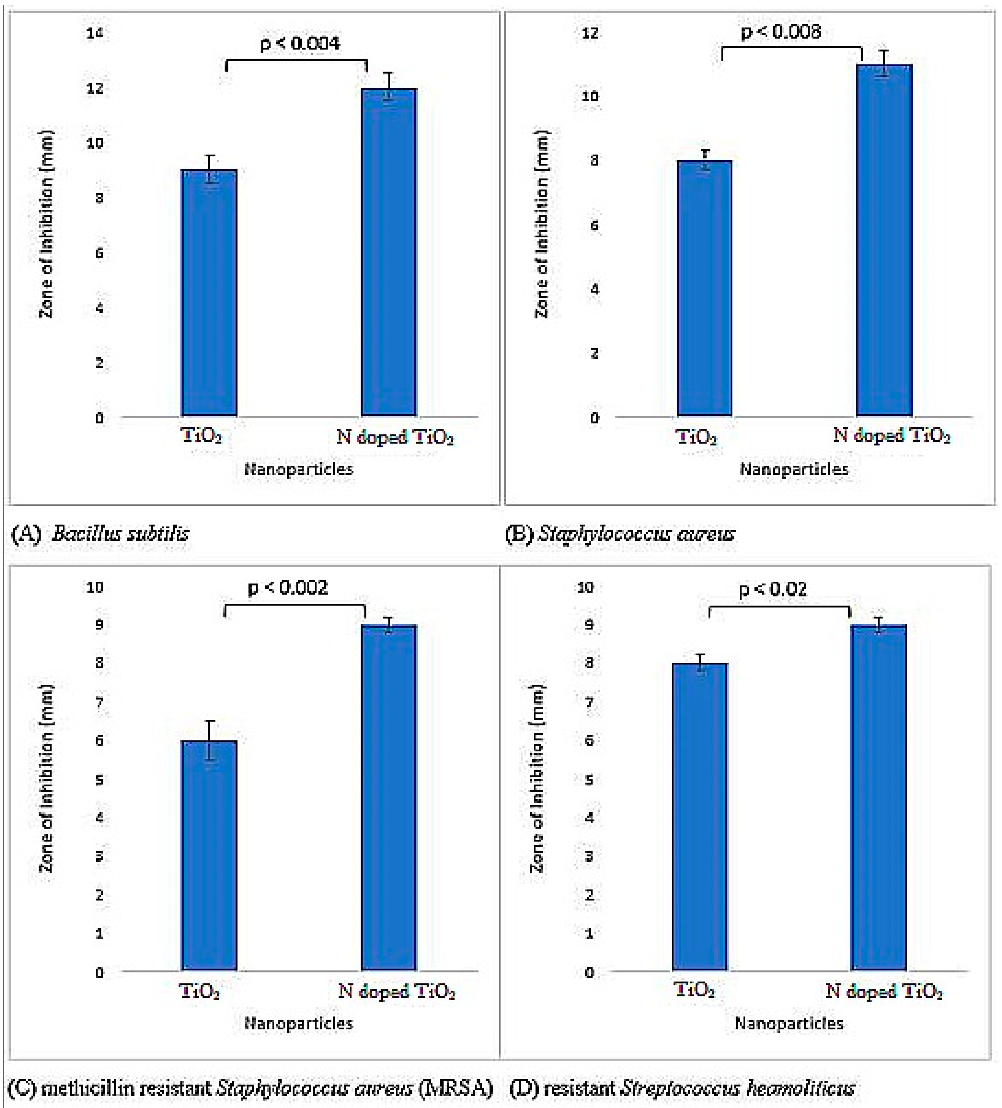
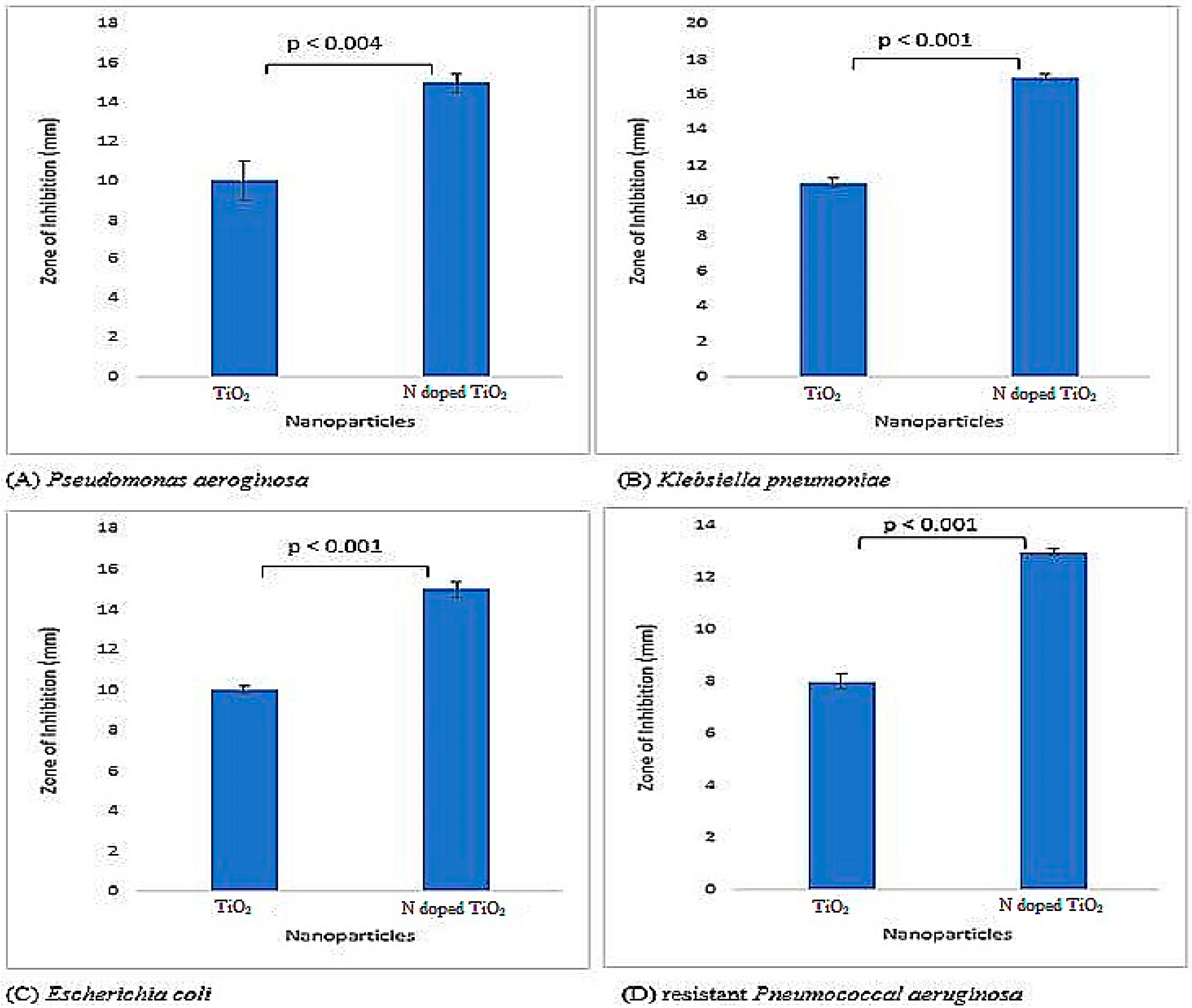
2.4. Antibacterial Activity
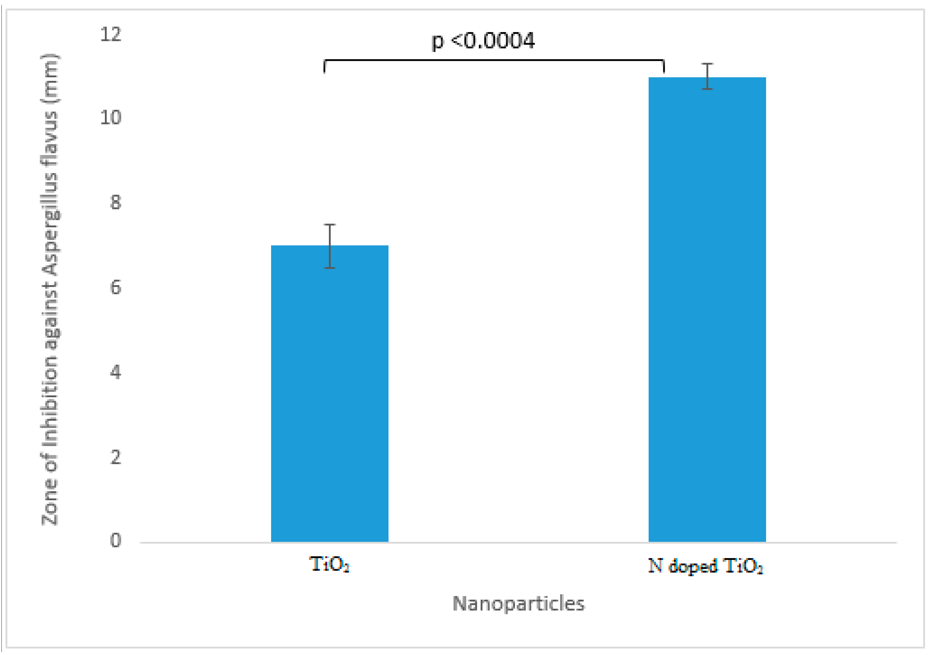
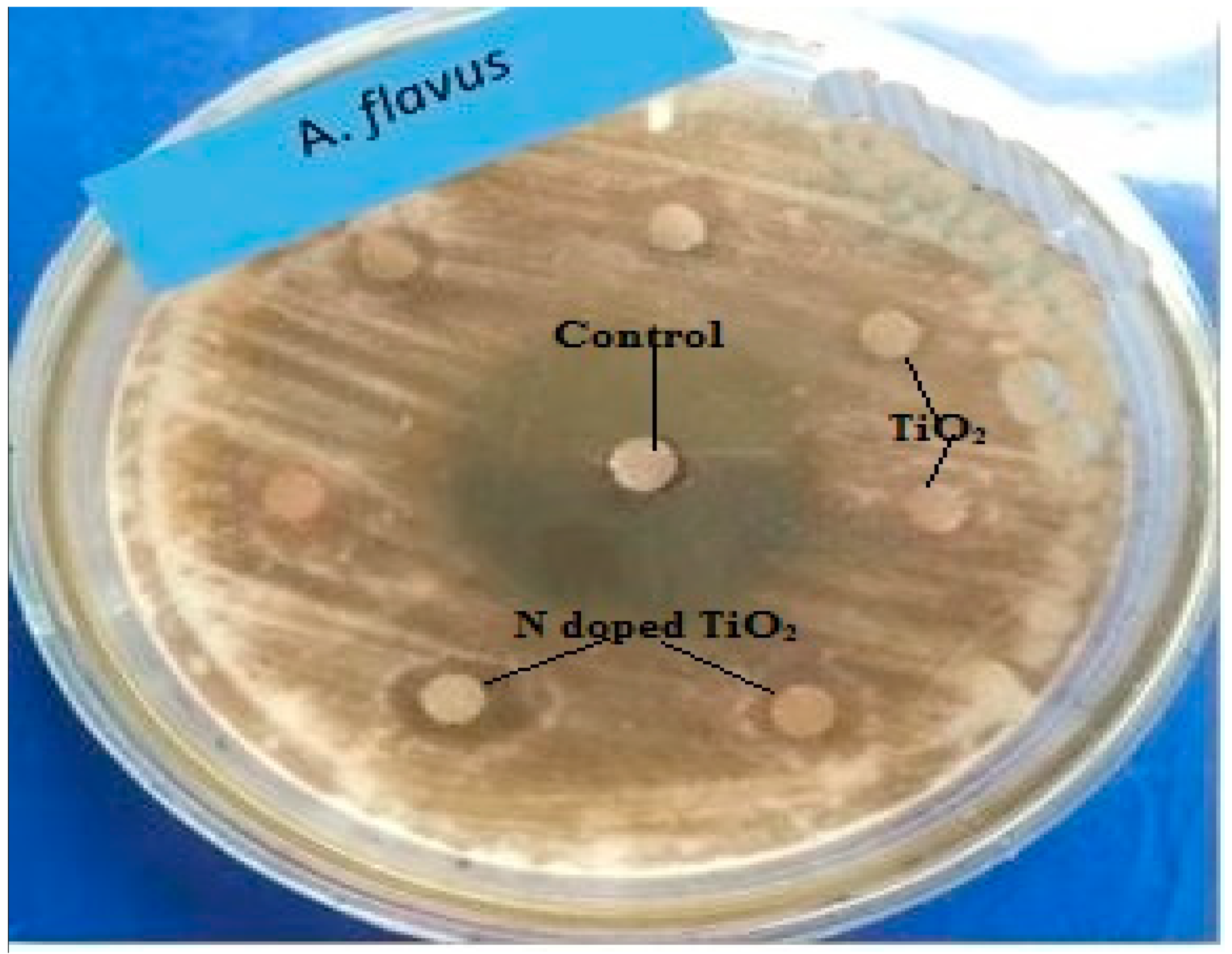
2.5. Antifungal Activity
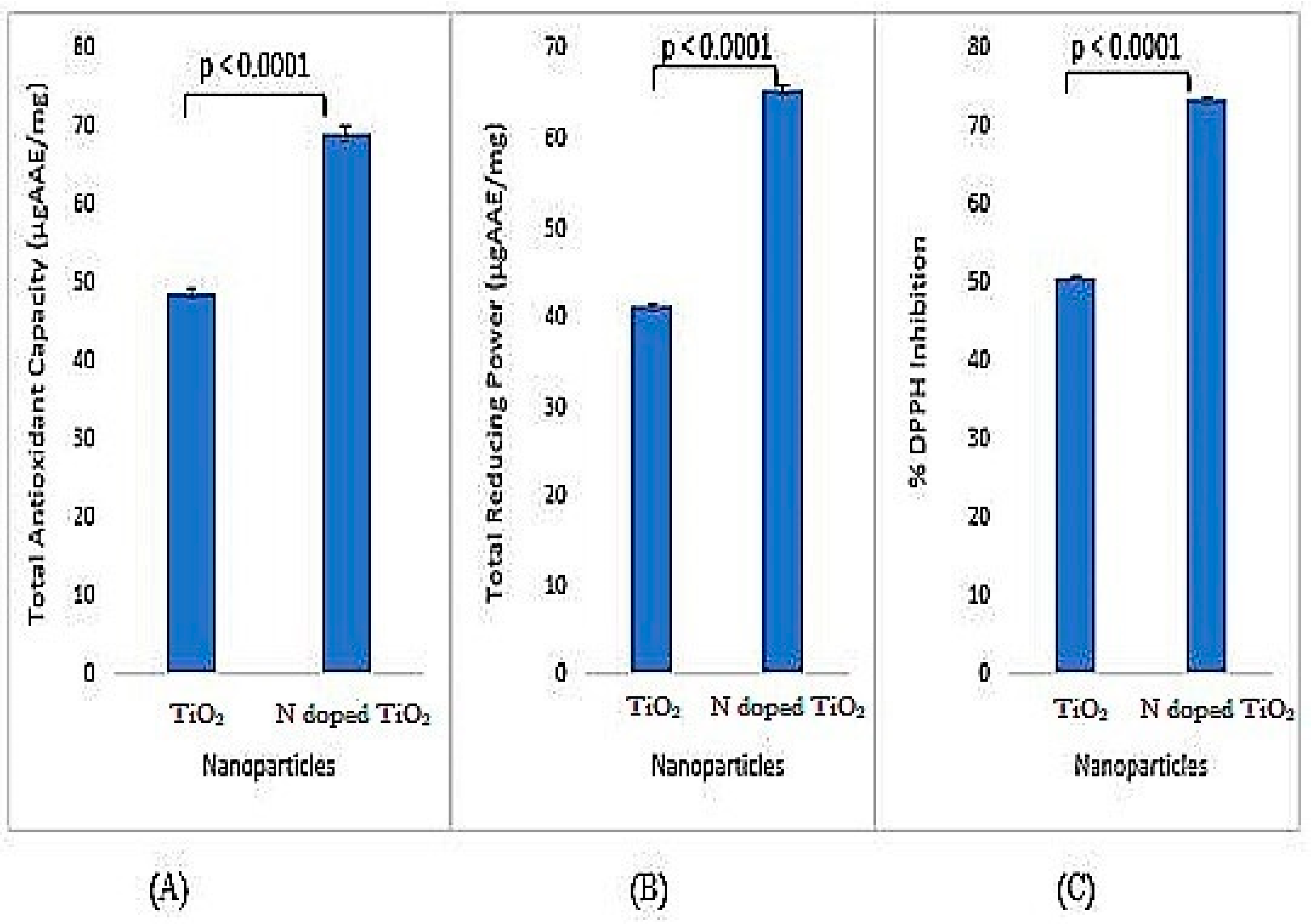
2.6. Antioxidant Activity
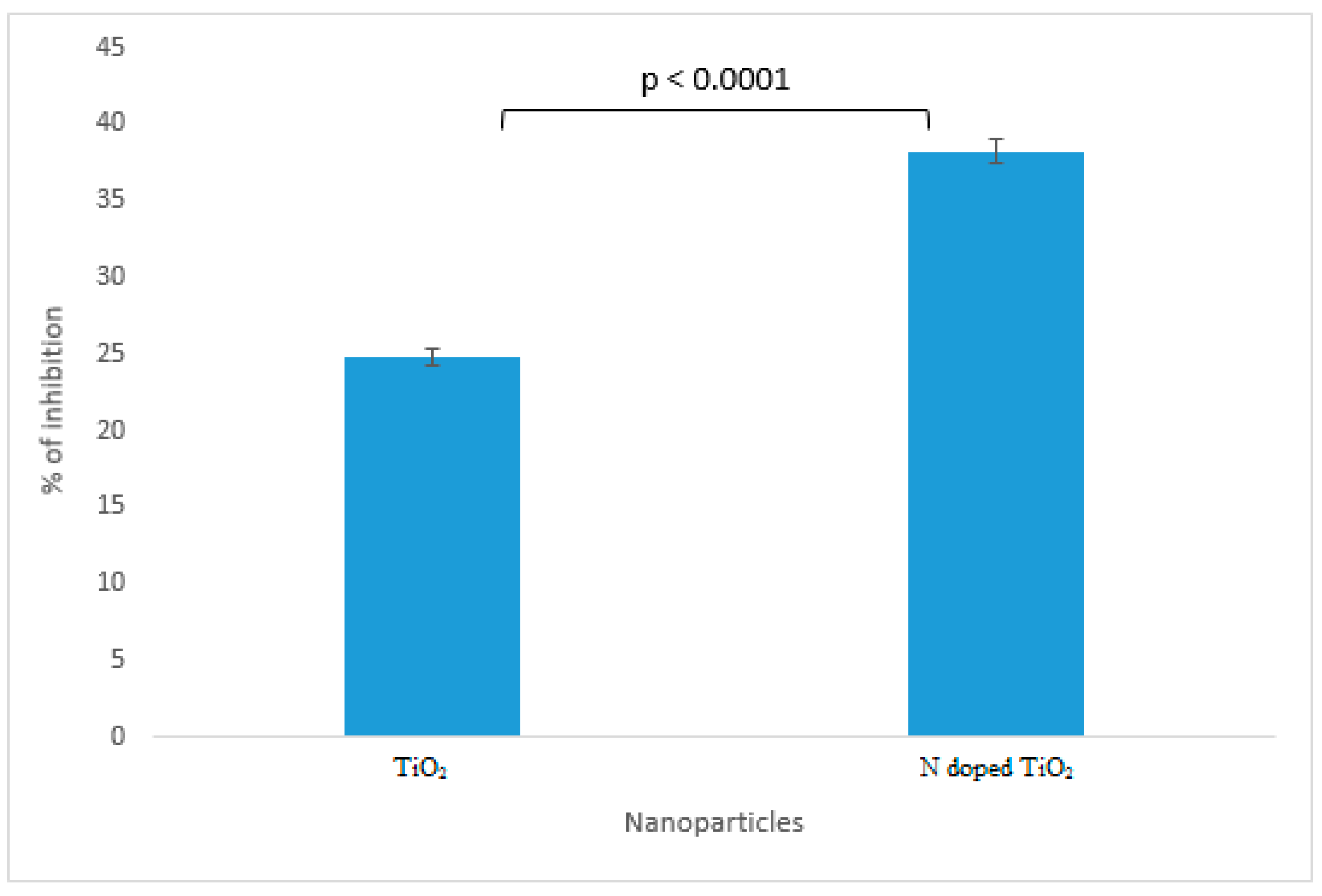
2.7. Antidiabetic Activity
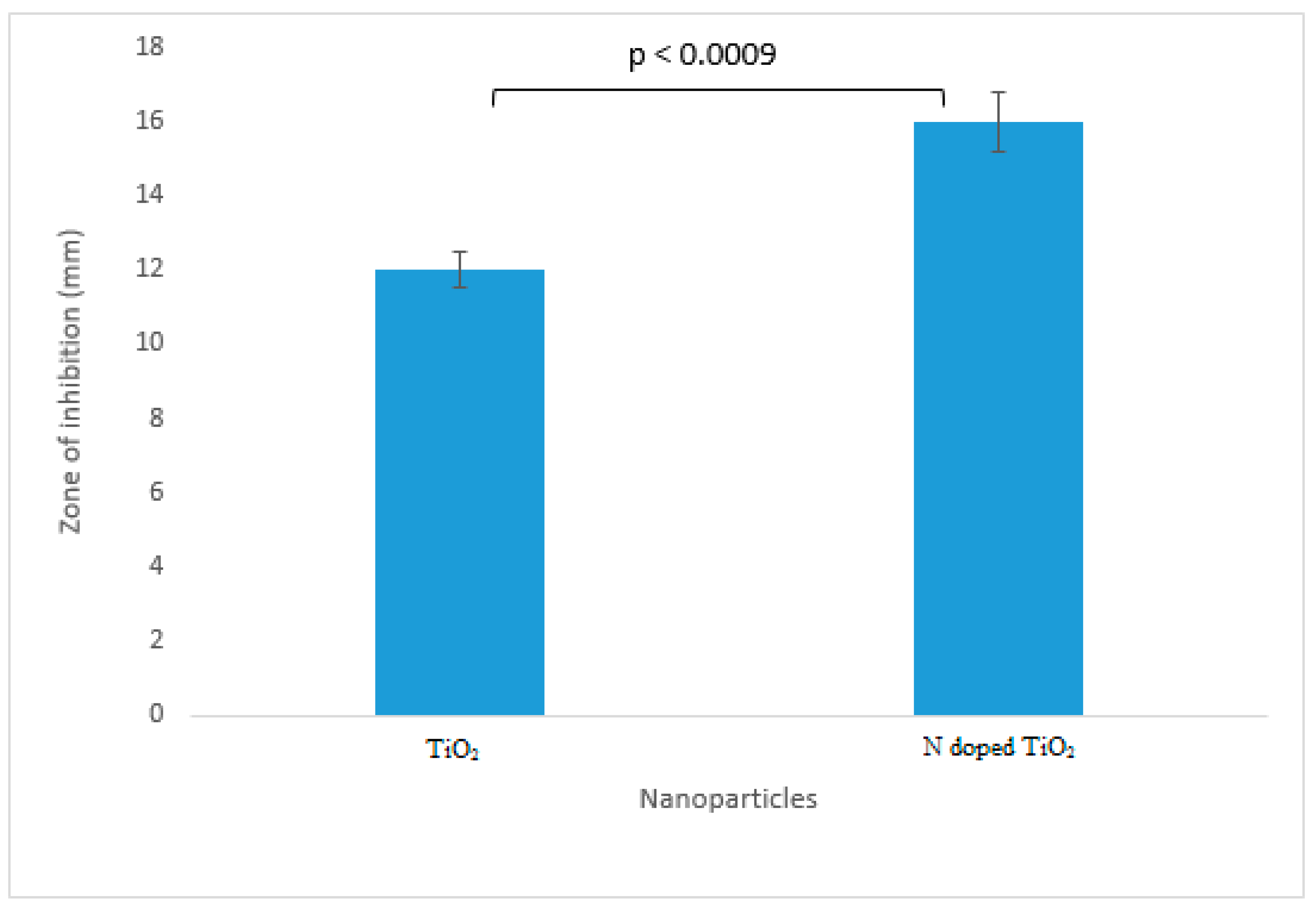
2.8. Protein Kinase Inhibition Activity
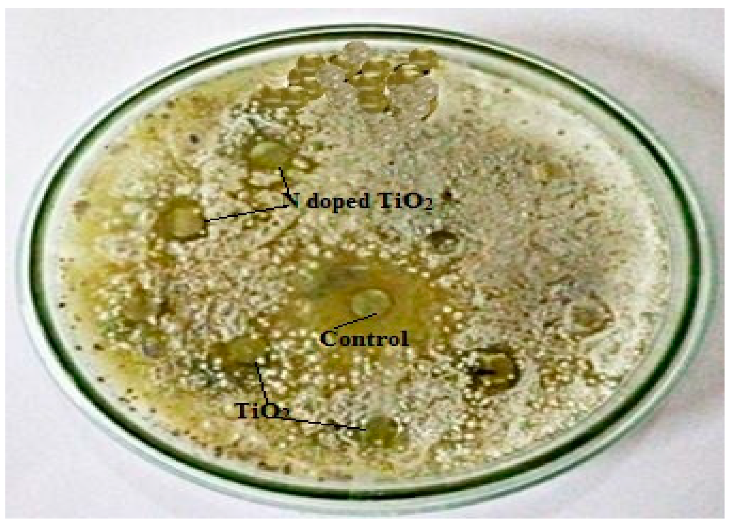
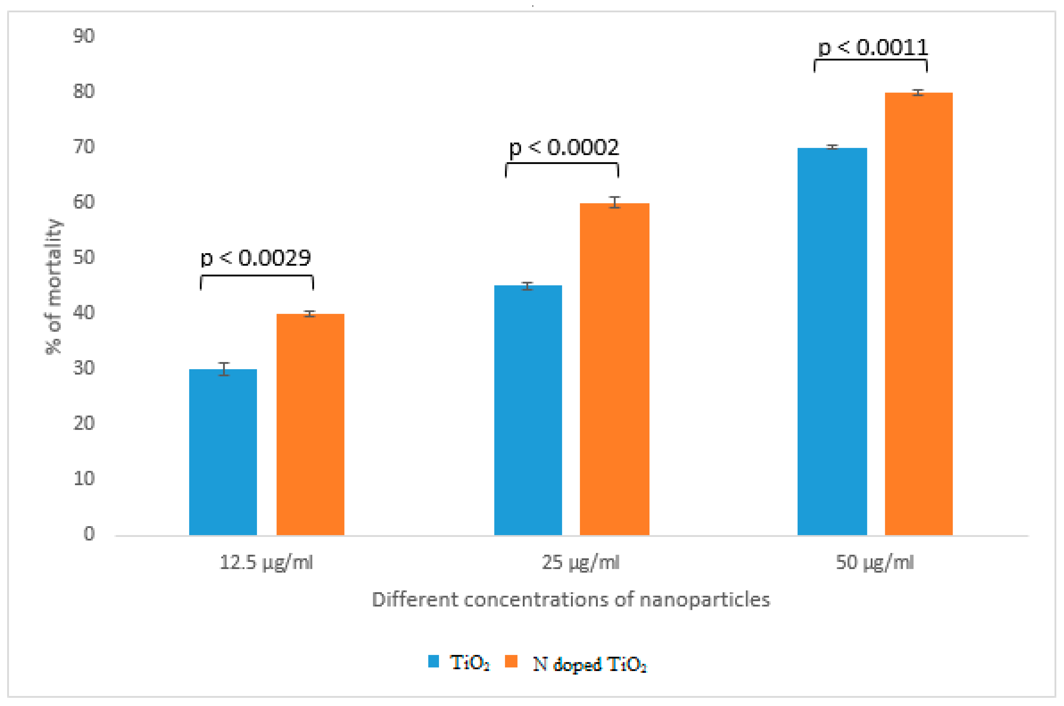
2.9. Cytotoxic Activity
3. Material and Methods
3.1. Chemical Fabrication of TiO2 Nanoparticles and N-Doped TiO2 Nanoparticles
3.2. Characterization of TiO2 Nanoparticles and N-doped TiO2 Nanoparticles
3.2.1. X-Ray Diffraction (XRD)
3.2.2. Scanning Electron Microscopy (SEM) and Energy Dispersive X-Ray (EDX)
3.2.3. Transmission Electron Microscopy (TEM)
3.3. Biological Potential of TiO2 Nanoparticles and N-Doped TiO2 Nanoparticles
3.3.1. Antibacterial Activity
3.3.2. Antifungal Activity
3.4. Antioxidant Activity
3.4.1. Total Antioxidant Capacity (TAC)
3.4.2. Total Reducing Power (TRP)
3.5. % DPPH Inhibition
3.6. Antidiabetic Activity
3.7. Protein Kinase Inhibition Activity
3.8. Cytotoxic Activity
3.9. Statistical Analysis
4. Conclusions
Author Contributions
Funding
Acknowledgments
Conflicts of Interest
References
- McNamara, K.; Tofail, S.A.M. Nanoparticles in biomedical applications. Adv. Phys. X 2017, 2, 54–88. [Google Scholar] [CrossRef]
- Ramos, A.P.; Cruz, M.A.E.; Tovani, C.B.; Ciancaglini, P. Biomedical applications of nanotechnology. Biophys. Rev. 2017, 9, 79–89. [Google Scholar] [CrossRef] [PubMed]
- Ali, A.; Ambreen, S.; Javed, R.; Tabassum, S.; Ul Haq, I.; Zia, M. ZnO nanostructure fabrication in different solvents transforms physio-chemical, biological and photodegradable properties. Mater. Sci Eng C Mater. Biol. Appl. 2017, 74, 137–145. [Google Scholar] [CrossRef] [PubMed]
- Javed, R.; Ahmed, M.; Haq, I.u.; Nisa, S.; Zia, M. PVP and PEG doped CuO nanoparticles are more biologically active: Antibacterial, antioxidant, antidiabetic and cytotoxic perspective. Mater. Sci. Eng. C 2017, 79, 108–115. [Google Scholar] [CrossRef] [PubMed]
- Ali, T.; Tripathi, P.; Azam, A.; Raza, W.; Ahmed, A.S.; Ahmed, A.; Muneer, M. Photocatalytic performance of Fe-doped TiO2 nanoparticles under visible-light irradiation. Mater. Res. Express 2017, 4, 015022. [Google Scholar] [CrossRef]
- Buraso, W.; Lachom, V.; Siriya, P.; Laokul, P. Synthesis of TiO2 nanoparticles via a simple precipitation method and photocatalytic performance. Mater. Res. Express 2018, 5, 115003. [Google Scholar] [CrossRef]
- Eadi, S.B.; Kim, S.; Jeong, S.W.; Jeon, H.W. Novel Preparation of Fe Doped TiO2 Nanoparticles and Their Application for Gas Sensor and Photocatalytic Degradation. Adv. Mater. Sci. Eng. 2017, 2017, 1–6. [Google Scholar] [CrossRef]
- Huang, J.; Guo, X.; Wang, B.; Li, L.; Zhao, M.; Dong, L.; Liu, X.; Huang, Y. Synthesis and Photocatalytic Activity of Mo-Doped TiO2 Nanoparticles. J. Spectrosc. 2015, 2015, 1–8. [Google Scholar] [CrossRef]
- Krishnakumar, V.; Boobas, S.; Jayaprakash, J.; Rajaboopathi, M.; Han, B.; Louhi-Kultanen, M. Effect of Cu doping on TiO2 nanoparticles and its photocatalytic activity under visible light. J. Mater. Sci. Mater. Electron. 2016, 27, 7438–7447. [Google Scholar] [CrossRef]
- Mahshid, S.; Askari, M.; Ghamsari, M.S. Synthesis of TiO2 nanoparticles by hydrolysis and peptization of titanium isopropoxide solution. J. Mater. Process. Technol. 2007, 189, 296–300. [Google Scholar] [CrossRef]
- Manzoor, M.; Rafiq, A.; Ikram, M.; Nafees, M.; Ali, S. Structural, optical, and magnetic study of Ni-doped TiO2 nanoparticles synthesized by sol–gel method. Int. Nano Lett. 2018, 8, 1–8. [Google Scholar] [CrossRef]
- Khairy, M.; Zakaria, W. Effect of metal-doping of TiO2 nanoparticles on their photocatalytic activities toward removal of organic dyes. Egypt. J. Pet. 2014, 23, 419–426. [Google Scholar] [CrossRef]
- Nithya, N.; Bhoopathi, G.; Magesh, G.; Kumar, C.D.N. Neodymium doped TiO2 nanoparticles by sol-gel method for antibacterial and photocatalytic activity. Mater. Sci. Semicond. Process. 2018, 83, 70–82. [Google Scholar] [CrossRef]
- Zahid, M.; Papadopoulou, E.L.; Suarato, G.; Binas, V.D.; Kiriakidis, G.; Gounaki, I.; Moira, O.; Venieri, D.; Bayer, I.S.; Athanassiou, A. Fabrication of Visible Light-Induced Antibacterial and Self-Cleaning Cotton Fabrics Using Manganese Doped TiO2 Nanoparticles. ACS Appl. Bio. Mater. 2018, 1, 1154–1164. [Google Scholar] [CrossRef]
- Viana, M.M.; Soares, V.F.; Mohallem, N.D.S. Synthesis and characterization of TiO2 nanoparticles. Ceram. Int. 2010, 36, 2047–2053. [Google Scholar] [CrossRef]
- Ahamed, M.; Khan, M.A.M.; Akhtar, M.J.; Alhadlaq, H.A.; Alshamsan, A. Ag-doping regulates the cytotoxicity of TiO2 nanoparticles via oxidative stress in human cancer cells. Sci. Rep. 2017, 7, 17662. [Google Scholar] [CrossRef]
- Ahmad, J.; Siddiqui, M.; Akhtar, M.; Alhadlaq, H.; Alshamsan, A.; Khan, S.; Wahab, R.; Al-Khedhairy, A.; Al-Salim, A.; Musarrat, J.; et al. Copper doping enhanced the oxidative stress–mediated cytotoxicity of TiO2 nanoparticles in A549 cells. Hum. Exp. Toxicol. 2018, 37, 496–507. [Google Scholar] [CrossRef]
- Caratto, V.; Locardi, F.; Alberti, S.; Villa, S.; Sanguineti, E.; Martinelli, A.; Balbi, T.; Canesi, L.; Ferretti, M. Different sol–gel preparations of iron-doped TiO2 nanoparticles: Characterization, photocatalytic activity and cytotoxicity. J. Sol.-Gel Sci. Technol. 2016, 80, 152–159. [Google Scholar] [CrossRef]
- Cheng, X.; Yu, X.; Xing, Z.; Yang, L. Synthesis and characterization of N-doped TiO2 and its enhanced visible-light photocatalytic activity. Arab. J. Chem. 2016, 9, S1706–S1711. [Google Scholar] [CrossRef]
- Ansari, S.A.; Khan, M.M.; Ansari, M.O.; Cho, M.H. Nitrogen-doped titanium dioxide (N-doped TiO2) for visible light photocatalysis. New J. Chem. 2016, 40, 3000–3009. [Google Scholar] [CrossRef]
- Kim, T.H.; Go, G.-M.; Cho, H.-B.; Song, Y.; Lee, C.-G.; Choa, Y.-H. A Novel Synthetic Method for N-doped TiO2 Nanoparticles Through Plasma-Assisted Electrolysis and Photocatalytic Activity in the Visible Region. Front. Chem. 2018, 6. [Google Scholar] [CrossRef] [PubMed]
- Zane, A.; Zuo, R.; Villamena, F.A.; Rockenbauer, A.; Digeorge Foushee, A.M.; Flores, K.; Dutta, P.K.; Nagy, A. Biocompatibility and antibacterial activity of nitrogen-doped titanium dioxide nanoparticles for use in dental resin formulations. Int. J. Nanomed. 2016, 11, 6459–6470. [Google Scholar] [CrossRef] [PubMed]
- Salehi, P.; Babanouri, N.; Roein-Peikar, M.; Zare, F. Long-term antimicrobial assessment of orthodontic brackets coated with nitrogen-doped titanium dioxide against Streptococcus mutans. Prog. Orthod. 2018, 19, 35. [Google Scholar] [CrossRef] [PubMed]
- Atchudan, R.; Edison, T.N.J.I.; Perumal, S.; Vinodh, R.; Lee, Y.R. In-situ green synthesis of nitrogen-doped carbon dots for bioimaging and TiO2 nanoparticles@nitrogen-doped carbon composite for photocatalytic degradation of organic pollutants. J. Alloy. Compd. 2018, 766, 12–24. [Google Scholar] [CrossRef]
- Li, Z.; Mi, L.; Wang, P.-N.; Chen, J.-Y. Study on the visible-light-induced photokilling effect of nitrogen-doped TiO2 nanoparticles on cancer cells. Nanoscale Res. Lett. 2011, 6, 356. [Google Scholar] [CrossRef]
- Katoueizadeh, E.; Zebarjad, S.M.; Janghorban, K. Synthesis and enhanced visible-light activity of N-doped TiO2 nano-additives applied over cotton textiles. J. Mater. Res. Technol. 2018, 7, 204–211. [Google Scholar] [CrossRef]
- Alavi, M.; Karimi, N. Characterization, antibacterial, total antioxidant, scavenging, reducing power and ion chelating activities of green synthesized silver, copper and titanium dioxide nanoparticles using Artemisia haussknechtii leaf extract. Artif. Cells Nanomed. Biotechnol. 2017, 46, 2066–2081. [Google Scholar] [CrossRef]
- Arora, B.; Murar, M.; Dhumale, V. Antimicrobial potential of TiO2 nanoparticles against MDR Pseudomonas aeruginosa. J. Exp. Nanosci. 2015, 10, 819–827. [Google Scholar] [CrossRef]
- Kalyanasundaram, S.; Prakash, M.J. Biosynthesis and Characterization of Titanium Dioxide Nanoparticles Using Pithecellobium Dulce and Lagenaria Siceraria Aqueous Leaf Extract and Screening their Free Radical Scavenging and Antibacterial Properties. ILCPA 2015, 50, 80–95. [Google Scholar] [CrossRef]
- Santhoshkumar, T.; Rahuman, A.A.; Jayaseelan, C.; Rajakumar, G.; Marimuthu, S.; Kirthi, A.V.; Velayutham, K.; Thomas, J.; Venkatesan, J.; Kim, S.-K. Green synthesis of titanium dioxide nanoparticles using Psidium guajava extract and its antibacterial and antioxidant properties. Asian Pac. J. Trop. Med. 2014, 7, 968–976. [Google Scholar] [CrossRef]
- Senarathna, U.L.N.H.; Fernando, S.S.N.; Gunasekara, T.D.C.P.; Weerasekera, M.M.; Hewageegana, H.G.S.P.; Arachchi, N.D.H.; Siriwardena, H.D.; Jayaweera, P.M. Enhanced antibacterial activity of TiO2 nanoparticle surface modified with Garcinia zeylanica extract. Chem. Cent. J. 2017, 11, 7. [Google Scholar] [CrossRef] [PubMed]
- Burello, E.; Worth, A.P. A theoretical framework for predicting the oxidative stress potential of oxide nanoparticles. Nanotoxicology 2011, 5, 228–235. [Google Scholar] [CrossRef] [PubMed]
- Caratto, V.; Ball, L.; Sanguineti, E.; Insorsi, A.; Firpo, I.; Alberti, S.; Ferretti, M.; Pelosi, P. Antibacterial activity of standard and N-doped titanium dioxide-coated endotracheal tubes: An in vitro study. Rev. Bras. De Ter. Intensiva 2017, 29, 55–62. [Google Scholar] [CrossRef] [PubMed]
- Wang, L.; Hu, C.; Shao, L. The antimicrobial activity of nanoparticles: Present situation and prospects for the future. IJN 2017, Volume 12, 1227–1249. [Google Scholar] [CrossRef]
- Javed, R.; Usman, M.; Tabassum, S.; Zia, M. Effect of capping agents: Structural, optical and biological properties of ZnO nanoparticles. Appl. Surf. Sci. 2016, 386, 319–326. [Google Scholar] [CrossRef]
- Ripolles-Avila, C.; Martinez-Garcia, M.; Hascoët, A.-S.; Rodríguez-Jerez, J.J. Bactericidal efficacy of UV activated TiO2 nanoparticles against Gram-positive and Gram-negative bacteria on suspension. CyTA-J. Food 2019, 17, 408–418. [Google Scholar] [CrossRef]
- Maheswari, P.; Ponnusamy, S.; Harish, S.; Ganesh, M.R.; Hayakawa, Y. Hydrothermal synthesis of pure and bio modified TiO2: Characterization, evaluation of antibacterial activity against gram positive and gram negative bacteria and anticancer activity against KB Oral cancer cell line. Arab. J. Chem. 2018, S1878535218302508. [Google Scholar] [CrossRef]
- Haghighi, F.; Mohammadi, S.R.; Mohammadi, P.; Hosseinkhani, S. Antifungal Activity of TiO2 nanoparticles and EDTA on Candida albicans Biofilms. Infect. Epidemiol. Med. 2013, 1, 33–38. [Google Scholar]
- Niska, K.; Inkielewicz-Stepniak, I.; Pyszka, K.; Tukaj, C.; Wozniak, M.; Radomski, M. Titanium dioxide nanoparticles enhance production of superoxide anion and alter the antioxidant system in human osteoblast cells. IJN 2015, 10, 1095–1107. [Google Scholar]
- Langle, A.; González-Coronel, M.A.; Carmona-Gutiérrez, G.; Moreno-Rodríguez, J.A.; Venegas, B.; Muñoz, G.; Treviño, S.; Díaz, A. Stevia rebaudiana loaded titanium oxide nanomaterials as an antidiabetic agent in rats. Rev. Bras. Farmacogn. 2015, 25, 145–151. [Google Scholar] [CrossRef]
- Bogdan, J.; Pławińska-Czarnak, J.; Zarzyńska, J. Nanoparticles of Titanium and Zinc Oxides as Novel Agents in Tumor Treatment: A Review. Nanoscale Res. Lett 2017, 12, 225. [Google Scholar] [CrossRef] [PubMed]
- Ji, J.; Yang, H.; Liu, Y.; Chen, H.; Kong, J.; Liu, B. TiO2-assisted silver enhanced biosensor for kinase activity profiling. Chem. Commun. 2009, 1508. [Google Scholar] [CrossRef] [PubMed]
- Latha, S.T.; Reddy, M.C.; Muthukonda, S.V.; Srikanth, V.V.S.S.; Lomada, D. In vitro and in vivo evaluation of anti-cancer activity: Shape-dependent properties of TiO 2 nanostructures. Mater. Sci. Eng.: C 2017, 78, 969–977. [Google Scholar] [CrossRef] [PubMed]
- Yan, Z.; Deng, P.; Liu, Y. Recent Advances in Protein Kinase Activity Analysis Based on Nanomaterials. IJMS 2019, 20, 1440. [Google Scholar] [CrossRef]
- He, F.; Yu, W.; Fan, X.; Jin, B. In vitro cytotoxicity of biosynthesized titanium dioxide nanoparticles in human prostate cancer cell lines. Trop. J. Pharm. Res. 2018, 16, 2793–2799. [Google Scholar] [CrossRef][Green Version]
- Chellappa, M.; Anjaneyulu, U.; Manivasagam, G.; Vijayalakshmi, U. Preparation and evaluation of the cytotoxic nature of TiO2 nanoparticles by direct contact method. IJN 2015, 10, 31–40. [Google Scholar]
- Kukia, R.N.; Rasmi, Y.; Abbasi, A.; Koshoridze, N.; Shirpoor, A.; Burjanadze, G.; Saboory, E. Bio-Effects of TiO2 Nanoparticles on Human Colorectal Cancer and Umbilical Vein Endothelial Cell Lines. Asian Pac. J. Cancer Prev. 2018, 19, 2821–2829. [Google Scholar]
- Koca, F.D.; Duman, F. Genotoxic and cytotoxic activity of green synthesized TiO2 nanoparticles. Appl. Nanosci. 2019, 9, 815–823. [Google Scholar] [CrossRef]
- Horie, M.; Sugino, S.; Kato, H.; Tabei, Y.; Nakamura, A.; Yoshida, Y. Does photocatalytic activity of TiO2 nanoparticles correspond to photo-cytotoxicity? Cellular uptake of TiO2 nanoparticles is important in their photo-cytotoxicity. Toxicol. Mech. Methods 2016, 26, 284–294. [Google Scholar] [CrossRef]
- Huerta-García, E.; Zepeda-Quiroz, I.; Sánchez-Barrera, H.; Colín-Val, Z.; Alfaro-Moreno, E.; Ramos-Godinez, M.; López-Marure, R. Internalization of Titanium Dioxide Nanoparticles Is Cytotoxic for H9c2 Rat Cardiomyoblasts. Molecules 2018, 23, 1955. [Google Scholar] [CrossRef]
- Zhu, Y.; Eaton, J.W.; Li, C. Titanium Dioxide (TiO2) Nanoparticles Preferentially Induce Cell Death in Transformed Cells in a Bak/Bax-Independent Fashion. PLoS ONE 2012, 7. [Google Scholar] [CrossRef] [PubMed]
- Shajudheen, V.P.M.; Viswanathan, K.; Anitha, R.K.; Uma, M.A.; Saravana, K.S. A Simple Chemical Precipitation Method of Titanium Dioxide Nanoparticles Using Polyvinyl Pyrrolidone As A Capping Agent And Their Characterization. Int. J. Chem. Mol. Eng. 2016, 10. [Google Scholar]
- Haq, I.U.; Mannan, A.; Ahmed, I.; Hussain, I.; Jamil, M.; Mirza, B. ANTIBACTERIAL ACTIVITY AND BRINE SHRIMP TOXICITY OF ARTEMISIA DUBIA EXTRACT. Pak. J. Bot. 2012, 44, 1487–1490. [Google Scholar]
- Akhtar, N.; Ihsan-ul-Haq; Mirza, B. Phytochemical analysis and comprehensive evaluation of antimicrobial and antioxidant properties of 61 medicinal plant species. Arab. J. Chem. 2018, 11, 1223–1235. [Google Scholar] [CrossRef]
- Khan, S.; Ur-Rehman, T.; Mirza, B.; Ul-Haq, I.; Zia, M. Antioxidant, Antimicrobial, Cytotoxic and Protein Kinase Inhibition Activities of Fifteen Traditional Medicinal Plants From Pakistan. Pharm. Chem. J. 2017, 51, 391–398. [Google Scholar] [CrossRef]
- Kim, J.-S.; Kwon, Y.-S.; Chun, W.-J.; Kim, T.-Y.; Sun, J.; Yu, C.-Y.; Kim, M.-J. Rhus verniciflua Stokes flavonoid extracts have anti-oxidant, anti-microbial and α-glucosidase inhibitory effect. Food Chem. 2010, 120, 539–543. [Google Scholar] [CrossRef]
- Yao, G.; Sebisubi, F.M.; Voo, L.Y.C.; Ho, C.C.; Tan, G.T.; Chang, L.C. Citrinin derivatives from the soil filamentous fungus Penicillium sp. H9318. J. Braz. Chem. Soc. 2011, 22, 1125–1129. [Google Scholar] [CrossRef]
- Apu, A.S.; Bhuyan, S.H.; Khatun, F.; Liza, M.S.; Matin, M.; Hossain, F. Assessment of cototoxic activity of two medicinal plants using brine shrimp (artemia salina) as an erperimental tool. IJPSR 2013, 4, 1125–1130. [Google Scholar]
Sample Availability: Samples of the compounds are not available from the authors. |


| Methods of Characterization and Biological Potential | TiO2 Nanoparticles | N-doped TiO2 Nanoparticles |
|---|---|---|
| Characterization | ||
| XRD | Size: 25 nm | Size: 17.5 nm |
| SEM | Morphology: Spherical but lesser clarity of shape due to more aggregation | Morphology: Spherical and more clarity of shape due to lesser aggregation |
| EDX | O: 73.42% | O: 63.95% |
| Ti: 26.58% | Ti: 35.32% | |
| - | N: 0.73% | |
| TEM | Size: 20–25 nm | Size: 10–15 nm |
| Biological Potential | ||
| Antibacterial activity against Gram-positive bacteria: | Zone of Inhibition Gram-positive bacteria: | Zone of Inhibition Gram-positive bacteria: |
| Bacillus subtilis | 9 mm | 12 mm |
| Staphylococcus aureusmethicillin resistant | 8 mm | 11 mm |
| Staphylococcus aureus resistant | 6 mm | 9 mm |
| Streptococcus haemoliticus | 8 mm | 9 mm |
| Gram-negative bacteria: | Gram-negative bacteria: | Gram-negative bacteria: |
| Pseudomonas aeruginosa | 10 mm | 15 mm |
| Klebsiella pneumoniae | 11 mm | 17 mm |
| Escherichia coli resistant | 10 mm | 15 mm |
| Pneumococcal aeruginosa | 8 mm | 13 mm |
| Antifungal activity against Aspergillus flavus | Zone of Inhibition: 7 mm | Zone of Inhibition: 11 mm |
| Antioxidant activity | ||
| TAC | 48.9 µgAAE/mg | 69.1 µgAAE/mg |
| TRP | 41.2 µgAAE/mg | 65.5 µgAAE/mg |
| % DPPH Inhibition | 50.8% | 73.6% |
| Antidiabetic activity against alpha amylase enzyme | % of Inhibition: 24.8% | % of Inhibition: 38.2% |
| Protein kinase inhibitory activity | Zone of Inhibition: 12 mm | Zone of Inhibition: 16 mm |
| Cytotoxic activity at | % of mortality | % of mortality |
| 12.5 µg/mL | 30% | 40% |
| 25 µg/mL | 45% | 60% |
| 50 µg/mL | 70% | 80% |
© 2019 by the authors. Licensee MDPI, Basel, Switzerland. This article is an open access article distributed under the terms and conditions of the Creative Commons Attribution (CC BY) license (http://creativecommons.org/licenses/by/4.0/).
Share and Cite
Ahmad, M.A.; Yuesuo, Y.; Ao, Q.; Adeel, M.; Hui, Z.Y.; Javed, R. Appraisal of Comparative Therapeutic Potential of Undoped and Nitrogen-Doped Titanium Dioxide Nanoparticles. Molecules 2019, 24, 3916. https://doi.org/10.3390/molecules24213916
Ahmad MA, Yuesuo Y, Ao Q, Adeel M, Hui ZY, Javed R. Appraisal of Comparative Therapeutic Potential of Undoped and Nitrogen-Doped Titanium Dioxide Nanoparticles. Molecules. 2019; 24(21):3916. https://doi.org/10.3390/molecules24213916
Chicago/Turabian StyleAhmad, Muhammad Arslan, Yang Yuesuo, Qiang Ao, Muhammad Adeel, Zhang Yan Hui, and Rabia Javed. 2019. "Appraisal of Comparative Therapeutic Potential of Undoped and Nitrogen-Doped Titanium Dioxide Nanoparticles" Molecules 24, no. 21: 3916. https://doi.org/10.3390/molecules24213916
APA StyleAhmad, M. A., Yuesuo, Y., Ao, Q., Adeel, M., Hui, Z. Y., & Javed, R. (2019). Appraisal of Comparative Therapeutic Potential of Undoped and Nitrogen-Doped Titanium Dioxide Nanoparticles. Molecules, 24(21), 3916. https://doi.org/10.3390/molecules24213916