Food-Grade Encapsulation Systems for (−)-Epigallocatechin Gallate

 , and
, and
Abstract
1. Introduction
2. EGCG in Tea
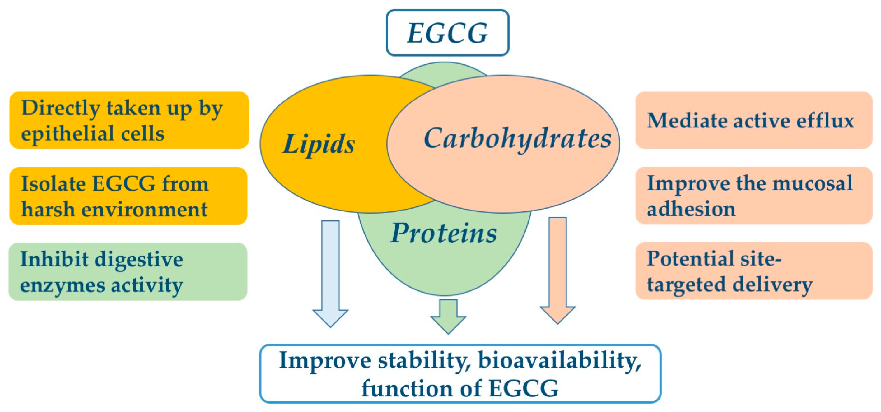
3. Food-Grade Encapsulation System
3.1. Proteins for EGCG Encapsulation
3.2. Carbohydrates for EGCG Microencapsulation
3.3. Lipids for EGCG Microencapsulation
3.4. Food Grade Systems for EGCG Delivery and Their Possible Mechanisms of Action
4. Challenges
5. Conclusions and Expectations
Acknowledgments
Author Contributions
Conflicts of Interest
References
- Fu, Q.Y.; Li, Q.S.; Lin, X.M.; Qiao, R.Y.; Yang, R.; Li, X.M.; Dong, Z.B.; Xiang, L.P.; Zheng, X.Q.; Lu, J.L.; et al. Antidiabetic effects of tea. Molecules 2017, 22, 849. [Google Scholar] [CrossRef] [PubMed]
- Thitimuta, S.; Pithayanukul, P.; Nithitanakool, S.; Bavovada, R.; Leanpolchareanchai, J.; Saparpakorn, P. Camellia sinensis l. Extract and its potential beneficial effects in antioxidant, anti-inflammatory, anti-hepatotoxic, and anti-tyrosinase activities. Molecules 2017, 22, 401. [Google Scholar] [CrossRef] [PubMed]
- Kondo, K.; Kurihara, M.; Miyata, N.; Suzuki, T.; Toyoda, M. Mechanistic studies of catechins as antioxidants against radical oxidation. Arch. Biochem. Biophys. 1999, 362, 79–86. [Google Scholar] [CrossRef] [PubMed]
- Xiang, L.P.; Wang, A.; Ye, J.H.; Zheng, X.Q.; Polito, C.A.; Lu, J.L.; Li, Q.S.; Liang, Y.R. Suppressive effects of tea catechins on breast cancer. Nutrients 2016, 8, 458. [Google Scholar] [CrossRef] [PubMed]
- Du, L.L.; Fu, Q.Y.; Xiang, L.P.; Zheng, X.Q.; Lu, J.L.; Ye, J.H.; Li, Q.S.; Polito, C.A.; Liang, Y.R. Tea polysaccharides and their bioactivities. Molecules 2016, 21, 1449. [Google Scholar] [CrossRef] [PubMed]
- Shi, M.; Nie, Y.; Zheng, X.Q.; Lu, J.L.; Liang, Y.R.; Ye, J.H. Ultraviolet b (uvb) photosensitivities of tea catechins and the relevant chemical conversions. Molecules 2016, 21, 1345. [Google Scholar] [CrossRef] [PubMed]
- Liu, C.; Zheng, X.Q.; Xiang, L.P.; Lu, J.L.; Polito, C.A.; Liang, Y.R. Protective effect of (−)-epigallocatechin gallate on ultraviolet b-induced skin damage in hairless mice. Trop. J. Pharm. Res. 2016, 15, 1183–1189. [Google Scholar] [CrossRef]
- Nagle, D.G.; Ferreira, D.; Zhou, Y.D. Epigallocatechin-3-gallate (egcg): Chemical and biomedical perspectives. Phytochemistry 2006, 67, 1849–1855. [Google Scholar] [CrossRef] [PubMed]
- Krupkova, O.; Ferguson, S.J.; Wuertz-Kozak, K. Stability of (−)-epigallocatechin gallate and its activity in liquid formulations and delivery systems. J. Nutr. Biochem. 2016, 37, 1–12. [Google Scholar] [CrossRef] [PubMed]
- Yang, C.S.; Sang, S.M.; Lambert, J.D.; Lee, M.J. Bioavailability issues in studying the health effects of plant polyphenolic compounds. Mol. Nutr. Food Res. 2008, 52, S139–S151. [Google Scholar] [CrossRef] [PubMed]
- Lam, P.L.; Gambari, R. Advanced progress of microencapsulation technologies: In vivo and in vitro models for studying oral and transdermal drug deliveries. J. Control. Release 2014, 178, 25–45. [Google Scholar] [CrossRef] [PubMed]
- Kuang, S.S.; Oliveira, J.C.; Crean, A.M. Microencapsulation as a tool for incorporating bioactive ingredients into food. Crit. Rev. Food Sci. 2010, 50, 951–968. [Google Scholar] [CrossRef] [PubMed]
- Nesterenko, A.; Alric, I.; Silvestre, F.; Durrieu, V. Vegetable proteins in microencapsulation: A review of recent interventions and their effectiveness. Ind. Crops Prod. 2013, 42, 469–479. [Google Scholar] [CrossRef]
- Higdon, J.V.; Frei, B. Tea catechins and polyphenols: Health effects, metabolism, and antioxidant functions. Crit. Rev. Food Sci. Nutr. 2003, 43, 89–143. [Google Scholar] [CrossRef] [PubMed]
- Miketova, P.; Schram, K.H.; Whitney, J.; Li, M.; Huang, R.; Kerns, E.; Valcic, S.; Timmermann, B.N.; Rourick, R.; Klohr, S. Tandem mass spectrometry studies of green tea catechins. Identification of three minor components in the polyphenolic extract of green tea. J. Mass Spectrom. 2000, 35, 860–869. [Google Scholar] [CrossRef]
- Cai, Y.; Zhang, J.; Chen, N.G.; Shi, Z.; Qiu, J.; He, C.; Chen, M. Recent advances in anticancer activities and drug delivery systems of tannins. Med. Res. Rev. 2017, 37, 665–701. [Google Scholar] [CrossRef] [PubMed]
- Thangapandiyan, S.; Miltonprabu, S. Epigallocatechin gallate effectively ameliorates fluoride-induced oxidative stress and DNA damage in the liver of rats. Can. J. Physiol. Pharmacol. 2013, 91, 528–537. [Google Scholar] [CrossRef] [PubMed]
- Babu, P.V.; Si, H.; Liu, D. Epigallocatechin gallate reduces vascular inflammation in db/db mice possibly through an nf-kappab-mediated mechanism. Mol. Nutr. Food Res. 2012, 56, 1424–1432. [Google Scholar] [CrossRef] [PubMed]
- Tedeschi, E.; Suzuki, H.; Menegazzi, M. Antiinflammatory action of egcg, the main component of green tea, through stat-1 inhibition. Ann. N. Y. Acad. Sci. 2002, 973, 435–437. [Google Scholar] [CrossRef] [PubMed]
- Valcic, S.; Muders, A.; Jacobsen, N.E.; Liebler, D.C.; Timmermann, B.N. Antioxidant chemistry of green tea catechins. Identification of products of the reaction of (−)-epigallocatechin gallate with peroxyl radicals. Chem. Res. Toxicol. 1999, 12, 382–386. [Google Scholar] [CrossRef] [PubMed]
- Fujiki, H.; Yoshizawa, S.; Horiuchi, T.; Suganuma, M.; Yatsunami, J.; Nishiwaki, S.; Okabe, S.; Nishiwaki-Matsushima, R.; Okuda, T.; Sugimura, T. Anticarcinogenic effects of (−)-epigallocatechin gallate. Prev. Med. 1992, 21, 503–509. [Google Scholar] [CrossRef]
- Li, N.; Taylor, L.S.; Ferruzzi, M.G.; Mauer, L.J. Kinetic study of catechin stability: Effects of ph, concentration, and temperature. J. Agric. Food Chem. 2012, 60, 12531–12539. [Google Scholar] [CrossRef] [PubMed]
- Wang, R.; Zhou, W.B.; Wen, R.A.H. Kinetic study of the thermal stability of tea catechins in aqueous systems using a microwave reactor. J. Agric. Food Chem. 2006, 54, 5924–5932. [Google Scholar] [CrossRef] [PubMed]
- Zhu, Q.Y.; Zhang, A.Q.; Tsang, D.; Huang, Y.; Chen, Z.Y. Stability of green tea catechins. J. Agric. Food Chem. 1997, 45, 4624–4628. [Google Scholar] [CrossRef]
- Zeng, L.; Ma, M.J.; Li, C.; Luo, L.Y. Stability of tea polyphenols solution with different ph at different temperatures. Int. J. Food Prop. 2017, 20, 1–18. [Google Scholar] [CrossRef]
- Fan, F.Y.; Shi, M.; Nie, Y.; Zhao, Y.; Ye, J.H.; Liang, Y.R. Differential behaviors of tea catechins under thermal processing: Formation of non-enzymatic oligomers. Food Chem. 2016, 196, 347–354. [Google Scholar] [CrossRef] [PubMed]
- Sang, S.M.; Lee, M.J.; Hou, Z.; Ho, C.T.; Yang, C.S. Stability of tea polyphenol (−)-epigallocatechin-3-gallate and formation of dimers and epimers under common experimental conditions. J. Agric. Food Chem. 2005, 53, 9478–9484. [Google Scholar] [CrossRef] [PubMed]
- Wang, R.; Zhou, W.; Jiang, X. Reaction kinetics of degradation and epimerization of epigallocatechin gallate (egcg) in aqueous system over a wide temperature range. J. Agric. Food Chem. 2008, 56, 2694–2701. [Google Scholar] [CrossRef] [PubMed]
- Suzuki, M.; Sano, M.; Yoshida, R.; Degawa, M.; Miyase, T.; Maeda-Yamamoto, M. Epimerization of tea catechins and o-methylated derivatives of (−)-epigallocatechin-3-o-gallate: Relationship between epimerization and chemical structure. J. Agric. Food Chem. 2003, 51, 510–514. [Google Scholar] [CrossRef] [PubMed]
- Onoue, S.; Ochi, M.; Yamada, S. Development of (−)-epigallocatechin-3-gallate (egcg)-loaded enteric microparticles with intestinal mucoadhesive property. Int. J. Pharm. 2011, 410, 111–113. [Google Scholar] [CrossRef] [PubMed]
- Green, R.J.; Murphy, A.S.; Schulz, B.; Watkins, B.A.; Ferruzzi, M.G. Common tea formulations modulate in vitro digestive recovery of green tea catechins. Mol. Nutr. Food Res. 2007, 51, 1152–1162. [Google Scholar] [CrossRef] [PubMed]
- Krook, M.A.; Hagerman, A.E. Stability of polyphenols epigallocatechin gallate and pentagalloyl glucose in a simulated digestive system. Food Res. Int. 2012, 49, 112–116. [Google Scholar] [CrossRef] [PubMed]
- Lambert, J.D.; Lee, M.J.; Diamond, L.; Ju, J.Y.; Bose, M.; Newmark, H.L.; Yang, C.S. Dose-dependent levels of epigallocatechin-3-gallate in human colon cancer cells and mouse plasma and tissues. Drug Metab. Dispos. 2006, 34, 8–11. [Google Scholar] [CrossRef] [PubMed]
- Shi, M.; Huang, L.Y.; Nie, N.; Ye, J.H.; Zheng, X.Q.; Lu, J.L.; Liang, Y.R. Binding of tea catechins to rice bran protein isolate: Interaction and protective effect during in vitro digestion. Food Res. Int. 2017, 93, 1–7. [Google Scholar] [CrossRef] [PubMed]
- Shim, S.M.; Yoo, S.H.; Ra, C.S.; Kim, Y.K.; Chung, J.O.; Lee, S.J. Digestive stability and absorption of green tea polyphenols: Influence of acid and xylitol addition. Food Res. Int. 2012, 45, 204–210. [Google Scholar] [CrossRef]
- Xie, Y.L.; Kosinska, A.; Xu, H.R.; Andlauer, W. Milk enhances intestinal absorption of green tea catechins in in vitro digestion/caco-2 cells model. Food Res. Int. 2013, 53, 793–800. [Google Scholar] [CrossRef]
- Zhang, L.; Chow, M.S.S.; Zuo, Z. Effect of the co-occurring components from green tea on the intestinal absorption and disposition of green tea polyphenols in caco-2 monolayer model. J. Pharm. Pharmacol. 2006, 58, 37–44. [Google Scholar] [CrossRef] [PubMed]
- Chan, K.Y.; Zhang, L.; Zuo, Z. Intestinal efflux transport kinetics of green tea catechins in caco-2 monolayer model. J. Pharm. Pharmacol. 2007, 59, 395–400. [Google Scholar] [CrossRef] [PubMed]
- Kadowaki, M.; Sugihara, N.; Tagashira, T.; Terao, K.; Furuno, K. Presence or absence of a gallate moiety on catechins affects their cellular transport. J. Pharm. Pharmacol. 2008, 60, 1189–1195. [Google Scholar] [CrossRef] [PubMed]
- Hong, J.; Lu, H.; Meng, X.F.; Ryu, J.H.; Hara, Y.; Yang, C.S. Stability, cellular uptake, biotransformation, and efflux of tea polyphenol (−)-epigallocatechin-3-gallate in ht-29 human colon adenocarcinoma cells. Cancer Res. 2002, 62, 7241–7246. [Google Scholar] [PubMed]
- Gao, S.; Hu, M. Bioavailability challenges associated with development of anti-cancer phenolics. Mini Rev. Med. Chem. 2010, 10, 550–567. [Google Scholar] [CrossRef] [PubMed]
- Hong, J.; Lambert, J.D.; Lee, S.H.; Sinko, P.J.; Yang, C.S. Involvement of multidrug resistance-associated proteins in regulating cellular levels of (−)-epigallocatechin-3-gallate and its methyl metabolites. Biochem. Biophys. Res. Commun. 2003, 310, 222–227. [Google Scholar] [CrossRef] [PubMed]
- Augustin, M.A.; Udabage, P. Influence of processing on functionality of milk and dairy proteins. Adv. Food Nutr. Res. 2007, 53, 1–38. [Google Scholar] [PubMed]
- Sabouri, S.; Geng, J.H.; Corredig, M. Tea polyphenols association to caseinate-stabilized oil-water interfaces. Food Hydrocoll. 2015, 51, 95–100. [Google Scholar] [CrossRef]
- Sabouri, S.; Corredig, M. Acid induced destabilization of emulsions prepared with sodium caseinate-epigallocatechin-gallate complexes. Food Hydrocoll. 2016, 61, 113–118. [Google Scholar] [CrossRef]
- Sabouri, S.; Wright, A.J.; Corredig, M. In vitro digestion of sodium caseinate emulsions loaded with epigallocatechin gallate. Food Hydrocoll. 2017, 69, 350–358. [Google Scholar] [CrossRef]
- Paximada, P.; Echegoyen, Y.; Koutinas, A.A.; Mandala, I.G.; Lagaron, J.M. Encapsulation of hydrophilic and lipophilized catechin into nanoparticles through emulsion electrospraying. Food Hydrocoll. 2017, 64, 123–132. [Google Scholar] [CrossRef]
- Ru, Q.M.; Yu, H.L.; Huang, Q.R. Encapsulation of epigallocatechin-3-gallate (egcg) using oil-in-water (o/w) submicrometer emulsions stabilized by iota-carrageenan and beta-lactoglobulin. J. Agric. Food Chem. 2010, 58, 10373–10381. [Google Scholar] [CrossRef] [PubMed]
- Liang, J.; Yan, H.; Yang, H.J.; Kim, H.W.; Wan, X.C.; Lee, J.; Ko, S. Synthesis and controlled-release properties of chitosan/beta-lactoglobulin nanoparticles as carriers for oral administration of epigallocatechin gallate. Food Sci. Biotechnol. 2016, 25, 1583–1590. [Google Scholar] [CrossRef]
- Fan, Y.T.; Zhang, Y.Z.; Yokoyama, W.; Yi, J. Beta-lactoglobulin-chlorogenic acid conjugate-based nanoparticles for delivery of (−)-epigallocatechin-3-gallate. RSC Adv. 2017, 7, 21366–21374. [Google Scholar] [CrossRef]
- Shutava, T.G.; Balkundi, S.S.; Vangala, P.; Steffan, J.J.; Bigelow, R.L.; Cardelli, J.A.; O’Neal, D.P.; Lvov, Y.M. Layer-by-layer-coated gelatin nanoparticles as a vehicle for delivery of natural polyphenols. ACS Nano 2009, 3, 1877–1885. [Google Scholar] [CrossRef] [PubMed]
- Shutava, T.G.; Balkundi, S.S.; Lvov, Y.M. (−)-epigallocatechin gallate/gelatin layer-by-layer assembled films and microcapsules. J. Colloid Interfaces Sci. 2009, 330, 276–283. [Google Scholar] [CrossRef] [PubMed]
- Gomez-Mascaraque, L.G.; Lagaron, J.M.; Lopez-Rubio, A. Electrosprayed gelatin submicroparticles as edible carriers for the encapsulation of polyphenols of interest in functional foods. Food Hydrocoll. 2015, 49, 42–52. [Google Scholar] [CrossRef]
- Garcia, J.P.D.; Hsieh, M.F.; Doma, B.T.; Peruelo, D.C.; Chen, I.H.; Lee, H.M. Synthesis of gelatin-gamma-polyglutamic acid-based hydrogel for the in vitro controlled release of epigallocatechin gallate (egcg) from camellia sinensis. Polymers 2014, 6, 39–58. [Google Scholar] [CrossRef]
- Gomez-Mascaraque, L.G.; Soler, C.; Lopez-Rubio, A. Stability and bioaccessibility of egcg within edible micro-hydrogels. Chitosan vs. Gelatin, a comparative study. Food Hydrocoll. 2016, 61, 128–138. [Google Scholar] [CrossRef]
- Donsi, F.; Voudouris, P.; Veen, S.J.; Velikov, K.P. Zein-based colloidal particles for encapsulation epigallocatechin gallate and delivery of epigallocatechin gallate. Food Hydrocoll. 2017, 63, 508–517. [Google Scholar] [CrossRef]
- Liang, J.; Yan, H.; Wang, X.L.; Zhou, Y.B.; Gao, X.L.; Puligundla, P.; Wan, X.C. Encapsulation of epigallocatechin gallate in zein/chitosan nanoparticles for controlled applications in food systems. Food Chem. 2017, 231, 19–24. [Google Scholar] [CrossRef] [PubMed]
- Yang, R.; Liu, Y.; Meng, D.; Chen, Z.; Blanchard, C.L.; Zhou, Z. Urea-driven epigallocatechin gallate (egcg) permeation into the ferritin cage, an innovative method for fabrication of protein-polyphenol co-assemblies. J. Agric. Food Chem. 2017, 65, 1410–1419. [Google Scholar] [CrossRef] [PubMed]
- Peres, I.; Rocha, S.; Gomes, J.; Morais, S.; Pereira, M.C.; Coelho, M. Preservation of catechin antioxidant properties loaded in carbohydrate nanoparticles. Carbohyd. Polym. 2011, 86, 147–153. [Google Scholar] [CrossRef]
- Rocha, S.; Generalov, R.; Pereira, M.D.; Peres, I.; Juzenas, P.; Coelho, M.A.N. Epigallocatechin gallate-loaded polysaccharide nanoparticles for prostate cancer chemoprevention. Nanomedicine 2011, 6, 79–87. [Google Scholar] [CrossRef] [PubMed]
- Park, S.J.; Garcia, C.V.; Shin, G.H.; Kim, J.T. Fabrication and optimization of egcg-loaded nanoparticles by high pressure homogenization. J. Appl. Polym. Sci. 2016, 133. [Google Scholar] [CrossRef]
- Goncalves, V.S.S.; Poejo, J.; Matias, A.A.; Rodriguez-Rojo, S.; Cocero, M.J.; Duarte, C.M.M. Using different natural origin carriers for development of epigallocatechin gallate (egcg) solid formulations with improved antioxidant activity by pgss-drying. RSC Adv. 2016, 6, 67599–67609. [Google Scholar] [CrossRef]
- Hong, Z.Y.; Xu, Y.Q.; Yin, J.F.; Jin, J.C.; Jiang, Y.W.; Du, Q.Z. Improving the effectiveness of (−)-epigallocatechin gallate (egcg) against rabbit atherosclerosis by egcg-loaded nanoparticles prepared from chitosan and polyaspartic acid. J. Agric. Food Chem. 2014, 62, 12603–12609. [Google Scholar] [CrossRef] [PubMed]
- Zou, L.Q.; Peng, S.F.; Liu, W.; Chen, X.; Liu, C.M. A novel delivery system dextran sulfate coated amphiphilic chitosan derivatives-based nanoliposome: Capacity to improve in vitro digestion stability of (-)-epigallocatechin gallate. Food Res. Int. 2015, 69, 114–120. [Google Scholar] [CrossRef]
- Hu, B.; Wang, Y.; Xie, M.H.; Hu, G.L.; Ma, F.G.; Zeng, X.X. Polymer nanoparticles composed with gallic acid grafted chitosan and bioactive peptides combined antioxidant, anticancer activities and improved delivery property for labile polyphenols. J. Funct. Foods 2015, 15, 593–603. [Google Scholar] [CrossRef]
- Hu, B.; Ting, Y.W.; Zeng, X.X.; Huang, Q.R. Bioactive peptides/chitosan nanoparticles enhance cellular antioxidant activity of (−)-epigallocatechin-3-gallate. J. Agric. Food Chem. 2013, 61, 875–881. [Google Scholar] [CrossRef] [PubMed]
- Hu, B.; Ting, Y.W.; Yang, X.Q.; Tang, W.P.; Zeng, X.X.; Huang, Q.R. Nanochemoprevention by encapsulation of (−)-epigallocatechin-3-gallate with bioactive peptides/chitosan nanoparticles for enhancement of its bioavailability. Chem. Commun. 2012, 48, 2421–2423. [Google Scholar] [CrossRef] [PubMed]
- Dube, A.; Nicolazzo, J.A.; Larson, I. Chitosan nanoparticles enhance the intestinal absorption of the green tea catechins (+)-catechin and (−)-epigallocatechin gallate. Eur. J. Pharm. Sci. 2010, 41, 219–225. [Google Scholar] [CrossRef] [PubMed]
- Liu, F.; Majeed, H.; Antoniou, J.; Li, Y.; Ma, Y.; Yokoyama, W.; Ma, J.G.; Zhong, F. Ph and temperature stability of (−)-epigallocatechin-3-gallate-beta-cyclodextrin inclusion complex-loaded chitosan nanoparticles. Carbohyd. Polym. 2016, 149, 340–347. [Google Scholar] [CrossRef] [PubMed]
- Liang, J.; Cao, L.; Zhang, L.; Wan, X.C. Preparation, characterization, and in vitro antitumor activity of folate conjugated chitosan coated egcg nanoparticles. Food Sci. Biotechnol. 2014, 23, 569–575. [Google Scholar] [CrossRef]
- Jang, K.I.; Lee, H.G. Stability of chitosan nanoparticles for l-ascorbic acid during heat treatment in aqueous solution. J. Agric. Food Chem. 2008, 56, 1936–1941. [Google Scholar] [CrossRef] [PubMed]
- Hu, B.; Ma, F.G.; Yang, Y.K.; Xie, M.H.; Zhang, C.; Xu, Y.; Zeng, X.X. Antioxidant nanocomplexes for delivery of epigallocatechin-3-gallate. J. Agric. Food Chem. 2016, 64, 3422–3429. [Google Scholar] [CrossRef] [PubMed]
- Fang, J.Y.; Lee, W.R.; Shen, S.C.; Huang, Y.L. Effect of liposome encapsulation of tea catechins on their accumulation in basal cell carcinomas. J. Dermatol. Sci. 2006, 42, 101–109. [Google Scholar] [CrossRef] [PubMed]
- Luo, X.; Guan, R.; Chen, X.; Tao, M.; Ma, J.; Zhao, J. Optimization on condition of epigallocatechin-3-gallate (egcg) nanoliposomes by response surface methodology and cellular uptake studies in caco-2 cells. Nanoscale Res. Lett. 2014, 9, 291. [Google Scholar] [CrossRef] [PubMed]
- Zou, L.Q.; Peng, S.F.; Liu, W.; Gan, L.; Liu, W.L.; Liang, R.H.; Liu, C.M.; Niu, J.; Cao, Y.L.; Liu, Z.; et al. Improved in vitro digestion stability of (−)-epigallocatechin gallate through nanoliposome encapsulation. Food Res. Int. 2014, 64, 492–499. [Google Scholar] [CrossRef]
- Liang, R.; Chen, L.; Yokoyama, W.; Williams, P.A.; Zhong, F. Niosomes consisting of tween-60 and cholesterol improve the chemical stability and antioxidant activity of (−)-epigallocatechin gallate under intestinal tract conditions. J. Agric. Food Chem. 2016, 64, 9180–9188. [Google Scholar] [CrossRef] [PubMed]
- Rashidinejad, A.; Birch, E.J.; Sun-Waterhouse, D.; Everett, D.W. Effect of liposomal encapsulation on the recovery and antioxidant properties of green tea catechins incorporated into a hard low-fat cheese following in vitro simulated gastrointestinal digestion. Food Bioprod. Process. 2016, 100, 238–245. [Google Scholar] [CrossRef]
- Rashidinejad, A.; Birch, E.J.; Sun-Waterhouse, D.; Everett, D.W. Delivery of green tea catechin and epigallocatechin gallate in liposomes incorporated into low-fat hard cheese. Food Chem. 2014, 156, 176–183. [Google Scholar] [CrossRef] [PubMed]
- Radhakrishnan, R.; Kulhari, H.; Pooja, D.; Gudem, S.; Bhargava, S.; Shukla, R.; Sistla, R. Encapsulation of biophenolic phytochemical egcg within lipid nanoparticles enhances its stability and cytotoxicity against cancer. Chem. Phys. Lipids 2016, 198, 51–60. [Google Scholar] [CrossRef] [PubMed]
- Istenic, K.; Korosec, R.C.; Ulrih, N.P. Encapsulation of (−)-epigallocatechin gallate into liposomes and into alginate or chitosan microparticles reinforced with liposomes. J. Sci. Food Agric. 2016, 96, 4623–4632. [Google Scholar] [CrossRef] [PubMed]
- Granja, A.; Vieira, A.C.; Chaves, L.L.; Nunes, C.; Neves, A.R.; Pinheiro, M.; Reis, S. Folate-targeted nanostructured lipid carriers for enhanced oral delivery of epigallocatechin-3-gallate. Food Chem. 2017, 237, 803–810. [Google Scholar] [CrossRef] [PubMed]
- Frias, I.; Neves, A.R.; Pinheiro, M.; Reis, S. Design, development, and characterization of lipid nanocarriers-based epigallocatechin gallate delivery system for preventive and therapeutic supplementation. Drug Des. Dev. Ther. 2016, 10, 3519–3528. [Google Scholar] [CrossRef] [PubMed]
- Nakai, M.; Fukui, Y.; Asami, S.; Toyoda-Ono, Y.; Iwashita, T.; Shibata, H.; Mitsunaga, T.; Hashimoto, F.; Kiso, Y. Inhibitory effects of oolong tea polyphenols on pancreatic lipase in vitro. J. Agric. Food Chem. 2005, 53, 4593–4598. [Google Scholar] [CrossRef] [PubMed]
- Rohn, S.; Rawel, H.M.; Kroll, J. Inhibitory effects of plant phenols on the activity of selected enzymes. J. Agric. Food Chem. 2002, 50, 3566–3571. [Google Scholar] [CrossRef]
- Wolfram, S.; Raederstorff, D.; Wang, Y.; Teixeira, S.R.; Elste, V.; Weber, P. Teavigo (epigallocatechin gallate) supplementation prevents obesity in rodents by reducing adipose tissue mass. Ann. Nutr. Metab. 2005, 49, 54–63. [Google Scholar] [CrossRef] [PubMed]
- Liang, J.; Yan, H.; Puligundla, P.; Gao, X.L.; Zhou, Y.B.; Wan, X.C. Applications of chitosan nanoparticles to enhance absorption and bioavailability of tea polyphenols: A review. Food Hydrocoll. 2017, 69, 286–292. [Google Scholar] [CrossRef]
- Dube, A.; Nicolazzo, J.A.; Larson, I. Chitosan nanoparticles enhance the plasma exposure of (−)-epigallocatechin gallate in mice through an enhancement in intestinal stability. Eur. J. Pharm. Sci. 2011, 44, 422–426. [Google Scholar] [CrossRef] [PubMed]
- Mo, R.; Jin, X.; Li, N.; Ju, C.; Sun, M.; Zhang, C.; Ping, Q. The mechanism of enhancement on oral absorption of paclitaxel by n-octyl-o-sulfate chitosan micelles. Biomaterials 2011, 32, 4609–4620. [Google Scholar] [CrossRef] [PubMed]
- Werle, M. Natural and synthetic polymers as inhibitors of drug efflux pumps. Pharm. Res. 2008, 25, 500–511. [Google Scholar] [CrossRef] [PubMed]
- Hu, B.; Ting, Y.W.; Zeng, X.X.; Huang, Q.R. Cellular uptake and cytotoxicity of chitosan-caseinophosphopeptides nanocomplexes loaded with epigallocatechin gallate. Carbohyd. Polym. 2012, 89, 362–370. [Google Scholar] [CrossRef] [PubMed]
- Hu, B.; Wang, S.S.; Li, J.; Zeng, X.X.; Huang, Q.R. Assembly of bioactive peptide-chitosan nanocomplexes. J. Phys. Chem. B 2011, 115, 7515–7523. [Google Scholar] [CrossRef] [PubMed]
- Corstens, M.N.; Berton-Carabin, C.C.; de Vries, R.; Troost, F.J.; Masclee, A.A.M.; Schroen, K. Food-grade micro-encapsulation systems that may induce satiety via delayed lipolysis: A review. Crit. Rev. Food Sci. 2017, 57, 2218–2244. [Google Scholar] [CrossRef] [PubMed]
- Renouf, M.; Guy, P.; Marmet, C.; Longet, K.; Fraering, A.L.; Moulin, J.; Barron, D.; Dionisi, F.; Cavin, C.; Steiling, H.; Williamson, G. Plasma appearance and correlation between coffee and green tea metabolites in human subjects. Br. J. Nutr. 2010, 104, 1635–1640. [Google Scholar] [CrossRef] [PubMed]
- Henning, S.M.; Wang, P.W.; Abgaryan, N.; Vicinanza, R.; de Oliveira, D.M.; Zhang, Y.J.; Lee, R.P.; Carpenter, C.L.; Aronson, W.J.; Heber, D. Phenolic acid concentrations in plasma and urine from men consuming green or lack tea and potential chemopreventive properties for colon cancer. Mol. Nutr. Food Res. 2013, 57, 483–492. [Google Scholar] [CrossRef] [PubMed]


| Wall Material | ET | APS (nm) | EE (%) | ZP | PI | Activity | Reference |
|---|---|---|---|---|---|---|---|
| Sodium caseinate | Emulsion | 230~250 | / | −41~−43 | / | / | Sabouri et al., 2015 [44] |
| Sodium caseinate Pectin | Emulsion | 240 | / | / | / | No inhibition of proteolytic; Reduce free fatty acid release | Sabouri et al., 2017 [46] |
| Whey protein; Bacterial cellulose | Emulsion; electrospray | 253~3226 | 56~97 | / | 0.5~2.9 | Protection against moisture, heating and dissolution during storage | Paximada et al., 2017 [47] |
| β-LG; l-carrageenan | Emulsion | ~400 | / | / | / | Enhanced in vitro anticancer activity | Ru et al., 2010 [48] |
| β-LG; Chitosan | Ionic gelation | 100~500 | 54.2~60.7 | 10~35 | 0.13 | Controlled release of EGCG | Liang et al., 2016 [49] |
| β-LG-chlorogenic acid | Covalent grafting; freeze dry | 105~110 | 71.8~73.5 | −44~−48 | / | Protection against degradation or oxidation | Fan et al., 2017 [50] |
| Gelatin | Self-assembly | 50~300 | / | / | / | Retain antioxidant activity; High loading (30%, w/w) | Shutava et al., 2009 [51,52] |
| Gelatin | Electrospray | 470 | 96 | / | / | Increase in vitro antioxidant activity | Gomez-Mascaraque et al., 2015 [53] |
| Gelatin; γ-PGA | ionic gelation | / | 62.7~68.7 | / | / | Sustained release of EGCG | Garcia et al., 2014 [54] |
| Gelatin | Electrospray | ~5000 | 95 | / | / | Delay EGCG release and degradation; Protection against oxidation | Gomez-Mascaraque et al., 2016 [55] |
| Zein; Sodium caseinate | Antisolvent; precipitation | 170~250 | 37~46 | −30~−42 | <0.15 | Control EGCG release and fat digestion | Donsi et al., 2017 [56] |
| Zein; Chitosan | Vacuum evaporation; freeze dry | 156~241 | 61.4~80.7 | 21.1~35.1 | 0.09~0.23 | Protection against oxidation | Liang et al., 2017 [57] |
| Ferritin; Urea | Self-assembly | 12 | 17.6 | / | / | Improve EGCG stability | Yang et al., 2017 [58] |
| Wall Material | ET | APS (nm) | EE (%) | ZP | PI | Activity | Reference |
|---|---|---|---|---|---|---|---|
| Gum arabic-maltodextrin | Homogenization; spray dry | 40~400 | 96 | −36 | 0.58 | Preserve EGCG antioxidant properties | Peres et al., 2011 [59] |
| Gum arabic-maltodextrin | Homogenization; spray dry | 120 | 85 | −12.3 | 0.45 | Retain EGCG inhibitory effects on prostate cancer cells proliferative | Rocha et al., 2011 [60] |
| Alginate; chitosan | Homogenization | 293 | 80.1 | +37.49 | / | Higher DPPH radical scavenge activity | Park et al., 2016 [61] |
| OSA-starch | Precipitation; GSSD | 2000 | 80.5 | / | / | Higher storage ability; Higher antioxidant activity | Goncalves et al., 2016 [62] |
| β-Glucan | Precipitation; GSSD | 20,900 | 77.4 | / | / | Higher storage and antioxidant ability; Improve EGCG intracellular activity | Goncalves et al., 2016 [62] |
| Chitosan; polyaspartic acid | Self-assembly | 102.4 | 25 | +33.3 | 0.224 | Improve the ability against rabbit atherosclerosis | Hong et al., 2014 [63] |
| Amphiphilic chitosan; dextran sulfate; cholesterol | Self-assembly | 64.5~189.8 | 90.8~95.1 | +40.6~+51.8 | 0.323~0.422 | Sustaining release and protect EGCG from degradation | Zou et al., 2015 [64] |
| CPP; chitosan; gallic acid | Self-assembly; freeze dry | ~300 | 84~90 | / | / | Controlled release; Prevent degradation and amplify anticancer against caco-2 cells | Hu et al., 2015 [65] |
| Bioactive peptides; CPP; chitosan | Ionic gelation | 143.7 | 70.5~81.7 | 30.8 | 0.08~ 0.13 | Controlled release and increase cellular antioxidant | Hu et al., 2013 [66] |
| Chitosan; CPP | Ionic gelation | 150 | / | 32.2 | 0.05~0.14 | Enhance EGCG intestinal permeability | Hu et al., 2012 [67] |
| Chitosan; tripolyphosphate | Ionic gelation | 440 | / | 25 | / | Enhance EGCG concentration in stomach, jejunum and plasma of mice | Dube et al., 2010 [68] |
| CSH-SBE-β-CD | Ionic gelation | 150~12,000 | / | −5~+30 | / | Decrease antioxidant activity | Liu et al., 2016 [69] |
| CMC; folate | Ionic gelation | 401.3 | 75 | +36.6 | / | Greater tumor inhibition effect | Liang et al., 2014 [70] |
| Caffeic acid; chitosan; CPP | Ionic gelation | 273.8 | 88.1 | +27.9 | 0.268 | Controlled release; Prevent EGCG degradation under neutral or alkaline | Hu et al., 2016 [71] |
| Ferulic acid; chitosan; CPP | Ionic gelation | 251.3 | 90.4 | +25.7 | 0.386 | Controlled release; Prevent EGCG degradation under neutral or alkaline | Hu et al., 2016 [71] |
| Wall Material | ET | APS (nm) | EE (%) | ZP | PI | Activity | Reference |
|---|---|---|---|---|---|---|---|
| Egg; PC, cholesterol | Organic solvent evaporation | 104.6~378.2 | 84.6~99 | −0.9~−26.2 | / | Protection of EGCG against degradation; Increase EGCG uptake by tumor | Fang et al., 2006 [73] |
| PC; cholesterol | Reverse-phase evaporation | 180 | 85.79 | / | / | More stable in vitro digestion; Modulate the growth of Caco-2 tumor cells | Luo et al., 2014 [74] |
| Phospholipid, cholesterol | Ethanol injection; DHPM | 71.7 | 92.1 | −10.8 | 0.286 | Sustained release of EGCG | Zou et al., 2014 [75] |
| Cholesterol | Ethanol injection | ~60 | 76.4 | / | 0.110 | Increase in vitro digestion antioxidant ability | Liang et al., 2016 [76] |
| Soybean lecithin | Precipitation gas saturated solution drying | 8100 | 75.8 | / | / | Higher storage ability and higher antioxidant activity; Improve the intracellular activity of EGCG | Goncalves et al., 2016 [59] |
| Soy lecithin | Homogenization | 153~173 | 53.1~70.9 | −42.4~−46.1 | / | High retention in a low-fat hard cheese system; High recovery from digestion | Rashidinejad et al., 2014, 2016 [77,78] |
| Soy lecithin glycerol monostearate; stearic acid | Emulsion-solvent evaporation | 112.5~157.4 | 67.2~89.5 | −30.1~−37.2 | 0.14~0.268 | Higher cytotoxicity against human breast cancer and prostate cancer cells | Radhakrishnan et al., 2016 [79] |
| Phospholipon 90 G, alginate, chitosan | Proliposome method; freeze dry | / | 97.5 | / | / | Higher stability in alkaline medium | Istenic et al., 2016 [80] |
| NLC; Folate | Homogenization; ultrasonic freeze dry | 359 | 85 | −28 | 0.18 | Controlled release of EGC; Storage stability up to 8 weeks | Granja et al., 2017 [81] |
| SLN | Homogenization; ultrasonic | 364 | 83 | −24 | 0.19 | Stable for at least 3 months; High stability and a slower release in vitro digestion system | Frias et al., 2016 [82] |
| NLC | Homogenization; ultrasonic | 300 | 90 | −28 | 0.15 | Stable for at least 3 months; High stability and a slower release of EGCG in vitro digestion system | Frias et al., 2016 [82] |
| Encapsulation System | In Vitro Digestion System | EGCG Release in Vitro Digestion | Reference |
|---|---|---|---|
| β-LG; Chitosan | GP: pH 2.0 IP: pH 6.8 | GP: % (t): ~30% (2 h) IP:% (t): ~70% (4 h) | Liang et al., 2016 [49] |
| β-LG-chlorogenic acid | GP: pH 1.5 IP: pH 6.5 | GP: % (t): 14.2% (3 h) IP: % (t): 32.7% (4 h) | Fan et al., 2017 [50] |
| Gelatin | GP: pH 3 IP: pH 7 | GP: RSA% (t): 23% (2 h) IP: RSA% (t): 36% (2 h) | Gomez-Mascaraqueet al., 2016. [55] |
| Chitosan | GP: pH 3 IP: pH 7 | GP: RSA% (t): 15% (2 h) IP: RSA% (t): 5% (2 h) | Gomez-Mascaraque et al., 2016. [55] |
| Zein; sodium caseinate | GP: pH 1.2 IP: pH 7.4 | GP: % (t): ~90% (2 h) IP: % (t): ~90% (2 h) | Donsi et al., 2017 [56] |
| Amphiphilic chitosan; dextran sulfate; cholesterol | IP: pH 7.4 | IP: % (t): 63%(2 h) 51% (3 h) | Zou et al., 2015 [64] |
| Chitosan; CPP | GP: pH 1.2 IP: pH 7.4 | GP: % (t): ~45% (2~8 h) IP: % (t): 35~40% (2 h) 32~25% (3~8 h) | Hu et al., 2015 [65] |
| Chitosan; CPP; gallic acid | GP: pH 1.2 IP: pH 7.4 | GP: % (t): ~40% (2~8 h) IP: % (t): 35~40% (2 h) 32~25% (3~8 h) | Hu et al., 2015 [65] |
| Chitosan; CPP | GP: pH1.2 | GP: % (t): ~35% (2 h) ~40% (4–8 h) | Hu et al., 2016 [72] |
| Chitosan; CPP; caffeic acid | GP: pH 1.2 | GP: % (t): ~25% (2 h) ~30%(4–8 h) | Hu et al., 2016 [72] |
| Chitosan; CPP; ferulic acid | GP: pH 1.2 | GP: % (t): ~30% (2 h) ~35% (4–8 h) | Hu et al., 2016 [72] |
| PC; cholesterol | GP: pH 1.3 IP: pH 7.5 | GP: % (t): 21% (4 h) IP: % (t): 35% (4 h) | Luo et al., 2014 [74] |
| Phospholipid; cholesterol | GP: pH 1.2 IP: pH 7.4 | GP: % (t): 94% (3 h) IP: % (t): 94~7.8% (0~3 h) | Zou et al., 2014 [75] |
| Cholesterol | GP: pH 2.0 IP: pH 7.4 | GP: % (t): 99% (1 h) IP: % (t): 49% (2 h) | Liang et al., 2016 [76] |
| NLC; folic acid | GP: pH 1.6 IP: pH 6.5 | GP: % (t): 13% (3 h) IP: % (t): 19~48% (3~21 h) | Granja et al., 2017 [81] |
© 2018 by the authors. Licensee MDPI, Basel, Switzerland. This article is an open access article distributed under the terms and conditions of the Creative Commons Attribution (CC BY) license (http://creativecommons.org/licenses/by/4.0/).
Share and Cite
Shi, M.; Shi, Y.-L.; Li, X.-M.; Yang, R.; Cai, Z.-Y.; Li, Q.-S.; Ma, S.-C.; Ye, J.-H.; Lu, J.-L.; Liang, Y.-R.; et al. Food-Grade Encapsulation Systems for (−)-Epigallocatechin Gallate. Molecules 2018, 23, 445. https://doi.org/10.3390/molecules23020445
Shi M, Shi Y-L, Li X-M, Yang R, Cai Z-Y, Li Q-S, Ma S-C, Ye J-H, Lu J-L, Liang Y-R, et al. Food-Grade Encapsulation Systems for (−)-Epigallocatechin Gallate. Molecules. 2018; 23(2):445. https://doi.org/10.3390/molecules23020445
Chicago/Turabian StyleShi, Meng, Yun-Long Shi, Xu-Min Li, Rui Yang, Zhuo-Yu Cai, Qing-Sheng Li, Shi-Cheng Ma, Jian-Hui Ye, Jian-Liang Lu, Yue-Rong Liang, and et al. 2018. "Food-Grade Encapsulation Systems for (−)-Epigallocatechin Gallate" Molecules 23, no. 2: 445. https://doi.org/10.3390/molecules23020445
APA StyleShi, M., Shi, Y.-L., Li, X.-M., Yang, R., Cai, Z.-Y., Li, Q.-S., Ma, S.-C., Ye, J.-H., Lu, J.-L., Liang, Y.-R., & Zheng, X.-Q. (2018). Food-Grade Encapsulation Systems for (−)-Epigallocatechin Gallate. Molecules, 23(2), 445. https://doi.org/10.3390/molecules23020445