
Skin Color Analysis of Various Body Parts (Forearm, Upper Arm, Elbow, Knee, and Shin) and Changes with Age in 53 Korean Women, Considering Intrinsic and Extrinsic Factors


Abstract

1. Introduction
2. Materials and Methods
2.1. Clinical Study and Human Subjects
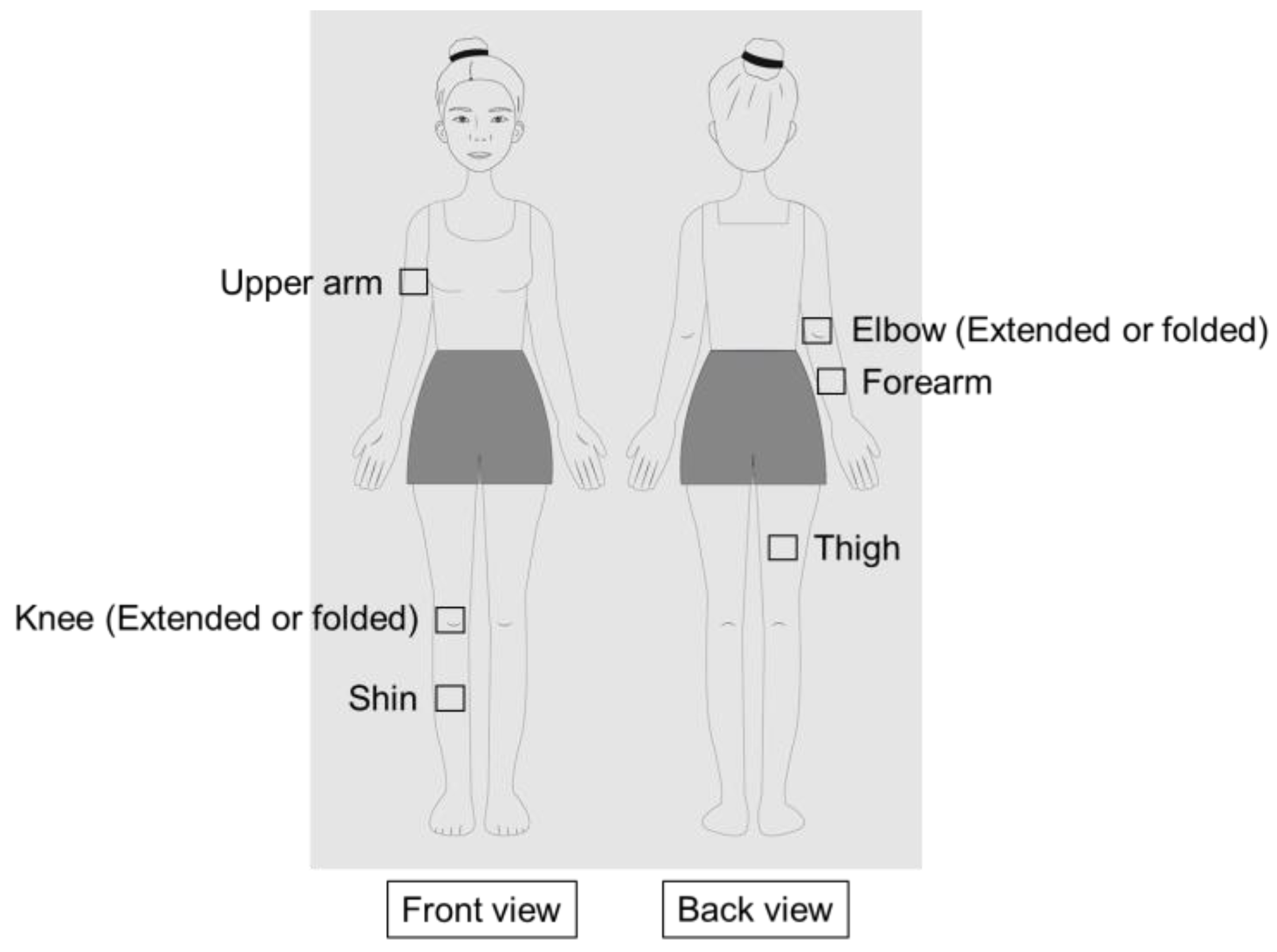
2.2. Body Sites for Skin Color Assessment
2.3. Skin Color Assessment Methods
2.4. Statistical Analysis
3. Results
3.1. Information of Human Subjects
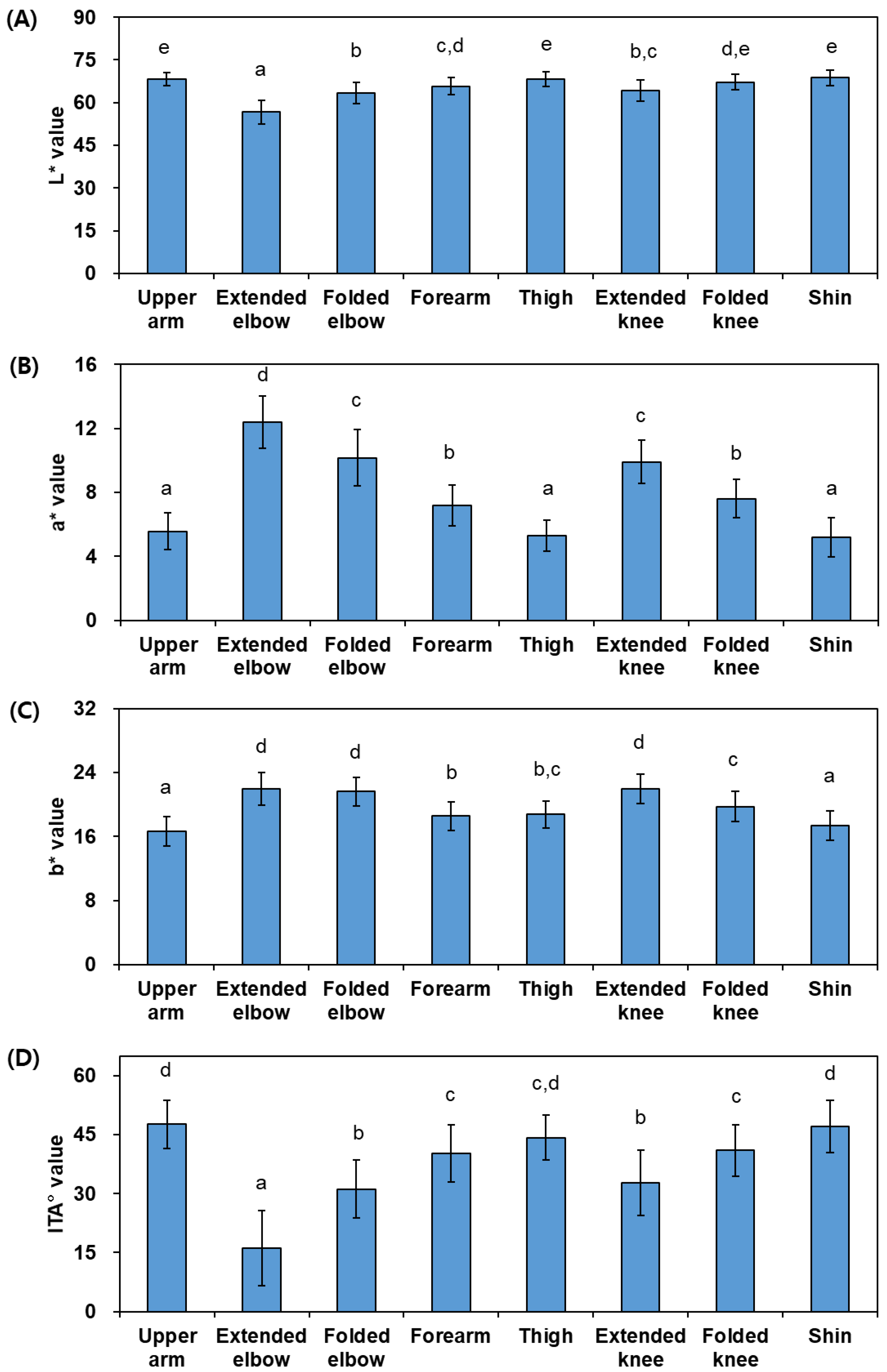
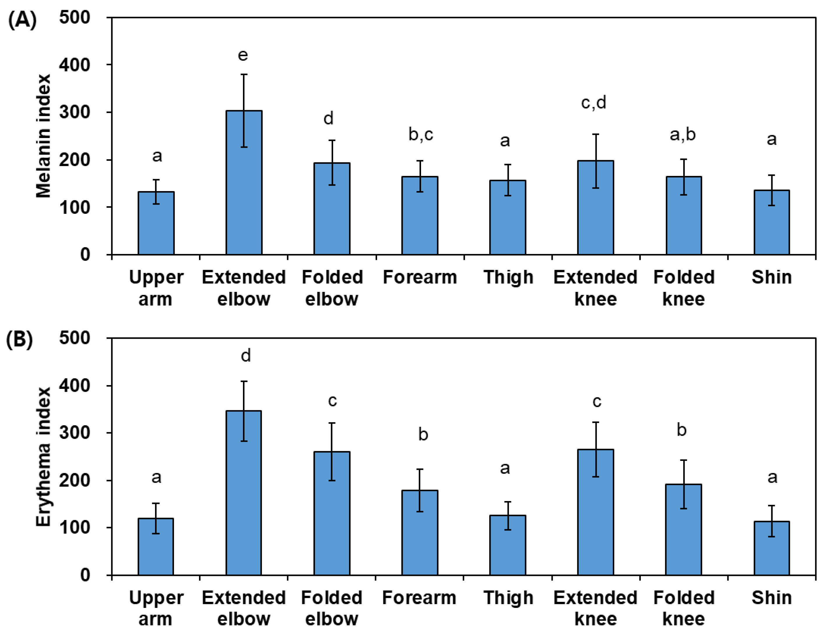
3.2. Differences in Skin Colors of Various Body Sites
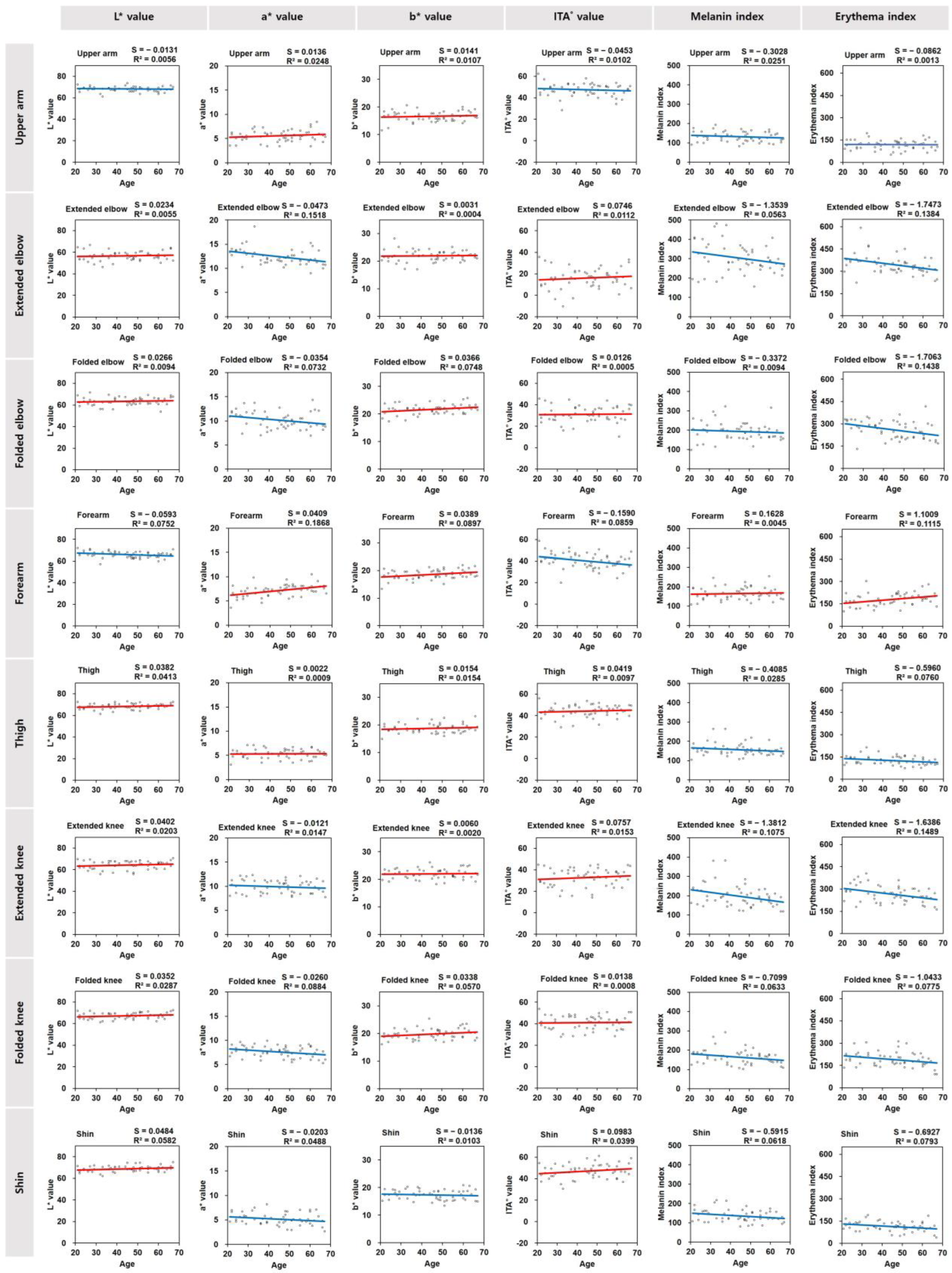
3.3. Age-Dependent Changes of Skin Colors of Various Body Sites
3.4. Correlation of Skin Color Parameter Values and the Ages of the Subjects
4. Discussion
5. Conclusions
Author Contributions
Funding
Institutional Review Board Statement
Informed Consent Statement
Data Availability Statement
Conflicts of Interest
References
- Humphrey, S.; Brown, S.M.; Cross, S.J.; Mehta, R. Defining Skin Quality: Clinical Relevance, Terminology, and Assessment. Dermatol. Surg. 2021, 47, 974–981. [Google Scholar] [CrossRef] [PubMed]
- Perreira, K.M.; Wassink, J.; Harris, K.M. Beyond Race/Ethnicity: Skin Color, Gender, and the Health of Young Adults in the United States. Popul. Res. Policy Rev. 2019, 38, 271–299. [Google Scholar] [CrossRef] [PubMed]
- Deng, L.; Xu, S.H. Adaptation of human skin color in various populations. Hereditas 2017, 155, 1. [Google Scholar] [CrossRef] [PubMed]
- Sandby-Moller, J.; Poulsen, T.; Wulf, H.C. Epidermal thickness at different body sites: Relationship to age, gender, pigmentation, blood content, skin type and smoking habits. Acta Derm. Venereol. 2003, 83, 410–413. [Google Scholar] [CrossRef] [PubMed]
- Han, K.W.; Choi, T.; Son, D.G. Skin color of Koreans: Statistical evaluation of affecting factors. Ski. Res. Technol. 2006, 12, 170–177. [Google Scholar] [CrossRef] [PubMed]
- Del Bino, S.; Duval, C.; Bernerd, F. Clinical and Biological Characterization of Skin Pigmentation Diversity and Its Consequences on UV Impact. Int. J. Mol. Sci. 2018, 19, 2688. [Google Scholar] [CrossRef] [PubMed]
- Maranduca, M.A.; Branisteanu, D.; Serban, D.N.; Branisteanu, D.C.; Stoleriu, G.; Manolache, N.; Serban, I.L. Synthesis and physiological implications of melanic pigments. Oncol. Lett. 2019, 17, 4183–4187. [Google Scholar] [CrossRef] [PubMed]
- Moreiras, H.; Seabra, M.C.; Barral, D.C. Melanin Transfer in the Epidermis: The Pursuit of Skin Pigmentation Control Mechanisms. Int. J. Mol. Sci. 2021, 22, 4466. [Google Scholar] [CrossRef]
- Lu, Y.Y.; Tonissen, K.F.; Di Trapani, G. Modulating skin colour: Role of the thioredoxin and glutathione systems in regulating melanogenesis. Biosci. Rep. 2021, 41, BSR20210427. [Google Scholar] [CrossRef]
- Yamaguchi, Y.; Hearing, V.J. Physiological factors that regulate skin pigmentation. Biofactors 2009, 35, 193–199. [Google Scholar] [CrossRef]
- Bastonini, E.; Kovacs, D.; Picardo, M. Skin Pigmentation and Pigmentary Disorders: Focus on Epidermal/Dermal Cross-Talk. Ann. Dermatol. 2016, 28, 279–289. [Google Scholar] [CrossRef] [PubMed]
- Thawabteh, A.M.; Jibreen, A.; Karaman, D.; Thawabteh, A.; Karaman, R. Skin Pigmentation Types, Causes and Treatment—A Review. Molecules 2023, 28, 4839. [Google Scholar] [CrossRef] [PubMed]
- Boo, Y.C. Metabolic Basis and Clinical Evidence for Skin Lightening Effects of Thiol Compounds. Antioxidants 2022, 11, 503. [Google Scholar] [CrossRef] [PubMed]
- Fang, B.; Card, P.D.; Chen, J.J.; Li, L.J.; Laughlin, T.; Jarrold, B.; Zhao, W.Z.; Benham, A.M.; Määttä, A.T.; Hawkins, T.J.; et al. A Potential Role of Keratinocyte-Derived Bilirubin in Human Skin Yellowness and Its Amelioration by Sucrose Laurate/Dilaurate. Int. J. Mol. Sci. 2022, 23, 5884. [Google Scholar] [CrossRef]
- Zonios, G.; Bykowski, J.; Kollias, N. Skin melanin, hemoglobin, and light scattering properties can be quantitatively assessed in vitro using diffuse reflectance spectroscopy. J. Investig. Dermatol. 2001, 117, 1452–1457. [Google Scholar] [CrossRef] [PubMed]
- Alkawaz, M.H.; Mohamad, D.; Saba, T.; Basori, A.H.; Rehman, A. The Correlation Between Blood Oxygenation Effects and Human Emotion Towards Facial Skin Colour of Virtual Human. 3D Res. 2015, 6, 13. [Google Scholar] [CrossRef]
- Chen, C.Y.; Zhang, J.Q.; Li, L.; Guo, M.M.; He, Y.F.; Dong, Y.M.; Meng, H.; Yi, F. Advanced Glycation End Products in the Skin: Molecular Mechanisms, Methods of Measurement, and Inhibitory Pathways. Front. Med. 2022, 9, 837222. [Google Scholar] [CrossRef] [PubMed]
- Yardman-Frank, J.M.; Fisher, D.E. Skin pigmentation and its control: From ultraviolet radiation to stem cells. Exp. Dermatol. 2021, 30, 560–571. [Google Scholar] [CrossRef] [PubMed]
- Boo, Y.C. Emerging Strategies to Protect the Skin from Ultraviolet Rays Using Plant-Derived Materials. Antioxidants 2020, 9, 637. [Google Scholar] [CrossRef]
- Shenoy, A.; Madan, R. Post-Inflammatory Hyperpigmentation: A Review of Treatment Strategies. J. Drugs Dermatol. 2020, 19, 763–768. [Google Scholar] [CrossRef]
- Boo, Y.C. Mechanistic Basis and Clinical Evidence for the Applications of Nicotinamide (Niacinamide) to Control Skin Aging and Pigmentation. Antioxidants 2021, 10, 1315. [Google Scholar] [CrossRef] [PubMed]
- Choi, S.Y.; Lee, Y.; Kim, S.S.; Ju, H.M.; Baek, J.H.; Park, C.S.; Lee, D.H. Inhibitory effect of corn silk on skin pigmentation. Molecules 2014, 19, 2808–2818. [Google Scholar] [CrossRef] [PubMed]
- Boo, Y.C.; Jo, D.J.; Oh, C.M.; Lee, S.Y.; Kim, Y.M. The First Human Clinical Trial on the Skin Depigmentation Efficacy of Glycinamide Hydrochloride. Biomedicines 2020, 8, 257. [Google Scholar] [CrossRef] [PubMed]
- Jeong, H.; Kim, S.; Seo, Y.; Koh, J.; Baek, J. Investigation of symptoms of hand skin changes with aging in Korean women and development of a new standard grading scale for hand aging. Ski. Res. Technol. 2020, 26, 788–793. [Google Scholar] [CrossRef] [PubMed]
- Schmalwieser, A.W.; Götzinger, S.; Schwabel, F. Exploratory study on the body distribution of skin color, pigmentation and, degree of tan in Central European Caucasian Women. Photochem. Photobiol. Sci. 2024, 23, 493–502. [Google Scholar] [CrossRef] [PubMed]
- Yun, I.S.; Lee, W.J.; Rah, D.K.; Kim, Y.O.; Park, B.Y.Y. Skin color analysis using a spectrophotometer in Asians. Ski. Res. Technol. 2010, 16, 311–315. [Google Scholar] [CrossRef] [PubMed]
- Vernez, D.; Milon, A.; Vuilleumier, L.; Bulliard, J.L. Anatomical exposure patterns of skin to sunlight: Relative contributions of direct, diffuse and reflected ultraviolet radiation. Br. J. Dermatol. 2012, 167, 383–390. [Google Scholar] [CrossRef] [PubMed]
- Hsieh, C.Y.; Tsai, T.F. Friction-Induced Skin Disorders—A Review. Dermatitis 2023, 34, 278–286. [Google Scholar] [CrossRef] [PubMed]
- Jeon, C.; Agbai, O.; Butler, D.; Murase, J. Dermatologic conditions in patients of color who are pregnant. Int. J. Womens Dermatol. 2017, 3, 30–36. [Google Scholar] [CrossRef]
- Bernatchez, S.F.; Bichel, J. The Science of Skin: Measuring Damage and Assessing Risk. Adv. Wound Care 2023, 12, 187–204. [Google Scholar] [CrossRef]
- Tan, C.; Xia, L.L. Dermoscopy of frictional asymptomatic darkening of the extensor surfaces. Postep. Dermatol. I Alergol. 2019, 36, 232–233. [Google Scholar] [CrossRef] [PubMed]
- Venkatesh, S.; Maymone, M.B.C.; Vashi, N.A. Aging in skin of color. Clin. Dermatol. 2019, 37, 351–357. [Google Scholar] [CrossRef] [PubMed]
- Pierard, G.E. EEMCO guidance for the assessment of skin colour. J. Eur. Acad. Dermatol. Venereol. 1998, 10, 1–11. [Google Scholar] [CrossRef] [PubMed]
- Wilkes, M.; Wright, C.Y.; du Plessis, J.L.; Reeder, A. Fitzpatrick Skin Type, Individual Typology Angle, and Melanin Index in an African Population: Steps Toward Universally Applicable Skin Photosensitivity Assessments. JAMA Dermatol. 2015, 151, 902–903. [Google Scholar] [CrossRef] [PubMed]
- Ly, B.C.K.; Dyer, E.B.; Feig, J.L.; Chien, A.L.; Del Bino, S. Research Techniques Made Simple: Cutaneous Colorimetry: A Reliable Technique for Objective Skin Color Measurement. J. Investig. Dermatol. 2020, 140, 3–12. [Google Scholar] [CrossRef] [PubMed]
- Clarys, P.; Alewaeters, K.; Lambrecht, R.; Barel, A.O. Skin color measurements: Comparison between three instruments: The Chromameter®, the DermaSpectrometer® and the Mexameter®. Ski. Res. Technol. 2000, 6, 230–238. [Google Scholar] [CrossRef]
- Weihs, P.; Helletzgruber, S.; Kranewitter, S.; Langer, L.; Lumerding, Z.; Luschin, V.; Schmidt, P.; Heydenreich, J.; Schmalwieser, A.W. UV Exposure during Cycling as a Function of Solar Elevation and Orientation. Atmosphere 2024, 15, 215. [Google Scholar] [CrossRef]
- Bao, S. Mechanical stress. Handb. Clin. Neurol. 2015, 131, 367–396. [Google Scholar]
- Boo, Y.C. Can Plant Phenolic Compounds Protect the Skin from Airborne Particulate Matter? Antioxidants 2019, 8, 379. [Google Scholar] [CrossRef]
- Russell-Goldman, E.; Murphy, G.F. The Pathobiology of Skin Aging: New Insights into an Old Dilemma. Am. J. Pathol. 2020, 190, 1356–1369. [Google Scholar] [CrossRef]
- Amin, R.; Volzer, B.; Genedy-Kalyoncu, M.E.; Blume-Peytavi, U.; Kottner, J. The prevalence and severity of dry skin and related skin care in older adult residents in institutional long-term care: A cross-sectional study. Geriatr. Nurs. 2023, 54, 331–340. [Google Scholar] [CrossRef] [PubMed]
- Bentov, I.; Reed, M.J. The effect of aging on the cutaneous microvasculature. Microvasc. Res. 2015, 100, 25–31. [Google Scholar] [CrossRef] [PubMed]

| Age Groups | N | Mean | SD | Minimum | Maximum |
|---|---|---|---|---|---|
| 20s | 11 | 25.36 | 2.91 | 21 | 29 |
| 30s | 10 | 36.20 | 2.70 | 32 | 39 |
| 40s | 12 | 46.42 | 2.35 | 41 | 49 |
| 50s | 10 | 54.30 | 2.95 | 51 | 59 |
| 60s | 10 | 62.90 | 2.73 | 60 | 67 |
| Total | 53 | 44.72 | 13.49 | 21 | 67 |
| Item | Classification | All Subjects | 20s Subjects | 30s Subjects | 40s Subjects | 50s Subjects | 60s Subjects | ||||||
|---|---|---|---|---|---|---|---|---|---|---|---|---|---|
| N | % | N | % | N | % | N | % | N | % | N | % | ||
| Occupation | Workers | 14 | 26 | 5 | 45 | 6 | 60 | 1 | 8 | 1 | 10 | 1 | 10 |
| Student | 4 | 8 | 4 | 36 | 0 | 0 | 0 | 0 | 0 | 0 | 0 | 0 | |
| None | 2 | 4 | 2 | 18 | 0 | 0 | 0 | 0 | 0 | 0 | 0 | 0 | |
| Housewife | 33 | 62 | 0 | 0 | 4 | 40 | 11 | 92 | 9 | 90 | 9 | 90 | |
| Workplace | Indoor | 45 | 85 | 11 | 100 | 9 | 90 | 9 | 75 | 8 | 80 | 8 | 80 |
| Outdoor | 0 | 0 | 0 | 0 | 0 | 0 | 0 | 0 | 0 | 0 | 0 | 0 | |
| Both | 8 | 15 | 0 | 0 | 1 | 10 | 3 | 25 | 2 | 20 | 2 | 20 | |
| Average sleep duration | Less than 5 h | 2 | 4 | 1 | 9 | 0 | 0 | 0 | 0 | 0 | 0 | 1 | 10 |
| 5 to 8 h | 47 | 89 | 7 | 64 | 10 | 100 | 12 | 100 | 9 | 90 | 9 | 90 | |
| 8 h or longer | 4 | 8 | 3 | 27 | 0 | 0 | 0 | 0 | 1 | 10 | 0 | 0 | |
| Exposure to sunlight or UV rays | Less than 1 h | 28 | 53 | 9 | 82 | 3 | 30 | 7 | 58 | 4 | 40 | 5 | 50 |
| 1 to 3 h | 24 | 45 | 2 | 18 | 7 | 70 | 4 | 33 | 6 | 60 | 5 | 50 | |
| 3 h or longer | 1 | 2 | 0 | 0 | 0 | 0 | 1 | 8 | 0 | 0 | 0 | 0 | |
| Exposure to repeated friction | Yes | 5 | 9 | 0 | 0 | 0 | 0 | 4 | 33 | 0 | 0 | 1 | 10 |
| No | 47 | 89 | 10 | 91 | 10 | 100 | 8 | 67 | 10 | 100 | 9 | 90 | |
| History | 1 | 2 | 1 | 9 | 0 | 0 | 0 | 0 | 0 | 0 | 0 | 0 | |
| Smoking cigarettes | None | 53 | 100 | 11 | 100 | 10 | 100 | 12 | 100 | 10 | 100 | 10 | 100 |
| Less than 10 | 0 | 0 | 0 | 0 | 0 | 0 | 0 | 0 | 0 | 0 | 0 | 0 | |
| 10 or more | 0 | 0 | 0 | 0 | 0 | 0 | 0 | 0 | 0 | 0 | 0 | 0 | |
| Dominant hand | Left | 5 | 9 | 1 | 9 | 0 | 0 | 0 | 0 | 1 | 10 | 3 | 30 |
| Right | 31 | 58 | 7 | 64 | 6 | 60 | 10 | 83 | 6 | 60 | 2 | 20 | |
| Both | 17 | 32 | 3 | 27 | 4 | 40 | 2 | 17 | 3 | 30 | 5 | 50 | |
| Item | Classification | All Subjects | 20s Subjects | 30s Subjects | 40s Subjects | 50s Subjects | 60s Subjects | |||||||
|---|---|---|---|---|---|---|---|---|---|---|---|---|---|---|
| N | % | N | % | N | % | N | % | N | % | N | % | |||
| Facial skin types | Dry skin | 24 | 45 | 1 | 9 | 1 | 10 | 9 | 75 | 4 | 40 | 9 | 90 | |
| Neutral skin | 14 | 26 | 4 | 36 | 4 | 0 | 1 | 8 | 5 | 50 | 0 | 0 | ||
| Oily skin | 1 | 2 | 0 | 0 | 1 | 70 | 0 | 0 | 0 | 0 | 0 | 0 | ||
| Dry-oily skin | 13 | 25 | 5 | 45 | 4 | 20 | 2 | 17 | 1 | 10 | 1 | 10 | ||
| Problematic | 1 | 2 | 1 | 9 | 0 | 0 | 0 | 0 | 0 | 0 | 0 | 0 | ||
| Body skin moisture | Arm | Moist | 1 | 2 | 0 | 0 | 0 | 0 | 0 | 0 | 0 | 0 | 1 | 10 |
| Normal | 25 | 47 | 8 | 73 | 5 | 50 | 3 | 25 | 4 | 40 | 5 | 50 | ||
| Dry | 27 | 51 | 3 | 27 | 5 | 50 | 9 | 75 | 6 | 60 | 4 | 40 | ||
| Leg | Moist | 0 | 0 | 0 | 0 | 0 | 0 | 0 | 0 | 0 | 0 | 0 | 0 | |
| Normal | 14 | 26 | 6 | 55 | 3 | 30 | 2 | 17 | 1 | 10 | 2 | 20 | ||
| Dry | 39 | 74 | 5 | 45 | 7 | 70 | 10 | 83 | 9 | 90 | 8 | 80 | ||
| Body skin oil | Arm | Excessive | 1 | 2 | 1 | 9 | 0 | 0 | 0 | 0 | 0 | 0 | 0 | 0 |
| Normal | 32 | 60 | 6 | 55 | 7 | 70 | 5 | 42 | 7 | 70 | 7 | 70 | ||
| Insufficient | 20 | 38 | 4 | 36 | 3 | 30 | 7 | 58 | 3 | 30 | 3 | 30 | ||
| Leg | Excessive | 1 | 2 | 1 | 9 | 0 | 0 | 0 | 0 | 0 | 0 | 0 | 0 | |
| Normal | 26 | 49 | 5 | 45 | 7 | 70 | 6 | 50 | 3 | 30 | 5 | 50 | ||
| Insufficient | 26 | 49 | 5 | 45 | 3 | 30 | 6 | 50 | 7 | 70 | 5 | 50 | ||
| Body skin texture | Arm | Soft | 9 | 17 | 2 | 18 | 2 | 20 | 3 | 25 | 0 | 0 | 3 | 30 |
| Average | 38 | 72 | 9 | 82 | 7 | 70 | 7 | 58 | 9 | 90 | 6 | 60 | ||
| Rough | 6 | 11 | 0 | 0 | 1 | 10 | 2 | 17 | 1 | 10 | 1 | 10 | ||
| Leg | Soft | 6 | 11 | 2 | 18 | 2 | 20 | 0 | 0 | 0 | 0 | 2 | 20 | |
| Average | 36 | 68 | 7 | 64 | 6 | 60 | 10 | 83 | 8 | 80 | 5 | 50 | ||
| Rough | 11 | 21 | 2 | 18 | 2 | 20 | 2 | 17 | 2 | 20 | 3 | 30 | ||
| Body skin thickness | Arm | Thin | 16 | 30 | 3 | 27 | 4 | 40 | 3 | 25 | 2 | 20 | 4 | 40 |
| Average | 32 | 60 | 6 | 55 | 5 | 50 | 7 | 58 | 8 | 80 | 6 | 60 | ||
| Thick | 5 | 9 | 2 | 18 | 1 | 10 | 2 | 17 | 0 | 0 | 0 | 0 | ||
| Leg | Thin | 14 | 26 | 3 | 27 | 2 | 20 | 3 | 25 | 2 | 20 | 4 | 40 | |
| Average | 32 | 60 | 6 | 55 | 6 | 60 | 7 | 58 | 8 | 80 | 5 | 50 | ||
| Thick | 7 | 13 | 2 | 18 | 2 | 20 | 2 | 17 | 0 | 0 | 1 | 10 | ||
| Body skin pigmentation | Arm | High | 11 | 21 | 2 | 18 | 2 | 20 | 1 | 8 | 3 | 30 | 3 | 30 |
| Normal | 36 | 68 | 8 | 73 | 6 | 60 | 9 | 75 | 6 | 60 | 7 | 70 | ||
| Low | 6 | 11 | 1 | 9 | 2 | 20 | 2 | 17 | 1 | 10 | 0 | 0 | ||
| Leg | High | 9 | 17 | 2 | 18 | 1 | 10 | 1 | 8 | 2 | 20 | 3 | 30 | |
| Normal | 35 | 66 | 8 | 73 | 5 | 50 | 10 | 83 | 6 | 60 | 6 | 60 | ||
| Low | 9 | 17 | 1 | 9 | 4 | 40 | 1 | 8 | 2 | 20 | 1 | 10 | ||
| Skin Sites | The L* Value | The a* Value | The b* Value | The ITA° Value | Melanin Index | Erythema Index | ||||||
|---|---|---|---|---|---|---|---|---|---|---|---|---|
| r | p | r | p | r | p | r | p | r | p | r | p | |
| Upper arm | −0.075 | 0.596 | 0.158 | 0.258 | 0.103 | 0.462 | −0.101 | 0.473 | −0.158 | 0.257 | −0.036 | 0.796 |
| Extended elbow | 0.074 | 0.597 | −0.390 | 0.004 | 0.021 | 0.881 | 0.106 | 0.451 | −0.237 | 0.087 | −0.372 | 0.006 |
| (weak, negative) | (weak, negative) | |||||||||||
| Folded elbow | 0.097 | 0.488 | −0.271 | 0.050 | 0.274 | 0.047 | 0.023 | 0.870 | −0.097 | 0.489 | −0.379 | 0.005 |
| (weak) | (weak, negative) | |||||||||||
| Forearm | −0.274 | 0.047 | 0.432 | 0.001 | 0.299 | 0.029 | −0.293 | 0.033 | 0.067 | 0.632 | 0.334 | 0.015 |
| (weak, negative) | (moderate) | (weak) | (weak, negative) | (weak) | ||||||||
| Thigh | 0.203 | 0.144 | 0.030 | 0.830 | 0.124 | 0.376 | 0.099 | 0.482 | −0.169 | 0.226 | −0.276 | 0.046 |
| (weak, negative) | ||||||||||||
| Extended knee | 0.142 | 0.309 | −0.122 | 0.385 | 0.044 | 0.754 | 0.124 | 0.378 | −0.328 | 0.017 | −0.386 | 0.004 |
| (weak, negative) | (weak, negative) | |||||||||||
| Folded Knee | 0.169 | 0.226 | −0.297 | 0.031 | 0.239 | 0.085 | 0.028 | 0.840 | −0.252 | 0.069 | −0.278 | 0.044 |
| (weak, negative) | (weak, negative) | |||||||||||
| Shin | 0.241 | 0.082 | −0.221 | 0.111 | -0.102 | 0.470 | 0.200 | 0.152 | −0.249 | 0.073 | −0.282 | 0.041 |
| (weak, negative) | ||||||||||||
Disclaimer/Publisher’s Note: The statements, opinions and data contained in all publications are solely those of the individual author(s) and contributor(s) and not of MDPI and/or the editor(s). MDPI and/or the editor(s) disclaim responsibility for any injury to people or property resulting from any ideas, methods, instructions or products referred to in the content. |
© 2024 by the authors. Licensee MDPI, Basel, Switzerland. This article is an open access article distributed under the terms and conditions of the Creative Commons Attribution (CC BY) license (https://creativecommons.org/licenses/by/4.0/).
Share and Cite
Lee, E.J.; Ryu, J.H.; Baek, J.H.; Boo, Y.C. Skin Color Analysis of Various Body Parts (Forearm, Upper Arm, Elbow, Knee, and Shin) and Changes with Age in 53 Korean Women, Considering Intrinsic and Extrinsic Factors. J. Clin. Med. 2024, 13, 2500. https://doi.org/10.3390/jcm13092500
Lee EJ, Ryu JH, Baek JH, Boo YC. Skin Color Analysis of Various Body Parts (Forearm, Upper Arm, Elbow, Knee, and Shin) and Changes with Age in 53 Korean Women, Considering Intrinsic and Extrinsic Factors. Journal of Clinical Medicine. 2024; 13(9):2500. https://doi.org/10.3390/jcm13092500
Chicago/Turabian StyleLee, Eun Ju, Ja Hyun Ryu, Ji Hwoon Baek, and Yong Chool Boo. 2024. "Skin Color Analysis of Various Body Parts (Forearm, Upper Arm, Elbow, Knee, and Shin) and Changes with Age in 53 Korean Women, Considering Intrinsic and Extrinsic Factors" Journal of Clinical Medicine 13, no. 9: 2500. https://doi.org/10.3390/jcm13092500
APA StyleLee, E. J., Ryu, J. H., Baek, J. H., & Boo, Y. C. (2024). Skin Color Analysis of Various Body Parts (Forearm, Upper Arm, Elbow, Knee, and Shin) and Changes with Age in 53 Korean Women, Considering Intrinsic and Extrinsic Factors. Journal of Clinical Medicine, 13(9), 2500. https://doi.org/10.3390/jcm13092500