
 Open Access
 - Review

33 Pages
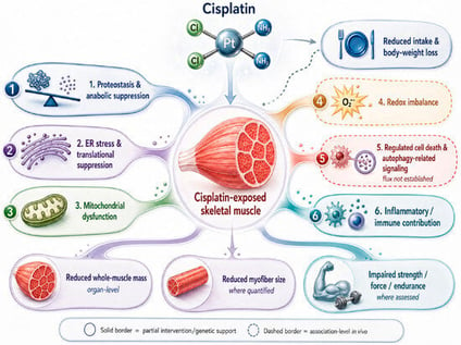
Cisplatin causes skeletal muscle wasting in tumor-free rodents, reducing muscle mass, myofiber size and, where measured, strength. This critical narrative review appraises that phenotype and its mechanistic basis. Tumor-bearing models show that the loss also occurs under tumor burden, but most cannot separate drug from tumor effects. Melatonin influences mitochondrial, redox, regenerative, and structural outcomes in skeletal muscle under non-cisplatin conditions. Direct melatonin–cisplatin evidence is limited to pretreatment studies in C2C12 cells from one research group. No in vivo melatonin-plus-cisplatin skeletal-muscle study and no clinical study of melatonin against cisplatin-associated muscle loss were identified in searches through 22 September 2026. Biological plausibility is supported, but protection has not been demonstrated. A controlled tumor-free study would provide a more interpretable test of whether melatonin protects skeletal muscle during cisplatin exposure. Translational validation would then require a separate tumor-bearing study testing whether protection persists under tumor burden and whether cisplatin’s antitumor activity is preserved.
Muscles
16 October 2026
![Proposed integrative model (hypothetical) of the relationship between the afferent signal, its laboratory estimate, and the systems that set the rate of accumulation. Group III/IV muscle afferents respond to protons, ATP, and lactate via acid-sensing ion channels, P2X, and TRPV1 receptors, and their central projection reduces motor output. Blocking that projection with lumbar intrathecal fentanyl raises motor output and accelerates the decline in contractile force. We propose that exercise ends when this feedback, summed with afferent traffic from the respiratory muscles and the corollary discharge accompanying the motor command, becomes intolerable; this termination rule is inferred from converging observations and has not been demonstrated directly. The quantity proposed to be regulated is that summed signal; the potentiated-twitch decrement estimates it only in proportion to the metabolite-driven share of contractile impairment, because the afferents sense the metabolites and not the impairment itself. The afferent agonists [2] and the effects of fentanyl blockade [15,31,32] rest on direct experiments; the summation across sources and the termination rule are inferences. The diagram is schematic: no values are plotted, and no fixed end-exercise threshold of contractile impairment is depicted or implied, consistent with the argument developed in Section 3.1, Section 3.2, Section 3.3, Section 3.4, Section 3.5, Section 3.6, Section 3.7 and Section 3.8. In the studies reviewed, arterial oxygen delivery, respiratory muscle work, substrate availability, and perceived effort changed the rate at which the signal accumulates more than its end-exercise value.](https://mdpi-res.com/cdn-cgi/image/width=281%2Cheight=192/https://mdpi-res.com/muscles/muscles-05-00074/article_deploy/html/images/muscles-05-00074-g001-550.jpg)
![Overview of the bioanalytical strategy to determine the tongue proteome. The experimental workflow and the findings from the bioinformatic PANTHER (http://pantherdb.org) [62] analysis of protein classes in the tongue proteome are shown.](https://mdpi-res.com/cdn-cgi/image/width=281%2Cheight=192/https://mdpi-res.com/muscles/muscles-05-00073/article_deploy/html/images/muscles-05-00073-g001-550.jpg)