Advances in Cornea, Cataract, and Refractive Surgery

A topical collection in Medicina (ISSN 1648-9144). This collection belongs to the section "Ophthalmology".

Viewed by 11129

Editor


E-Mail Website
Collection Editor
Department of Ophthalmology, University of Geneva, Rue de Alcide-Jentzer 22, 1205 Geneva, Switzerland
Interests: corneal grafts; cataract surgery; refractive surgery; LASIK; PRK; intraocular lenses; phakic lenses
Special Issues, Collections and Topics in MDPI journals

Topical Collection Information

Dear Colleagues,

I am delighted to present a Topical Collection on the topic of “Advances in Cornea, Cataract, and Refractive Surgery”

The latest technological developments and advanced surgical techniques have radically changed the daily practice of eye surgeons, with new treatment options making surgery safer, faster, and more precise. The advent of new diagnostic tools especially in the anterior segment enables better patient management and precise monitoring of disease progression, leading to an improvement in postoperative surgical outcomes.

This Topical Collection is a multidisciplinary forum on the role of diagnostic and surgical procedures in this subfield of ophthalmology.

The published papers will describe new developments in these areas. This Topical Collection accepts high-quality articles containing original research results, as well as review articles of exceptional merit.

Dr. Ivo Guber
Collection Editor

Manuscript Submission Information

Manuscripts should be submitted online at www.mdpi.com by registering and logging in to this website. Once you are registered, click here to go to the submission form. Manuscripts can be submitted until the deadline. All submissions that pass pre-check are peer-reviewed. Accepted papers will be published continuously in the journal (as soon as accepted) and will be listed together on the collection website. Research articles, review articles as well as short communications are invited. For planned papers, a title and short abstract (about 100 words) can be sent to the Editorial Office for announcement on this website.

Submitted manuscripts should not have been published previously, nor be under consideration for publication elsewhere (except conference proceedings papers). All manuscripts are thoroughly refereed through a single-blind peer-review process. A guide for authors and other relevant information for submission of manuscripts is available on the Instructions for Authors page. Medicina is an international peer-reviewed open access monthly journal published by MDPI.

Please visit the Instructions for Authors page before submitting a manuscript. The Article Processing Charge (APC) for publication in this open access journal is 2200 CHF (Swiss Francs). Submitted papers should be well formatted and use good English. Authors may use MDPI's English editing service prior to publication or during author revisions.

Published Papers (7 papers)

2024

Jump to: 2023, 2022

5 pages, 5118 KiB  
Case Report
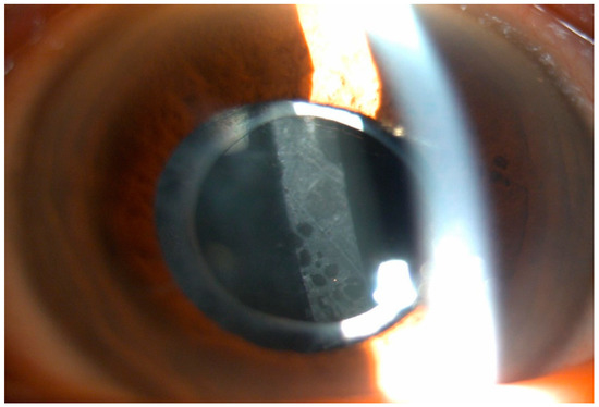
A Case of Parinaud Oculoglandular Syndrome in Which Bartonella DNA Was Detected in the Cornea and Conjunctiva by Polymerase Chain Reaction
by Junya Saito, Akira Machida, Daisuke Inoue, Masumi Suzuki Shimizu, Kohsuke Matsui, Kohei Harada, Mao Kusano, Yasser Helmy Mohamed and Masafumi Uematsu
Medicina 2024, 60(9), 1425; https://doi.org/10.3390/medicina60091425 - 31 Aug 2024
Viewed by 471
Abstract
Background and Objectives: Parinaud oculoglandular syndrome (POS) is unilateral granulomatous follicular conjunctivitis with ipsilateral afferent lymphadenopathy, primarily caused by cat-scratch disease, tularemia, and sporotrichosis. We report a case of POS in which Bartonella DNA was detected using polymerase chain reaction (PCR) in [...] Read more.
Background and Objectives: Parinaud oculoglandular syndrome (POS) is unilateral granulomatous follicular conjunctivitis with ipsilateral afferent lymphadenopathy, primarily caused by cat-scratch disease, tularemia, and sporotrichosis. We report a case of POS in which Bartonella DNA was detected using polymerase chain reaction (PCR) in corneal and conjunctival specimens. Methods: A 29-year-old man, who started keeping a stray cat two months prior, became aware of right preauricular lymphadenopathy and right ocular conjunctival hyperemia one month prior. Subsequently, he developed a fever of approximately 37.9 °C, with a purulent ocular discharge appearing 1 week before being referred to our department for a detailed ophthalmological examination. The patient’s right eye showed hyperemia and edema in the bulbar conjunctiva, along with palpebral conjunctival hyperemia, follicles, and white ulcers. Two weeks later, his serum IgM titer for Bartonella henselae was 1:20, and Bartonella DNA was detected by PCR in the corneal and conjunctival specimens. Based on these findings, the patient was diagnosed with POS caused by cat-scratch disease (CSD). Oral doxycycline, rifampicin, topical gatifloxacin, betamethasone phosphate, and erythromycin eye ointments were prescribed. Results: After 2 weeks of oral treatment and 2 months of eye drop treatment, the deterioration of the cornea and conjunctiva improved when the patient recovered good visual acuity. Conclusions: PCR assays of corneal and conjunctival specimens are useful for the diagnosis of CSD presenting with POS. These results suggested that Bartonella may be directly involved in the ocular surface pathogenesis of POS. Full article
Show Figures

Figure 1

11 pages, 2578 KiB  
Article
Intradevice Repeatability and Interdevice Comparison of Two Specular Microscopy Devices in a Real-Life Setting: Tomey EM-4000 and Nidek CEM-530
by Mateusz Kecik, Martina Kropp, Gabriele Thumann, Bojan Pajic, Josef Guber and Ivo Guber
Medicina 2024, 60(7), 1110; https://doi.org/10.3390/medicina60071110 - 9 Jul 2024
Viewed by 809
Abstract
Background and Objectives: The purpose of this study was to compare two commercially available specular microscopes (Tomey EM-4000 and Nidek CEM-530) in a real-life clinical setting in terms of intra- and interdevice variability. The study was conducted on all patients seen in [...] Read more.
Background and Objectives: The purpose of this study was to compare two commercially available specular microscopes (Tomey EM-4000 and Nidek CEM-530) in a real-life clinical setting in terms of intra- and interdevice variability. The study was conducted on all patients seen in a clinical practice specializing in anterior segment pathologies, regardless of the purpose of their visit. Materials and Methods: In total, 112 eyes of 56 patients (age 23–85 years old) were included in the study. Each eye was measured three times with each device (for a total of six measurements), and results for central corneal thickness (CCT) and corneal endothelial cell density (ECD) were recorded. The results were then evaluated with the D’Agostino–Pearson normality test and compared with a Wilcoxon signed-rank test, t-test, ANOVA or Mann–Whitney test for intra- and interdevice variability. Results: Both specular microscopes produced very reliable reproducible intradevice results: The Tomey EM-4000 measured an ECD of 2390 ± 49.57 cells/mm2 (mean ± standard error of mean); the range was 799–3010 cells/mm2. The determined CCT was 546 ± 5.104 µm (mean ± standard error of mean [SEM]); the range was 425–615 µm. The measurements with the Nidek CEM-530 revealed an ECD of 2417 ± 0.09 cells/mm2 (mean ± SEM); the range was 505–3461 cells/mm2 (mean ± SEM). The mean CCT detected was 546.3 ± 4.937 µm (mean ± SEM); the range was 431–621 µm. The interdevice differences were statistically significant for both parameters, ECD (p = 0.0175) and CCT (p = 0.0125) (p < 0.05). Conclusions: The Nidek CEM-530 and the Tomey EM-4000 both produced reliable and reproducible results in terms of ECD and CCT. The absolute measurements were statistically significantly different for CCT and ECD for both devices; the Nidek produces slightly higher values. Full article
Show Figures

Figure 1

12 pages, 1799 KiB  
Article
Visual Outcomes in Ectopia Lentis in Marfan Syndrome: A Study of Four Surgical Techniques in Children and Adults
by Anca Cristina Dogaroiu, Maria Dudau, Catalin Dogaroiu and Calin Petru Tataru
Medicina 2024, 60(7), 1098; https://doi.org/10.3390/medicina60071098 - 5 Jul 2024
Viewed by 866
Abstract
Background/Objectives: To evaluate how the surgical technique and type of implanted intraocular lens influence the postoperative visual acuity and complications in ectopia lentis associated to Marfan syndrome patients. Materials and Methods: The medical records and videos of ectopia lentis surgeries in [...] Read more.
Background/Objectives: To evaluate how the surgical technique and type of implanted intraocular lens influence the postoperative visual acuity and complications in ectopia lentis associated to Marfan syndrome patients. Materials and Methods: The medical records and videos of ectopia lentis surgeries in patients (children and adults) with Marfan syndrome, were retrospectively reviewed and compared. The study included 33 eyes that underwent four different intraocular lens implantation (IOL) techniques: IOL in conjunction with a simple capsular tension ring, IOL in conjunction with a Cionni modified capsular tension ring (m-CTR), two-point scleral IOL fixation and IOL with one haptic in the bag and one haptic sutured to the sclera. Results: Vision significantly improved from a mean preoperative visual acuity of 0.1122 to a mean postoperative visual acuity of 0.4539 in both age groups (p < 0.0001), with no difference in the primary outcome between children and adults. The most common surgical technique used in both age groups was IOL in conjunction with an m-CTR. There was only one major postoperative complication requiring additional surgery. Conclusions: Zonular weakness mainly influenced by age was the most important selection criterion for the surgical approach. Regardless of the technique employed, the postoperative visual acuity was improved in both adults and children. Full article
Show Figures

Figure 1

2023

Jump to: 2024, 2022

10 pages, 727 KiB  
Article
Comparison of the Incidence of Nd:YAG Laser Capsulotomy Based on the Type of Intraocular Lens
by Yuri Lee, Jae Suk Kim, Bum Gi Kim, Je Hyung Hwang, Min Ji Kang and Jee Hye Lee
Medicina 2023, 59(12), 2173; https://doi.org/10.3390/medicina59122173 - 14 Dec 2023
Cited by 1 | Viewed by 1671
Abstract
Background and Objectives: Posterior capsular opacification (PCO) is the most common long-term complication of successful cataract surgery and can cause visual impairment. We aimed to investigate the effects of intraocular lens (IOL) characteristics on PCO by comparing the incidence of neodymium-doped yttrium [...] Read more.
Background and Objectives: Posterior capsular opacification (PCO) is the most common long-term complication of successful cataract surgery and can cause visual impairment. We aimed to investigate the effects of intraocular lens (IOL) characteristics on PCO by comparing the incidence of neodymium-doped yttrium aluminum garnet (Nd:YAG) laser capsulotomy for different types of intraocular lenses. Materials and Methods: A retrospective analysis was performed on 2866 eyes that underwent cataract surgery between January 2010 and December 2017, with at least 5 years of follow-up. The IOLs used for surgery were the hydrophobic lenses SN60WF (Alcon, Fort Worth, TX, USA), ZCB00 (Johnson & Johnson Vision, Santa Ana, CA, USA), and MX60 (Bausch & Lomb, Rochester, NY, USA), and the hydrophilic lens MI60 (Bausch & Lomb, Rochester, NY, USA). We analyzed the incidence of Nd:YAG laser capsulotomy according to the type of IOL used. Results: The incidence of Nd:YAG laser capsulotomy was significantly higher with MI60 lenses (31.70%, 175/552 eyes) compared to SN60WF (7.90%, 113/1431 eyes), ZCB00 (10.06%, 64/636 eyes), and MX60 (10.57%, 13/123 eyes; p < 0.001) lenses. The incidence of Nd:YAG laser capsulotomy was significantly lower with the hydrophobic IOLs (8.68%, 190/2190 eyes) than with the hydrophilic IOL (31.70%, 175/552 eyes; p < 0.001). Over time, the rate of increase in the cumulative number of Nd:YAG laser capsulotomy cases was the highest with MI60. The cumulative rate of Nd:YAG laser capsulotomy during the first 3 years was 4.90% with SN60WF (70/1431 eyes), 6.76% with ZCB00 (43/636 eyes), 8.94% with MX60 (11/123 eyes), and 26.10% with MI60 (144/552 eyes) lenses. Conclusions: The incidence of PCO is influenced by the material of the IOLs. The hydrophilic IOL was associated with a higher rate of Nd:YAG laser capsulotomy than the hydrophobic IOLs, with a shorter time to Nd:YAG laser capsulotomy. Full article
Show Figures

Figure 1

15 pages, 3386 KiB  
Article
Flexural and Cell Adhesion Characteristic of Phakic Implantable Lenses
by Kazuo Ichikawa, Kei Ichikawa, Naoki Yamamoto and Rie Horai
Medicina 2023, 59(7), 1282; https://doi.org/10.3390/medicina59071282 - 10 Jul 2023
Cited by 1 | Viewed by 1752
Abstract
Background and Objectives: In this study, we aimed to compare the physical properties of hole-implantable collamer lenses (H-ICLs) and implantable phakic contact lenses (IPCLs) and investigate their flexural and cell adhesion characteristics. Materials and Methods: Transverse compression load to achieve lens [...] Read more.
Background and Objectives: In this study, we aimed to compare the physical properties of hole-implantable collamer lenses (H-ICLs) and implantable phakic contact lenses (IPCLs) and investigate their flexural and cell adhesion characteristics. Materials and Methods: Transverse compression load to achieve lens flexion and static Young’s modulus were measured in H-ICLs and IPCLs using designated equipment. Load was measured both with and without restraining the optic section of the lenses. Adhesion of iHLEC-NY2 cells to the lens surfaces was examined using phase-contrast microscopy, and cell proliferation activity was evaluated using WST-8 assay. Results: The H-ICL showed a greater tendency for transverse compression load compared to IPCL, while the IPCL showed a higher Young’s modulus with respect to the force exerted on the center of the anterior surface of the optic section. The joint between the optic section and haptic support in the IPCL was found to mitigate the effects of transverse compression load. Both lens types showed minimal cell adhesion. Conclusions: Our findings indicate that H-ICLs and IPCLs exhibit distinct physical properties and adhesive characteristics. The IPCL demonstrated higher Young’s modulus and unique structural features, while the H-ICL required greater transverse compression load to achieve the flexion required to tuck the haptic supports into place behind the iris to fix the lens. The observed cell non-adhesive properties for both lens types are promising in terms of reducing complications related to cell adhesion. However, further investigation and long-term observation of IPCL are warranted to assess its stability and potential impact on the iris. These findings contribute to a better understanding of the performance and potential applications of H-ICLs and IPCLs in ophthalmology. Full article
Show Figures

Figure 1

11 pages, 2573 KiB  
Article
Comparison of Conventional and Femtosecond Laser-Assisted Cataract Surgery Regarding Macula Behavior and Thickness
by Filip Slezak, Gabriele Thumann, Martina Kropp, Zeljka Cvejic, Eline Elodie Barbara De Clerck, Giorgio Enrico Bravetti, Ivo Guber and Bojan Pajic
Medicina 2023, 59(4), 639; https://doi.org/10.3390/medicina59040639 - 23 Mar 2023
Cited by 1 | Viewed by 1835
Abstract
Background: The aim of the study was to compare macular thickness behavior and clinical outcomes after femtosecond laser-assisted cataract surgery (FLACS) versus phacoemulsification conventional surgery (PCS). Methods: Macular Optical Coherence Tomography OCT was analyzed in 42 patients preoperatively, 1 day, 12 [...] Read more.
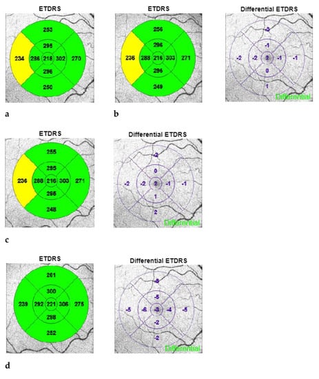
Background: The aim of the study was to compare macular thickness behavior and clinical outcomes after femtosecond laser-assisted cataract surgery (FLACS) versus phacoemulsification conventional surgery (PCS). Methods: Macular Optical Coherence Tomography OCT was analyzed in 42 patients preoperatively, 1 day, 12 days, 4 weeks and 6 weeks postoperatively according to the 9-field Early Treatment Diabetic Retinopathy Study (ETDRS) grid. Clinical findings were collected in both the FLACS group and the PCS group. Results: There was no significant difference in macular thickness between the FLACS and PCS groups (p > 0.05). However, from postoperative day 12 onwards, there was a significant increase in macular thickness observed in both groups (p < 0.001). In the FLACS group, a significant increase in visual acuity was observed on the first postoperative day, as compared to the PCS group (p = 0.006). Conclusions: The use of a low-energy high-frequency femtosecond laser has potentially no effect on postoperative macular thickness. In the FLACS group, visual rehabilitation was significantly faster as compared to the PCS group. No complications occurred intraoperatively in either group. Full article
Show Figures

Figure 1

2022

Jump to: 2024, 2023

7 pages, 1486 KiB  
Case Report
Successful Phakic Intraocular Lens Implantation with the Usage of Topical Ascorbic Acid in Patient with Reduced Corneal Endothelial Cell Density
by Hung-Chi Chen, Chia-Yi Lee, Chao-Min Cheng, Yi-Jen Hsueh, Chao-Kai Chang and Wei-Chi Wu
Medicina 2022, 58(10), 1367; https://doi.org/10.3390/medicina58101367 - 28 Sep 2022
Cited by 2 | Viewed by 2075
Abstract
We aimed to describe the use of topical ascorbic acid (AA) in a patient with reduced endothelial cells density (ECD) who was scheduled for phakic intraocular lens (pIOL) implantation. A 28-year-old woman presenting with dry eye and reduced ECD would like to have [...] Read more.
We aimed to describe the use of topical ascorbic acid (AA) in a patient with reduced endothelial cells density (ECD) who was scheduled for phakic intraocular lens (pIOL) implantation. A 28-year-old woman presenting with dry eye and reduced ECD would like to have her high myopia (spherical equivalence >−15.0 D) corrected. The procedure of laser refractive surgery or even pIOL was not indicated for the reduced ECD of 1865/mm2 in the right eye and 2188/mm2 in the left eye, as well as level 3 dry eye. Fortunately, the ECD increased to 3144/mm2 in the right eye and 2538/mm2 in the left eye after topical AA was prescribed for one year preoperatively and one month postoperatively, with concomitant improvement of dry eye to level 1. Finally, bilateral pIOL implantation was performed smoothly and no sign of corneal decompensation was found postoperatively. Three months postoperatively, the ECD showed a satisfactory level of 2983/mm2 in the right eye and 3003/mm2 in the left eye. In conclusion, topical AA instillation might increase and maintain the density of central human corneal endothelial cells (HCECs) even after pIOL implantation. Full article
Show Figures

Figure 1

Back to TopTop