Skip to Content

Journal of Developmental Biology

Journal of Developmental Biology is an international, peer-reviewed, open access journal on the development of multicellular organisms at the molecule, cell, tissue, organ and whole organism levels published quarterly online by MDPI.

Indexed in PubMed | Quartile Ranking JCR - Q2 (Developmental Biology)

Get Alerted

Add your email address to receive forthcoming issues of this journal.

All Articles (435)

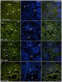
Background/Objectives: Growing evidence indicates that melatonin contributes to kidney development and function, while disruptions of fetal circadian signaling have been linked to congenital anomalies of the kidney and urinary tract (CAKUT). This study aimed to characterize the developmental and spatial expression patterns of melatonin receptors MTNR1A and MTNR1B in normal human fetal kidneys and in CAKUT phenotypes. Methods: This study analyzed 40 human fetal kidney specimens, including healthy controls and CAKUT cases (horseshoe kidneys, duplex kidneys, and dysplastic kidneys), obtained from spontaneous abortions and pregnancy terminations. Samples were classified into developmental phases Ph2–Ph4 according to established morphological criteria. Immunofluorescence staining was used to visualize MTNR1A and MTNR1B expression. Quantitative analysis was performed using ImageJ, measuring the fluorescence area percentage. Statistical comparisons were conducted using a two-way ANOVA. Results: In control kidneys, MTNR1A expression was predominantly observed in glomeruli and interstitial cells and showed a descending trend across developmental stages, whereas MTNR1B was localized to glomeruli and strongly to the apical membranes of tubules, particularly distal tubules, without substantial developmental variation. CAKUT phenotypes exhibited higher expression of both receptors compared to controls. Significant phase-dependent differences in MTNR1A expression were observed in horseshoe, duplex, and dysplastic kidneys. MTNR1B expression decreased across developmental stages in dysplastic kidneys and differed significantly between Ph3 and Ph4 in duplex kidneys. At Ph3, duplex kidneys showed the highest MTNR1B expression. Conclusions: Altered developmental expression patterns of MTNR1A and MTNR1B in CAKUT suggest an association between melatonin signaling and abnormal human kidney development.

15 April 2026

Immunofluorescence staining of (a) control human fetal kidneys (CTRL), (b) horseshoe kidneys (HK), (c) duplex kidneys (DK), and (d) dysplastic kidneys (DYS) using an antibody against melatonin receptor 1A (MTNR1A). Arrows indicate MTNR1A staining in the glomeruli (g), distal convoluted tubules (dct), dysplastic tubules (dt), and mild staining in the proximal convoluted tubules (pct), as indicated in the corresponding 4′,6-diamidino-2-phenylindole (DAPI)-stained images. All images were acquired at 40× magnification; scale bar is 50 µm.

Ribosomes are widely recognized as large intracellular macromolecular complexes responsible for protein synthesis. However, in recent years, numerous studies have revealed that ribosomal proteins possess non-canonical functions beyond translation, including roles in cell fate regulation, development, and disease. This review outlines emerging concepts surrounding the extracellular functions of ribosomes, with a particular focus on ribosome-induced cellular plasticity and transdifferentiation. Our studies have demonstrated that the incorporation of exogenous ribosomes reprograms somatic cells into a multipotent state and promotes differentiation into multiple lineages. These findings represent an alternative perspective to the conventional view of ribosomes as merely translational components. Furthermore, we discuss the biological significance of factors secreted by ribosome-incorporated cells by integrating the paracrine hypothesis with ribosome-mediated cell fate conversion. Finally, we explore the potential applications of ribosomes in regenerative medicine and cell-cultured food production. By redefining ribosomes as active regulators of cellular identity, this review provides a conceptual framework for understanding ribosome-driven cell fate regulation and its potential applications in sustainable biotechnology.

9 April 2026

Ribosome incorporation induces stem cell-like cluster formation and multilineage transdifferentiation of somatic cells. Somatic cells incorporate exogenous ribosomes, leading to the formation of stem cell-like cell clusters characterized by increased cellular plasticity. These ribosome-incorporated clusters subsequently exhibit multilineage differentiation potential and can give rise to diverse cell types, including neurons, cardiomyocytes, adipocytes, osteoblasts, and chondrocytes, under appropriate lineage-specific induction conditions. This process occurs without genetic modification and suggests that ribosome incorporation reconfigures the cellular state of somatic cells, enabling broad transdifferentiation capacity.

Mammals and reptiles possess a metanephric kidney as the terminal renal organ for homeostasis of solutes and waste products. The development of the metanephric kidney has primarily been studied in mammalian model systems. Little is known about the conservation of metanephric kidney formation in non-mammalian species such as reptiles. Uniquely, reptiles maintain kidney progenitor cell populations throughout life and continually develop new nephrons, the functional unit of the kidney. The red-eared slider turtle, Trachemys scripta elegans, was utilized to investigate the conservation of reptilian metanephric kidney development. The nephron progenitor cell (NPC) marker, Six2, was detected in whole-mount turtle kidneys in a similar pattern to mammals. However, there were differences in progenitor cell niche morphology where turtle NPC populations formed distinct elongated rows instead of the rosette-like morphology found in the mouse. The pattern of NPC populations in the embryonic turtle kidney was maintained in the adult turtle. Whole-genome bisulfite sequencing was performed on cortical tissue containing the NPC populations from adult turtle kidneys and compared to those of adult mice. Significant conservation of gene methylation was detected in adult cortical tissue between the two species, although unique signatures were detected in turtle samples related to DNA repair and β-catenin signaling. This suggests a high level of conservation of metanephric kidney development at the genetic level.

7 April 2026

Relationship of renal organ size during embryonic development. (a,b) Representative craniocaudal (long axis) and mediolateral (short axis) axes at stage 23 of mesonephric (a) and metanephric (b) kidneys. Scale bars = 50 µm. (c,d) Average lengths of craniocaudal and mediolateral axes for mesonephric (c) and metanephric (d) kidneys at different developmental stages. Single kidneys were measured from five independent embryos for each stage. Error bars represent standard deviation. (e) Mesonephros to metanephros ratio of craniocaudal and mediolateral axis measurements. Average lengths for the mesonephros were divided by average lengths for the metanephros for each axis.

Serotonin is a critical morphogen in early development, yet the mechanisms regulating its homeostasis in the preimplantation embryo remain unclear, particularly under conditions of maternal antidepressant exposure. Here, we investigated embryonic serotonergic autonomy using mouse models of pharmacological transport blockade (maternal fluoxetine treatment) and in vitro treatment with the monoamine oxidase inhibitor pargyline. We employed immunofluorescence, RT-qPCR, and live-cell imaging to assess metabolic flux, gene expression, and physiological health. We demonstrate that monoamine oxidase functions as a metabolic firewall, progressively maturing from zygote to blastocyst to degrade excess amines. Paradoxically, maternal serotonin transporter blockade triggered significant intracellular serotonin hyper-accumulation in blastocysts, associated with a trend toward a compensatory upregulation of the biosynthetic gene Ddc. While this serotonin overload did not compromise morphology, mitochondrial function, or pluripotency marker expression, it induced a robust epigenetic response. Excess serotonin promoted elevated H3Q5ser immunoreactivity in both nuclear and cytoplasmic compartments via a transglutaminase-dependent mechanism. These findings reveal that the preimplantation embryo possesses a resilient, autonomous serotonergic system capable of compensatory synthesis. However, environmental fluctuations are chemically recorded via transglutaminase-mediated serotonylation, representing an epigenetic mark that warrants further long-term study within the Developmental Origins of Health and Disease (DOHaD) framework.

3 April 2026

Regulation of intracellular serotonin (5-HT) levels during preimplantation development via monoamine oxidase (MAO) activity. (a–c) Representative confocal images showing immunolocalization of MAO-A in a zygote (a), non-compact morula (b), and blastocyst (c). Note the punctate cytoplasmic distribution and dense clusters in the interblastomere zones, inner cell mass (ICM), and trophectoderm (TE). (d) Negative control (secondary antibodies only). (e–h) 5-HT turnover in blastocysts incubated in control KSOM medium (e), 1 μM 5-HT (f), 5 μM pargyline (MAO inhibitor) (g), or 5-HT + pargyline (h). (i) Quantification of 5-HT fluorescence intensity in blastocysts and 2-cell embryos (n = 13 independent litters each). Data are presented as mean ± SEM. Asterisks indicate significant differences compared to the control group (Friedman test with Dunn’s multiple comparisons test: ** p < 0.01). (j–m) 5-HT turnover in 2-cell embryos treated as in panels (e–h). Scale bars: 10 μm.

News & Conferences

Volumes

Latest Issues

Open for Submission

Editor's Choice

XFacebookLinkedIn
J. Dev. Biol. - ISSN 2221-3759