Recent Advances in Retina Surgery
A project collection of Journal of Clinical Medicine (ISSN 2077-0383). This project collection belongs to the section "Ophthalmology".
Papers displayed on this page all arise from the same project. Editorial decisions were made independently of project staff and handled by the Editor-in-Chief or qualified Editorial Board members.
Submission Status:
Closed
|
Viewed by 11686
Share This Project Collection
Editor
 Prof. Dr. Francisco Javier Ascaso
Prof. Dr. Francisco Javier Ascaso
E-Mail
Website
Guest Editor
1. Head of the Department of Ophthalmology, Hospital Clínico Universitario Lozano Blesa, Zaragoza, Spain
2. Associate Professor of Ophthalmology, Department of Surgery, School of Medicine, University of Zaragoza, Zaragoza, Spain
3. Aragón Health Research Institute (IIS Aragón), Zaragoza, Spain
Interests: medical retina; surgical retina; history of ophthalmology
Project Overview
Dear Colleagues,
On behalf of the Journal of Clinical Medicine Editorial Team, I am delighted to present a Special Issue on the topic “Recent Advances in Retina Surgery”.
In recent years, we can report numerous improvements in the management of vitreoretinal disorders. Therapeutical approaches have undergone a profound evolution thanks to a continuous implementation of the diagnostic and surgical instrumentations, and some new surgical technique for the management of advanced retinal pathologies such as inherited retinal diseases, advanced age-related macular degeneration, ocular cancer and vitreomacular diseases.
This Special Issue aims at creating a multidisciplinary forum of discussion about the role of surgical procedures in different retinal pathologies. The published papers will describe new developments in these areas. This Special Issue accepts high-quality articles containing original research results as well as review articles of exceptional merit.
Prof. Dr. Francisco Javier Ascaso
Guest Editor
Manuscript Submission Information
Manuscripts should be submitted online at www.mdpi.com by registering and logging in to this website. Once you are registered, click here to go to the submission form. All submissions that pass pre-check are peer-reviewed. Accepted papers will be published continuously in the journal (as soon as accepted) and will be listed together on the collection website. Research articles, review articles as well as short communications are invited. For planned papers, a title and short abstract (about 250 words) can be sent to the Editorial Office for assessment.
Submitted manuscripts should not have been published previously, nor be under consideration for publication elsewhere (except conference proceedings papers). All manuscripts are thoroughly refereed through a single-blind peer-review process. A guide for authors and other relevant information for submission of manuscripts is available on the Instructions for Authors page. Journal of Clinical Medicine is an international peer-reviewed open access semimonthly journal published by MDPI.
Please visit the Instructions for Authors page before submitting a manuscript.
The Article Processing Charge (APC) for publication in this open access journal is 2600 CHF (Swiss Francs).
Submitted papers should be well formatted and use good English. Authors may use MDPI's
English editing service prior to publication or during author revisions.
Keywords
- Choroidal tumors
- Diabetic retinopathy
- Inherited retinal diseases
- Macular surgery
- Ocular diagnostic tools
- Ocular trauma
- Retinal detachment
- Retinal surgery
- Vitreoretinal diseases
Published Papers (3 papers)
Open AccessReview
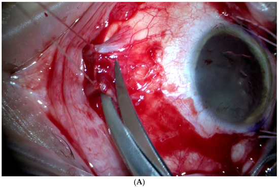
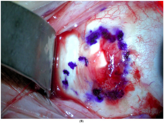
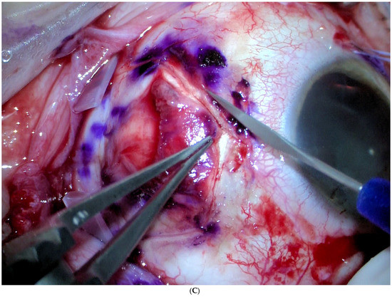
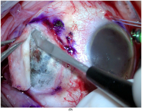
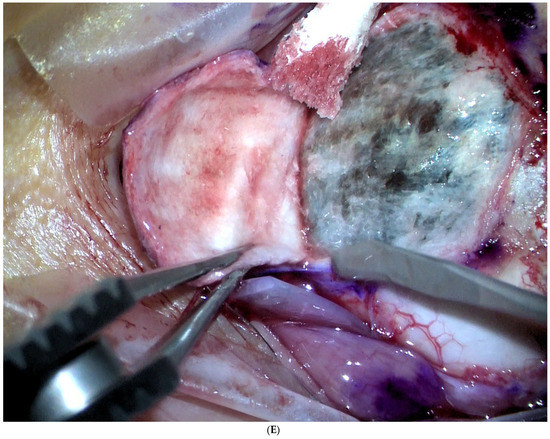
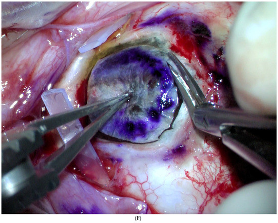
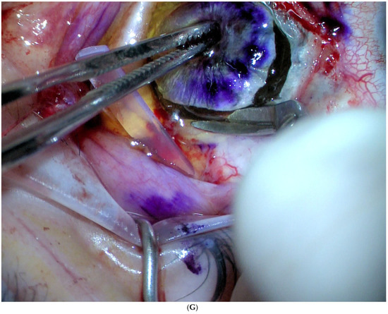
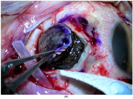
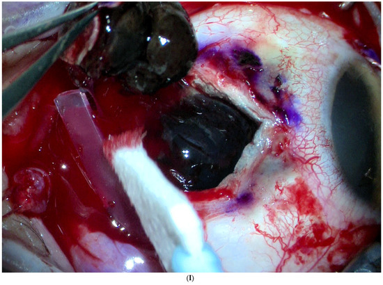
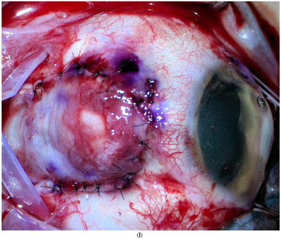
Local Resection in Choroidal Melanoma: A Review
by
Josep Maria Caminal, Daniel Lorenzo, Cristina Gutierrez, Andrea Slocker, Josep Maria Piulats, Estefania Cobos, Pere Garcia-Bru, Rahul Morwani, Juan Francisco Santamaria and Luis Arias
Cited by 17 | Viewed by 3857
Abstract
Surgical resection is widely used to treat small tumours located in the iris and the ciliary body, due to the accessibility of these sites. By contrast, surgical removal of choroidal tumours is substantially more challenging, which is why this procedure is performed only
[...] Read more.
Surgical resection is widely used to treat small tumours located in the iris and the ciliary body, due to the accessibility of these sites. By contrast, surgical removal of choroidal tumours is substantially more challenging, which is why this procedure is performed only at specialised centres. In the present article, we review the literature on surgical resection of choroidal tumours, which can be performed as endoresection (ab interno) or transscleral resection (ab externo). An important aim of this review is to describe and compare the two approaches in terms of visual outcomes, survival rates, and complications. Both approaches are indicated for the removal of large tumours (thickness > 8 mm) with small base diameters. Surgical resection of the tumour allows clinicians to obtain valuable histopathologic and cytogenetic data from the specimen and eliminates the risks associated with radiotherapy. However, both of these surgical approaches are technically challenging procedures involving the risk of severe early and late postoperative complications.
Full article
►▼
Show Figures
Open AccessArticle
Long-Term Follow-Up of Macular Perfusion Evaluated by Optical Coherence Tomography Angiography after Rhegmatogenous Retinal Detachment Surgery
by
Isabel Bartolomé-Sesé, María D. Díaz-Barreda, Elvira Orduna-Hospital, Ana Boned-Murillo, Francisco J. Ascaso and Isabel Pinilla
Cited by 5 | Viewed by 2970
Abstract
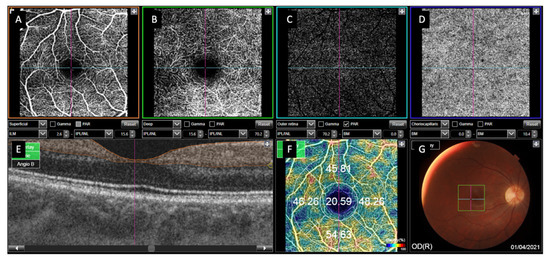
Background: The goal of this study was to investigate macular microvascular changes using optical coherence tomography angiography (OCTA) at one year after successful rhegmatogenous retinal detachment (RRD) surgery. Methods: We performed a cross-section study including RRD treated by pars plana vitrectomy (PPV) with
[...] Read more.
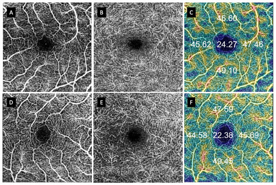
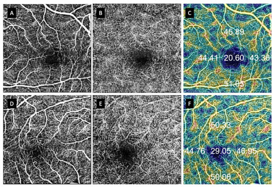
Background: The goal of this study was to investigate macular microvascular changes using optical coherence tomography angiography (OCTA) at one year after successful rhegmatogenous retinal detachment (RRD) surgery. Methods: We performed a cross-section study including RRD treated by pars plana vitrectomy (PPV) with or without scleral buckling and SF6 tamponade. After 12 months, DRI-Triton SS-OCTA was performed. Superficial and deep retinal capillary plexuses (SCP and DCP), choriocapillaris (CC) vessel density (VD), and foveal avascular zone (FAZ) morphology were analyzed. Results were compared with the unaffected contralateral eye. Results: Sixty eyes were included. We observed an increase in VD in the central area of both the SCP and DCP in macula-off eyes treated with PPV + SB and in the SCP of macula-off eyes treated with PPV. Macula-off eyes had a diminished VD for both plexuses in the superior quadrant and in the SCP inferior quadrant in those treated with PPV + SB. The CC flow was diminished in the temporal quadrant of macular-off eyes treated with PPV + SB. Healthy eyes presented higher diameter values than macula-off eyes treated with PPV + SB. FAZ horizontal and vertical diameters were smaller in patients with macula-off RRD vs. macula-on RRD and control groups. Conclusion: Macular vascularity remains almost unchanged one year after successful RRD surgery, irrespective of the surgical technique or prior macular status.
Full article
►▼
Show Figures
Open AccessArticle
OCT Angiography Findings in Macula-ON and Macula-OFF Rhegmatogenous Retinal Detachment: A Prospective Study
by
Francesco Barca, Daniela Bacherini, Francesco Dragotto, Ruggero Tartaro, Chiara Lenzetti, Lucia Finocchio, Gianni Virgili, Tomaso Caporossi, Fabrizio Giansanti, Alfonso Savastano and Stanislao Rizzo
Cited by 28 | Viewed by 3178
Abstract
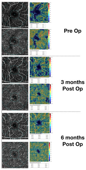
Background: The aim of the study was to evaluate pre-operative and post-operative retinal vasculature using optical coherence tomography angiography (OCTA) in patients who underwent rhegmatogenous retinal detachment (RRD) surgery repair.
Materials and Methods: A total of 33 eyes were included in this prospective
[...] Read more.
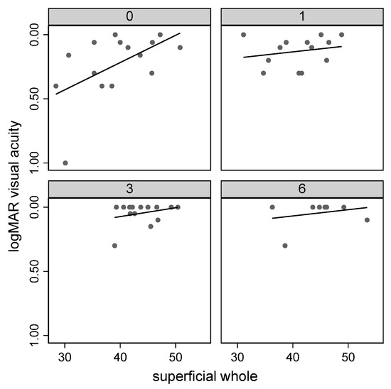
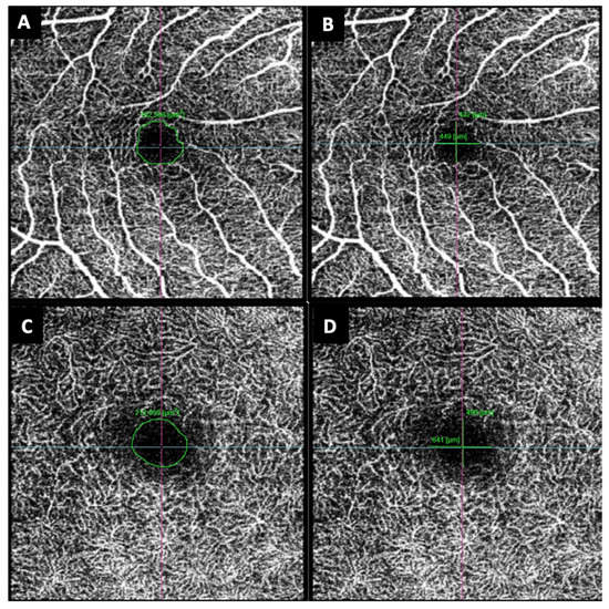
Background: The aim of the study was to evaluate pre-operative and post-operative retinal vasculature using optical coherence tomography angiography (OCTA) in patients who underwent rhegmatogenous retinal detachment (RRD) surgery repair.
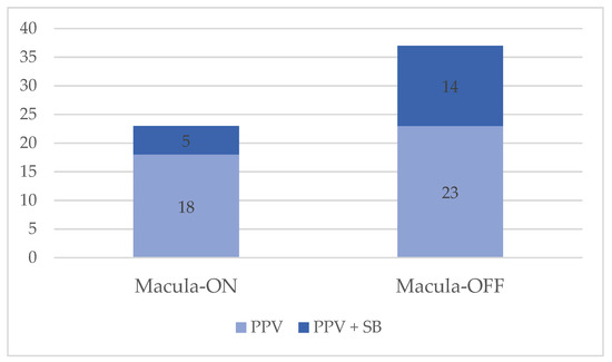
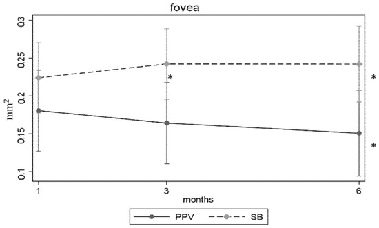
Materials and Methods: A total of 33 eyes were included in this prospective consecutive observational study: 15 affected by macula-ON and 18 by macula-OFF RRD. Superficial (SCP), deep capillary plexus (DCP), and foveal avascular zone (FAZ) area variations were evaluated by OCTA and correlated with visual acuity (VA) during a six-month follow-up.
Results: In the macula-ON group, the preoperative vascular density (VD) of the whole SCP (wSCP) on affected eyes was lower than that of the fellow eyes (
p < 0.05); this difference disappeared at 6 months after surgery (
p = 0.88). The wSCP VD and the parafoveal SCP (pfSCP) VD increased during follow-up (
p < 0.05); moreover, the higher the preoperative wSCP and pfSCP VD, the better the baseline VA (
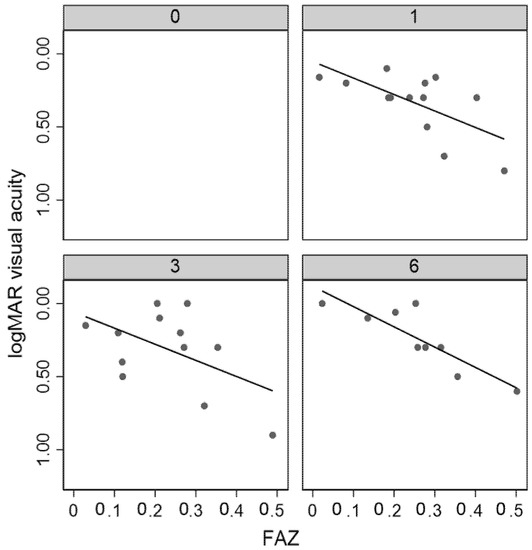
p < 0.05). In the macula-OFF group, at the first and sixth months after surgery, the larger the FAZ, the lower the VA (
p < 0.05).
Conclusions: Macula-ON SCP VD affected preoperative VA, and it was lower than the fellow eye, but recovered over time. In the macula-OFF group, a larger FAZ area was related to a worse VA, as is the case in diabetes and in retinal vein occlusion (RVO).
Full article
►▼
Show Figures


