- Technical Note

Expanding Horizons in Craniomaxillofacial Reconstruction: The Role of Exoscopic Microsurgery in Head and Neck Surgery
- Khalid Abdel-Galil and
- Kemal Mustafa Tekeli
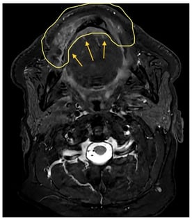
Exoscopic systems are increasingly used as an alternative to the operating microscope in microsurgical reconstruction, offering high-definition visualisation, shared operative viewing, and greater flexibility in surgeon positioning. This retrospective case series describes the use of exoscopic visualisation during microsurgical reconstruction in five illustrative head and neck and reconstructive cases. Different commercially available exoscopic platforms were utilised, and feasibility, workflow integration, and surgeon-perceived ergonomic aspects were assessed descriptively. Exoscopic visualisation was feasible for completion of microvascular anastomoses across a range of complex reconstructions. From the surgeons’ perspective, exoscopy allowed a more flexible working posture during prolonged microsurgical tasks and may offer advantages in training environments, particularly for junior surgeons. Further studies incorporating objective outcome measures are required to better define the role of exoscopy in microsurgical practice.
3 February 2026