Skip to Content
  • 42 days
    Time to First Decision

Arthropoda

Arthropoda - formerly Entomology - is an international, peer-reviewed, open access journal on the study of arthropods published quarterly online by MDPI.

Get Alerted

Add your email address to receive forthcoming issues of this journal.

All Articles (65)

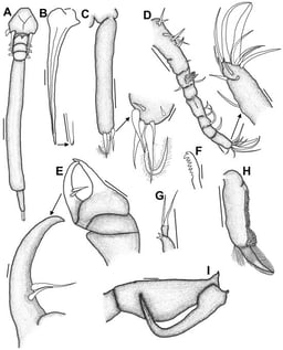
Kroyeria species (Siphonostomatoida: Kroyeriidae) are currently represented by 20 valid species. One of these is K. minuta described from the milk shark off India. However, the original description has some uncertainties and atypical features and therefore a re-description is needed. Kroyeria species were collected from milk sharks off South Africa, and amongst these were some specimens of K. minuta as well as a new species of Kroyeria. This work therefore combines a re-description of K. minuta as well as a description of K. confusa n. sp. from the milk shark off South Africa. The most distinguishing feature of K. minuta is the structure of the legs, especially that of leg 2 in the female. The new species shares characteristics with K. rhophemophaga and K. triakos but differs mainly with respect to the morphology of the legs. Thus, the number of valid Kroyeria species is now 21, and the number of reported Kroyeria species from South African waters increases to 13 with the addition of K. confusa n. sp. and the newly reported K. minuta.

28 May 2026

Kroyeria minuta Pillai, 1968, adult female. (A) Dorsal habitus; (B) dorsal stylet → tip; (C) caudal ramus → tip (ventral view); (D) antennule → last segment; (E) antenna → last segment; (F) tip of mandible; (G) maxillule; (H) maxilla; (I) maxilliped. Scale bars: (A) 0.25 mm; (B) 50 µm → tip 50 µm; (C) 50 µm → tip 10 µm; (D) 50 µm → tip 10 µm; (E) 50 µm → last segment 10 µm; (F) 10 µm; (G) 50 µm; (H) 50 µm; (I) 50 µm.
  • Brief Report
  • Open Access

Detection of Penaeus vannamei Pathogens from Water and Sediment eDNA Using a Universal Conventional PCR Approach

  • Mriya López-Galicia,
  • Roberto Cruz-Flores and
  • Jorge Cáceres-Martínez
  • + 2 authors

Environmental DNA (eDNA) offers a promising, non-invasive approach for monitoring infectious agents in aquaculture. While molecular techniques for detecting shrimp pathogens are well established in host tissues, there is a lack of standardized protocols for pathogen detection from environmental samples using conventional PCR. In this study, we developed and validated a universal conventional PCR protocol for monitoring DNA from major viral and bacterial shrimp pathogens within pond water and sediment samples. The method was applied to two commercial shrimp farms in Mexico, where eDNA was extracted from field-collected water and sediment. Using published primer sets, we successfully amplified DNA sequences corresponding to six key pathogens—Infectious hypodermal and hematopoietic necrosis virus (IHHNV), Baculovirus penaei (BP), Monodon baculovirus (MBV), Shrimp hemocyte iridescent virus (SHIV), Candidatus Hepatobacter penaei (NHP-B), and Acute hepatopancreatic necrosis disease (AHPND)-causing Vibrio spp.—in environmental samples. Sequencing of PCR amplicons confirmed 93–100% identity to previously reported pathogen strains, highlighting the method’s reliability. Pathogen detection rates varied by site, sample type, and date, with the percentage of positive samples ranging from 11.1% to 77.7%. Notably, this is the first report of SHIV DNA detection from environmental samples in the Americas, highlighting its value for pathogen surveillance even in the absence of documented outbreaks. This protocol offers a cost-effective and scalable tool for pathogen surveillance in shrimp aquaculture, enhancing early disease detection and contributing to improved biosecurity and risk assessment frameworks.

20 April 2026

Detection and percentage of positive samples of shrimp pathogens utilizing eDNA. (A) Detection of an ~420 bp amplicon (black arrows) of the NHP-B genome from water (top row) and sediment (bottom row) samples. (B,C) Percentage of positive samples of IHHNV, MBV, SHIV, NHP-B and AHPND in water and sediment from Farm 1. (D,E) Percentage of positive samples of IHHNV, MBV, NHP-B and AHPND in water and sediment from Farm 2.

Five new species of Schizomida are described from the Ecuadorian Andes and the Amazonian ecoregion in three different genera: in Colombiazomus, Colombiazomus karsticus n. sp. (female); in Hansenochrus, Hansenochrus maicun. sp. (male) and Hansenochrus pastaza n. sp. (female); and in Surazomus, Surazomus colonso, n. sp. (male, female) and Surazomus yasunin. sp. (male, female). A distribution map of all newly described species is presented.

1 April 2026

Colombiazomus karsticus n. sp., female holotype. (A) Habitus, dorsal view. (B) Propeltidium, mesopeltidium and metapeltidium, dorsal view. (C) Habitus, ventral view. Scale bars 0.5 mm.

In penaeids, the major yolk protein precursor vitellogenin is synthesized in both the hepatopancreas and the ovary. While ovarian vitellogenin expression is clearly regulated by hormones from the X-organ/sinus gland in the eyestalk, regulation in the hepatopancreas remains poorly understood. Here, we performed transcriptome profiling stratified by endogenous hepatopancreatic vitellogenin gene (Maj-Vg1) expression levels in immature kuruma prawn Marsupenaeus japonicus. Pathway enrichment analysis identified the insulin, mechanistic target of rapamycin (mTOR), glucagon, and AMP-activated protein kinase (AMPK) pathways as candidate modules associated with the control of hepatopancreatic Maj-Vg1 expression. Analysis of differentially expressed genes identified slit-like (Slit) and calreticulin (Calr) as genes potentially involved in the regulation of Maj-Vg1 expression. In ex vivo hepatopancreas explants, insulin-like peptide 1 from this species induced Maj-Vg1 and was accompanied by the upregulation of lipogenic markers (Max-like protein X (Mlx) and acetyl-CoA carboxylase (Acc)), consistent with vitellogenin’s lipid-transport role. Expression patterns of Calr, tuberous sclerosis complex 1 and 2 (Tsc1 and Tsc2) suggest regulatory inputs beyond insulin signaling, indicating context-dependent regulation. Taken together, these data identify metabolic status as an important contributor to hepatopancreatic Maj-Vg1 expression and define further research targets, including mTOR, AMPK, glucagon, and the Slit/Roundabout axis, for understanding vitellogenesis in penaeids.

23 March 2026

Maj-Vg1 and Maj-Vg2 expression levels in the hepatopancreas of the 18 previtellogenic individuals. Each symbol represents one individual plotted as Maj-Vg1 (y-axis) versus Maj-Vg2 (x-axis). The x-axis is shown on a logarithmic scale for clarity. Upward and downward triangles indicate samples selected for the high- and low-Maj-Vg1 groups, respectively, for mRNA-seq. Open squares denote individuals not used for mRNA-seq. Individuals marked with a cross that showed high Maj-Vg1 expression were excluded due to insufficient RNA quality or atypically high Maj-Vg2 expression. Although Maj-Vg2 expression serves as an indicator of vitellogenic stage, histological observations ultimately confirmed that all six selected individuals were at the previtellogenic stage (Figure S1).

News & Conferences

Volumes

Latest Issues

Open for Submission

Editor's Choice

XFacebookLinkedIn
Arthropoda - ISSN 2813-3323