Article

Omics Description (Metabolome and Microbiome) from Centuroides suffusus and Centuroides vittatus (Arachnida: Scorpiones)

by
Mariana Lizbeth Jiménez-Martínez
1,†,
Patricio Adrián Zapata-Morin
2,†,
María de Lourdes Ramírez-Ahuja
1,
Manuel de Luna
3,
Ivan Meneses-Morales
4,
Gerardo de Jesús Trujillo-Rodríguez
1,
Estela Ruiz-Baca
4,
Laura Elia Martínez-Garza
5,
Cesaré Moises Ovando-Vazquez
6,
Carlos Solis-Rojas
7,
Antonio Guzman-Velasco
8,
Margarita L. Martinez-Fierro
9,
Ivan Delgado-Enciso
10,
Adriana E. Flores-Suarez
11,
Angelica Lopez-Rodriguez
4,* and
Iram P. Rodríguez-Sánchez
1,*
1
Laboratorio de Fisiologia Molecular y Estructural, Facultad de Ciencias Biologicas, Universidad Autonoma de Nuevo Leon, San Nicolas de los Garza, Nuevo Leon 66455, Mexico
2
Department of Microbiology and Immunology, School of Biological Sciences, Universidad Autónoma de Nuevo Leon, San Nicolas de los Garza, Nuevo Leon 66455, Mexico
3
Laboratorio de Entomologia Forestal, Facultad de Ciencias Forestales, Universidad Autonoma de Nuevo Leon, Linares, Nuevo Leon 67700, Mexico
4
Facultad de Ciencias Quimicas, Universidad Juarez del Estado de Durango, Durango 34120, Mexico
5
Genetics Department, Hospital Universitario Dr. Jose Eleuterio Gonzalez Universidad Autonoma de Nuevo Leon, Monterrey, Nuevo Leon 64460, Mexico
6
CNS IPICYT, Laboratorio de Bioinformatica e Inteligencia Artificial, San Luis Potosí, S.L.P. 78218, Mexico
7
Laboratorio de Entomologia y Artropodos, Facultad de Ciencias Biologicas, Universidad Autonoma de Nuevo Leon, San Nicolas de los Garza, Nuevo Leon 66455, Mexico
8
Laboratorio de Conservacion de Vida Silvestre y Desarrollo Sustentable, Facultad de Ciencias Biologicas, Universidad Autonoma de Nuevo Leon, San Nicolas de los Garza, Nuevo Leon 66455, Mexico
9
Molecular Medicine Laboratory, Unidad Académica de Medicina Humana y C.S, Universidad Autónoma de Zacatecas, Zacatecas 98000, Mexico
10
Facultad de Medicina, Universidad de Colima & Instituto Estatal de Cancerologia, Servicios de Salud del Estado de Colima 28040, Mexico
11
Laboratorio de Entomología Médica, Departamento de Zoología de Invertebrados, Facultad de Ciencias Biológicas, Universidad Autónoma de Nuevo León, San Nicolás de los Garza 66455, Mexico
*
Authors to whom correspondence should be addressed.
These authors contributed equally to this work.
Arthropoda 2025, 3(4), 14; https://doi.org/10.3390/arthropoda3040014
Submission received: 12 April 2025 / Revised: 22 July 2025 / Accepted: 14 October 2025 / Published: 17 October 2025

Abstract

Scorpions are characterized by their venomous adaptations, including specialized stingers, and their ecological diversity. Some families, such as Buthidae, have medically significant species and their venoms possess a diverse array of chemicals. In Mexico, Centruroides suffusus and Centruroides vittatus coexist, with C. suffusus considered medically important due to its highly toxic venom. This study describes the metabolomic and microbiomic profiles of C. suffusus and C. vittatus. The metabolomic profiling (12 amino acids and 28 acylcarnitines) reveals significant differences between the two species, hinting at metabolic and ecological variations. Ornithine (ORN) and arginine (ARG) were the most abundant in C. vittatus, while tyrosine (TYR) was the most abundant amino acid molecule in C. suffusus. The microbiome analysis (by Next-Generation Sequencing of the 16S ribosomal gene) indicates similarities in gut bacteria composition between the two species (Phyla: Actinobacteria, Bacteroidetes, Cyanobacteria, Firmicutes, Proteobacteria and Tenericutes).

1. Introduction

Scorpions (Arachnida: Scorpiones) are venomous arachnids with an opisthosoma divided into two regions. The terminal appendage is used to deliver venom to both kill prey and help the scorpion defend itself against potential predators [1,2].
The venom of scorpions is composed of a wide array of chemicals, as they are rich in small peptides, mucopolysaccharides, and enzymes [3,4], all of which represent valuable biomedical and biotechnological resources. Among these, medically significant species are found mainly within Buthidae and Hemiscorpiidae (its only genus, Hemiscorpius Peters, 1861), while Nebo (Diplocentridae, Simon, 1878) has been mentioned in isolated reports but lacks confirmed evidence of clinical importance [5]. Neurotoxins that facilitate prey capture by targeting ion channels. In humans, they can bind to sodium, potassium, chloride, or calcium channels, disrupting nerve impulse transmission and causing symptoms such as intense pain, hypersalivation, muscle spasms, asphyxia, and paralysis, which may be fatal in severe cases [6,7,8]. The Mexican scorpion fauna comprises 310 species grouped into eight families [9]; among these families, Buthidae stands out as the only one that has species of medical importance [10]. This family is represented in Mexico by two genera: the ubiquitous genus Centruroides Marx, 1889, of which 54 species are known to inhabit the country [9], and the much more geographically restricted species of the endemic genus Chaneke Francke, Teruel & Santibañez-López, 2014, of which there are only four in Mexican territory [11]. The state of Durango harbors a notable scorpion diversity influenced by its environmental heterogeneity, which includes arid and semi-arid zones as well as temperate forests [12]. This landscape variation shapes scorpion distribution, as observed for the two Centruroides species with medical importance in Durango: C. suffusus and C. infamatus [13]. These two species, C. suffusus and C. vittatus, were selected for this study because they were the most frequently encountered during field collections in Durango and are of medical relevance in the region. Additionally, these species differ ecologically and morphologically, with C. suffusus typically larger and associated with arid habitats, while C. vittatus is smaller and occupies a broader range of environments.
By DNA sequencing techniques, it has been possible to compare the mitochondrial genome of several species of scorpions, including C. suffusus and C. vittatus, of which there are partial sequences in GenBank [14,15,16,17,18]; nonetheless, there is a lack of information regarding the integration of physiological data, which can help unveil mechanistic divergence among species. Growing evidence suggests that ecological and developmental factors, such as metabolism, affect multiple physiological processes in animals [19,20]. In this context, acylcarnitines play a key role and are essential for the transport of short, medium, and long-chain fatty acids across the internal mitochondrial membrane for their subsequent β-oxidation.
The study of the metabolome in arachnids and insects, especially amino acid and acylcarnitine profiles, is essential to understanding their metabolic processes. Tandem mass spectrometry (MS/MS) is a key technique for identifying and quantifying these metabolites. However, to date, no metabolic profiles of amino acids and acylcarnitines have been described for any scorpion species. In insects such as Culex quinquefasciatus, changes in these profiles were observed after exposure to insecticides, underlining their importance in the response to environmental stress [21]. Although most metabolomic studies to date have focused on vertebrates, such as horses [22] and bears [23,24], these examples illustrate the broad applicability of metabolomics in evaluating physiological and biochemical profiles across diverse taxa. However, comparable studies in arthropods, particularly arachnids, remain scarce. The microbiota of scorpions, which includes communities of bacteria and other microorganisms, plays a crucial role in their biology. Recent studies have identified unique bacteria in the telson of scorpions, such as Mollicutes, suggesting that these bacteria may influence venom composition [25]. These component organisms of the microbiota can affect the production and effectiveness of poisons [26].
This study aims to characterize the metabolomic and microbiomic profiles of C. suffusus and C. vittatus. We focused on amino acids and acylcarnitines due to their known roles in energy metabolism, oxidative stress response, and detoxification pathways. Previous studies have identified these metabolites as informative biomarkers in insects. To our knowledge, this is the first study to characterize the metabolomic profiles of scorpions, providing novel insights into their amino acid and acylcarnitine composition. However, comparable information in scorpions remains scarce. Ecologically, C. suffusus is commonly found in arid and semi-arid environments and is known for its larger size and more potent venom, while C. vittatus occupies a broader range of habitats across North America and is generally smaller in size. Our dual approach seeks to uncover species-specific biochemical patterns and microbial associations that may contribute to physiological or ecological differentiation.

2. Materials and Methods

2.1. Scorpions Sample Collection

We collected adult specimens of C. suffusus and C. vittatus from urban habitats in Victoria de Durango (24°01′13.2″ N 104°39′27.2″ W), state of Durango, Mexico, using an aspirator during manual night searches. Permission for scorpion field collection was obtained from the Secretariat of Environment and Natural Resources (SEMARNAT—scientific collection license: No. SPARN/DGVS/12661/23).
The collection and sample preparation took place in October 2023. The specimens were deposited in the Arachnid Collection of the School of Biological Sciences at UANL. Additionally, 10 females and 10 males for each species were used to extract the muscle tissue from the dorsal mesosoma for the metabolomic analysis and the intestines for the microbiome analysis. Specimens were dissected under sterile conditions using autoclaved dissection tools and disposable gloves to minimize contamination. Muscle tissue was carefully excised from the dorsal mesosoma, and intestines were removed using forceps and micro-scissors. All dissections were conducted in a laminar flow hood to reduce environmental contamination. Although muscle tissue was carefully dissected from the dorsal mesosoma under sterile conditions, we acknowledge the potential for minor contamination from surrounding tissues or gut content due to the anatomical compactness of scorpions.

2.2. Metabolome Analysis

Muscle tissue samples (0.30 g) from adult specimens of C. suffusus and C. vittatus were macerated and homogenized in 500 μL of ddH2O. Homogenates were placed in 1.5 mL conical tubes and centrifuged for 10 s at 15,339× g to separate debris. The aqueous phase was recovered with a syringe, filtered through a 0.22 μm acrodisc filter, and transferred to a clean tube. Then, 30 μL of the filtered sample was spotted onto S&S903 filter paper (15 repetitions with 15 min intervals). Dried spots were punched (3.2 mm diameter) using a Wallac DBS Puncher (PerkinElmer, Waltham, MA, USA).
The NeoBase Non-derivatized LC-MS/MS kit (PerkinElmer, Walthman, MA, USA), as previously applied by Martin-Park et al. [21], was used to obtain the metabolite profiles comprising 12 amino acids and 28 acylcarnitines. The values were normalized using references of internal standards labeled with stable isotopes and done to analyzed in LC-MS/MS (API 2000, ABSciex, Framingham, MA, USA) integrated into a micropump and an automatic sampler (Series 2000, Perkin Elmer, Richmond, CA, USA). The results were analyzed by Analyst 1.6.2 Software (ABSciex) and the NeoBase database.
In our study, we conducted statistical analyses using R programming (version 3.6.2) [27], this is to explore the metabolites showing a significant concentration difference at an alpha of 0.05, utilizing normalization to ensure comparability across samples. This involved adjusting each sample’s total measured values to equal one and employing a logarithmic transformation to mitigate the influence of extreme values. We used the Shapiro–Wilk test to assess data distribution, resorting to the Mann–Whitney U test for non-normally distributed data and ANOVA for normally distributed data, with a significance threshold set at 0.05. A graph was generated to represent these metabolites visually; we employed the smallest concentration of each metabolite as the baseline of zero, where the left side depicted the highest concentrations for C. vittatus and the right side for C. suffusus, using fold change (FC), i.e., Log2 (higher/lower) to indicate the direction and magnitude of concentration differences. The complete raw dataset and additional tables that underpin our analysis are available in the (Supplementary Material Tables S8–S10 for amino acids and Tables S12–S14 for acylcarnitines), providing further insight and context to our findings.

2.3. Microbiome Determination

Specimens of C. suffusus and C. vittatus were sterilized with 96% ethanol for 3 min and rinsed three times with sterile deionized water before dissection. The entire digestive tract was carefully dissected under a stereomicroscope under sterile conditions. Petri dishes containing 3 mL of sterile phosphate-buffered solution (10 mmol/L, pH 7.4) were used with a pair of flame-sterilized forceps. The extracted guts were placed in a 1.5 mL sterile conic tube with a 1 mL DNA shield (DNA/RNA Shield™ Reagent, Zymo Research, Irvine, CA, USA) and stored at –70 °C until nucleic acid extraction. The bacterial DNA was extracted with the ZymoBIOMICS kit, and the quality (see electrophoresis in Figure S15) and quantity (by microvolume spectrophotometer) of the extracted DNA were verified before sending the samples to be sequenced. Bacterial 16S rRNA gene sequencing was conducted using V1–V2 or V3–V4 region primers (oligonucleotides designed by Zymo Research) and the Quick-16STM NGS Library Prep Kit. Sequencing was carried out through the Targeted Metagenomic Sequencing Service on an Illumina MiSeq™ platform using a v3 reagent kit (2 × 300 bp paired-end reads, 600 cycles total) (Zymo Research, Irvine, CA, USA). The total number of raw sequences obtained for C. suffusus (773,276) and for C. vittatus (765,992) represents the merged forward and reverse reads after quality filtering. Data was processed using the Zymo Research bioinformatics pipeline [28]. Additional details are provided in Supplementary Table S16. Data mining was conducted in the R programming language [27]. Taxonomic assignments were made using the DADA2 pipeline [28] with SILVA v138.1 [29], retaining species-level identifications only when classification confidence exceeded 97%, recognizing the general resolution limits of 16S rRNA sequencing. Plots were generated with the R programing (version 3.6.2) using the packages data.tree [30], treemap [31], ape [32], ggtree and tidytree [33], and ggplot2 [34]. The script is available in the link: https://github.com/cesaremov/scorpions_iram/blob/main/plot_cladogram.R (accessed on 10 April 2025).

3. Results

Metabolome
The metabolomic profile was determined by tandem mass spectrometry (MS/MS) and encompassed 12 amino acids and 28 acylcarnitines.
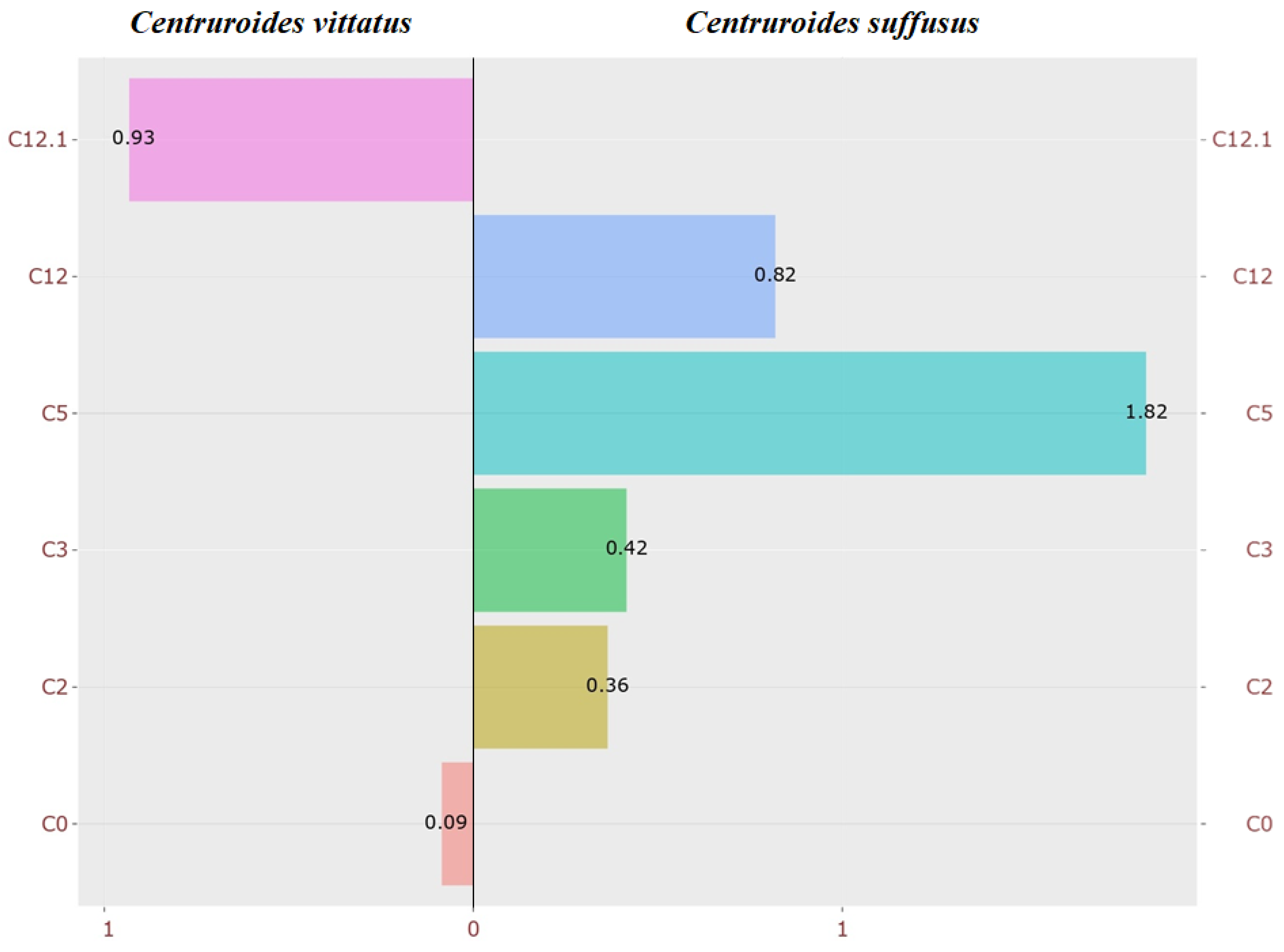
Statistical analyses were performed with an alpha of 0.05 to identify metabolites exhibiting significant differences between species across triplicates. Figure 1 and Figure 2 display only the amino acids and acylcarnitines with statistically significant differences, presented as fold changes (FCs), calculated using the log2 ratio of higher to lower concentrations, with the metabolite of lower concentration serving as baseline reference (zero).
Among the amino acids analyzed, tyrosine (TYR) was significantly more abundant in C. suffusus (2.89-fold), whereas ornithine (ORN, 2.5-fold) and arginine (ARG, 1.13-fold) were higher in C. vittatus (Figure 1). It is important to note that ORN values showed variability among replicates, with one of the three samples presenting a markedly different concentration, which could have influenced the overall mean and fold change calculation.
Detailed percentage data for each amino acid are provided in Supplementary Table S9, with additional statistical analyses and mean differences presented in Supplementary Table S11.
For acylcarnitines, C5 showed the greatest difference, with higher levels in C. suffusus (0.93-fold). Additionally, acylcarnitines, such as C2, C3, and C0, also showed species-specific variations (Figure 2). Percentage data for acylcarnitines are available in Supplementary Table S13, and statistical analyses in Table S15.
It is important to note that “non-significant” in this context refers to the lack of statistical difference between species, rather than indicating a lack of biological relevance. Full data for amino acids and acylcarnitines, including both significant and non-significant metabolites, are provide in Supplementary Tables S11 and S15, respectively.
Microbiome
Next Generation Sequencing (NGS) of the 16S rRNA gene revealed the gut microbiota composition of C. suffusus and C. vittatus. In total, 15 bacterial OTUs (Operational Taxonomic Unit), representing clusters of similar sequences that may include one or more species, had relative abundances greater than 0.5% were identified, accounting for 95.42% of total bacterial abundance in C. suffusus (65,189 genome copies/μL) and 97.76% in C. vittatus (64,119 genome copies/μL) (Figure S1). It is important to note that OTUs represent sequence clusters that may include multiple species, and while genus-level assignments are robust with V1-V2 or V3-V4 regions, species-level resolution is generally limited.
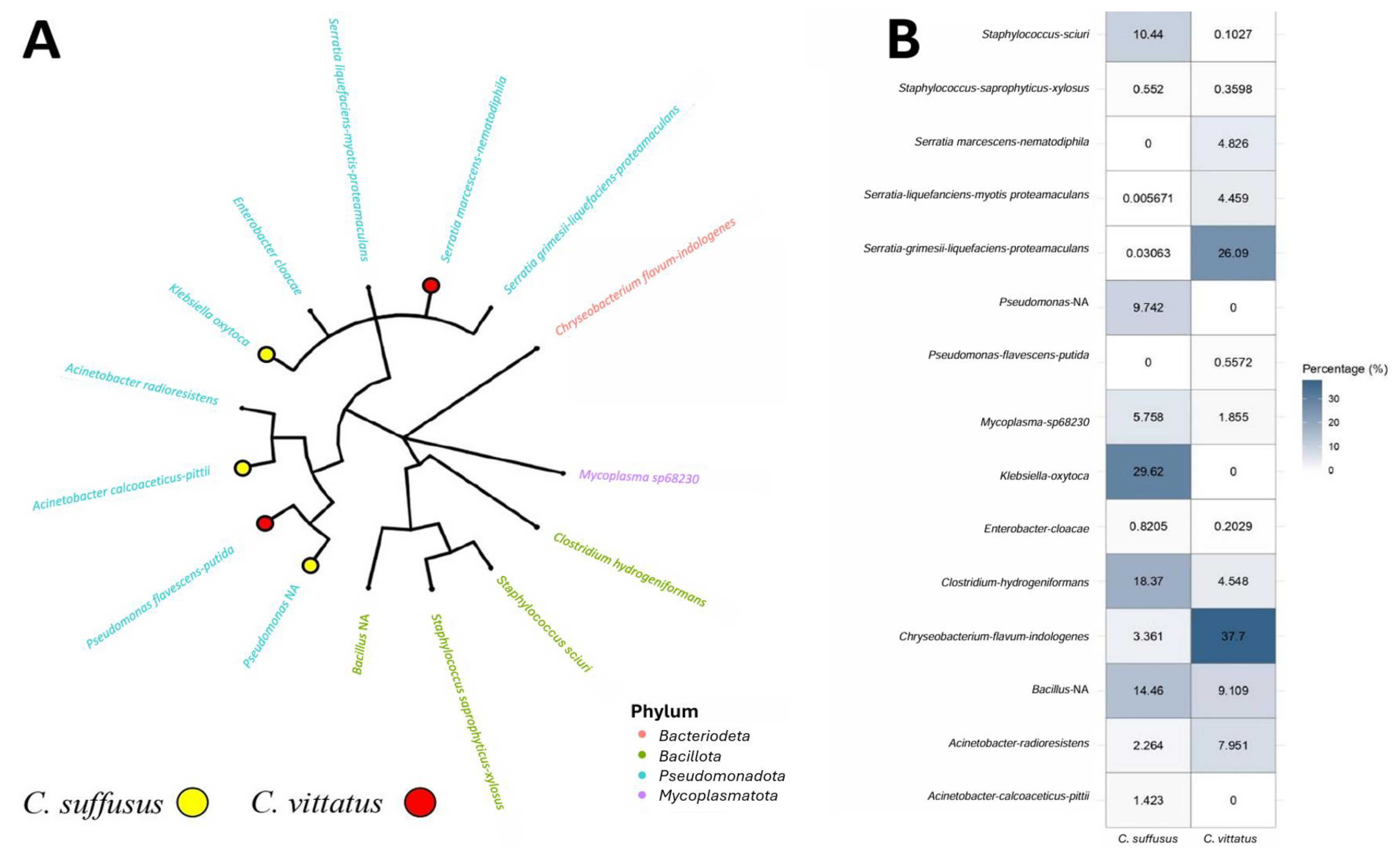
Figure 3 shows a comparative overview of bacterial taxa identified in both species. Panel A shows the phylogenetic relationships among the dominant OTUs identified, highlighting that Proteobacteria-related genera such as Serratia and Pseudomonas clustered closely, while Firmicutes genera such as Staphylococcus formed distinct branches, reflecting their taxonomic divergence. Panel B presents the relative abundances by phylum, demonstrating that Proteobacteria was dominant in both scorpions, followed by Firmicutes, Tenericutes, and Bacteroidetes. Notably, C. suffusus showed high abundances of Klebsiella oxytoca (29.62%) and Pseudomonas NA (9.74%), while C. vittatus was dominated by Chryseobacterium flavum-indologenes (37.7%) and Serratia grimesii-liquefaciens-proteamaculans (26.09%).
Table 1 provides a detailed comparison of bacterial taxa presence between the two species. Notably, C. suffusus showed exclusive presence of Pseudomonas NA, Acinetobacter calcoaceticus-pittii, and Klebsiella oxytoca, while C. vittatus harbored Pseudomonas flavescens-putida and Serratia marcescens-nematodiphila exclusively. Shared genera included Acinetobacter radioresistens, Enterobacter cloacae, Serratia grimesii-liquefaciens-proteamaculans, Clostridium hydrogeniformans, Staphylococcus sciuri, Staphylococcus saprophyticus-xylosus, Bacillus NA, and Mycoplasma sp.68230.”
Alpha diversity (Figure S14) was higher in C. suffusus, indicating greater bacterial richness compared to C. vittatus.
These results reveal a shared core microbiota dominated by Proteobacteria and Firmicutes, with species-specific differences in bacterial community composition. Detailed taxonomic distributions by phylum, class, order, family, genus, and species are provided in Supplementary Tables S1–S6, and heatmap clustering results are shown in Figures S8–S13.

4. Discussion

4.1. Metabolome

A comparative analysis of the metabolomic profiles of C. suffusus and C. vittatus revealed distinct patterns in the relative abundance of amino acids and acylcarnitines.

4.1.1. Amino Acid Profile

Marked differences were observed in the amino acid composition between species. Ornithine (ORN) and arginine (ARG) were more abundant in C. vittatus, both of which are key components of the urea cycle, a metabolic pathway that converts ammonia, a toxic byproduct of protein degradation, into urea, a non-toxic compound [35]. In vertebrates, this cycle occurs in the liver and contributes to the synthesis of vasodilators [36]. Conversely, tyrosine (TYR) was the most abundant amino acid molecule in C. suffusus, a precursor of thyroid hormones, catecholamines (adrenaline, dopamine, and norepinephrine), and melanin.
Tyrosine has been reported to participate in the biosynthesis of ubiquitin, a molecule associated with venom gland regulation in scorpions [37]. The elevated levels of tyrosine observed in C. suffusus, compared to C. vittatus, may be attributed to differences in venom gland physiology [38]. While all scorpions produce venom [39], only C. suffusus among the two species analyzed is considered medically important, due to the presence of a venom component that affects vertebrate systems [40]. C. vittatus, although not considered a threat to humans, likely exhibits venom activity specialized in immobilizing arthropod prey. Additionally, the formation of a ubiquitin–proteasome complex has been identified as a regulatory element in the venom glands of Tityus serrulatus Lutz & Mello, 1922, supporting a possible functional role of tyrosine-derived molecules in venom modulation [41]. Tyrosine, derived from phenylalanine, is involved in several downstream metabolic pathways that include the production of aromatic compounds and energy intermediates. Although the KEGG phenylalanine metabolism pathway in C. sculpturatus does not explicitly link tyrosine to cuticle formation, its role in the generation of phenolic derivates suggests potential involvement in physiological processes such as pigmentation or structural modification [42]. Arginine, like proline, serves as an energy source in invertebrates, specially under conditions of metabolic stress of high energy demand [43]. The distinct amino acid profiles between C. suffusus and C. vittatus may reflect species-specific metabolic patterns influenced by physiological needs or environmental factors [43].
Given that ornithine directly participates in the biosynthesis of arginine, the elevated ornithine levels observed in C. vittatus, are consistent with its higher arginine content. In contrast, C. suffusus, exhibited lower levels of both ornithine and arginine, suggesting reduced activity in the biosynthetic pathway. Additionally, both arginine and ornithine are involved in the metabolism of glutathione, a compound associated with cellular detoxification and resistance to insecticides [44].
Citrulline has been reported to actively participate in the urea cycle of the scorpion C. sculpturatus [45], functioning as a precursor to arginine, which in turn leads to the production of ornithine. These three amino acids, citrulline, arginine, and ornithine, are key components of the urea cycle, a pathway responsible for the elimination of nitrogenous waste products from the organism. The significant differences in their relative abundance between C. vittatus and C. suffusus may reflect species-specific physiological adaptations and geographical influences. In particular, C. vittatus may require more frequent nitrogen waste removal, possibly due to higher metabolic activity [46]. Fumarate then enters the Krebs cycle, linking nitrogen metabolism with energy production. Thus, the elevated levels of citrulline, arginine, and ornithine in C. vittatus may indicate a greater reliance on this metabolic pathway compared to C. suffusus.

4.1.2. Differential Display from Acylcarnitines Between Centruroides

In the acylcarnitine profile, several metabolites stood out: free carnitine (C0), acetylcarnitine (C2), propionylcarnitine (C3), isovalerylcarnitine (C5), dodecanoylcarnitine (C12), and monounsaturated dodecanoylcarnitine (C12:1). Although some average differences were observed between species, the variability across triplicate measurements limited the statistical significance of these differences. Nonetheless, a distinct pattern emerged: short- and medium-chain acylcarnitines (C2, C4, C5, and C12) showed elevated levels in C. suffusus, while free carnitine (C0) and monounsaturated dodecanoylcarnitine (C12.1) were more elevated in C. vittatus. These play essential roles in the transport of fatty acids into mitochondria, enabling their subsequent β-oxidation and ATP generation.
In scorpions, the differences in acylcarnitine levels are likely influenced by diet and the fitness cost associated with acquiring or metabolizing specific fatty acids, depending on the species. These differential metabolite expressions may reflect broader metabolic [47] disparities between species, including variations in dietary preferences, habitat use, evolutionary adaptations, or predator–prey dynamics. For instance, the elevated tyrosine levels observed in C. suffusus could be associated with greater efficiency in capturing prey rich in specific amino acids or metabolic precursors.
The composition of scorpion venom is influenced by multiple factors, including diet, metabolic activity, and species-specific physiological traits [48]. Given the observed differences in metabolic profiles of C. vittatus and C. suffusus, it is plausible that discrepancies in their venom compositions also differ.
Acylcarnitines are essential intermediates in fatty acid metabolism, facilitating the transport and catabolism of fatty acids and promoting ATP production via beta-oxidation [49]. Fatty acids and their derivatives may fulfill various biological functions, including the modulation of venom component activity [50,51]. Therefore, variations in fatty acid profiles may significantly influence the biological activity and toxicity of scorpion venom. These findings raise the question of whether elevated acylcarnitine levels correlate with venom composition, supporting the need to characterize the metabolic profiles of medically important scorpion venoms.

4.2. Microbiome

This study presents the first characterization of the gut microbiota in C. suffusus and C. vittatus. The gut microbiota is a dynamic ecosystem that is essential for host health and homeostasis [52,53,54]. Its composition is influenced by multiple factors, including diet [48], developmental stage [55,56], geographic location [57,58], and host physiology. These microbial symbionts contribute to physiological stability, supporting key processes such as immunity, metabolism, and nutrient absorption [59]. Certain bacteria have been associated with longevity and immunity [60], as well as host behavior [61] and reproduction [62,63].
In arthropods, gut microbiota play diverse roles, including the maintenance of the peritrophic membrane [64], degradation of polysaccharides [65,66], nitrogen fixation and recycling [67,68], detoxification [69], and protection against pathogens [70,71,72].
Scorpions have a broad diet that includes arthropods and small vertebrates. They are highly tolerant to environmental stressors, including prolonged periods of food and water deprivation, due to their ability to consume large quantities at once [2,73]. Additionally, they can tolerate extreme temperatures [74]. These physiological adaptations may influence their metabolic profiles and gut microbiota composition, as observed in this study.
The microbiota of Centruroides limpidus (Karsch, 1879) and Vaejovis smithi (Pocock, 1902) (Vaejovidae) have been previously described, showing partial overlap with that of C. suffusus and C. vittatus [75]. However, in contrast to the interspecific differences observed between C. limpidus and V. smithi, C. suffusus and C. vittatus share an exceptionally similar gut microbiota, differing only in five species of bacteria. This high similarity may be influenced by their phylogenetic proximity, as both belong to the genus Centuroides, as well as by ecological factors such as similar diet or microhabitat. Additionally, because both species were collected from the same geographical region, shared environmental conditions could also contribute to microbiome similarity.
Two species of bacteria were exclusive to C. vittatus: Serratia marcescens and Pseudomonas flavescens. Both are Gram-negative bacilli, ubiquitous in soil, water, and plant surfaces, and commonly found in moist environments such as bathrooms, drains, and sinks. Despite overlapping ecological niches, they differ in pigmentation: S. marcescens produces a brick-red pigment (prodigiosin) at 28 °C, but not at 37 °C, whereas P. flavescens is capable of producing yellow or green pigments.
In contrast, a Pseudomonas species, along with Acinetobacter calcoaceticus-pittii and Klebsiella oxytoca, was found in the digestive tract of C. suffusus. These bacteria are notable for their ability to thrive in diverse environments, their potential involvement in human and animal infections, and their relevance in both clinical and environmental contexts. Pseudomonas spp. is known for its resistance to adverse conditions and its ability to degrade complex organic compounds, making it valuable for bioremediation. A. calcoaceticus-pittii is recognized for its multidrug resistance and role as an opportunistic pathogen in hospital settings. K. oxytoca, a member of the human intestinal microbiota, can also cause nosocomial infections, being capable of surviving in different environments and developing resistance to antibiotics. These bacteria exhibit adaptability to diverse ecological niches and possess pathogenic potential, important to both human and animal health. Their presence highlights their importance in microbiological and ecological research. Additionally, the detection of Firmicutes, Actinobacteria, and Spirochaetes, commonly found in insect gut microbiota [76,77], confirms that these groups are also components of the scorpion gut microbiome.
The 16S gene of the microbiota inhabiting the digestive tract, telson, and gonads of Androctonus australis was analyzed using SANGER sequencing. The identified sequences were classified into seven bacterial taxonomic groups: Firmicutes, Betaproteobacteria, Gammaproteobacteria, Flavobacteria, Actinobacteria, Mollicutes, and Cyanobacteria [78]. Among the Firmicutes, the order Lactobacillales included Enterococcus faecium, Enterococcus faecalis, and Lactobacillus plantarum, lactic-acid-producing bacteria commonly found in A. australis as well as other arthropods and chordates [79]. These symbiotic bacteria may benefit the host by producing bacteriocins that inhibit pathogens or by contributing to other physiological processes [80,81,82].
Previous studies in scorpions, including Centruroides limpidus and Vaejovis smithi [76] Hadrurus arizonensis [25] have consistently reported Mollicutes, Firmicutes, and Proteobacteria as dominant taxa. Our findings are consistent with this general pattern, suggesting conserved symbiotic relationships within scorpions, although the specific genera and relative abundances vary among species.
Although some bacterial taxa were assigned at the species level with high confidence scores, we recognize that 16S gene sequencing typically resolves taxa at the genus level, and species-level assignments should be interpreted cautiously.

5. Conclusions

This study provides the first integrated analysis of metabolomic and microbiome profiles in C. suffusus and C. vittatus. Significant interspecific differences were identified in amino acid concentrations, notably citrulline, ornithine, and arginine, key components of the urea cycle, which were more abundant in C. vittatus. In contrast, tyrosine, associated with venom biosynthesis and cuticle development, was more abundant in C. suffusus. Acylcarnitine profiles also showed variation, suggesting species-specific differences in lipid metabolism and energy metabolism. The microbiome analysis revealed highly similar gut communities, with only minor taxonomic differences. These findings underscore the value of combining metabolomic and microbiomics data to enhance our understanding of the biochemical characteristics and microbial associations in scorpions.

Supplementary Materials

The following supporting information can be downloaded at: https://doi.org/10.5281/zenodo.16294828 (accessed on 10 April 2025).

Author Contributions

Conceptualization, I.P.R.-S., M.L.J.-M. and A.L.-R.; carried out the experiments: I.P.R.-S., M.L.J.-M., M.d.L.R.-A., I.M.-M., A.L.-R., G.d.J.T.-R., E.R.-B., L.E.M.-G., C.S.-R. and A.G.-V.; collected the data: M.d.L., P.A.Z.-M., M.d.L.R.-A., I.M.-M., G.d.J.T.-R., C.M.O.-V., E.R.-B., L.E.M.-G. and C.S.-R.; conducted the data analysis: P.A.Z.-M., I.M.-M., M.L.J.-M. and C.M.O.-V.; supervised the findings of the work: I.P.R.-S., A.L.-R., A.E.F.-S., C.M.O.-V., L.E.M.-G., M.L.M.-F. and I.D.-E.; discussed the results: I.P.R.-S., M.L.J.-M., P.A.Z.-M., M.d.L.R.-A., M.d.L., A.L.-R., A.E.F.-S., L.E.M.-G., C.S.-R., A.G.-V., M.L.M.-F. and I.D.-E.; wrote the final manuscript version: I.P.R.-S., M.L.J.-M., P.A.Z.-M., M.d.L.R.-A., M.d.L., A.L.-R., A.E.F.-S., L.E.M.-G., A.G.-V., M.L.M.-F. and I.D.-E. All authors have read and agreed to the published version of the manuscript.

Funding

This research received no external funding.

Institutional Review Board Statement

The animal study protocol was approved by the Ethics Committee of the Instituto Estatal de Cancerología de Colima (Approval Code: CEICANCL2024-ESCORP-12; Approval Date: 12 April 2024; valid until 12 April 2026; Registro CONBIOÉTICA-06-CEI-001-20200721).

Data Availability Statement

The datasets generated during and/or analyzed during the current study are available in the Zenodo repository at https://doi.org/10.5281/zenodo.16294828 (accessed on 10 April 2025).

Conflicts of Interest

The authors declare no conflicts of interest.

References

  1. Beccaloni, J. Arachnids; University of California Press: Berkeley, CA, USA, 2009; p. 320. [Google Scholar]
  2. Polis, G.A. The Biology of Scorpions; Stanford University Press: Redwood City, CA, USA, 1990; p. 587. [Google Scholar]
  3. Cao, Z.; Yu, Y.; Wu, Y.; Hao, P.; Di, Z.; He, Y.; Chen, Z.; Yang, W.; Shen, Z.; He, X.; et al. The genome of Mesobuthus martensii reveals a unique adaptation model of arthropods. Nat. Commun. 2013, 4, 2602. [Google Scholar] [CrossRef]
  4. He, Y.; Zhao, R.; Di, Z.; Li, Z.; Xu, X.; Hong, W.; Wu, Y.; Zhao, H.; Li, W.; Cao, Z. Molecular diversity of Chaerilidae venom peptides reveals the dynamic evolution of scorpion venom components from Buthidae to non-Buthidae. J. Proteom. 2013, 89, 1–14. [Google Scholar] [CrossRef]
  5. Ward, M.J.; Ellsworth, S.A.; Nystrom, G.S. A global accounting of medically significant scorpions: Epidemiology, major toxins, and comparative resources in harmless counterparts. Toxicon 2018, 151, 137–155. [Google Scholar] [CrossRef]
  6. Santos, M.S.; Silva, C.G.; Neto, B.S.; Grangeiro-Júnior, C.R.; Lopes, V.H.; Teixeira-Júnior, A.G.; Bezerra, D.A.; Luna, J.V.C.; Crdeiro, J.B.; Goncalves Junior, J.; et al. Clinical and epidemiological aspects of scorpionism in the world: A systematic review. Wilderness Environ. Med. 2016, 27, 504–518. [Google Scholar] [CrossRef]
  7. Niermann, C.N.; Tate, T.G.; Suto, A.L.; Barajas, R.; White, H.A.; Guswiler, O.D.; Secor, S.M.; Rowe, A.H.; Rowe, M.P. Defensive venoms: Is pain sufficient for predator deterrence? Toxins 2020, 12, 260. [Google Scholar] [CrossRef] [PubMed]
  8. Santibáñez-López, C.E.; Aharon, S.; Ballesteros, J.A.; Gainett, G.; Baker, C.M.; González-Santillán, E.; Harvey, M.S.; Kassan, M.K.; Almaaty, A.H.A.; Aldeyarbi, S.M.; et al. Phylogenomics of scorpions reveal contemporaneous diversification of scorpion mammalian predators and mammal-active sodium channel toxins. Syst. Biol. 2022, 71, 1281–1289. [Google Scholar] [CrossRef] [PubMed]
  9. Ponce-Saavedra, J.; Linares-Guillén, J.W.; Quijano-Ravell, A.F. Una nueva especie de alacrán del género Centruroides Marx (Scorpiones: Buthidae) de la costa noroeste de México. Acta Zool. Mex. 2022, 38, 1–24. [Google Scholar] [CrossRef]
  10. Ponce-Saavedra, J.; Francke, O.F. Clave para la identificación de especies de alacranes del género Centruroides Marx 1890 (Scorpiones: Buthidae) en el centro occidente de México. Biológicas 2013, 15, 52–62. [Google Scholar]
  11. Kovařík, F.; Teruel, R.; Lowe, G. Two new scorpions of the genus Chaneke Francke, Teruel et Santibáñez-López, 2014 (Scorpiones: Buthidae) from southern Mexico. Euscorpius 2016, 218, 220–232. [Google Scholar] [CrossRef]
  12. Santibáñez-López, C.E.; Francke, O.F.; Ureta, C.; Possani, L.D. Scorpions from Mexico: From species diversity to venom complexity. Toxins 2016, 8, 2. [Google Scholar] [CrossRef]
  13. Riaño-Umbarila, L.; Gómez-Ramírez, I.V.; García-Arredondo, A.; Rodríguez-Rodríguez, E.R.; Possani, L.D. Identification and molecular modeling of novel toxins from scorpion venom gland transcriptomes. Toxicon 2017, 138, 130–137. [Google Scholar] [CrossRef] [PubMed]
  14. Corzo, G.; Prochnicka-Chalufour, A.; García, B.I.; Possani, L.D.; Delepierre, M. Solution structure of Cn5, a crustacean toxin found in the venom of the scorpions Centruroides noxius and Centruroides suffusus suffusus. Biochim. Biophys. Acta Proteins Proteom. 2009, 1794, 1591–1598. [Google Scholar] [CrossRef] [PubMed]
  15. Espino-Solis, G.; Estrada, P.; Olamendi-Portugal, G.; Villegas, T.; Zamudio, E.; Cestele, F.; Possani, L.; Corzo, G. Isolation and molecular cloning of beta-neurotoxins from the venom of the scorpion Centruroides suffusus suffusus. Toxicon 2011, 57, 739–746. [Google Scholar] [CrossRef] [PubMed]
  16. Yamashita, T.; Rhoads, D.; Pummill, J. The complete mitochondrial genome of the scorpion Centruroides vittatus (Arachnida: Scorpiones). Mitochondrial DNA Part B 2017, 2, 841–842. [Google Scholar] [CrossRef]
  17. Esposito, L.A.; Prendini, L. Island ancestors and new world biogeography: A case study from the scorpions (Buthidae: Centruroidinae). Sci. Rep. 2019, 9, 3500. [Google Scholar] [CrossRef]
  18. Moreno-Carmona, M.; Montaña-Lozano, P.; Quiroga, C.F.P.; Baeza, J.A. Comparative analysis of mitochondrial genomes reveals family-specific architectures and molecular features in scorpions (Arthropoda: Arachnida: Scorpiones). Gene 2023, 859, 147189. [Google Scholar] [CrossRef]
  19. Botana, L.M.; Hess, P.; Munday, R.; Nathalie, A.; DeGrasse, S.L.; Feeley, M.; Toshiyuki, S.; van den Berg, M.; Fattori, V.; Garrido Gamarro, E.; et al. Derivation of toxicity equivalency factors for marine biotoxins associated with Bivalve Molluscs. Trends Food Sci. Technol. 2017, 59, 15–24. [Google Scholar] [CrossRef]
  20. Street, M.E.; Bernasconi, S. Endocrine-disrupting chemicals in human fetal growth. Int. J. Mol. Sci. 2020, 21, 1430. [Google Scholar] [CrossRef]
  21. Martin-Park, A.; Gomez-Govea, M.A.; Lopez-Monroy, B.; Treviño-Alvarado, V.M.; Torres-Sepúlveda, M.D.R.; López-Uriarte, G.A.; Villanueva-Segura, O.K.; Ruiz-Herrera, M.d.C.; Martinez-Fierro, M.d.l.L.; Delgado-Enciso, I.; et al. Profiles of amino acids and acylcarnitines related with insecticide exposure in Culex quinquefasciatus (Say). PLoS ONE 2017, 12, e0169514. [Google Scholar]
  22. Rodríguez-Sánchez, I.P.; Treviño-Alvarado, V.M.; Torres-Sepúlveda, M.D.R.; López-Saldaña, L.A.; Ponce-García, G.; López-Uriarte, G.A.; Ruiz-Herrera, M.d.C.; Zamora-Avila, D.E.; Villareal-Perez, J.; Davalos-Guillermo, G.; et al. Reference values for amino acids and acylcarnitines in peripheral blood in Quarter horses and American Miniature horses. Acta Vet. Scand. 2015, 57, 62. [Google Scholar]
  23. Abellan-Borja, A.; Rodriguez-Sanchez, I.P.; Carrera-Treviño, R.; Villanueva-Segura, O.K.; Zapata-Morin, P.A.; Martinez-de-Villareal, L.E.; Barboza-Aranda, L.J.; Gomez-Govea, M.A.; MArtinez-Fierro, M.L.; Delgado-Enciso, I.; et al. Free amino acid and acylcarnitine values in Ursus americanus Pallas 1780 (black bear) from Northeastern Mexico. PLoS ONE 2023, 18, e0272979. [Google Scholar] [CrossRef]
  24. Villarreal-Pérez, J.Z.; Villarreal-Martínez, J.Z.; Lavalle-González, F.J.; Torres-Sepúlveda, M.D.R.; Ruiz-Herrera, C.; Cerda-Flores, R.M.; Castillo-Garcia, E.R.; Rostriguez-Sanchez, I.P.; Martinez de Villareal, L.E. Plasma and urine metabolic profiles are reflective of altered beta-oxidation in non-diabetic obese subjects and patients with type 2 diabetes mellitus. Diabetol. Metab. Syndr. 2014, 6, 129. [Google Scholar] [CrossRef] [PubMed]
  25. Shimwell, C.; Atkinson, L.; Graham, M.R.; Murdoch, B. A first molecular characterization of the scorpion telson microbiota of Hadrurus arizonensis and Smeringurus mesaensis. PLoS ONE 2023, 18, 1. [Google Scholar] [CrossRef] [PubMed]
  26. Cabezas-Cruz, A. Grand challenges in arachnid microbiota and diseases. Front. Arachnid Sci. 2023, 2, 1215831. [Google Scholar] [CrossRef]
  27. R Core Team. R: A Language and Environment for Statistical Computing; R Foundation for Statistical Computing: Vienna, Austria, 2012; Available online: https://www.R-project.org/ (accessed on 10 April 2025).
  28. Callahan, B.J.; McMurdie, P.J.; Rosen, M.J.; Han, A.W.; Johnson, A.J.A.; Holmes, S.P. DADA2: High-resolution sample inference from Illumina amplicon data. Nat. Methods. 2016, 13, 581–583. [Google Scholar] [CrossRef]
  29. Quast, C.; Pruesse, E.; Yilmaz, P.; Gerken, J.; Schweer, T.; Yarza, P.; Peplies, J.; Glöckner, F.O. The SILVA ribosomal RNA gene database project: Improved data processing and web-based tools. Nucleic Acids Res. 2013, 41, D590–D596. [Google Scholar] [CrossRef]
  30. Glur, C. data.tree: To Manage Hierarchical Data and Tree Structures. R Package Version 1.0.0. Available online: https://CRAN.R-project.org/package=data.tree (accessed on 10 April 2025).
  31. Tennekes, M. treemap: Treemap Visualization. R Package Version 2.4-2. Available online: https://CRAN.R-project.org/package=treemap (accessed on 10 April 2025).
  32. Paradis, E.; Schliep, K. ape 5.0: An environment for modern phylogenetics and evolutionary analyses in R. Bioinformatics 2019, 35, 526–528. [Google Scholar] [CrossRef]
  33. Yu, G. Data Integration, Manipulation and Visualization of Phylogenetic Trees; Chapman and Hall/CRC: Boca Raton, FL, USA, 2022. [Google Scholar]
  34. Wickham, H. ggplot2: Elegant Graphics for Data Analysis; Springer: New York, NY, USA, 2016; ISBN 978-3-319-24277-4. Available online: https://ggplot2.tidyverse.org (accessed on 10 April 2025).
  35. Martins, J.C.S.; Assunção Romão, H.A.; Canettieri, C.K.; Cercilian, A.C.; Oliveira, P.R.O.; Ferreira, C.; Terra, W.R.; de Oliveira Dias, R. The loss of the urea cycle and ornithine metabolism in different insect orders: An omics approach. Insect Mol. Biol. 2025, 34, 632–644. [Google Scholar] [CrossRef]
  36. LeMoine, C.M.; Walsh, P.J. Evolution of urea transporters in vertebrates: Adaptation to urea’s multiple roles and metabolic sources. J. Exp. Biol. 2015, 218 Pt 12, 1936–1945. [Google Scholar] [CrossRef]
  37. Sarkar, A.; Foderaro, T.; Kramer, L.; Markley, A.L.; Lee, J.; Traylor, M.J.; Fox, J.M. Evolution-guided biosynthesis of terpenoid inhibitors. ACS Synth. Biol. 2022, 11, 9. [Google Scholar] [CrossRef]
  38. McElroy, T.; McReynolds, C.N.; Gulledge, A.; Knight, K.R.; Smith, W.E.; Albrecht, E.A. Differential toxicity and venom gland gene expression in Centruroides vittatus. PLoS ONE 2017, 12, e0184695. [Google Scholar] [CrossRef]
  39. van Aardt, W.J.; le Roux, J.M.; Lindeque, J.Z.; Mason, S.; Louw, R. The effect of temperature on the respiration and metabolism of the African burrowing scorpion (Opistophthalmus latimanus). Comp. Biochem. Physiol. D Genom. Proteom. 2016, 20, 50–56. [Google Scholar] [CrossRef]
  40. White, J. Venomous animals: Clinical toxinology. EXS 2010, 100, 233–291. [Google Scholar] [CrossRef] [PubMed]
  41. de Oliveira, U.C.; Nishiyama, M.Y., Jr.; Dos Santos, M.B.; Pinto, J.R.A.S.; Souza-Imberg, A.; Lopes, A.H.; Junqueira-de-Azevedo, I.L.M. Proteomic endorsed transcriptomic profiles of venom glands from Tityus obscurus and T. serrulatus scorpions. PLoS ONE 2018, 13, e0193739. [Google Scholar] [CrossRef] [PubMed]
  42. Kanehisa Laboratories. KEGG Phenylalanine Metabolism-Centruroides sculpturatus (Bark Scorpion). Available online: https://www.kegg.jp/pathway/cscu00360 (accessed on 10 April 2025).
  43. Laparie, M.; Larvor, V.; Frenot, Y.; Renault, D. Starvation resistance and effects of diet on energy reserves in a predatory ground beetle (Merizodus soledadinus; Carabidae) invading the Kerguelen Islands. Comp. Biochem. Physiol. A Mol. Integr. Physiol. 2012, 161, 122–129. [Google Scholar] [CrossRef] [PubMed]
  44. Ponce, G.; Cantú, P.C.; Flores, A.; Badii, M.; Zapata, R.; López Ildefonso, B. Modo de Acción de los Insecticidas. Available online: https://respyn.uanl.mx/index.php/respyn/article/view/178/160 (accessed on 10 April 2025).
  45. Kanehisa Laboratories. KEGG Tyrosine Metabolism-Centruroides sculpturatus (Bark Scorpion). Available online: https://www.kegg.jp/pathway/cscu00350 (accessed on 10 April 2025).
  46. Barmore, W.; Azad, F.; Stone, W. Physiology, Urea Cycle. In StatPearls; StatPearls Publishing: Treasure Island, FL, USA, 2025. Available online: https://www.ncbi.nlm.nih.gov/books/NBK513323/ (accessed on 10 April 2025).
  47. Dambrova, M.; Liepinsh, E. Acylcarnitines in health and disease: Biomarkers and drug targets. Maced. Pharm. Bull. 2022, 68 (Suppl. S1), 19–20. [Google Scholar] [CrossRef]
  48. Tobassum, S.; Tahir, H.M.; Zahid, M.T.; Gardner, Q.A.; Ahsan, M.M. Effect of Milking Method, Diet, and Temperature on Venom Production in Scorpions. J. Insect Sci. 2018, 18, 19. [Google Scholar] [CrossRef]
  49. Indiveri, C.; Iacobazzi, V.; Tonazzi, A.; Giangregorio, N.; Infantino, V.; Convertini, P.; Console, L.; Palmieri, F. The mitochondrial carnitine/acylcarnitine carrier: Function, structure and physiopathology. Mol. Aspects Med. 2011, 32, 223–233. [Google Scholar] [CrossRef]
  50. Pucca, M.B.; Cerni, F.A.; Pinheiro Junior, E.L.; Bordon, K.C.; Amorim, F.G.; Cordeiro, F.A.; Longhim, H.T.; Cremonez, C.M.; Oliveira, G.H.; Arantes, E.C. Tityus serrulatus venom—A lethal cocktail. Toxicon 2015, 108, 272–284. [Google Scholar] [CrossRef]
  51. Ghezellou, P.; Jakob, K.; Atashi, J.; Ghassempour, A.; Spengler, B. Mass-spectrometry-based lipidome and proteome profiling of Hottentotta saulcyi (Scorpiones: Buthidae) venom. Toxins 2022, 14, 370. [Google Scholar] [CrossRef]
  52. Schloss, P.D.; Delalibera, I.; Handelsman, J.O.; Raffa, K.F. Bacteria associated with the guts of two wood-boring beetles: Anoplophora glabripennis and Saperda vestita (Cerambycidae). Environ. Entomol. 2006, 35, 625–629. [Google Scholar] [CrossRef]
  53. Tang, X.; Freitak, D.; Vogel, H.; Ping, L.; Shao, Y.; Cordero, E.A.; Andersen, G.; Westermann, M.; Heckel, D.; Boland, W. Complexity and variability of gut commensal microbiota in polyphagous lepidopteran larvae. PLoS ONE 2012, 7, e36978. [Google Scholar] [CrossRef]
  54. Wong, A.C.; Chaston, J.M.; Douglas, A.E. The inconstant gut microbiota of Drosophila species revealed by 16S rRNA gene analysis. ISME J. 2013, 7, 1922–1932. [Google Scholar] [CrossRef]
  55. Huang, E.Y.; Leone, V.A.; Devkota, S.; Wang, Y.; Brady, M.J.; Chang, E.B. Composition of dietary fat source shapes gut microbiota architecture and alters host inflammatory mediators in mouse adipose tissue. JPEN J. Parenter. Enteral Nutr. 2013, 37, 746–754. [Google Scholar] [CrossRef]
  56. Vasanthakumar, A.; Handelsman, J.O.; Schloss, P.D.; Bauer, L.S.; Raffa, K.F. Gut microbiota of an invasive subcortical beetle, Agrilus planipennis Fairmaire, across various life stages. Environ. Entomol. 2008, 37, 1344–1353. [Google Scholar] [CrossRef]
  57. Wong, C.N.A.; Ng, P.; Douglas, A.E. Low-diversity bacterial community in the gut of the fruitfly Drosophila melanogaster. Environ. Microbiol. 2011, 13, 1889–1900. [Google Scholar] [CrossRef] [PubMed]
  58. Corby-Harris, V.; Pontaroli, A.C.; Shimkets, L.J.; Bennetzen, J.L.; Habel, K.; Promislow, D.E.L. Geographical distribution and diversity of bacteria associated with natural populations of Drosophila melanogaster. Appl. Environ. Microbiol. 2007, 73, 3470–3479. [Google Scholar] [CrossRef] [PubMed]
  59. Adams, M.D.; Chan, E.R.; Molyneaux, N.D.; Bonomo, R.A. Genomewide analysis of divergence of antibiotic resistance determinants in closely related isolates of Acinetobacter baumannii. Antimicrob. Agents Chemother. 2010, 54, 3569–3577. [Google Scholar] [CrossRef] [PubMed]
  60. Klepzig, K.D.; Adams, A.S.; Handelsman, J.; Raffa, K.F. Symbioses: A key driver of insect physiological processes, ecological interactions, evolutionary diversification, and impacts on humans. Environ. Entomol. 2009, 38, 67–77. [Google Scholar] [CrossRef]
  61. Guo, X.; Liu, S.; Wang, Z.; Zhang, X.X.; Li, M.; Wu, B. Metagenomic profiles and antibiotic resistance genes in gut microbiota of mice exposed to arsenic and iron. Chemosphere 2014, 112, 1–8. [Google Scholar] [CrossRef]
  62. Ridley, E.V.; Wong, A.C.; Westmiller, S.; Douglas, A.E. Impact of the resident microbiota on the nutritional phenotype of Drosophila melanogaster. PLoS ONE 2012, 7, e36765. [Google Scholar] [CrossRef]
  63. Werren, J.H.; Baldo, L.; Clark, M.E. Wolbachia: Master manipulators of invertebrate biology. Nat. Rev. Microbiol. 2008, 6, 741–751. [Google Scholar] [CrossRef] [PubMed]
  64. Sharon, G.; Segal, D.; Ringo, J.M.; Hefetz, A.; Zilber-Rosenberg, I.; Rosenberg, E. Commensal bacteria play a role in mating preference of Drosophila melanogaster. Proc. Natl. Acad. Sci. USA 2010, 107, 20051–20056. [Google Scholar] [CrossRef] [PubMed]
  65. Narasimhan, S.; Rajeevan, N.; Liu, L.; Zhao, Y.O.; Heisig, J.; Pan, J.; Eppler-Epstein, R.; Deponte, K.; Fish, D.; Fikrig, E. Gut microbiota of the tick vector Ixodes scapularis modulate colonization of the Lyme disease spirochete. Cell Host Microbe 2014, 15, 58–71. [Google Scholar] [CrossRef] [PubMed]
  66. Brune, A.; Ohkuma, M. Role of the Termite Gut Microbiota in Symbiotic Digestion. In Biology of Termites: A Modern Synthesis; Springer: Dordrecht, The Netherlands, 2011; pp. 439–475. [Google Scholar]
  67. Lee, S.C.; Tang, M.S.; Lim, Y.A.; Choy, S.H.; Kurtz, Z.D.; Cox, L.M.; Gundra, U.M.; Cho, I.; Bonneau, R.; Blaser, M.J.; et al. Helminth colonization is associated with increased diversity of the gut microbiota. PLoS Negl. Trop. Dis. 2014, 8, e2880. [Google Scholar] [CrossRef]
  68. Ohkuma, M. Symbioses of flagellates and prokaryotes in the gut of lower termites. Trends Microbiol. 2008, 16, 345–352. [Google Scholar] [CrossRef]
  69. Morales-Jiménez, J.; Vera-Ponce de León, A.; García-Domínguez, A.; Martínez-Romero, E.; Zúñiga, G.; Hernández-Rodríguez, C. Nitrogen-fixing and uricolytic bacteria associated with the gut of Dendroctonus rhizophagus and Dendroctonus valens (Curculionidae: Scolytinae). Microb. Ecol. 2013, 66, 200–210. [Google Scholar] [CrossRef]
  70. Kikuchi, Y.; Hayatsu, M.; Hosokawa, T.; Nagayama, A.; Tago, K.; Fukatsu, T. Symbiont-mediated insecticide resistance. Proc. Natl. Acad. Sci. USA 2012, 109, 8618–8622. [Google Scholar] [CrossRef]
  71. Gonzalez-Ceron, L.; Santillan, F.; Rodriguez, M.H.; Mendez, D.; Hernandez-Avila, J.E. Bacteria in midguts of field-collected Anopheles albimanus block Plasmodium vivax sporogonic development. J. Med. Entomol. 2003, 40, 371–374. [Google Scholar] [CrossRef]
  72. Dillon, R.J.; Vennard, C.T.; Buckling, A.; Charnley, A.K. Diversity of locust gut bacteria protects against pathogen invasion. Ecol. Lett. 2005, 8, 1291–1298. [Google Scholar] [CrossRef]
  73. Cirimotich, C.M.; Ramirez, J.L.; Dimopoulos, G. Native microbiota shape insect vector competence for human pathogens. Cell Host Microbe 2011, 10, 307–310. [Google Scholar] [CrossRef] [PubMed]
  74. Hadley, N.F. Water relations of the desert scorpion, Hadrurus arizonensis. J. Exp. Biol. 1970, 53, 547–558. [Google Scholar] [CrossRef]
  75. Hadley, N.F. Adaptational biology of desert scorpions. J. Arachnol. 1974, 2, 11–23. [Google Scholar]
  76. Bolaños, L.M.; Rosenblueth, M.; Castillo-Ramírez, S.; Figuier-Huttin, G.; Martinez-Romero, E. Species-specific diversity of novel bacterial lineages and differential abundance of predicted pathways for toxic compound degradation in scorpion gut microbiota. Environ. Microbiol. 2016, 18, 1364–1378. [Google Scholar] [CrossRef]
  77. Engel, P.; Moran, N.A. The gut microbiota of insects–diversity in structure and function. FEMS Microbiol. Rev. 2013, 37, 699–735. [Google Scholar] [CrossRef]
  78. Yun, J.H.; Roh, S.W.; Whon, T.W.; Jung, M.J.; Kim, M.S.; Park, D.S.; Yoon, C.; Nam, Y.; Kim, Y.; Choi, J.; et al. Insect gut bacterial diversity determined by environmental habitat, diet, developmental stage, and phylogeny of host. Appl. Environ. Microbiol. 2014, 80, 5254–5264. [Google Scholar] [CrossRef]
  79. Elmnasri, K.; Hamdi, C.; Ettoumi, B.; Crotti, E.; Guesmi, A.; Najjari, A.; Doudoumis, V.; Boudabous, A.; Daffonchio, D.; Tsiamis, G.; et al. Highly divergent Mollicutes symbionts coexist in the scorpion Androctonus australis. J. Basic Microbiol. 2018, 58, 827–835. [Google Scholar] [CrossRef]
  80. Palmer, K.L.; Carniol, K.; Manson, J.M.; Heiman, D. High-quality draft genome sequences of 28 Enterococcus sp. isolates. J. Bacteriol. 2010, 192, 2469–2470. [Google Scholar] [CrossRef]
  81. Ellegaard, K.M.; Tamarit, D.; Javelind, E.; Olofsson, T.C. Extensive intra-phylotype diversity in lactobacilli and bifidobacteria from the honeybee gut. BMC Genom. 2015, 16, 284. [Google Scholar] [CrossRef]
  82. Vilanova, C.; Baixeras, J.; Latorre, A.; Porcar, M. The generalist inside the specialist: Gut bacterial communities of two insect species feeding on toxic plants are dominated by Enterococcus sp. Front. Microbiol. 2016, 7, 1005. [Google Scholar] [CrossRef]
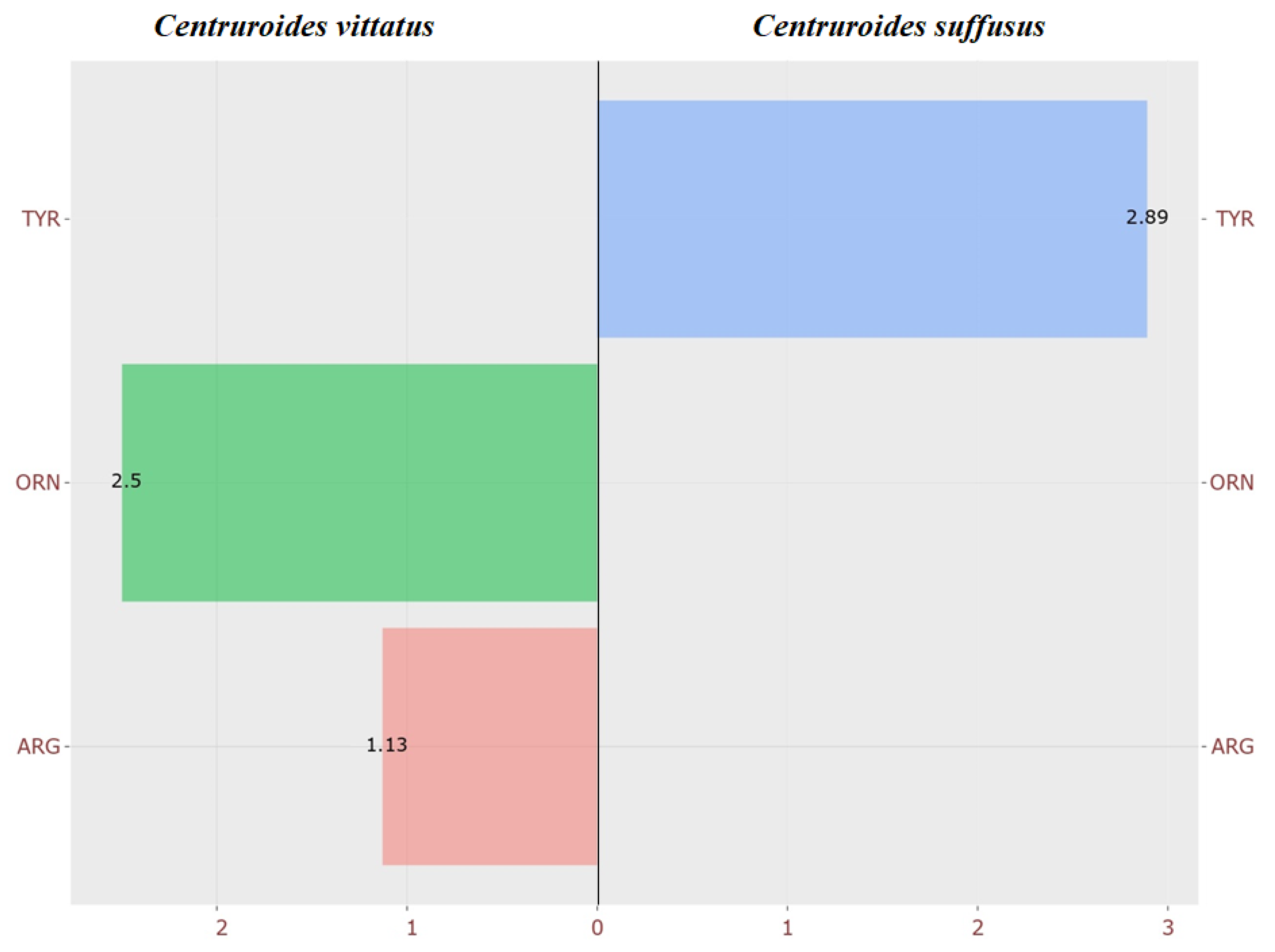
Figure 1. Relative abundances of selected amino acids in Centruroides vittatus and Centruroides suffusus. The bar plot shows the fold changes in tyrosine (TYR), ornithine (ORN), and arginine (ARG) between the two species. Full amino acid percentage data are presented in Supplementary Table S9.
Arthropoda 03 00014 g001
Figure 2. Relative abundances of selected acylcarnitines in Centruroides vittatus and Centruroides suffusus. Bar plot showing fold changes in C0, C2, C3, C5, C12, and C12.1 between the two species. Y. Detailed percentage data for acylcarnitines are presented in Supplementary Table S13.
Arthropoda 03 00014 g002
Figure 3. Comparative microbiota distribution between Centruroides vittatus and Centruroides suffusus. (A) Phylogenetic tree of the microbiota of the two species. The phylogenetic tree was reconstructed using the Maximum Likelihood method based on 16S rRNA gene sequences, with bootstrap support values shown at nodes. Sequences without circles represent bacterial taxa shared between both species, while colored circles indicate species-specific taxa. (B) Bacterial species abundance percentage heatmaps. Note: The abbreviation “NA” refers to the fact that no data are available to assign specific names to that species. Percent identity values between sequences are provided in Supplementary Table S17 to support taxonomic assignments and highlight the similarity levels among detected bacterial taxa.
Arthropoda 03 00014 g003
Table 1. Bacterial comparative analysis display.
PhylumSpeciesC. suffususC. vittatus
BacteriodetaChryseobacterium flavum-indologenes××
PseudomonadotaPseudomonas NA ×
Pseudomonas flavescens-putida×
Acinetobacter calcoacetius-pittii ×
Acinetobacter radioresistens××
Klebsiella oxytoca ×
Enterobacter cloacae××
Serratia liquafaciens-myotis-protemaculans××
Serratia marcescens-nematodiphila×
Serratia grimesii-liquefaciens-protemaculans××
BacillotaClostridium hydrogeniformans××
Staphylococcus sciuri××
Staphylococcus saprophyticus-xylosus××
Bacillus NA××
MycoplasmatotaMycoplasma sp.68230××
Disclaimer/Publisher’s Note: The statements, opinions and data contained in all publications are solely those of the individual author(s) and contributor(s) and not of MDPI and/or the editor(s). MDPI and/or the editor(s) disclaim responsibility for any injury to people or property resulting from any ideas, methods, instructions or products referred to in the content.