applsci-logo

Journal Browser

Journal Browser

Digital Dentistry: Computer-Aid Diagnosis and Treatment

A special issue of Applied Sciences (ISSN 2076-3417). This special issue belongs to the section "Applied Dentistry and Oral Sciences".

Deadline for manuscript submissions: closed (20 October 2023) | Viewed by 11425

Special Issue Editors


E-Mail Website
Guest Editor
Department of Dental Surgery, Faculty of Dental Surgery, University of Malta, MSD 2080 Msida, Malta
Interests: digital dentistry; oral implantology; bone imaging
Special Issues, Collections and Topics in MDPI journals

E-Mail Website
Guest Editor
Faculdade de Odontologia, University of São Paulo, Sao Paulo 05508-060, Brazil
Interests: dental radiology

E-Mail Website
Guest Editor
Dept. Buccofacial Prostheses, Univ. Complutense Madrid, Plaza Ramon & Cajal S-N, E-28040 Madrid, Spain
Interests: odontología

Special Issue Information

Dear Colleagues,

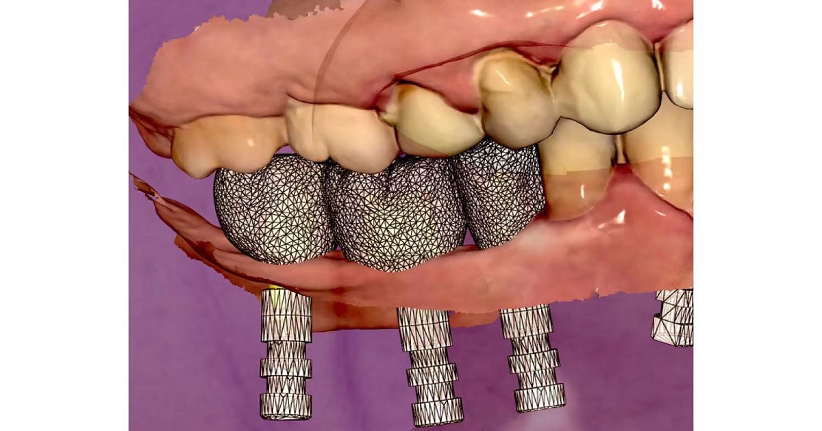
The advent of digital technologies in dentistry has led to shorter treatment times and higher predictability of treatment outcomes. Among the most important technologies used in digital workflows in dentistry is computer-aided design and computer-aided manufacturing (CAD-CAM). Scientific evidence has been consecutively presented, showing the usefulness of three-dimensional (3D) images not only for diagnosis but also for treatment planning. Current cone beam computed tomography (CBCT) scanners can offer higher contrast and spatial resolution for bone images, as well as lower radiation doses than before. Similarly, intraoral scanners have been validated to perform digital impressions, which can replace the conventional impressions usually performed with a series of impression materials. The integration of these 3D images enables the creation of a virtual patient, enhancing the multidisciplinary treatment plan.

Prof. Dr. Arthur R. G. Cortes
Prof. Dr. Claudio Costa
Prof. Dr. Guillermo Pradies
Guest Editors

Manuscript Submission Information

Manuscripts should be submitted online at www.mdpi.com by registering and logging in to this website. Once you are registered, click here to go to the submission form. Manuscripts can be submitted until the deadline. All submissions that pass pre-check are peer-reviewed. Accepted papers will be published continuously in the journal (as soon as accepted) and will be listed together on the special issue website. Research articles, review articles as well as short communications are invited. For planned papers, a title and short abstract (about 100 words) can be sent to the Editorial Office for announcement on this website.

Submitted manuscripts should not have been published previously, nor be under consideration for publication elsewhere (except conference proceedings papers). All manuscripts are thoroughly refereed through a single-blind peer-review process. A guide for authors and other relevant information for submission of manuscripts is available on the Instructions for Authors page. Applied Sciences is an international peer-reviewed open access semimonthly journal published by MDPI.

Please visit the Instructions for Authors page before submitting a manuscript. The Article Processing Charge (APC) for publication in this open access journal is 2400 CHF (Swiss Francs). Submitted papers should be well formatted and use good English. Authors may use MDPI's English editing service prior to publication or during author revisions.

Keywords

  • computer-aided design
  • computer-aided manufacturing
  • CAD-CAM
  • cone beam computed tomography
  • digital workflow
  • image-guided surgery

Benefits of Publishing in a Special Issue

  • Ease of navigation: Grouping papers by topic helps scholars navigate broad scope journals more efficiently.
  • Greater discoverability: Special Issues support the reach and impact of scientific research. Articles in Special Issues are more discoverable and cited more frequently.
  • Expansion of research network: Special Issues facilitate connections among authors, fostering scientific collaborations.
  • External promotion: Articles in Special Issues are often promoted through the journal's social media, increasing their visibility.
  • e-Book format: Special Issues with more than 10 articles can be published as dedicated e-books, ensuring wide and rapid dissemination.

Further information on MDPI's Special Issue polices can be found here.

Published Papers (4 papers)

Order results
Result details
Select all
Export citation of selected articles as:

Editorial

Jump to: Research, Review

3 pages, 197 KiB  
Editorial
Digital versus Conventional Workflow in Oral Rehabilitations: Current Status
by Arthur Rodriguez Gonzalez Cortes
Appl. Sci. 2022, 12(8), 3710; https://doi.org/10.3390/app12083710 - 7 Apr 2022
Cited by 6 | Viewed by 2694
Abstract
In recent years, computer-aided design and computer-aided manufacturing (CAD-CAM) technology has developed along with its applications in dentistry, including several new techniques that are used in oral rehabilitation applications [...] Full article
(This article belongs to the Special Issue Digital Dentistry: Computer-Aid Diagnosis and Treatment)

Research

Jump to: Editorial, Review

11 pages, 1446 KiB  
Article
Dimensional Changes in Extraction Sockets: A Pilot Study Evaluating Differences between Digital and Conventional Impressions
by Min-Woo Baek, Hyun-Chang Lim, Kwantae Noh, Seong-Ho Choi and Dong-Woon Lee
Appl. Sci. 2022, 12(15), 7662; https://doi.org/10.3390/app12157662 - 29 Jul 2022
Cited by 1 | Viewed by 3791
Abstract
Traditionally, soft tissue records are obtained by dental impression using impression materials; however, accurately recording the soft tissue immediately after tooth extraction is difficult. We measured the tissue changes after tooth extraction and compared two impression modalities (digital versus conventional) by measuring the [...] Read more.
Traditionally, soft tissue records are obtained by dental impression using impression materials; however, accurately recording the soft tissue immediately after tooth extraction is difficult. We measured the tissue changes after tooth extraction and compared two impression modalities (digital versus conventional) by measuring the changes at the soft tissue level. In this case, 15 patients with 17 single extraction sites were enrolled. Conventional impression (CI) using vinyl polysiloxane material and digital impression (DI) using an intraoral scanner were prepared immediately after extraction (T0) and at 2 months post-extraction (T1). Standard tessellation language files were generated for superimposition of the tissue surface. The tissue changes and discrepancies were measured on the superimposed surfaces. The differences in the changes and the discrepancy between the tissue surface impression at each time point were compared. At all measuring levels, the total tissue change was significantly different between groups DI and CI (p < 0.05). DI exhibited a more pronounced tissue surface at both time points, and the total discrepancy was statistically significantly greater at T0 than at T1 (p < 0.05). The values from DI and CI demonstrated small but significantly different for the same study material. The interpretation of such differences may depend on the clinical situation or scientific value. Full article
(This article belongs to the Special Issue Digital Dentistry: Computer-Aid Diagnosis and Treatment)
Show Figures

Figure 1

13 pages, 4751 KiB  
Article
Three-Dimensional Distance Mapping Method to Evaluate Mandibular Symmetry and Morphology of Adults with Unilateral Premolar Scissors Bite
by Yajuan Xie, Xinwei Lyu, Yuyao Liu, Runling Zeng, Yuwei Liao and Jiali Tan
Appl. Sci. 2022, 12(12), 5814; https://doi.org/10.3390/app12125814 - 8 Jun 2022
Cited by 1 | Viewed by 1688
Abstract
(1) Objective: This study aimed to evaluate the association between unilateral premolar scissors bite and mandibular symmetry of adults via the 3D distance mapping method. (2) Methods: A total of 53 cone-beam computed tomography (CBCT) images of adults with unilateral premolar scissors bite [...] Read more.
(1) Objective: This study aimed to evaluate the association between unilateral premolar scissors bite and mandibular symmetry of adults via the 3D distance mapping method. (2) Methods: A total of 53 cone-beam computed tomography (CBCT) images of adults with unilateral premolar scissors bite were set as study samples. A total of 53 age- and sex-matched samples without scissors bite were in the control group. Three-dimensional mandibular models and seven mandibular functional units, including condylar process (Co), coronoid process (Cr), mandibular ramus (Ra), mandibular angle (Ma), alveolar process (Ap), mandibular body (Mb), and chin process (Ch) were constructed and mirrored. After superimposition of the original and the mirrored models, 3D distance maps and deviation analysis were performed to evaluate the mandibular symmetry and morphology. (3) Results: In the study group, the matching percentages of the entire mandible (50.79 ± 10.38%), Ap (67.00 ± 12.68%), Mb (66.62 ± 9.44%), Ra (62.52 ± 11.00%), Ch (80.75 ± 9.86%), and Co (62.78 ± 13.56) were lower than that of the entire mandible (58.60 ± 5.52) (p < 0.01), Ap (73.83 ± 8.88%) (p < 0.01), Mb (72.37 ± 8.69%) (p < 0.01), Ra (68.60 ± 7.56%) (p < 0.01), Ch (85.23 ± 6.80%) (p < 0.01), and Co (67.58 ± 10.32%) (p < 0.05) in the control group. However, Cr and Ma showed no significant difference (p > 0.05). (4) Conclusions: The 3D distance mapping method provided a qualitative and quantitative mandibular symmetry and morphology assessment. Mandibular asymmetry was found in adults with unilateral premolar scissors bites. Mandibular functional units, including the alveolar process, mandibular body, mandibular ramus, chin process, and condylar process, showed significant differences, while no significant difference was observed in the coronoid process and mandibular angle. Full article
(This article belongs to the Special Issue Digital Dentistry: Computer-Aid Diagnosis and Treatment)
Show Figures

Figure 1

Review

Jump to: Editorial, Research

7 pages, 403 KiB  
Review
Factors Affecting Trueness of Intraoral Scans: An Update
by Arthur Rodriguez Gonzalez Cortes, Anne-Marie Agius and Juliana No-Cortes
Appl. Sci. 2022, 12(13), 6675; https://doi.org/10.3390/app12136675 - 1 Jul 2022
Cited by 5 | Viewed by 1780
Abstract
Background: Intraoral scanning (IOS) technologies have been constantly developed and improved. This systematic review aimed at assessing studies in the recent literature describing factors that influence the trueness of intraoral scans. Methods: Comparative in vitro and in vivo (i.e., interventional and observational) studies [...] Read more.
Background: Intraoral scanning (IOS) technologies have been constantly developed and improved. This systematic review aimed at assessing studies in the recent literature describing factors that influence the trueness of intraoral scans. Methods: Comparative in vitro and in vivo (i.e., interventional and observational) studies that quantitatively assessed the trueness of intraoral scans and that identified factors significantly affecting IOS trueness values were considered eligible for inclusion. The PUBMED and EMBASE databases were searched for articles published in the last two years (from February 2020 to February 2022). Data assessment and extraction were performed according to the guidelines of the PRISMA statement. Results: The present search strategy yielded 13 publications. An initial screening of the publications was performed using abstracts and key words, and after application of exclusion criteria, a total of nine studies were finally identified as eligible to be discussed. Several factors significantly affecting IOS were identified. Conclusions: Studies using current IOS technologies revealed that the device, scanning distance, operator experience, rescanning and post-processing scans, conditions of the preparations and presence of adjacent teeth are factors significantly affecting IOS trueness. Full article
(This article belongs to the Special Issue Digital Dentistry: Computer-Aid Diagnosis and Treatment)
Show Figures

Figure 1

Back to TopTop