Article

Three-Dimensional Distance Mapping Method to Evaluate Mandibular Symmetry and Morphology of Adults with Unilateral Premolar Scissors Bite

1
Hospital of Stomatology, Sun Yat-sen University, Guangzhou 510055, China
2
Guangdong Provincial Key Laboratory of Stomatology, Guangzhou 510080, China
3
Guanghua School of Stomatology, Sun Yat-sen University, Guangzhou 510080, China
*
Author to whom correspondence should be addressed.
Appl. Sci. 2022, 12(12), 5814; https://doi.org/10.3390/app12125814
Submission received: 19 April 2022 / Revised: 2 June 2022 / Accepted: 3 June 2022 / Published: 8 June 2022
(This article belongs to the Special Issue Digital Dentistry: Computer-Aid Diagnosis and Treatment)

Abstract

:

Featured Application

The present work was the first study to evaluate the association between unilateral premolar scissors bite and mandibular symmetry via a three-dimensional (3D) Distance Mapping technique. By this methodology, the mandibular morphology and symmetry can be visualized and accurately accessed. This computerized method reduced human error and saved measurement time.

Abstract

(1) Objective: This study aimed to evaluate the association between unilateral premolar scissors bite and mandibular symmetry of adults via the 3D distance mapping method. (2) Methods: A total of 53 cone-beam computed tomography (CBCT) images of adults with unilateral premolar scissors bite were set as study samples. A total of 53 age- and sex-matched samples without scissors bite were in the control group. Three-dimensional mandibular models and seven mandibular functional units, including condylar process (Co), coronoid process (Cr), mandibular ramus (Ra), mandibular angle (Ma), alveolar process (Ap), mandibular body (Mb), and chin process (Ch) were constructed and mirrored. After superimposition of the original and the mirrored models, 3D distance maps and deviation analysis were performed to evaluate the mandibular symmetry and morphology. (3) Results: In the study group, the matching percentages of the entire mandible (50.79 ± 10.38%), Ap (67.00 ± 12.68%), Mb (66.62 ± 9.44%), Ra (62.52 ± 11.00%), Ch (80.75 ± 9.86%), and Co (62.78 ± 13.56) were lower than that of the entire mandible (58.60 ± 5.52) (p < 0.01), Ap (73.83 ± 8.88%) (p < 0.01), Mb (72.37 ± 8.69%) (p < 0.01), Ra (68.60 ± 7.56%) (p < 0.01), Ch (85.23 ± 6.80%) (p < 0.01), and Co (67.58 ± 10.32%) (p < 0.05) in the control group. However, Cr and Ma showed no significant difference (p > 0.05). (4) Conclusions: The 3D distance mapping method provided a qualitative and quantitative mandibular symmetry and morphology assessment. Mandibular asymmetry was found in adults with unilateral premolar scissors bites. Mandibular functional units, including the alveolar process, mandibular body, mandibular ramus, chin process, and condylar process, showed significant differences, while no significant difference was observed in the coronoid process and mandibular angle.

1. Introduction

Scissors bite is defined as the maxillary posterior tooth completely located to the buccal aspect of the mandibular posterior tooth, without occlusal contact [1]. Scissors bite can be unilateral or bilateral, but unilateral is more common [2]. Researchers were concerned that scissors bite may be associated with occlusal disturbance, restricted lateral jaw movement, disturbed mastication, and temporomandibular disorders (TMD) [3,4,5]. Early orthodontic intervention or treatment was recommended for patients to prevent subsequent disease [2,6]. Previous studies mentioned that scissors bite can adversely affect jaw growth and appearance [3]. However, few studies accurately evaluated the association between unilateral premolar scissors bite and the mandibular morphology and symmetry.
Previous investigations reported the potential association between unilateral scissors bite and mandible position using conventional analysis methods. For example, Li et al. [7] evaluated the lateral cephalograms and found that patients with premolar scissor bite were observed to have less mandibular length. However, this method can only evaluate the mandible in a sagittal position. Due to the complexity of anatomical structures, two-dimensional (2D) X-ray techniques suffer from superimposition. The information obtained from 2D measurements is relatively limited for accurate evaluation [8]. Current CBCT scanners can offer higher contrast and spatial resolution for bone images [8,9]. Evangelista et al. [10] evaluated the morphologic and positional features of the mandible of patients with unilateral posterior crossbite via CBCT. Although the 3D models were constructed, the study still followed the 2D measurement rule to calculate the linear distance and angles. However, the bone itself is not a flat plane; the length of the line cannot completely represent the real bone surface. Furthermore, the manual work was time-consuming.
Based on the above literature, it was found that there were limitations of the previous methods, and few of them evaluated the mandibular symmetry of scissors bite via 3D measurements. Therefore, we attempted to propose a computer-aided method for more visual and accurate assessment, which was hypothesized to outperform the previous ones in the literature.
The purpose of this study was to introduce the 3D distance mapping technique to evaluate the mandibular symmetry and morphology of adult patients with unilateral premolar scissors bite.

2. Materials and Methods

2.1. Study Population

For this retrospective study, ethical approval was obtained from the ethics committee (KQEC-2021-36-01). CBCT records were selected from the image database of the Hospital of Stomatology, Guanghua School of Stomatology, Sun Yat-sen University, from 2014 to 2021. A total of 53 CBCT images of adults (≥18 years old) with unilateral premolar scissors bite were in the study group (male = 16, female = 37, mean age = 24.06 ± 4.64 years). The control group consisted of 53 CBCT images that had normal occlusion without scissors bite (male = 16, female = 37, mean age = 25.91 ± 4.98 years). The inclusion criteria of the study group were listed as follows: (1) unilateral premolar scissors bite involving at least one premolar; (2) an appropriate quality of CBCT scans without distortion or movement artifacts. The exclusion criteria were: (1) facial or dental trauma; (2) craniofacial anomalies, such as cleft lip palate; (3) previous orthodontic treatment, orthognathic treatment, and maxillofacial surgery; (4) dentition defect; (5) crossbite: mandibular teeth labially/buccally occlude to the maxillary teeth; (6) condylar degenerative disorders.

2.2. Workflow

Parameters of the CBCT scanner were set at 90.0 kV and 9 mA. The scanning time was set at 24 s, and the voxel size was 0.3 mm (Newtom VGi, Verona, Italy). Following the standard CBCT imaging operation protocols, patients were in intercuspation with the Frankfort horizontal plane parallel to the floor. All the data obtained were saved in Digital Imaging and Communications in Medicine (DICOM) format. To protect the confidentiality of the patients, all the information was de-identified. The patients signed informed consent before participation. In this study, the protocols for image segmentation, model rendering, and deviation analysis have been validated previously [11]. Examiners (Y.X. and R.Z.) have been specially trained for imaging processing. The flowchart in Figure 1 outlines the study procedure.

2.2.1. Segmenting Images

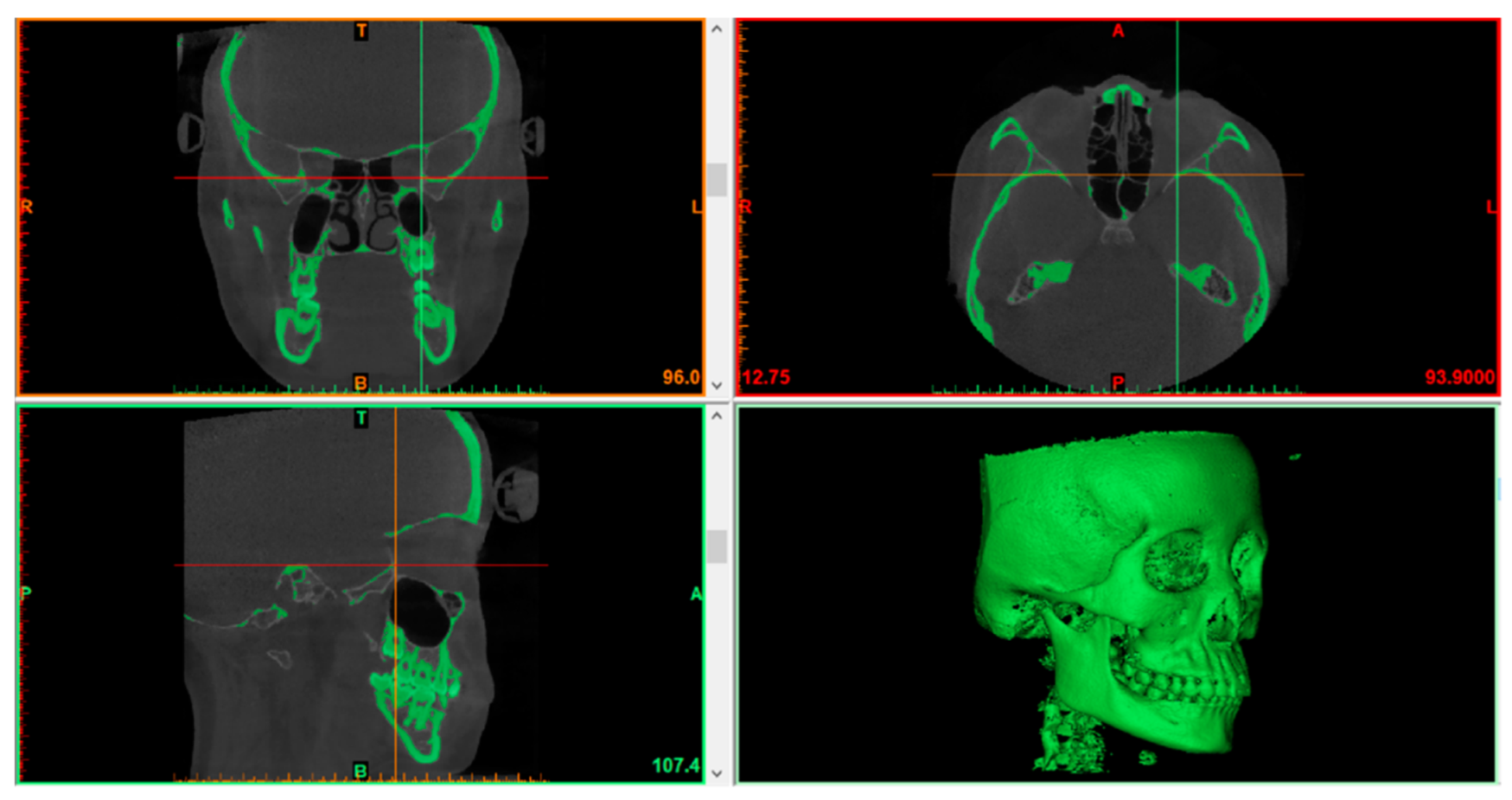
Firstly, DICOM data sets were imported to Mimics Research (version 20.0, Materialise NV, Liege, Belgium) for image segmentation. After the initial segmentation mask was developed, the predefined threshold was set to “bone” (226HU-3071HU), and then the parameter was adjusted to separate skeletal structures from noise (Figure 2). After that, the Mask Cropping tool and the Edit Mask tool were used to develop the mandible region. Finally, the Mask Smoothing tool and Contour Editing tool were taken to improve the quality of the mandible mask [12].

2.2.2. 3D Models Rendering and Mirroring

Still in the Mimics Research software, the mandible mask from Step1 was calculated and set as an original mandible part. Then, the original mandible was mirrored to obtain the mirrored mandible. After that, both the original mandible part and the mirrored mandible part were reconstructed into 3D models (.stl) [10,12].

2.2.3. Automatic Best-Fit Alignment of Mandibles

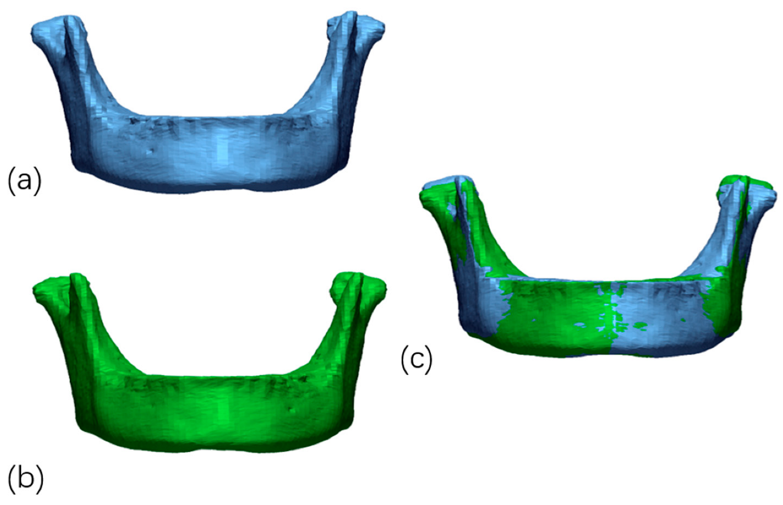
The two 3D models from Step 2 (the original mandible model and the mirrored mandible model) were exported from Mimics Research software and then imported to Geomagic Control X (version 2018.1.1, 3D Systems, Santa Clara, CA, USA). The original mandible model was defined as the reference data (Figure 3a), and the mirrored mandible model was defined as the measured data (Figure 3b). After that, the initial alignment was used to align measured data to reference data. Finally, the best-fit alignment was automatically conducted by computer. The precision of the registration was set to at least 0.1 mm. The polygon surface registration percentage was set to the maximum of 100% [12]. This best-fit alignment algorithm is concise, fast, and efficient so that it could provide automatic and reliable superimposition (Figure 3c).

2.2.4. Mandible Distance Mapping

After superimposition, the 3D Compare Tool of Geomagic Control X software automatically computed the distance distribution between the original and mirrored mandible. In the first stage, specify local data for this measurement by selecting predefined two vectors, which can be used for constructing a local coordinate system. Once local data were specified, searched deviation objects were projected and measured on a coordinate plane specified in this option. A total of 100% of the surface points were involved in the measurement, and the tolerance range was set to 0.5 mm. The difference between the original and mirrored mandible models was visualized by the distance mapping technique, which calculated the distance distribution between the two mandible surfaces [13,14]. The deviation analysis of the two mandibular models was displayed on a picture with a spectrum of several colors, which was similar to topographic maps. The Deviation Location tool helped to analyze the deviation and find locations where the deviations were out of the given tolerance. Each color represented a different distance range from the mirrored mandible to the original mandible surface. If the distance value was more significant than the positive limit (0.5 mm), the map appeared yellow and red. If the value was less than the negative limit −0.5 mm), blue was displayed. When the value was within the tolerance range, the map appeared green. The maximum deviation calculation was set to 3.0 mm [11].

2.2.5. Matching Percentage Calculation of Mandibles

Matching percentages (%) were the percentages of all the distance values within the tolerance ranges (0.5 mm) [12]. In this study, matching percentages represented the symmetry of the mandible. Higher values meant a minor difference between the scissors bite side and the non-scissors bite side for the study groups.

2.2.6. Construction of Mandibular Units

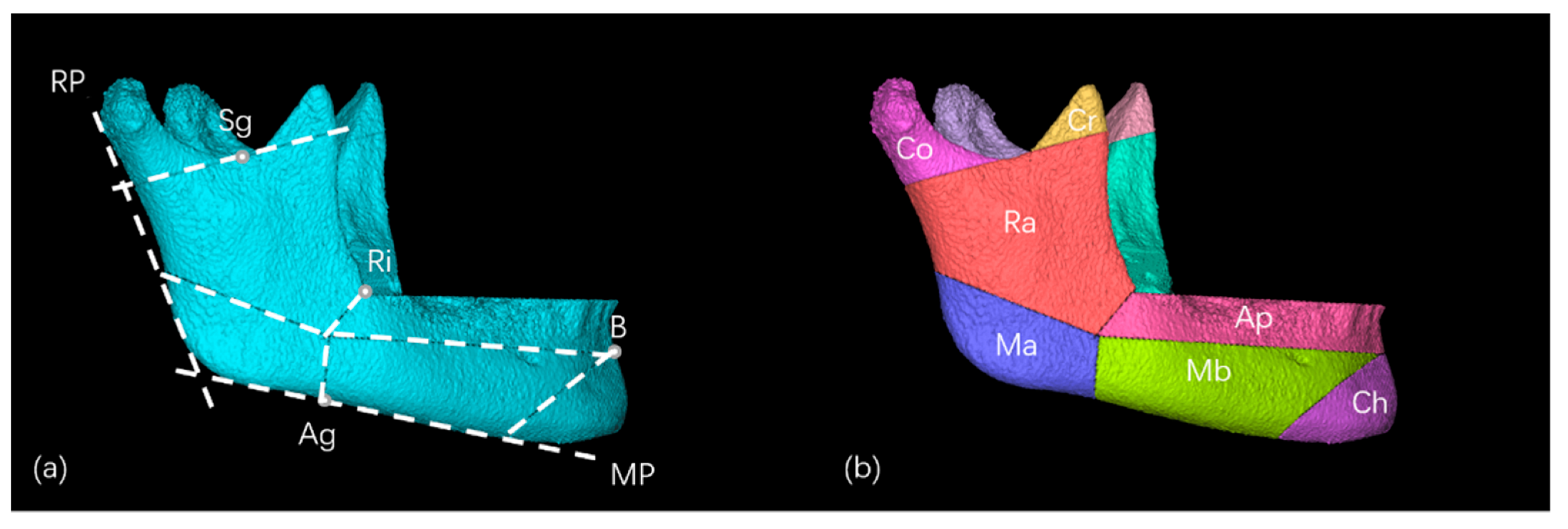
Based on the functional matrix theory [15], mandible growth relies on the contribution of mandibular functional units, including the alveolar process, mandibular body, condylar process, chin, coronoid process, mandibular ramus, and mandibular angle. This study reconstructed the mandibular functional units according to the landmarks on the 3D images [13,16] (Figure 4). Then each mandibular functional unit model was mirrored to obtain the mirrored models.

2.2.7. Automatic Best-Fit Alignment and Deviation Analysis of Mandibular Units

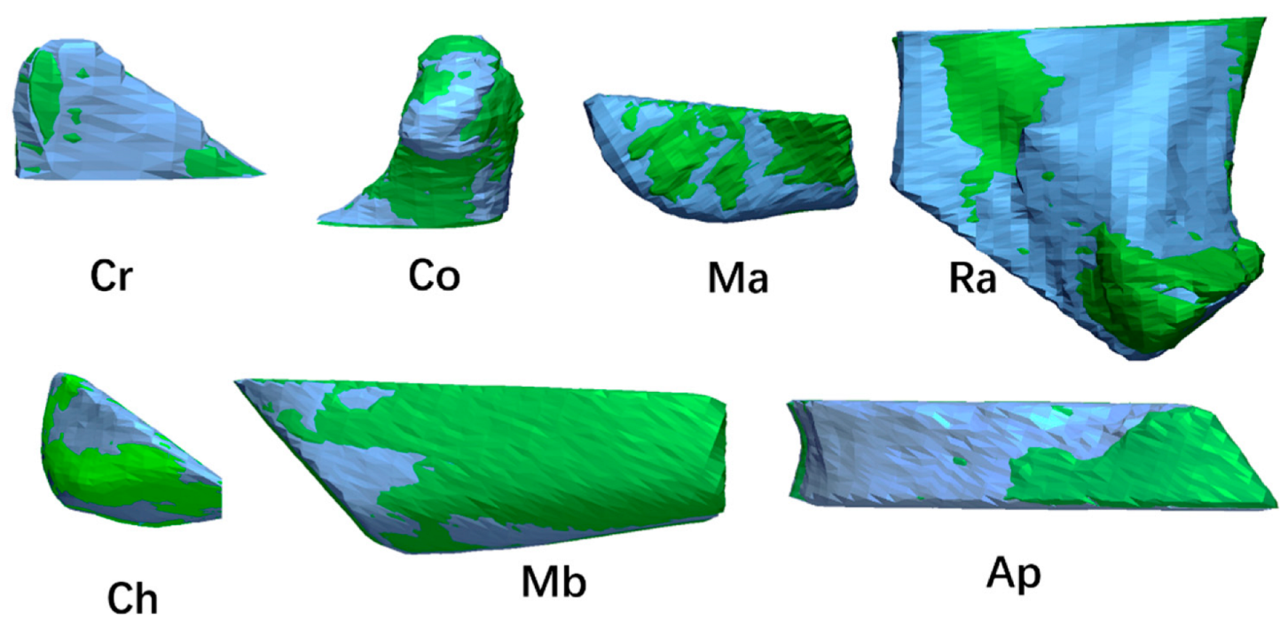
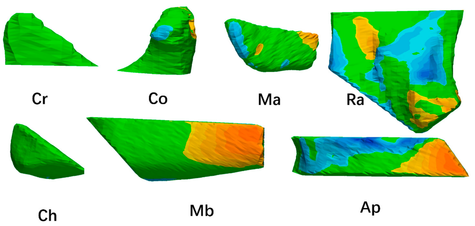
Same to the above method, each pair of original and mirrored functional units were conducted best-fit alignment by computer (Figure 5). After that, a deviation analysis of the units was also performed (Figure 6). The results of the 3D comparison were presented in colored maps. The tolerance range was set to 0.50 mm. Distance values higher than 0.50 mm were in yellow and red, values lower than −0.50 mm were in blue, and within tolerance range were in green. The maximum deviation calculation was set to 3.0 mm. After that, the matching percentages of each mandibular functional unit were calculated.

2.2.8. Statistical Analysis

All the measurement and calculation results were recorded in Microsoft Excel (2016, Microsoft, Redmond, WA, USA). The statistical analysis was performed with the SPSS (version 25.0; IBM, Armon, NY, USA). The Kolmogorov–Smirnov test was used to test the normality of the data. The data were normally distributed. The matching percentages obtained by deviation analysis were compared by an independent t-test. The entire mandible and mandibular functional units (alveolar process, mandibular body, condylar process, chin, coronoid process, mandibular ramus, and mandibular angle) were compared between the study group and the control group. p values < 0.05 were considered statistically significant. The intraclass correlation coefficient (ICC) values were used to check the intra-examiner and inter-examiner reliability after the steps were repeated.

3. Results

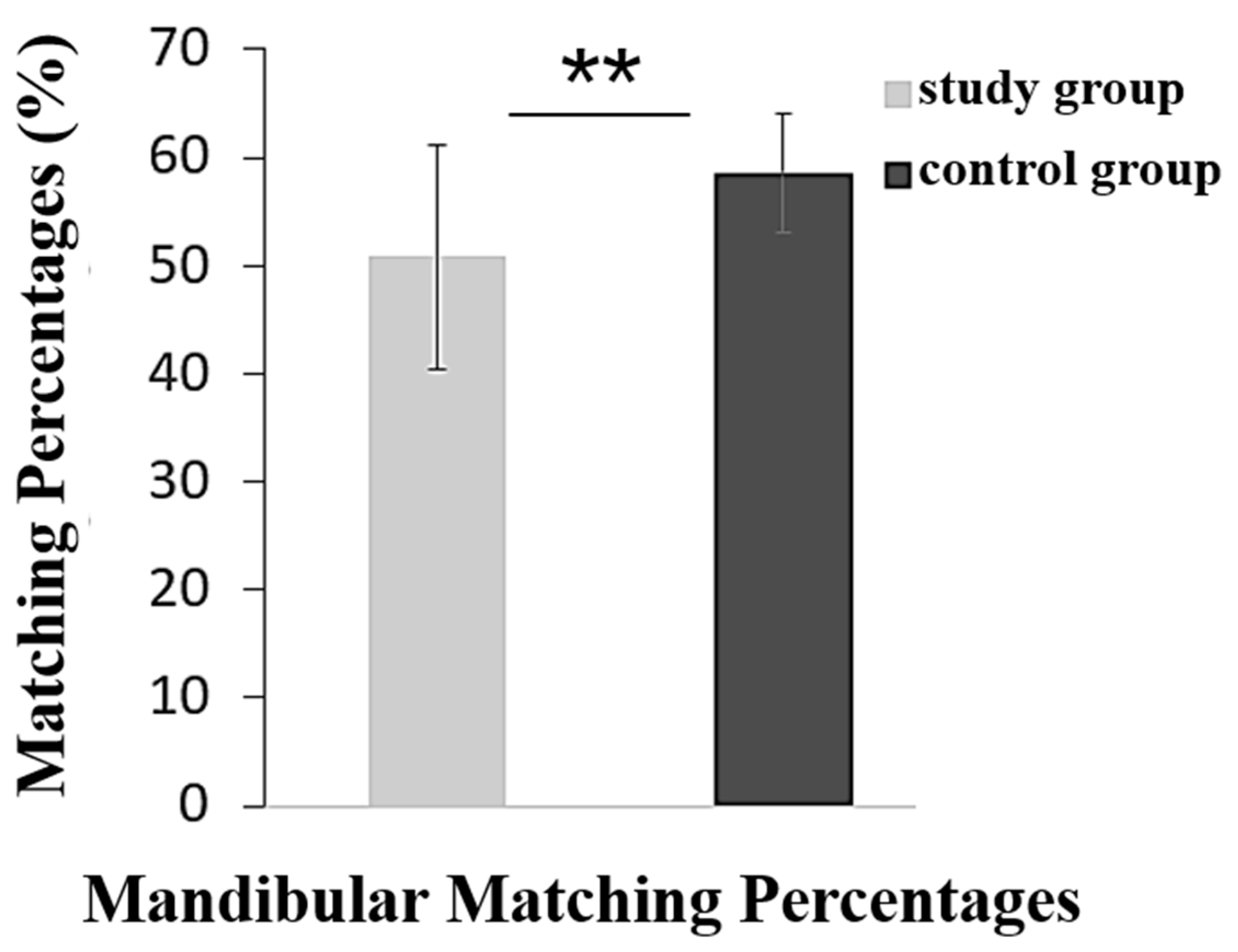
For the intra-examiner reliability, the ICC values of operator 1 ranged from 0.82 to 0.89 and ranged from 0.83 to 0.91 for operator 2. For the inter-examiner reliability, the ICC values ranged from 0.82 to 0.88. The results showed that the measurements were highly reliable. After the 3D deviation analysis, the matching percentages of the entire mandible were calculated in Figure 7. The matching percentage of the study group (50.79 ± 10.38%) was lower than the control group (58.60 ± 5.52%) (p < 0.01).
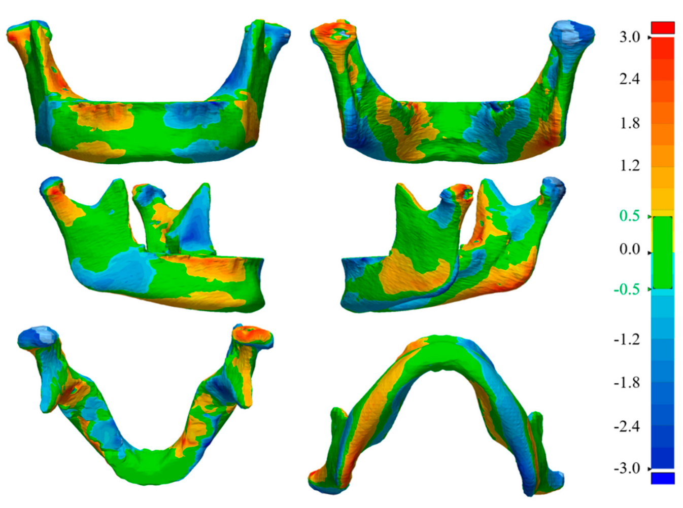
The 3D distance colored maps were observed in detail. The deviation analysis of the two mandible models was displayed on a picture with a spectrum of several colors. Each color represented a different distance range from the mirrored mandible to the original mandible surface. If the distance value was high than the positive limit (0.5 mm), the map appeared yellow and red. If the value was lower than the negative limit (−0.5 mm), blue was displayed. The scissors bite side and non-scissors bite can be compared via this method. As shown in Figure 8, the scissors bite side was the mandible’s right side. The yellow and red region on the right side (scissors bite side) represented that this part was smaller than the left side (non-scissors bite side). Meanwhile, the blue region on the right side (scissors bite side) represented that this part was wider than the other side. Therefore, the complex morphology of the bone can be observed. It was noticed that there were some regions of the mandibles showing more significant asymmetrical morphology.
Therefore, to further clarify this association, the whole mandible was delimitated into seven mandibular functional units, including the alveolar process, mandibular body, condylar process, chin, coronoid process, mandibular ramus, and mandibular angle, and the matching percentages were calculated in Table 1. The results revealed that some mandibular units of the study group showed lower matching percentages. These areas mainly localized at the alveolar process (67.00 ± 12.68%), mandibular ramus (62.52 ± 11.00%), mandibular body (66.62 ± 9.44%), chin units (80.75 ± 9.86%), and condylar process (62.78 ± 13.56%) (p < 0.05). No significant difference was found in the coronoid process and mandibular angle (p > 0.05).

4. Discussion

The results revealed that adult patients with unilateral scissors bite showed asymmetric characteristics in the mandible. The alveolar, condylar process, mandibular ramus, mandibular body, and chin units tended to be the most affected region. Only the coronoid process and mandibular angle showed no significant differences. To analyze whether the position of the premolar scissors bite tooth influenced the results, we compared the first premolar scissors bite, second premolar scissors bite, and two premolars scissors bite. However, no statistically significant difference was found among them (p > 0.05). Moreover, the matching percentage of the entire mandible was lower than each of the functional units. In this study, a lower matching percentage meant a more significant asymmetry. The higher matching percentages of the units represented relatively small changes. If all these small changes made by each functional unit were added up, there would be a more significant effect on the entire mandible.
Possible reasons will be discussed to explain the results. Previous studies have reported that occlusal disturbances have been considered the cause of mandibular asymmetry. Unilateral scissors bite malocclusion can make a cant to the occlusal plane [4]. The occlusal disturbances and functional lateral displacement can lead to dysfunctional jaw movement, skeletal mandibular deviation, and asymmetry in the condylar position, affecting the mandibular growth and development [17,18,19].
In addition, unilateral scissors bite may cause an imbalance between the muscles. The scissors bite teeth do not have the normal chewing function, and only the non-locked posterior teeth can be used. The chewing function is weakened, and the chewing efficiency is reduced, affecting the mandibular lateral movement and growth [20,21,22]. The jaw closing muscles’ surface electromyographic activities (SEMG) during centric maximal voluntary clenching (MVC) of patients with unilateral scissors bite have been studied. Compared with non-scissors bite, lower SEMG values for masseter, fewer contacts, and lower biting force distribution were found on the scissors bite side [23]. Also reported in another study, H. Tomonari et al. demonstrated the reduced contact of unilateral posterior scissors bite. They suggested that scissors bite malocclusion is associated with the masticatory chewing pattern and muscle activity, such as masseter and temporalis [1].
Furthermore, bones and muscles are in close crosstalk; a better understanding of the relationship between craniofacial deformities and muscles helps understand the relationship between malocclusion and mandible deformities [24,25]. Rodrigues et al. revealed that experimental removal of the masseter muscle during the growing period in rats induced atrophic changes and shortening of the whole mandible [26]. The mandibular condyle plays a vital role in bone remodeling and mandibular development [27]. Our study used the 3D distance mapping technique to evaluate. The results suggested the association between mandibular asymmetry and the condylar process. Miyazaki et al. also found that lateral imbalance of masseter muscle activity led to inhibition of chondrogenesis and induced asymmetric formation of the condylar cartilage during the growth period [28], which supported our results. So, we speculated that an asymmetrical activity of masticatory muscles between the scissors bite side and non-scissors bite sides. Moreover, in our study, no significant difference was found in the coronoid process and mandibular angle. The coronoid unit is affected by the temporalis muscle. Previous studies found that the volumes of the temporalis muscles were not significantly different between the two sides in patients with facial asymmetry [29,30]. This seemed to be basically in accordance with our study that no significant difference was found in the coronoid process. We speculated that mandibular asymmetry is possibly caused by the differences between the skeletal units, which may be related to the surrounding muscles. The asymmetrical muscle activities could transfer to the mandible. However, considering the complex role of the muscle, the exact causal relationship between the skeletal units and the muscles has not been fully clarified, and more research is required. Therefore, although certain trends were obvious, specific bone morphology may not necessarily be related to specific occlusal characteristics. The deep mechanism called for deep exploration.
Scissors bite cannot be corrected spontaneously and will gradually deteriorate as the maxillary teeth continue to erupt. The lateral movement of the mandible is limited in patients with unilateral scissors bite, which can induce temporomandibular joint disorders for some susceptible persons [24]. Therefore, researchers suggested early intervention for younger patients because of the growth potential [6]. Various non-surgical treatments and appliances have been advocated, such as maxillary contraction [31] and mandibular lingual transforce appliance [2]. However, the correction plans tend to be more complex and invasive in adulthood [32]. Implant anchorage is frequently used to correct the scissors bite on the one hand. On the other hand, it can also prevent the elongation of the locked teeth after correction [4,33]. However, severe scissors bite may only be corrected with orthognathic treatment to narrow the maxillary arch or widen the lower arch [34].
Previous researchers have used traditional two-dimensional radiographs to analyze the symmetry of the mandible. Lopatienė et al. [35] evaluated mandibular symmetry of 60 pre-orthodontic patients with unilateral posterior crossbite via panoramic radiographs. The heights of mandibular condylar, mandibular ramal, and mandibular condylar were measured to calculate Habets’ asymmetry index [36]. More asymmetric mandibular condylar and mandibular ramal on the crossbite side were observed. Apart from two-dimensional radiographs, there were also studies based on CBCT. Miresmaeili et al. [37] assessed the mandibular asymmetry of patients with unilateral posterior crossbite by CBCT images. The results showed that the length of the condyle and mandibular body on the crossbite side was reduced. Different from the above research, which adopted linear measurement, our study used the 3D distance mapping technique to analyze the surface. The results also demonstrated the asymmetry condylar process, mandibular ramus, and mandibular body. In addition, alveolar and chin were found affected. The mandible is an irregular surface, but lines are just chords of curved surfaces, and cannot fully represent the true shape of the mandible. The previous methods described above simply measured linear distances and angles, ignoring the contours of the mandible. Therefore, the method that can calculate the surface can better reflect the morphological characteristics of the mandible.
3D technology can reconstruct models, which are helpful in quantifying the displacement with more precise measurements [38,39]. The 3D mapping technique provided a user-friendly tool that converted the obtained data into a quantitative color-coded map. For example, Turow et al. used 3D computer tomography to describe acute and subacute scaphoid fracture morphology [40]. To the best of our knowledge, our present work was the first study to evaluate the association between unilateral premolar scissors bite and mandibular symmetry via the 3D distance mapping technique. The distance mapping algorithm computed the distance distribution between any two opposing surfaces by sampling the surface of the original mandible. Then, it identified the corresponding minimum distance to the mirrored mandible. The color-coded distance maps can be read similarly to topographic maps. Each color represented a different distance. So, the morphological differences can be quickly located. Moreover, different from Leonardi et al. [12], who only evaluated the entire mandible, this study also assessed mandibular functional units, respectively. Therefore, more detailed information can be identified and compared. Different from the previous measurements, this methodology will enable researchers to effectively interpret the imaging, save measuring time, reduce human error, and facilitate further medical research.
In this retrospective study, all the images were not taken specifically for this experiment. Subjects were selected from the image database of the Hospital of Stomatology, Guanghua School of Stomatology, Sun Yat-sen University, from 2014 to 2021. As a digital tool, the 3D distance mapping method itself is not risky when calculating and visualizing the data. Consideration of the risks of CBCT is still meaningful, even if the radiation dose is lower than before. Future research needs to follow and respect ALARA (As Low As Reasonably Achievable) principles [41] and other statements such as the guideline of the European Academy of DentoMaxilloFacial Radiology (EADMFR) [42]. To minimize the risk, improvements in hardware and software components can reduce the radiation dose to the patients, including changes in sensor technology and a smaller field of view depending upon the application [43]. Various exposure protocols can be chosen according to different groups, such as child mode, adult mode, high-resolution mode, high-definition mode, and endodontic mode [44]. Moreover, shielding devices are necessary to reduce doses for the patients [42].
The present study is limited to the control group. Because in practice, it was not realistic to find the ideal normal individuals in a dental hospital. However, to avoid affecting the results, the control samples used in this retrospective study were carefully selected from hospital imaging databases. Their occlusions were relatively normal, without scissors bite. Images with severe dental pathology were avoided in this experiment. Moreover, a longitudinal study would require if the study wants to evaluate the effects of growth and development in mandibular asymmetry. However, for ethical reasons, it is unrealistic to follow a malocclusion without treatment.
This study suggested that this technology may have potential applicability in various fields. The evaluation of bone structure with this method will be helpful for orthodontic treatment planning and clinical curative effect evaluation after treatment. Moreover, this technology also showed its potential and advantages in computer-aided design in orthognathic surgery, prosthodontics, airway analysis, etc. [45,46,47]. Of course, the clinical applications are not limited to mandible or dentistry, and they can also be applied in orthopedics.

5. Conclusions

The study revealed that unilateral premolar scissors bite correlated to mandible asymmetry via the 3D distance mapping technique. Moreover, mandibular functional units, including the alveolar process, mandibular body, condylar process, mandibular ramus, and chin units, presented a more asymmetric trend. The coronoid process and mandibular angle showed no significant difference. This 3D method can provide more accurate and visual information to study mandibular morphology and potentially facilitate the diagnosis, decision making, treatment, and follow-up of patients. In addition, for further development of this technology, it makes sense to explore more applications in the future.

Author Contributions

Conceptualization, methodology, software and writing—original draft preparation, Y.X.; validation and writing—review and editing, X.L.; formal analysis, Y.L. (Yuyao Liu); investigation and software, R.Z.; resources, Y.L. (Yuwei Liao); writing—review and editing, project administration and funding acquisition, J.T. All authors have read and agreed to the published version of the manuscript.

Funding

This research was funded by the Guangdong Basic and Applied Basic Research Foundation (2022A1515011094), Guangdong Financial Fund for High-Caliber Hospital Construction (174-2018-XMZC-0001-03-0125/C-05), and Science and Technology Planning Project of Guangdong Province, China (2020A0505100034).

Institutional Review Board Statement

The study was conducted in accordance with the Declaration of Helsinki and approved by the ethics committee of the Affiliated Stomatology Hospital of Sun Yat-sen University, Guangzhou, China (KQEC-2021-36-01).

Informed Consent Statement

Informed consent was obtained from all subjects involved in the study.

Data Availability Statement

The data sets used and/or analyzed during the current study are available from the corresponding author on reasonable request.

Acknowledgments

We appreciate the support from the funding and the participants.

Conflicts of Interest

The authors declare no conflict of interest.

References

  1. Tomonari, H.; Kubota, T.; Yagi, T.; Kuninori, T.; Kitashima, F.; Uehara, S.; Miyawaki, S. Posterior scissors-bite: Masticatory jaw movement and muscle activity. J. Oral Rehabil. 2014, 41, 257–265. [Google Scholar] [CrossRef]
  2. Doshi, U.H.; Chandorikar, H.; Nagrik, A.; Bhad, W.A.; Chavan, S.J. Early correction of unilateral scissor bite using transforce appliance and modified twin block appliance. J. Orthod. Sci. 2017, 6, 76–80. [Google Scholar] [CrossRef] [PubMed] [Green Version]
  3. Baik, U.-B.; Kim, Y.; Sugawara, J.; Hong, C.; Park, J.H. Correcting severe scissor bite in an adult. Am. J. Orthod. Dentofac. Orthop. 2019, 156, 113–124. [Google Scholar] [CrossRef]
  4. Lee, S.-A.; Chang, C.C.; Roberts, W.E. Severe unilateral scissors-bite with a constricted mandibular arch: Bite turbos and extra-alveolar bone screws in the infrazygomatic crests and mandibular buccal shelf. Am. J. Orthod. Dentofac. Orthop. 2018, 154, 554–569. [Google Scholar] [CrossRef] [PubMed] [Green Version]
  5. Türp, J.C.; Schindler, H. The dental occlusion as a suspected cause for TMDs: Epidemiological and etiological considerations. J. Oral Rehabil. 2012, 39, 502–512. [Google Scholar] [CrossRef]
  6. Favero, V.; Sbricoli, L.; Favero, L. Scissor bite in a young patient treated with an orthodontic-orthopedic device. A case report. Eur. J. Paediatr. Dent. 2013, 141, 153–155. [Google Scholar]
  7. Li, J.; Lu, S.-J.; Shi, Y.-J.; Lu, P.; Tian, Y.-L. Effect of premolars scissor bite on the sagittal position of mandible. Shanghai Kou Qiang Yi Xue 2019, 28, 179–183. [Google Scholar]
  8. Gil Choi, I.G.; Cortes, A.R.G.; Arita, E.S.; Georgetti, M.A.P. Comparison of conventional imaging techniques and CBCT for periodontal evaluation: A systematic review. Imaging Sci. Dent. 2018, 48, 79–86. [Google Scholar] [CrossRef]
  9. Kakumoto, T.; Barsoum, A.; Froum, S.J. Accuracy of Cone-Beam Computed Tomography Versus Periapical Radiography Measurements When Planning Placement of Implants in the Posterior Maxilla: A Retrospective Study. Compend. Contin. Educ. Dent. 2021, 42, e1–e4. [Google Scholar] [PubMed]
  10. Evangelista, K.; Valladares-Neto, J.; Silva, M.A.G.; Cevidanes, L.H.S.; Ruellas, A.C.D.O. Three-dimensional assessment of mandibular asymmetry in skeletal Class I and unilateral crossbite malocclusion in 3 different age groups. Am. J. Orthod. Dentofac. Orthop. 2020, 158, 209–220. [Google Scholar] [CrossRef] [PubMed]
  11. Muraglie, S.; Leonardi, R.; Aboulazm, K.; Stumpo, C.; Loreto, C.; Grippaudo, C. Evaluation of structural skeletal asymmetry of the glenoid fossa in adult patients with unilateral posterior crossbite using surface-to-surface matching on CBCT images. Angle Orthod. 2020, 90, 376–382. [Google Scholar] [CrossRef] [PubMed] [Green Version]
  12. Leonardi, R.; Muraglie, S.; Lo Giudice, A.; Aboulazm, K.S.; Nucera, R. Evaluation of mandibular symmetry and morphology in adult patients with unilateral posterior crossbite: A CBCT study using a surface-to-surface matching technique. Eur. J. Orthod. 2020, 42, 650–657. [Google Scholar] [CrossRef] [PubMed]
  13. Leonardi, R.; Muraglie, S.; Bennici, O.; Cavallini, C.; Spampinato, C. Three-dimensional analysis of mandibular functional units in adult patients with unilateral posterior crossbite: A cone beam study with the use of mirroring and surface-to-surface matching techniques. Angle Orthod. 2019, 89, 590–596. [Google Scholar] [CrossRef] [PubMed] [Green Version]
  14. Lintz, F.; Jepsen, M.; Netto, C.D.C.; Bernasconi, A.; Ruiz, M.; Siegler, S. Distance mapping of the foot and ankle joints using weightbearing CT: The cavovarus configuration. Foot Ankle Surg. 2021, 27, 412–420. [Google Scholar] [CrossRef]
  15. Moss, M.L.; Rankow, R.M. The role of the functional matrix in mandibular growth. Angle Orthod. 1968, 38, 95–103. [Google Scholar] [CrossRef]
  16. Mun, S.H.; Park, M.; Lee, J.; Lim, H.J.; Kim, B.C. Volumetric characteristics of prognathic mandible revealed by skeletal unit analysis. Ann. Anat. Anat. Anz. 2019, 226, 3–9. [Google Scholar] [CrossRef]
  17. Liu, C.; Kaneko, S.; Soma, K. Glenoid Fossa Responses to Mandibular Lateral Shift in Growing Rats. Angle Orthod. 2007, 77, 660–667. [Google Scholar] [CrossRef]
  18. Fourneron, M.; Morant, F.; Boutin, F.; Frapier, L. Is the Quad Helix more efficient to correct mandibular asymmetry before age 7? A retrospective comparative study. Int. Orthod. 2020, 18, 443–450. [Google Scholar] [CrossRef]
  19. Ramirez-Yañez, G.O.; Stewart, A.; Franken, E.; Campos, K. Prevalence of mandibular asymmetries in growing patients. Eur. J. Orthod. 2011, 33, 236–242. [Google Scholar] [CrossRef] [Green Version]
  20. Wiens, J.P.; Goldstein, G.R.; Andrawis, M.; Choi, M.; Priebe, J.W. Defining centric relation. J. Prosthet. Dent. 2018, 120, 114–122. [Google Scholar] [CrossRef] [PubMed]
  21. Manfredini, D.; Lombardo, L.; Siciliani, G. Temporomandibular disorders and dental occlusion. A systematic review of association studies: End of an era? J. Oral Rehabil. 2017, 44, 908–923. [Google Scholar] [CrossRef] [PubMed]
  22. Lim, W.H.; Choi, B.; Lee, J.-Y.; Ahn, S.-J. Dentofacial characteristics in orthodontic patients with centric relation–maximum intercuspation discrepancy. Angle Orthod. 2014, 84, 939–945. [Google Scholar] [CrossRef]
  23. Qi, K.; Guo, S.-X.; Xu, Y.; Deng, Q.; Liu, L.; Li, B.; Wang, M.-Q. An investigation of the simultaneously recorded occlusal contact and surface electromyographic activity of jaw-closing muscles for patients with temporomandibular disorders and a scissors-bite relationship. J. Electromyogr. Kinesiol. 2016, 28, 114–122. [Google Scholar] [CrossRef]
  24. Yamada, T.; Sugiyama, G.; Mori, Y. Masticatory muscle function affects the pathological conditions of dentofacial deformities. Jpn. Dent. Sci. Rev. 2020, 56, 56–61. [Google Scholar] [CrossRef] [PubMed]
  25. Sella-Tunis, T.; Pokhojaev, A.; Sarig, R.; O’Higgins, P.; May, H. Human mandibular shape is associated with masticatory muscle force. Sci. Rep. 2018, 8, 6042. [Google Scholar] [CrossRef] [Green Version]
  26. Rodrigues, L.; Traina, A.A.; Nakamai, L.F.; Luz, J.G.D.C. Effects of the unilateral removal and dissection of the masseter muscle on the facial growth of young rats. Braz. Oral Res. 2009, 23, 89–95. [Google Scholar] [CrossRef] [PubMed] [Green Version]
  27. Chouinard, A.-F.; Kaban, L.B.; Peacock, Z.S. Acquired Abnormalities of the Temporomandibular Joint. Oral Maxillofac. Surg. Clin. N. Am. 2018, 30, 83–96. [Google Scholar] [CrossRef] [PubMed]
  28. Miyazaki, M.; Yonemitsu, I.; Takei, M.; Kure-Hattori, I.; Ono, T. The imbalance of masticatory muscle activity affects the asymmetric growth of condylar cartilage and subchondral bone in rats. Arch. Oral Biol. 2016, 63, 22–31. [Google Scholar] [CrossRef] [Green Version]
  29. Kwon, T.-G.; Lee, K.-H.; Park, H.-S.; Ryoo, H.-M.; Kim, H.-J.; Lee, S.-H. Relationship Between the Masticatory Muscles and Mandibular Skeleton in Mandibular Prognathism With and Without Asymmetry. J. Oral Maxillofac. Surg. 2007, 65, 1538–1543. [Google Scholar] [CrossRef]
  30. Seo, S.-A.; Baik, H.-S.; Hwang, C.-J.; Yu, H.-S. Analysis of masseter muscle in facial asymmetry before and after orthognathic surgery using 3-dimensional computed tomography. Korean J. Orthod. 2009, 39, 18–27. [Google Scholar] [CrossRef] [Green Version]
  31. Hua, X.; Xiong, H.; Han, G.; Cheng, X. Correction of a dental arch-width asymmetric discrepancy with a slow maxillary contraction appliance. Am. J. Orthod. Dentofac. Orthop. 2012, 142, 842–853. [Google Scholar] [CrossRef]
  32. Deffrennes, G.; Deffrennes, D. Management of Brodie bite: Note on surgical treatment. Int. Orthod. 2017, 15, 640–676. [Google Scholar] [CrossRef] [PubMed]
  33. Ishihara, Y.; Kuroda, S.; Sugawara, Y.; Kurosaka, H.; Takano-Yamamoto, T.; Yamashiro, T. Long-term stability of implant-anchored orthodontics in an adult patient with a Class II Division 2 malocclusion and a unilateral molar scissors-bite. Am. J. Orthod. Dentofac. Orthop. 2014, 145, S100–S113. [Google Scholar] [CrossRef] [PubMed]
  34. Suda, N.; Tominaga, N.; Niinaka, Y.; Amagasa, T.; Moriyama, K. Orthognathic treatment for a patient with facial asymmetry associated with unilateral scissors-bite and a collapsed mandibular arch. Am. J. Orthod. Dentofac. Orthop. 2012, 141, 94–104. [Google Scholar] [CrossRef] [PubMed]
  35. Lopatienė, K.; Trumpytė, K. Relationship between unilateral posterior crossbite and mandibular asymmetry during late adolescence. Stomatologija 2018, 20, 90–95. [Google Scholar]
  36. Habets, L.L.M.H.; Bezuur, J.N.; Naeiji, M.; Hansson, T.L. The Orthopantomogram®, an aid in diagnosis of temporomandibular joint problems. II. The vertical symmetry. J. Oral Rehabil. 1988, 15, 465–471. [Google Scholar] [CrossRef]
  37. Miresmaeili, A.; Salehisaheb, H.; Farhadian, M.; Borjali, M. Mandibular asymmetry in young adult patients with unilateral posterior crossbite: A controlled retrospective CBCT study. Int. Orthod. 2021, 19, 433–444. [Google Scholar] [CrossRef] [PubMed]
  38. Leonardi, R.M.; Aboulazm, K.; Giudice, A.L.; Ronsivalle, V.; D’Antò, V.; Lagravère, M.; Isola, G. Evaluation of mandibular changes after rapid maxillary expansion: A CBCT study in youngsters with unilateral posterior crossbite using a surface-to-surface matching technique. Clin. Oral Investig. 2021, 25, 1775–1785. [Google Scholar] [CrossRef]
  39. Siegler, S.; Konow, T.; Belvedere, C.; Ensini, A.; Kulkarni, R.; Leardini, A. Analysis of surface-to-surface distance mapping during three-dimensional motion at the ankle and subtalar joints. J. Biomech. 2018, 76, 204–211. [Google Scholar] [CrossRef]
  40. Turow, A.; Bulstra, A.E.; Oldhoff, M.; Hayat, B.; Doornberg, J.; White, J.; Jaarsma, R.L.; Bain, G.I. 3D mapping of scaphoid fractures and comminution. Skelet. Radiol. 2020, 49, 1633–1647. [Google Scholar] [CrossRef]
  41. Hayashi, T.; Arai, Y.; Chikui, T.; Hayashi-Sakai, S.; Honda, K.; Indo, H.; Kawai, T.; Kobayashi, K.; Murakami, S.; Nagasawa, M.; et al. Clinical guidelines for dental cone-beam computed tomography. Oral Radiol. 2018, 34, 89–104. [Google Scholar] [CrossRef]
  42. Hiles, P.; Gilligan, P.; Damilakis, J.; Briers, E.; Candela-Juan, C.; Faj, D.; Foley, S.; Frija, G.; Granata, C.; Gala, H.D.L.H.; et al. European consensus on patient contact shielding. Insights Into Imaging 2021, 12, 194. [Google Scholar] [CrossRef]
  43. Ludlow, J.B.; Ivanovic, M. Comparative dosimetry of dental CBCT devices and 64-slice CT for oral and maxillofacial radiology. Oral Surg. Oral Med. Oral Pathol. Oral Radiol. Endodontol. 2008, 106, 106–114. [Google Scholar] [CrossRef] [PubMed]
  44. Jaju, P.P.; Jaju, S.P. Cone-beam computed tomography: Time to move from ALARA to ALADA. Imaging Sci. Dent. 2015, 45, 263–265. [Google Scholar] [CrossRef] [Green Version]
  45. Neagos, A.; Vrinceanu, D.; Dumitru, M.; Costache, A.; Cergan, R. Demographic, anthropometric, and metabolic characteristics of obstructive sleep apnea patients from Romania before the COVID-19 pandemic. Exp. Ther. Med. 2021, 22, 1487. [Google Scholar] [CrossRef] [PubMed]
  46. Ferlias, N.; Gjørup, H.; Doherty, M.A.; Haagerup, A.; Pedersen, T.K. Obstructive Sleep Apnoea in pycnodysostosis; a three-dimensional upper airway analysis. Orthod. Craniofacial Res. 2021. [Google Scholar] [CrossRef] [PubMed]
  47. Kiaee, B.; Nucci, L.; Sarkarat, F.; Talaeipour, A.R.; Eslami, S.; Amiri, F.; Jamilian, A. Three-dimensional assessment of airway volumes in patients with unilateral cleft lip and palate. Prog. Orthod. 2021, 22, 35. [Google Scholar] [CrossRef] [PubMed]
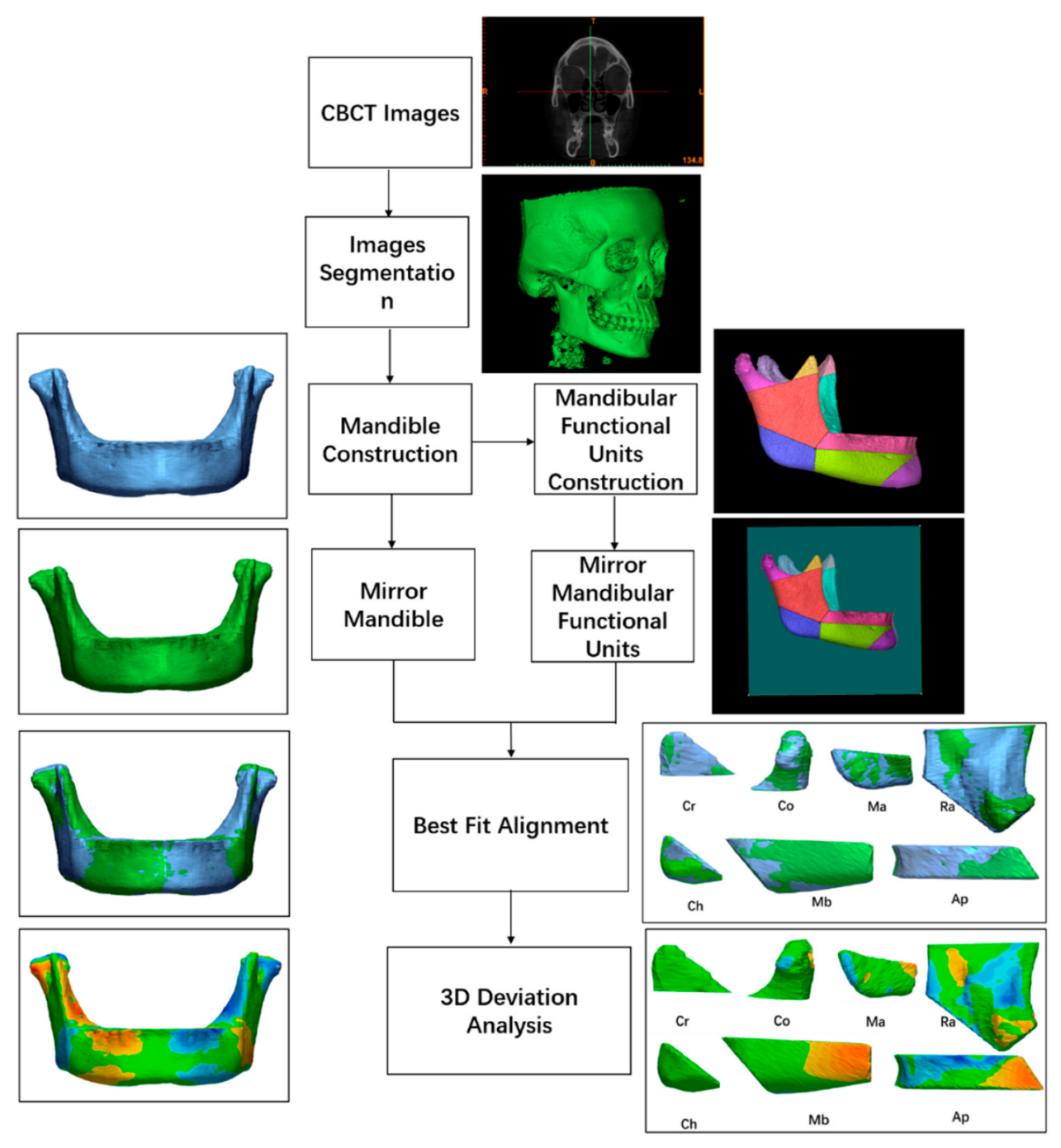
Figure 1. The flow chart of the whole image processing procedure.
Applsci 12 05814 g001
Figure 2. An initial segmentation mask from the CBCT data.
Applsci 12 05814 g002
Figure 3. (a) The original mandible model; (b) the mirrored mandible model; (c) the original mandible model and the mirrored mandible model were given best-fit alignment by computer automatically.
Applsci 12 05814 g003
Figure 4. (a) The landmarks were used to delimitate the mandibular functional units. Sg, the deepest point on the sigmoid notch; B, supramental, the most concave point on mandibular symphysis; Ri, ramus inflexion, the transition between the front edge of mandibular ramus and the mandibular body; Ag, the highest point in antegonial notch; MP, mandibular plane, the tangent to the lower edge of the mandible; RP ramal plane, the tangent to the condylar process and mandibular ramus. (b) The functional mandibular units were reconstructed. Co, condylar process; Cr, coronoid process; Ra, mandibular ramus; Ma, mandibular angle; Ap, alveolar process; Mb, mandibular body; Ch, chin process.
Applsci 12 05814 g004
Figure 5. Best-fit alignment of the mandibular units. Original mandibular units models (blue) and the mirrored units models (green).
Applsci 12 05814 g005
Figure 6. Surface deviation analysis of the mandibular units. The map showed the difference between the original model and the mirrored 3D models. The colored maps showed the negative (blue) and positive (yellow and red) deviation values. The ranges of tolerance at 0.5 mm are shown in green.
Applsci 12 05814 g006
Figure 7. Mandibular matching percentages of the study group and control group. p values were based on the independent t-test. The level of significance was set at 0.05; ** = p < 0.01.
Applsci 12 05814 g007
Figure 8. Surface deviation analysis of mandibles. The map showed the difference between the original mandible model and the mirrored mandible model. The tolerance range was set at 0.5 mm.
Applsci 12 05814 g008
Table 1. Matching percentages of the mandibular functional units.
Mandibular UnitsStudy GroupControl GroupSignificance
Mandibular ramus62.52 ± 11.0068.60 ± 7.56**
Mandibular angle69.16 ± 12.3971.44 ± 10.07NS
Condylar process62.78 ± 13.5667.58 ± 10.32*
Coracoid process87.03 ± 6.9989.73 ± 7.68NS
Alveolar process67.00 ± 12.6873.83 ± 8.88**
Mandibular body66.62 ± 9.4472.37 ± 8.69**
Chin80.75 ± 9.8685.23 ± 6.80**
p values were based on the independent t-test. The level of significance was set at 0.05. NS = no significance; * = p < 0.05; ** = p < 0.01.
Publisher’s Note: MDPI stays neutral with regard to jurisdictional claims in published maps and institutional affiliations.