Article

Computer-Aided Studies for Novel Arylhydantoin 1,3,5-Triazine Derivatives as 5-HT6 Serotonin Receptor Ligands with Antidepressive-Like, Anxiolytic and Antiobesity Action In Vivo

by
Rafał Kurczab
1,†,
Wesam Ali
2,3,†,
Dorota Łażewska
2,
Magdalena Kotańska
4,
Magdalena Jastrzębska-Więsek
5,
Grzegorz Satała
1,
Małgorzata Więcek
2,
Annamaria Lubelska
2,
Gniewomir Latacz
2,
Anna Partyka
5,
Małgorzata Starek
6,
Monika Dąbrowska
6,
Anna Wesołowska
5,
Claus Jacob
3,
Katarzyna Kieć-Kononowicz
2 and
Jadwiga Handzlik
2,*
1
Department of Medicinal Chemistry, Institute of Pharmacology, Polish Academy of Sciences, Smętna 12, PL 31-343 Cracow, Poland
2
Department of Technology and Biotechnology of Drugs, Jagiellonian University, Medical College, Medyczna 9, PL 30-688 Cracow, Poland
3
Division of Bioorganic Chemistry, School of Pharmacy, University of Saarland, Campus B2 1, D-66123 Saarbruecken, D-66123 Saarland, Germany
4
Department of Pharmacodynamics, Jagiellonian University, Medical College, Medyczna 9, PL 30-688 Cracow, Poland
5
Department of Clinical Pharmacy, Jagiellonian University, Medical College, Medyczna 9, PL 30-688 Cracow, Poland
6
Department of Inorganic Chemistry, Jagiellonian University, Medical College, Medyczna 9, PL 30-688 Cracow, Poland
*
Author to whom correspondence should be addressed.
These authors contributed equally to this work.
Molecules 2018, 23(10), 2529; https://doi.org/10.3390/molecules23102529
Submission received: 1 September 2018 / Revised: 22 September 2018 / Accepted: 24 September 2018 / Published: 3 October 2018

Abstract

:
This study focuses on the design, synthesis, biological evaluation, and computer-aided structure-activity relationship (SAR) analysis for a novel group of aromatic triazine-methylpiperazines, with an hydantoin spacer between 1,3,5-traizine and the aromatic fragment. New compounds were synthesized and their affinities for serotonin 5-HT6, 5-HT1A, 5-HT2A, 5-HT7, and dopamine D2 receptors were evaluated. The induced-fit docking (IFD) procedure was performed to explore the 5-HT6 receptor conformation space employing two lead structures. It resulted in a consistent binding mode with the activity data. For the most active compounds found in each modification line, anti-obesity and anti-depressive-like activity in vivo, as well as “druglikeness” in vitro, were examined. Two 2-naphthyl compounds (18 and 26) were identified as the most active 5-HT6R agents within each lead modification line, respectively. The 5-(2-naphthyl)hydantoin derivative 26, the most active one in the series (5-HT6R: Ki = 87 nM), displayed also significant selectivity towards competitive G-protein coupled receptors (6–197-fold). Docking studies indicated that the hydantoin ring is stabilized by hydrogen bonding, but due to its different orientation, the hydrogen bonds form with S5.44 and N6.55 or Q6.58 for 18 and 26, respectively. Compound 26 exerted anxiolytic-like and antidepressant-like activities. Importantly, it demonstrated anti-obesity properties in animals fed palatable feed, and did not show toxic effects in vitro.

Graphical Abstract

1. Introduction

The serotonin receptor 5-HT6 is one of the recently discovered members of the 5-HTRs family [1]. It is quite unique, since it includes a short third cytoplasmatic loop in parallel, with a long C-terminal tail, and one introne located in the middle of the third cytoplasmatic loop [2,3]. The localization of 5-HT6R is limited to the central nervous system, especially as it is located in the brain areas involved in learning and memory processes. Relatively limited, but intensive, research efforts in this area led to several families of potent 5-HT6R ligands [4,5], which have been proposed for potential treatment in the cognitive dysfunction associated with Alzheimer’s disease, anxiety or depression [5,6,7]. Over 20 compounds have qualified for clinical trials, most of which in studies following a cure for Alzheimer’s disease [5]. Nonetheless, none has reached the pharmaceutical market yet. Recent lines of evidence also point towards promising properties of some 5-HT6R ligands in the battle against obesity [8,9]. Therefore, the search for new ligands for the 5-HT6R, in particular, those with an action directed against obesity, seems to be a challenge for current medicinal chemistry.
Lines of evidence have identified several groups of chemical compounds, which displayed significant affinities for the serotonin receptors 5-HT6 [4,10,11]. The structure-activity relationship (SAR) analysis performed for the 5-HT6 ligands obtained previously enabled to identify pharmacophore features, including a triangle topology with tops of: A bulky hydrophobic area (HYD), positive ionizable nitrogen (PI), and hydrogen bond acceptor (HBA), as well as a central aromatic fragment (AR) (Figure 1) [12].
Taking into consideration the pharmacophore features, our previous studies have successfully found a new group of potent 5-HT6R agents among methylpiperazine derivatives of benzyl 2-amino-1,3,5-triazines which fit 3 out of 4 pharmacophore features. Two closed HBAs were the missing feature [13]. The computer-aided SAR analysis allowed us to modify the previous pharmacophore features and it has underlined the notable importance of aromatic substituents separated by a short linker from the 1,3,5-triazine. Thus, a bulky aromatic moiety, represented by the naphthyl (1) or m-chloro- (2) or m-methyl- (3) substituted phenyl ring (Figure 1) was beneficial for the kind of 5-HT6R affinity evaluated experimentally [13].
Taking into account both, the need to search for the 5-HT6R agents with new pharmacological possibilities and the results of previous SAR studies, we decided to explore a novel group of triazine-methylpiperazine derivatives (Figure 2), in which the spacer between 1,3,5-traizine and aromatic fragment is represented by a differently positioned hydantoin system containing both, two closed HBAs and HYD/AR features. In the first step, five pilot compounds (48) were synthesized and evaluated with regard to their affinity for the 5-HT6R. These initial studies resulted in two parallel lead structures 5 and 7 (Figure 2). Chemical modifications of the lead structures subsequently provided a series of compounds 927 (Table 1). Therefore, these studies are focused on the design, synthesis and biological evaluation, including affinity and selectivity for the 5-HT6R, as well as computer-aided SAR discussion for an entire series of hydantoin-triazines (427). For representative compounds, anti-obesity and anti-depressive activity in vivo, as well as their “drugability” properties, were also examined.

2. Results

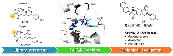
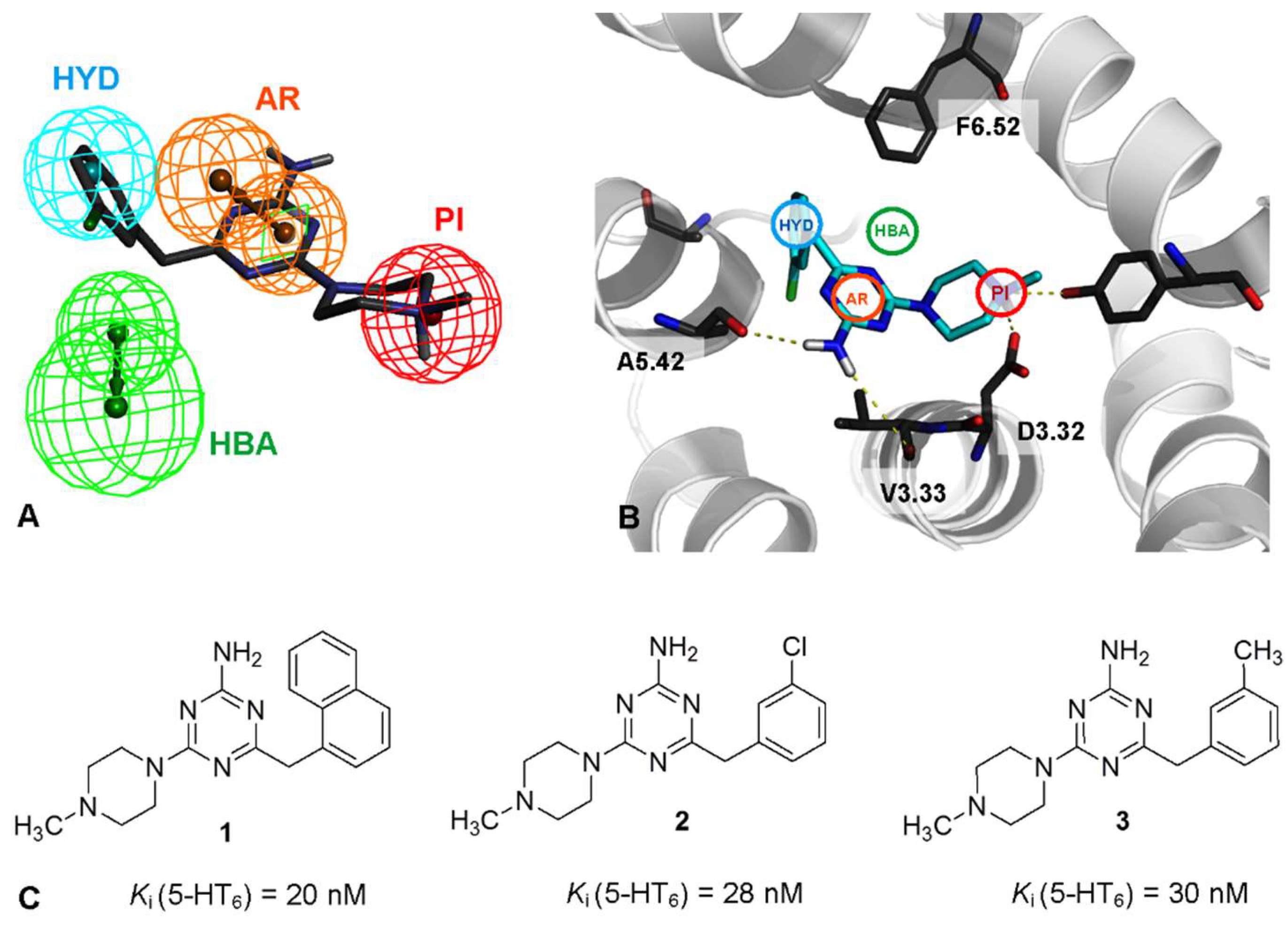
2.1. Identification of Lead Structures
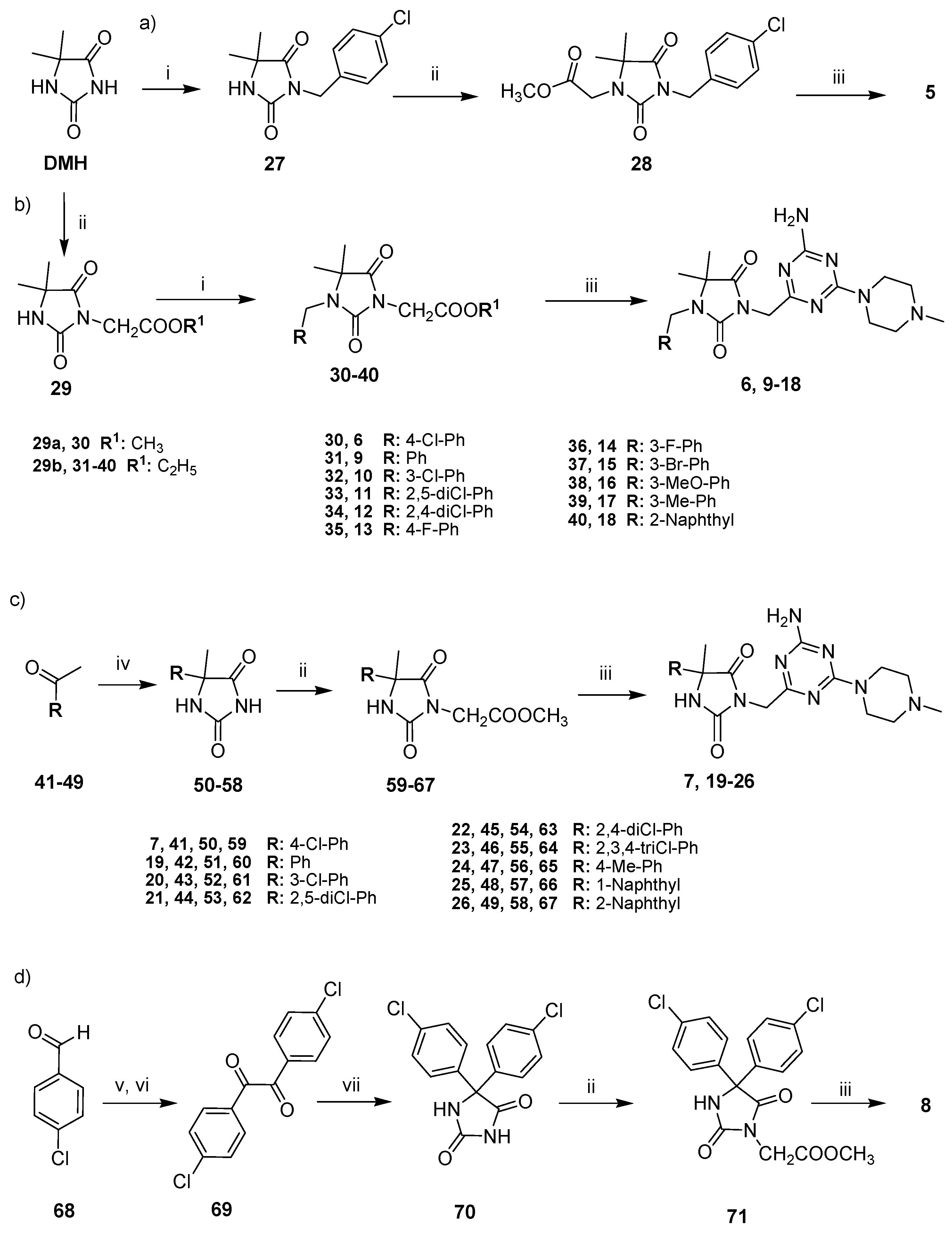
In the first step, a nonaromatic 5,5-dimethylhydantoin 1,3,5-triazine derivative 4 was found as a moderate 5-HT6R ligand after the radioligand binding assay and was therefore selected as an initial lead structure (Figure 2). Then, four different hydantoin moieties were taken into consideration as lead modifications, including: two N-substituted p-chlorobenzyl derivatives 5 and 6, with respect to position 3-N (5) and 1-N (6), the 5-(4-chlorophenyl)-5-methyl derivative 7 and 5,5-bis(4-chlorophenyl)hydantoin 8 (Figure 2).
Compounds 58 were synthesized and their affinity for the 5-HT6R was evaluated in the radioligand binding assays. Results displayed an increase of the 5-HT6R affinity in the case of compounds 6 and 7, whereas both 5 and 8 were much less active than the initial lead 4. Interestingly, the structure-activity relationship indicated a crucial role for the position of the 1,3,5-triazine substitution at hydantoin ring. Thus, 1,3,5-triazine linked by a methylene group at position 3 of hydantoin seems to be profitable (6), whilst that fragment situated at position 1 of hydantoin caused a huge decrease in activity (5). Taking into account these results, both 1-(4-chlorobenzyl)-3-((4-amino-6-(4-methylpiperazin-1-yl)-1,3,5-triazin-2-yl)methyl)-5,5-hydantoin (6), and the 3-((4-amino-6-(4-methylpiperazin-1-yl)-1,3,5-triazin-2-yl)methyl)-5-(4-chlorophenyl)-5-methylhydantoin (7) were selected in parallel as two particularly promising lead structures (Lead 1 and Lead 2, Figure 2). These two leads were subsequently modified within their aromatic area to generate a library of two distinct series of derivatives, i.e., compounds 918 and compounds 1926, respectively (Table 1).

2.2. Chemical Synthesis of Compounds 526

The final compounds 526 were obtained using four different synthesis pathways (Scheme 1a–d). The synthesis of 5,5-dimethylhydantoin triazine derivative 4 was described earlier [14]. As for the syntheses of the 3-benzyl derivative (5) and the 1-arylmethyl ones (6, 918), the commercial 5,5-dimethylhydantoin (DMH) was a starting material, which was substituted with an appropriate arylmethyl halide and methyl bromoacetate in a different order, respectively (a and b, Scheme 1). The reactions were performed as basic two-phase alkylations in acetone in the presence of a catalytic amount of benzyltriethylammonium chloride (TEBA). In case of 5, an alkylation with 4-chlorobenzyl at position 3 was the first step (27) [15], followed by the introduction of the methyl acetate fragment at position 1 (28, Scheme 1a). In contrast, the methyl ester 29a for the synthesis of 6 and the ethyl ester 29b, i.e., the common first intermediate for the synthesis of 918, were obtained by an alkylation of 5,5-dimethylhydantoin with a suitable alkyl bromoacetate at position 3. The ester 29 was substituted using commercial arylmethyl halides to give compounds 3040. Although a variety of purification methods was applied, including extraction, crystallization with charcoal and chromatography, only compounds 33, 34 and 40 gave precipitates (purity > 90%). Compounds 3032 and 3539 were obtained as glue-like residues with varying contents of the target product (44–81%) and were used for final cyclization in the crude form. In the case of 5-aryl-5-methylhydantoin derivatives (7, 1926), standard Bucherer-Bergs cyclic condensation of suitable commercial ketones (4149) was the first synthetic step providing 5-aryl-5-methylhydantoins 5058. Next, esters 5967 were synthesized via N-alkylation of 5058 in the hydantoin position 3 with methyl bromoacetate, under similar conditions to those for 29 and purification methods as those for 3040 (Scheme 1c). Among the esters (5967), compounds 60 and 66 were obtained in glue forms (purity 76–85%) with the remaining compounds (59, 6165 and 67) yielding white crystals (purity 95–100%). The ester intermediate for compound 8 was obtained within a 3-step synthesis, starting from the acyloin condensation of commercial 4-chlorobenzaldehyde (68), followed by oxidation to benzyl compound 69. The benzyl intermediate was used to perform a cyclic condensation with urea providing 5,5-bis(4-chlorophenyl)hydantoin (70), followed by an alkylation with methyl bromoacetate in position 3 of the hydantoin, in the aforementioned conditions of two-phase alkylation, to produce ester 71 (Scheme 1d). All ester hydantoin intermediates (28, 3040, 5967, and 71) were converted into the 1,3,5-triazine final products (526) via cyclic condensation with 1-(imino(4-methylpiperazin-1-yl)methyl)guanidine, which proceeded in sodium methanolate under the conditions corresponding to those for a series of benzyl-1,3,5-triazines described previously [13,14].

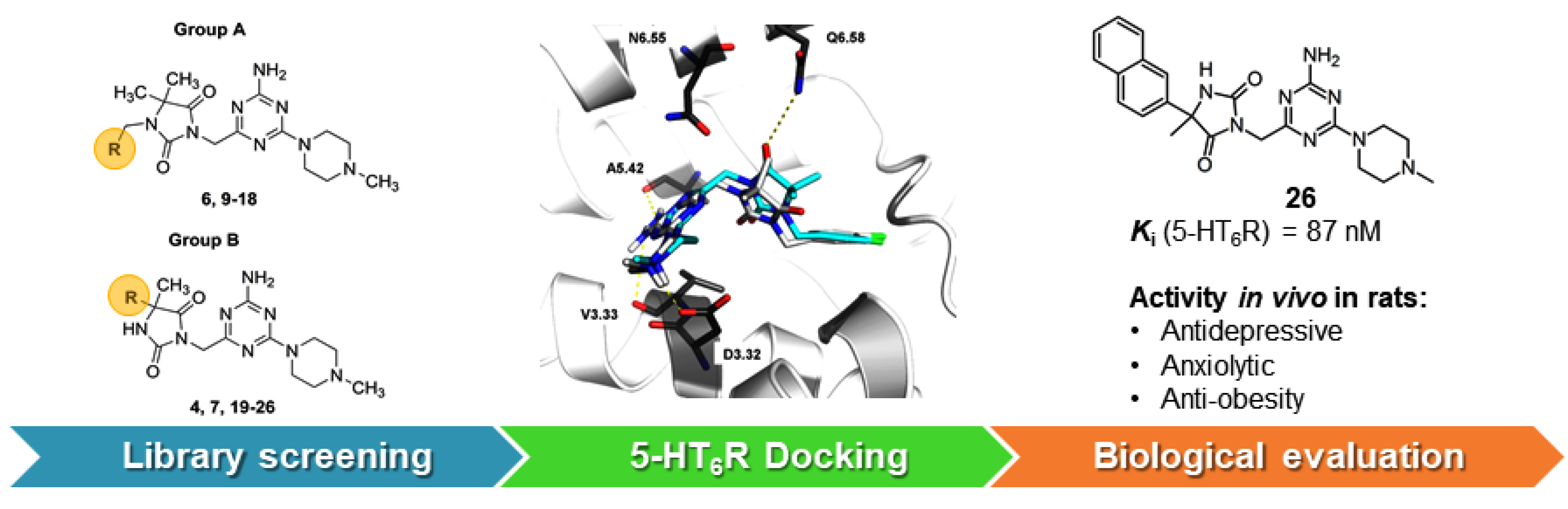
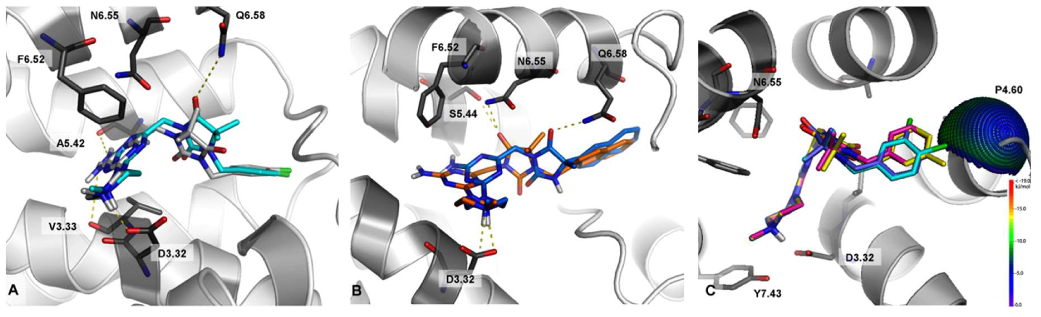
2.3. Molecular Modeling

Molecular docking was performed to gain an insight into the binding mode of the library of compounds synthetized (526) to the recently developed 5-HT6R homology models [13] built on the β2 adrenergic receptor template and optimized for the structures of Lead 1 and 2. The molecular docking indicated that newly synthesized compounds, generally, exhibited a very consistent binding mode with recently reported 5-HT6 ligands [16,17,18]. An influence of the topology of aromatic substituents and the position of the 1,3,5-triazine substitution at the hydantoin ring on the binding has been observed (Figure 2) and was in good agreement with results of the radioligand binding assay (see below).

2.4. Pharmacology

2.4.1. Radioligand Binding Assay

Compounds 526 were evaluated with respect to their affinity and selectivity for the target 5-HT6R in radioligand binding assays based on the human serotonin receptors 5-HT6, 5-HT1A, 5-HT2A, 5-HT7bR and dopaminergic D2LR, all of whom were expressed stably in HEK-293 cells. Five selective radioligands were employed (Table 1). Results expressed as Ki indicate that all derivatives of both leads displayed submicromolar affinities for the target 5-HT6R. More active agents (6, 7, 18, 23, 25 and 26) exerted Ki(5-HT6R) values lower than 200 nM. Compound 26 was the most active one (Ki = 87 nM) and also highly selective over 5-HT1AR, 5-HT2AR, and D2LR, with some selectivity over 5-HT7R (6-fold). Furthermore, a slightly less active 5-HT6R agent, compound 25, was highly selective with respect to all competitive GPCRs tested (Table 1). Most of the compounds displayed much weaker affinities for 5-HT1A, 5-HT2A and D2LR receptors than for 5-HT6R. Notably, compounds 811, 14, 17 and 24 were more potent for 5-HT7R than for the target 5-HT6R, in particular, compound 14 (5-HT7R; Ki = 88 nM), which was also selective toward 5-HT7R (Table 1).

2.4.2. Behavioral Tests In Vivo

The most potent 5-HT6R agent identified in the radioligand binding assays, i.e., compound 26, was selected for behavioral assays to determine its antidepressant- and anxiolytic-like properties in vivo in male Wistar rats.

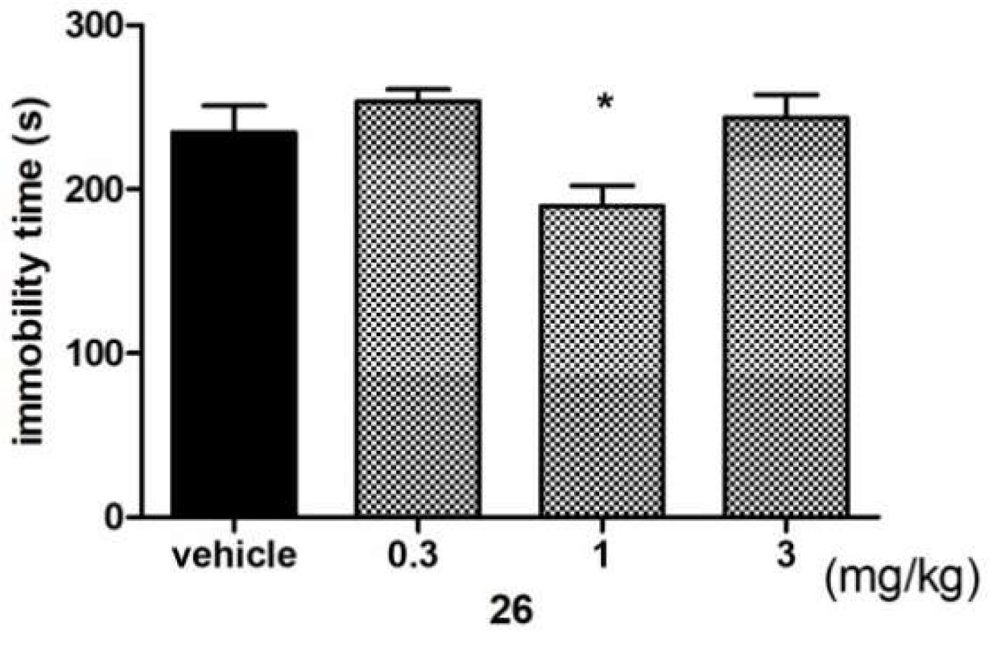
Antidepressant-Like Activity of Compound 26

In the forced swim test (FST), compound 26 significantly decreased immobility time about 20% (ANOVA: F(3,28) = 3.7674, p < 0.05) vs. vehicle treated group. A U-shaped dose-response in the FST test for compound 26 was observed (Figure 3).
This effect seems to be typical for some antidepressants with different mechanisms of action also producing U-shaped dose-response curves in some animal models and evaluated for antidepressant-like activity [19,20]. Moreover, this effect was also observed for other 5-HT6 ligands (i.e., SB258585) [21].

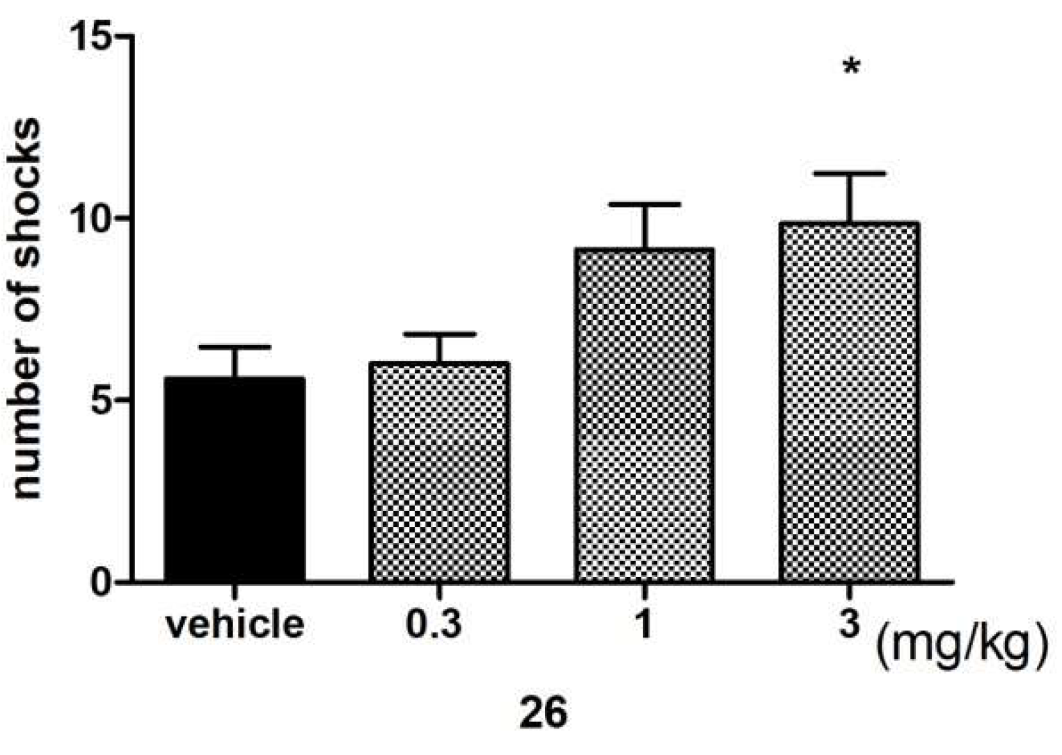
Anxiolytic-Like Activity of Compound 26

Anxiolytic-like activity was assessed using a Vogel conflict drinking test. As demonstrated in Figure 4, compound 26, administered at a dose of just 3 mg/kg, has significantly increased (by 76%) the number of accepted shocks (ANOVA: F(3,22) = 3.4967, p < 0.05) during the 5 min experimental session. Thus, compound 26 (at a dose of 3 mg/kg) exerted anxiolytic-like activity in this behavioral test.

2.4.3. Metabolic Assays In Vivo

Compounds 6 and 26, representing the most potent 5-HT6R agents of both groups, A (6) and B (26), were evaluated with regard to their influence on the body weight of male Wistar rats within metabolic assays in vivo (Figure 5). Animals that were fed palatable feed, and treated with 6, exhibited significantly less weight gain when compared to animals in the control group consuming a standard feed. From day 16 of the experiment, there was a significant difference between the groups. The body weight of rats treated with 6 having access to preferential feed differed significantly from the weight of control animals fed standard feed. Results are shown in Figure 3A,B.
Similarly, animals fed palatable feed and treated with 26 showed significantly less weight gain than animals in the control group consuming a preferential feed (Figure 5C,D), but there was a significant difference between the groups from day 12 of the experiment,. Additionally, the body weight of rats treated with 26 and having access to preferential feed did not differ significantly from the weight of control animals fed standard feed. No effects on body weight were noted in animals treated either with 6 or with 26 and consuming standard feed.

2.5. “Drug-Like” Properties

2.5.1. Lipophilicity

The lipophilicity for compounds considered in the search for lead structures (58) in particular for the most active compounds found as derivatives of both lead modifications (18, 23, 25 and 26) was determined with the standard RP-TLC method [22]. For comparison, the active arylmethyl-triazine compounds previously found (13) were also examined. Stationary phase RP-18 and mixtures of water/methanol (1:9, v/v) as a mobile phase were employed. RM values were calculated from the RF data obtained. It was found that the RM parameters decreased linearly with increasing amounts of the organic modifier (methanol) in the mobile phases tested. The regression coefficients (r2) determined for all compounds were higher than 0.95. On the basis of the linear relationship between RM values and the volume fraction of methanol, values of RM0 in the systems analyzed corresponding to 100% of water were obtained by extrapolation. RM0 values received for the compounds tested ranged from 3.14 to 4.49 (Figure 6).
The most lipophilic character is associated with compound 23 (RM0 = 4.49), while the least lipophilic hydantoin compound is a weak 5-HT6R agent 5 (RM0 = 3.22). The arylmethyl derivatives previously investigated (13) displayed slightly less lipophilic properties than those of the most active hydantoin agents (1826). Overall, these results indicate a good “drug-like” lipophilicity for the entire series of compounds investigated, taking into consideration either classical rules (Rules of Five, Ghose or Pfizer) or the latest CNS MPO approach [23].

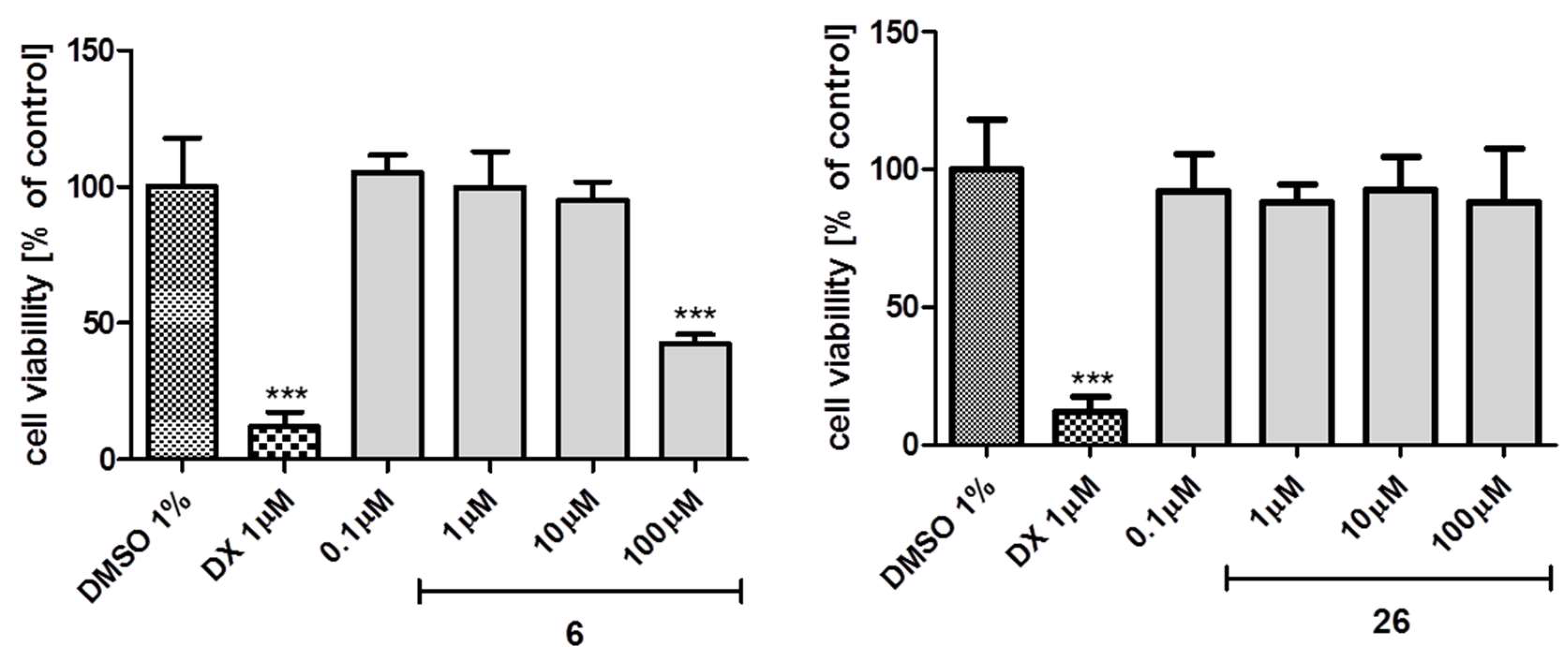
2.5.2. Toxicity In Vitro

The preliminary evaluation of the safety profile for the most promising 5-HT6R ligands 6 and 26 was performed in vitro with the HEK-293 eukaryotic cell line. The compounds examined were dissolved in culture growth media and incubated with cells for 72 h. Next, the MTS test was applied to determine the viability of the cells. As shown in Figure 7, a statistically significant decrease of viability (*** p < 0.001) was observed only for compound 6 and only at the highest concentration used, i.e., 100 μM. The reference cytostatic drug doxorubicin (DX) at the very low concentration 1 μM was also cytotoxic. Thus, the results obtained confirm a low or no significant toxicity for compounds 6 and 26, respectively.

2.5.3. Blood-Brain Barrier Permeability

In order to estimate an ability of the library synthetized (426) to penetrate the blood-brain barrier (BBB), the permeability QPlogBB parameter was calculated (Table 2) using QikProp from Schrodinger Suite 2017 [24]. QikProp predictions are for orally delivered drugs, and thus some natural neurotransmitters, e.g., dopamine and serotonin, are CNS negative, because they are extremely polar to cross the blood-brain barrier. Recommended values range for compounds that penetrate BBB is from −3.0 to 1.2. All the hydantoin-triazines investigated (426) are predicted as BBB-permeable (Table 2) with QPlogBB values from −0.98 (25) to −0.33 (12).

3. Computer-Aided SAR Analysis

In order to expand the current knowledge about structural properties responsible for 5-HT6R activity in the group of recently discovered aryl derivatives of 1,3,5-triazine, this study took into account the crucial role of the linker, which was found by comparative SAR analysis for the previously active arylmethyl- and inactive aryl-triazine molecules [13]. The new series of hydantoin-triazines (526) were designed as an extension of the methylene linker of the most active 2-arylmethyl-1,3,5-triaines, via an introduction of the inflexible, cyclic hydantoin insert, containing also hydrogen bond acceptors, and occurring in two topological variants found to be profitable in the preliminary lead identification (48). Both leads (6 and 7) and their derivatives (918 and 1926, respectively) displayed significant submicromolar affinities for the target 5-HT6 receptors, much more potent than those of previously investigated linker-free aryl-triazines [13], but slightly less potent if compared to the arylmethyl ones (13, Figure 1). Interestingly, the influence of the topology of the aromatic moieties in the group of 1,3,5-triazine derivatives seems to be distinctly linker-dependent. Although the bulky aromatic area of the naphthalene was beneficial for all the groups considered, it was distinctly predominant in the case of the 5-arylhydantoin (25, 26 vs. 7 and 1924; Table 1), comparable with the 3-methylphenyl and 3-chlorophenyl compounds in the group arylmethyl triazines (13, Figure 1), and slightly less favourable than the 4-chlorophenyl substituent within the 5,5-dimethylhydantoin group (18 vs. 6, Table 1). Furthermore, the preferable m-substitution within the phenyl ring, found for the benzyl 1,3,5-triazine 5-HT6R agents earlier [13], has not been confirmed in this study, either for the 5,5-dimethyl-1-benzylhydantoin- (6 vs. 10) or for the 5-phenyl-5-methylhydantoin triazine compounds (7 vs. 20). Consequently, an influence of both variants of the hydantoin linkers (1,3,5-substitution, group A or 3,5-substitution, group B) on the 5-HT6R activity was dependent on the properties of the aromatic moiety. Thus, both variants appear to be comparable, although the most active compound (26) is a member of the 5-aryl-5-methylhydantoin derivatives (group B).
Results of our docking studies have provided data useful to explain SAR on the molecular level (Figure 8).
In general, all of the newly synthesized derivatives of Lead 1 and Lead 2 exhibited a binding mode to 5-HT6R, which was very consistent with derivatives of 1,3,5-tiazines studied previously [13]. In addition to the crucial salt bridge interaction with D3.32, all of the docked ligands formed CH–π aromatic interactions with F6.52, and partially with F6.51. Additionally, the –NH2 group of the 1,3,5-triazine fragment was, usually, hydrogen-bonded with the carbonyl oxygen of A5.42 and V3.33 (Figure 8A). The methylene linker connecting 1,3,5-triazine and hydantoin rings, due to its tetrahedral conformation, forced the terminal aromatic group into a hydrophobic cavity formed by transmembrane helices 3–5 and extracellular loop 2. Thus, the structure-activity relationship in this series is determined by the different substituents at the terminal aromatic ring, and by the different orientation of hydantoin ring induced by its different positioning within the compound’s structure. A comparison of the analogues with different positions of the 1,3,5-triazine substitution at the hydantoin ring indicated that linking by the methylene group at position 3 of hydantoin is profitable (6, Ki = 127 nM), whereas a significant decrease of activity was noted for the derivative where the 1,3,5-triazine was linked with hydantoin via position 1 (5, Ki = 4275 nM). The analysis of the binding mode showed that a differently positioned hydantoin ring induces its different spatial orientation in the binding site (Figure 8A), and only the complex of 6 with the receptor is stabilized additionally by a hydrogen bond with a Q6.58 side chain.
Interestingly, within the group A and B, the best activity showed 2-naphthyl derivatives (18 and 26, respectively; Figure 8B). It should be noted, that apart from the length of the linker and the positioning of the hydantoin ring, the difference of activity between 18 and 26 is negligible—a comparison of its binding modes indicated that the most significant difference is noted for the hydantoin ring interaction. In both derivatives, the hydantoin ring is stabilized by hydrogen bonding, but due to its different orientation, the hydrogen bonds form with S5.44 and N6.55 or Q6.58 for 18 and 26, respectively. It is worth to note that the shift of the chlorine atom from 3- to 4-position resulted in a 4- (for modification B) to 5-fold (for modification A) increase of affinity for 5-HT6R compared to its unsubstituted analogues. This relationship was confirmed by molecular docking (Figure 8C) revealing that only the presence of chlorine atom in 4-position stabilized the L–R complex through the formation of halogen bond with the carbonyl oxygen of P4.60 residue (Cl∙∙∙O distance = 3.31 Å, σ-hole angle = 151.47°). As the halogen bond appeared to possess a highly directional nature, to explain the decrease in activity by shifting the chlorine atom from position 3 to 4 in the phenyl ring, the interaction sphere [25,26] was plotted onto the relevant backbone carbonyl oxygens (Figure 8C). The 4-chloro substituent was positioned within the energetically favorable areas of the sphere, whereas the 3-chloro substituent pointed outside of the sphere, indicating no ability to form halogen bond.

4. Experimental Section

4.1. Chemistry

Reagents were purchased from Alfa Aesar (Karlsruhe, Germany) or Sigma Aldrich (Darmstadt, Germany). Methanol was dried over calcium oxide Reaction progress was verified using thin layer chromatography (TLC), which was carried out on 0.2 mm Merck silica gel 60 F254 plates. Spots were visualized by UV light or treatment with Dragendorff reagent. Melting points (m.p.) were determined using MEL-TEMP II apparatus (LD Inc., Long Beach, CA, USA) and are uncorrected. The 1H-NMR and 13C-NMR spectra were obtained on a Mercury-VX 300 Mz spectrometer (Varian, Palo Alto, CA, USA) in DMSO-d6 or CDCl3. Chemical shifts in 1H-NMR spectra were reported in parts per million (ppm) on the δ scale using the solvent signal as an internal standard. Data are reported as follows: Chemical shift, multiplicity (s, singlet; br.s., broad singlet; d, doublet; t, triplet; m, multiplet), coupling constant J in Hertz (Hz), number of protons, proton’s position (Ind–indole, Naph—naphthalene, Ph—phenyl, Pp—piperazine). LC-MS were carried out on a system Water TQ Detector (Water Corporation, Milford, CT, USA) consisting of a Waters Acquity UPLC, coupled to a Waters TQD mass spectrometer. Retention times (tR) are given in minutes. The UPLC/MS purity of all final compounds was determined (%).
Synthesis of compounds 27, 29, 5058, 63, 65 and 6870 was obtained by our [15,27] or other research groups [28,29,30,31,32] earlier and are reported in Chemical Abstract Database (see Supplementary Information).

4.1.1. Synthesis of Methyl 2-(3-(4-chlorobenzyl)-5,5-dimethyl-2,4-dioxoimidazolidin-1-yl)acetate (28)

A mixture of 3-(4-chlorobenzyl)-5,5-dimethylimidazolidine-2,4-dione 27 (20 mmol, 5.05 g), TEBA (2.60 mmol, 0.60 g), K2CO3 (58 mmol, 8 g) in acetone (100 mL) was heated under reflux for 30 min. Methyl bromoacetate (20 mmol, 3.06 g) in acetone (20 mL) was added. The mixture was heated under reflux for 5 h and stirred at room temperature overnight. The inorganic precipitate was separated by filtration. The filtrate was evaporated, and the residue was crystallized with methanol. White solid, m.p. 78–80 °C, yield: 58% (11.60 mmol, 3.77 g), C15H17ClN2O4 (MW 324.76). 1H-NMR (300 MHz, DMSO-d6) δ = 7.38–7.41 (d def., 2H, Ar-3,5-H), 7.22–7.25 (d def., 2H, Ar-2,6-H), 4.57 (s, 2H, Ar-CH2), 4.18 (s, 2H, CH2COO), 3.68 (s, 3H, OCH3), 1.30 (s, 6H, 2× 5-CH3) ppm. LC/MS+: Purity: 98.40%, tR = 6.14, (ESI) m/z [M + H]+ 325.11.

4.1.2. General Procedure for the Synthesis of Alkyl 2-(1-arylmethyl-5,5-dimethyl-2,4-dioxoimidazolidin-3-yl)acetate (3040)

A mixture of alkyl 2-(5,5-dimethyl-2,4-dioxoimidazolidin-3-yl)acetate 29 (12 mmol, 29a: 2.40 g or 29b: 2.58 g), TEBA (1.20 mmol, 0.36 g), K2CO3 (12 mmol, 4.80 g) in acetone (60 mL) was heated under reflux for 30 min. The appropriate arylmethyl halide (12 mmol) in acetone (15 mL) was added. The mixture was heated under reflux for 5–7 h and stirred at room temperature for further 24–48 h. The inorganic precipitate was separated by filtration. The filtrate was collected, the solvent then evaporated and the residue was dissolved in 30 mL of DCM and washed with 1% NaOH (2 × 30 mL) and H2O (2 × 30 mL). The organic phase was dried on anhydrous Na2SO4 overnight, separated from the drying agent and the solvent was evaporated. Then, Methods A or B were applied for purification. Method A: The residue was crystallized with EtOH and charcoal to give crystals of the intermediates. Method B: If Method A was applied, but no crystal appeared, the ethanolic solution was concentrated to give crude ester intermediates in glue form. No ester intermediate (3040) was isolated in pure form (purity < 80%). The purity and identity of crude intermediates were evaluated using LC-MS. Taking into consideration the results of LC-MS a desirable ester was used for the synthesis of the final product in the amount suitable to its concentration in the crude product obtained (40–80%)
Ethyl 2-(1-(4-chlorobenzyl)-5,5-dimethyl-2,4-dioxoimidazolidin-3-yl)acetate (30). 1-Chloro-4-(chloromethyl)benzene (1.93 g) and 29a were used. Method A. White crystals, m.p. 88–90 °C, yield: 68% (8.16 mmol, 2.65 g), C15H17ClN2O4 (MW 324.76). 1H-NMR (300 MHz, DMSO-d6) δ = 7.36–7.37 (d def., 4H, Ar), 4.52 (s, 2H, Ar-CH2), 4.25 (s, 2H, CH2COO), 3.70 (s, 3H, OCH3), 1.25 (s, 6H, 2× CH3) ppm. LC/MS+: Purity: 81%, tR = 6.31, (ESI) m/z [M + H]+ 325.18.
Ethyl 2-(1-benzyl-5,5-dimethyl-2,4-dioxoimidazolidin-3-yl)acetate (31). Benzyl chloride (1.52 g) and 29a were used. Method B. Glue mixture containing 44% of 31, yield: 61.37%, C15H18N2O4 (MW 304.15). LC/MS+: Purity: 44%, tR = 6.09, (ESI) m/z [M + H]+ 304.14.
Ethyl 2-(1-(3-chlorobenzyl)-5,5-dimethyl-2,4-dioxoimidazolidin-3-yl)acetate (32). 1-Chloro-3-(chloromethyl) benzene (1.93 g) and 29a were used. Method B. Sticky liquid, yield: 85.22%, C15H19ClN2O4 (MW 338.59). 1H-NMR (300 MHz, DMSO-d6) δ = 7.41–7.33 (m, 1H, Ph-5-H), 7.31–7.28 (m, 3H, Ph-2,4,6-H), 4.54 (s, 2H, Ph-CH2), 4.23 (s, 2H, CH2COO), 4.10 (q, J = 7.05 Hz, 2H, CH3–CH2), 1.26 (s, 6H, 2× CH3), 1.16 (t, J = 7.35 Hz, 3H, CH3–CH2) ppm. LC/MS+: Purity: 67.03%, tR = 6.73, (ESI) m/z [M + H]+ 339.59.
Ethyl 2-(1-(2,5-dichlorobenzyl)-5,5-dimethyl-2,4-dioxoimidazolidin-3-yl) acetate (33). 1-Bromo-2,5-(di-chloro methyl) benzene (3.60 g) and 29a were used. Method A. yellowish Solid, m.p. 98 °C, yield: 98.21%, C16H18Cl2N2O4 (MW 373.04). 1H-NMR (300 MHz, DMSO-d6) δ = 7.52–7.41 (m, 1H, Ph-6-H), 7.39–7.40 (m, 2H, Ph-3,4-H), 4.58 (s, 2H, Ph-CH2), 4.24 (s, 2H, CH2COO), 4.10 (q, J = 7.00 Hz, 2H,CH3–CH2), 1.29 (s, 6H, 2× CH3), 1.17 (d, J = 7.00 Hz, 3H, CH3–CH2) ppm. LC/MS+: Purity: 93.90%, tR = 7.28, (ESI) m/z [M + H]+ 373.03.
Ethyl 2-(1-(2,4-dichlorobenzyl)-5,5-dimethyl-2,4-dioxoimidazolidin-3-yl)acetate (34). 1-Chloro-2,4-(di-chloro methyl) benzene (2.34 g) and 29a were used. Method B. White Solid, m.p. 105 °C, yield: 91.52%, C16H18Cl2N2O4 (MW 373.04). 1H-NMR (300 MHz, DMSO-d6) δ = 7.66 (d, J = 2.0 Hz, 1H, Ph-3-H), 7.47–7.37 (m, 2H-Ph-5,6-H), 4.60 (s, 2H-Ph-CH2), 4.26 (s, 2H, CH2COO), 4.16 (q, J = 7.1 Hz, 2H–CH3–CH2), 1.29 (s, 6H, 2× CH3), 1.21 (t, J = 7.1 Hz, 3H, CH3) ppm. 13C-NMR (75 MHz, DMSO-d6) δ = 175.83, 167.53, 154.84, 134.60, 133.22, 133.18, 130.71, 129.32, 127.98, 62.51, 61.83, 22.68, 14.38 ppm. LC/MS+: Purity: 91.74%, tR = 7.52, (ESI) m/z [M + H]+ 374.04.
Ethyl 2-(1-(4-fluorobenzyl)-5,5-dimethyl-2,4-dioxoimidazolidin-3-yl)acetate (35). 4-Flouro benzyl chloride (2.17 g) and 29a were used. Method B. Sticky liquid, yield: 90.72%, C16H19FN2O4 (MW 322.99). LC/MS+: Purity: 69.51%, tR = 7.52, (ESI) m/z [M + H]+ 323.99.
Ethyl 2-(1-(3-fluorobenzyl)-5,5-dimethyl-2,4-dioxoimidazolidin-3-yl)acetate (36). 3-Flouro benzyl chloride (1.74 g) and 29a were used. Method A. Sticky liquid, yield: 77.06%, C16H19FN2O4 (MW 322.99). 1H-NMR (300 MHz, DMSO-d6) δ = 7.41–7.36 (m, 1H, Ph-5-H), 7.19–7.13 (m, 3H, Ph-2,4,6-H), 4.55 (s, 2H, Ph-CH2), 4.23 (s, 2H, CH2COO), 4.10 (q, J = 7.05 Hz, 2H, CH3–CH2), 1.26 (s, 6H, 2× CH3), 1.16 (t, J = 7.05 Hz, 3H, CH3–CH2) ppm. LC/MS+: Purity: 69.43%, tR = 6.23, (ESI) m/z [M + H]+ 323.99.
Ethyl 2-(1-(3-bromobenzyl)-5,5-dimethyl-2,4-dioxoimidazolidin-3-yl)acetate (37). 3-Bromo benzyl chloride (3.75 g) and 29a were used. Method B. Sticky liquid, yield: 60%, C16H19BrN2O4 (MW 383.24). LC/MS+: Purity: 78.26%, tR = 6.85, (ESI) m/z [M + H]+ 384.24.
Ethyl 2-(1-(3-methoxybenzyl)-5,5-dimethyl-2,4-dioxoimidazolidin-3-yl)acetate (38). 3-Methoxy benzyl chloride (1.88 g) and 29a were used. Method B. Sticky liquid, yield: 68.94%, C17H22N2O5 (MW 334.38). LC/MS+: Purity: 88.03%, tR = 6.65, (ESI) m/z [M + H]+ 335.38.
Ethyl 2-(1-(3-methylbenzyl)-5,5-dimethyl-2,4-dioxoimidazolidin-3-yl)acetate (39). 3-Methyl benzyl chloride (1.69 g) and 29a were used. Method B. Sticky liquid, yield: 61.05%, C17H22N2O4 (MW 318.38). LC/MS+: Purity: 81.94%, tR = 6.12, (ESI) m/z [M + H]+ 319.38.
Ethyl 2-(1-(2-naphthyl)-5,5-dimethyl-2,4-dioxoimidazolidin-3-yl)acetate (40). 2-(Chloromethyl)naphthalene (2.65 g) and 29a were used. Method B. White solid, m.p. 95 °C, yield: 81.47%, C20H22N2O4 (MW 354.19). 1H-NMR (300 MHz, DMSO-d6) δ = 7.90–7.84 (m, 4H, naft-1,4,5,8-H), 7.52–7.45 (m, 3H, naft-3,6,7-H), 4.71 (s, 2H, Ph-CH2), 4.26 (s, 2H, CH2COO), 4.16–4.11 (q, J = 7.35 Hz, 2H, CH3–CH2), 1.26 (s, 6H, 2× CH3), 1.17(d, J = 7.00 Hz, 3H, CH3–CH2) ppm. LC/MS+: Purity: 90.56%, tR = 7.08, (ESI) m/z [M + H]+ 355.15.

4.1.3. General Procedures for the Synthesis of Methyl 2-(5-arylhydantoin-3-yl)acetates (6067 and 71)

A mixture of a suitable arylhydantoin 5058 (Scheme 1) or 70 (15–20 mmol) with K2CO3 (6–8 g), TEBA (0.45–0.60 g) in acetone (75–100 mL) was stirred and refluxed for 30 min. Then, the solution of methyl bromoacetate (15–20 mmol, 2.30–3.06 g) in acetone (15–20 mL) was added. The mixture was stirred and refluxed for 3–6 h, then, stirred at room temperature overnight. The inorganic components were separated by filtration. The filtrate was concentrated, and the residue was purified using the similar methods as those for compounds 30–40, but MeOH in place of EtOH was used for crystallization.
Methyl 2-(5-methyl-2,4-dioxo-5-phenylimidazolidin-3-yl)acetate (60). Compound 51 (2.85 g) and methyl bromoacetate (2.30 g) were used. Sticky liquid, yield: 48.6%, C13H14N2O4 (MW 262.26). 1H-NMR (300 MHz, DMSO-d6) δ = 9.06 (s, 1H, N1–H), 7.47–7.50 (d, J = 7.00 Hz, 2H, Ph-2,6-H), 7.33–7.42 (m, 3H Ph-3,4,5-H), 4.19 (s, 2H, N3–CH2), 3.67 (s, 3H, O-CH3), 1.70 (s, 3H, CH3) ppm. LC/MS+: Purity: 85.92%, tR = 4.43, (ESI) m/z [M + H]+ 263.11.
Methyl 2-(5-(3-chlorophenyl)-5-methyl-2,4-dioxoimidazolidin-3-yl)acetate (61). Compound 52 (4.49g) and methyl bromoacetate (3.06 g) were used. White Chrystal, m.p. 102 °C, yield: 51.10%, C13H13ClN2O4 (MW 296.71). 1H-NMR (300 MHz, DMSO-d6) δ = 9.13 (s, 1H, N1–H), 7.51 (s, 1H, Ph-6-H), 7.43–7.47 (m, 3H, Ph-2,4,5-H), 4.20 (s, 2H, N3-CH2), 3.66 (s, 3H, O–CH3), 1.70 (s, 3H, CH3) ppm. LC/MS+: Purity: 100%, tR = 5.25, (ESI) m/z [M + H]+ 297.07.
Methyl 2-(5-(2,5-dichlorophenyl)-5-methyl-2,4-dioxoimidazolidin-3-yl)acetate (62). Compound 53 (4.14 g) and methyl bromoacetate (2.45 g) were used. White solid, m.p. 214 °C, yield: 83.19%, C13H12Cl2N2O4 (MW 331.15). 1H-NMR (300 MHz, DMSO-d6) δ = 8.75 (s, 1H, N1-H), 7.69 (s, 2H, Ph-4-H), 7.52 (s, 2H, Ph-3,6-H), 4.25 (s, 2H, N3–CH2), 3.68 (s, 3H, O–CH3), 1.80 (s, 3H, CH3) ppm. LC/MS+: Purity: 99.10%, tR = 5.41, (ESI) m/z [M + H]+ 330.69.
Methyl 2-(5-(2,3,4-trichlorophenyl)-5-methyl-2,4-dioxoimidazolidin-3-yl)acetate (64). Compound 54 (5.18 g) and methyl bromoacetate (3.06 g) were used. White solid, m.p. 172 °C, yield: 37.76%, C13H12Cl2N2O4 (MW 331.15). 1H-NMR (300 MHz, DMSO-d6) δ = 8.75 (s, 1H, N1–H), 7.68–7.71 (d, J = 8.8 Hz, 1H, Ph-3-H), 7.65–7.66 (s, 1H, Ph-5-H), 7.48–7.52 (m, 1H, Ph-6-H), 4.25 (s, 2H, N3–CH2), 3.68 (s, 3H, O–CH3), 1.78 (s, 3H, CH3) ppm. LC/MS+: Purity: 96.70%, tR = 5.49, (ESI) m/z [M + H]+ 330.76.
Methyl 2-(5-methyl-5-(naphthalen-1-yl)-2,4-dioxoimidazolidin-3-yl)acetate (66). 5-Methyl-5-(naphthalen-1-yl)imidazolidine-2,4-dione 57 (20 mmol, 4.80 g), K2CO3 (8 g), TEBA (0.60 g) in acetone (100 mL) and methyl bromoacetate (20 mmol, 3.06 g) in acetone (20 mL) were stirred and refluxed for 6 h. Method B. White glue-crystals, m.p. 105–109 °C, yield: 78% (15.60 mmol, 4.86 g), C17H16N2O4 (MW 312.11). 1H-NMR (300 MHz, DMSO-d6) δ = 8.98 (s, 1H, NH), 7.94–8.01 (m, 3H, Ar-4,5,8), 7.75 (d, J = 7.44 Hz, 1H, Ar-2), 7.48–7.57 (m, 3H, Ar-3,6,7), 4.33–4.39 (m, 2H, –CH2CO), 3.73 (s, 3H, –OCH3), 1.94 (s, 3H, 5-CH3) ppm. LC/MS+: Purity: 65.46%, tR = 5.47, (ESI) m/z [M + H]+ 312.95.
Methyl 2-(5-methyl-5-(naphthalen-2-yl)-2,4-dioxoimidazolidin-3-yl)acetate (67). 5-Methyl-5-(naphthalen-2-yl)imidazolidine-2,4-dione 58 (15 mmol, 3.60 g), K2CO3 (6 g), TEBA (0.45 g) in acetone (75 mL) and methyl bromoacetate (15 mmol, 2.30 g) in acetone (15 mL) were stirred and refluxed for 5 h. Method A. White crystals, m.p. 134–138 °C, yield: 58% (8.7 mmol, 2.72 g), C17H16N2O4 (MW 312.11). 1H-NMR (300 MHz, DMSO-d6) δ = 9.15 (s, 1H, NH), 8.02 (s, 1H, Ar-1-H), 7.60 (dd, J1 = 8.72 Hz, J2 = 1.80 Hz, 2H, Ar-6,7-H), 7.87–7.99 (m, 3H, Ar-4,5,8-H), 7.51–7.57 (m, 1H, Ar-3), 4.23 (s, 2H, –CH2CO), 3.66 (s, 3H, –OCH3), 1.81 (s, 3H, 5-CH3) ppm. LC/MS+: Purity: 76.61%, tR = 5.63, (ESI) m/z [M + H]+ 312.82.
Methyl 2-(5,5-bis(4-chlorophenyl)-2,4-dioxoimidazolidin-3-yl)acetate (71). 5,5-bis(4-Chlorophenyl)-imidazolidin-2,4-dione 70 (15 mmol, 4.80 g), K2CO3 (6 g), TEBA (0.45 g) in acetone (75 mL) and methyl bromoacetate (15 mmol, 2.30 g) in acetone (15 mL) were stirred and refluxed for 4 h. Method A. Creamy crystals, m.p. 120–121 °C, yield: 62%, C18H14Cl2N2O4 (MW 392.03). 1H-NMR (300 MHz, DMSO-d6) δ = 7.53 (d, J = 8.7 Hz, 4H-ph), 7.37 (d, J = 8.7 Hz, 4H-ph), 4.31 (s, 2H–CH2), 3.70 (s, 3H-CH3), 1.04 (d, J = 6.1 Hz, 1H-NH) ppm. LC/MS+: Purity: 91.91%, tR = 7.26, (ESI) m/z [M + H]+ 393.10.

4.1.4. General Procedure for the Synthesis of 1,3,5-Triazine Final Products (526)

Sodium (8–10 mmol) was dissolved in 10 mL of absolute methanol, then 4-methylpiperazin-1-yl biguanidine × 2HCl (4–5 mmol) and an appropriate ester 28, 3040, 5967 or 71 (4–5 mmol) were added. The reaction mixture was refluxed for 15–30 h. After cooling to room temperature, water (10 mL) was added and the mixture was stirred for 0.5 h. The triazine product precipitated (526) was separated and purified using Methods C, D or E. Method C: Crystallization with methanol. Method D: crystallization with methanol and charcoal. Method E: crystallization with ethanol.
3-(4-Chlorobenzyl)-1-((4-amino-6-(4-methylpiperazin-1-yl)-1,3,5-triazin-2-yl)methyl)-5,5-dimethylimidazolidine-2,4-dione (5). Ester 28 (4.30 mmol, 1.40 g) and 4-methylpiperazin-1-yl biguanidine × 2HCl (4.30 mmol, 1.11 g) were used (48 h refluxed). Method C. White solid, m.p. 166–169 °C, yield: 34% (0.67 g), C21H27N8O2Cl (MW 458.95). 1H-NMR (300 MHz, DMSO-d6) δ 7.34–7.52 (m, 2H, Ph-3,5-H), 7.21–7.32 (m, 2H, Ph-2,6-H), 6.82 (br. s., 2H, NH2), 4.55 (s, 2H, Ph-CH2), 4.19 (s, 2H, TR-CH2), 3.72–3.37 (br s, 4H, Pp-2,6-H), 2.15 (s, 7H, Pp-3,5-H + Pp-CH3), 1.31 (s, 6H, 2× CH3) ppm. 13C-NMR (75 MHz, DMSO-d6) δ = 176.83, 173.77, 167.04, 164.65, 154.86, 136.23, 132.52, 129.73, 129.03, 62.11, 54.67, 46.19, 44.19, 42.75, 41.08, 22.84 ppm. LC/MS+: Purity: 99.46%, tR = 3.75, (ESI) m/z [M + H]+ 459.23.
1-(4-Chlorobenzyl)-3-((4-amino-6-(4-methylpiperazin-1-yl)-1,3,5-triazin-2-yl)methyl)-5,5-dimethylimidazolidine-2,4-dione (6). Ester 30 (4.30 mmol, 1.40 g) and 4-methylpiperazin-1-yl biguanidine × 2HCl (4.30 mmol, 1.11 g) were used (50 h refluxed). Method C. White solid, m.p. 204–205 °C, yield: 19% (0.38 g), C21H27N8O2Cl (MW 458.95). 1H-NMR (300 MHz, DMSO-d6) δ = 7.38 (s, 4H, Ph-2,3,5,6-H), 6.87 (br. s., 2H, NH2), 4.51 (s, 2H, Ph-CH2), 4.27 (s, 2H, TR-CH2), 3.55 (br. s., 4H, Pp-2,6-H), 2.15 (s, 7H, Pp-3,5-H + Pp-CH3), 1.26 (s, 6H, 2× CH3) ppm. LC/MS+: Purity: 100%, tR = 3.94, (ESI) m/z [M + H]+ 459.23.
3-((4-Amino-6-(4-methylpiperazin-1-yl)-1,3,5-triazin-2-yl)methyl)-5-(4-chlorophenyl)-5-methylimidazolidine-2,4-dione (7). Ester 59 (5 mmol, 1.48 g) and 4-methylpiperazin-1-yl biguanidine × 2HCl (4.30 mmol, 1.11 g) were used (13.5 h refluxed). Method C. White solid, m.p. 218–220 °C, yield: 23% (0.42 g), C19H23N8O2Cl (MW 430.89). 1H-NMR (300 MHz, DMSO-d6) δ = 9.02 (s, 1H, Hyd-1-H), 7.51–7.69 (m, 2H, Ph-3,5-H), 7.36–7.51 (m, 2H, Ph-2,6-H), 6.83 (br. s., 2H, NH2), 4.21 (s, 2H, TR-CH2), 3.53 (br. s., 2H, Pp-2,6-He), 3.27–3.32 (m, 2H, Pp-2,6-Ha), 2.13 (s, 7H, Pp-3,5-H + Pp-CH3), 1.68 (s, 3H, Hyd-CH3) ppm. 13C-NMR (75 MHz, DMSO-d6) δ = 175.25, 171.94, 166.94, 164.54, 155.78, 139.22, 133.15, 128.87, 127.93, 62.99, 54.57, 46.16, 42.63, 42.45, 25.92 ppm. LC/MS+: Purity: 100%, tR = 3.23, (ESI) m/z [M + H]+ 431.19.
3-((4-Amino-6-(4-methylpiperazin-1-yl)-1,3,5-triazin-2-yl)methyl)-5,5-bis(4-chlorophenyl)imidazolidine-2,4-dione (8). Ester 71 (4.80 mmol, 1.90 g) and 4-methylpiperazin-1-yl biguanidine × 2HCl (4 mmol, 1.03 g) were used (30 h refluxed). Method C. White solid, m.p. 266–268 °C, yield: 40% (0.83 g), C24H24N8O2Cl2 (MW 526.14). 1H-NMR (300 MHz, DMSO-d6) δ = 9.79 (s, 1H, Hyd-1-H), 7.34–7.59 (m, 8H, 2× Ph-2,3,5,6-H), 6.85 (br. s, 2H, NH2), 4.30 (s, 2H, TR-CH2), 3.55 (br. s, 2H, Pp-2,6-He), 3.09 (br. s, 2H, Pp-2,6-Ha), 1.76–2.35 (m, 7H, Pp-3,5-H + Pp-CH3) ppm. 13C-NMR (75 MHz, DMSO-d6) δ = 173.01, 171.74, 166.92, 164.52, 155.38, 138.75, 133.57, 129.12, 129.01, 68.59, 54.58, 46.16, 42.77, 42.53 ppm. LC/MS+: Purity: 100%, tR = 3.23, (ESI) m/z [M]+ 527.16.
3-((4-Amino-6-(4-methylpiperazin-1-yl)-1,3,5-triazin-2-yl)methyl)-1-benzyl-5,5-dimethylimidazolidine-2,4-dione (9). Ester 31 (3 mmol, 2.03 g) and 4-methylpiperazin-1-yl biguanidine × 2HCl (3 mmol, 0.77 g) were used (36 h refluxed). Method D. White solid, m.p. 206 °C, yield: 3.94%, C21H28N8O2 (MW 424.23). 1H-NMR (300 MHz, DMSO-d6) δ = 7.36–7.25 (m, 5H-Ph), 6.87 (s, 2H, NH2), 4.52 (s, 2H, CH2), 4.28 (s, 2H, TR-CH2), 3.62 (s, 4H, Pp-2,6-H), 2.15–2.23 (m, 7H, Pp-3,5-H, Pp-CH3), 1.25 (m, 6H, 2× CH3) ppm. LC/MS+: Purity: 98.30%, tR = 3.50, (ESI) m/z [M]+ 425.20.
1-(3-Chlorobenzyl)-3-((4-amino-6-(4-methylpiperazin-1-yl)-1,3,5-triazin-2-yl)methyl)-5,5-dimethylimidazolidine-2,4-dione (10). Ester 32 (5 mmol, 2.52 g) and 4-methylpiperazin-1-yl biguanidine × 2HCl (5 mmol, 0.92 g) were used (6 h refluxed), and stirred for another 24 h in a room temperature. Method D. White solid, m.p. 168 °C, yield: 24.81%, C21H28N8O2 (MW 458.19). 1H-NMR (300 MHz, DMSO-d6) δ = 7.42 (s, 1H, Ph-6-H), 7.33 (m, 3H, Ph-2,4,5-H), 6.86 (s, 2H, NH2), 4.52 (s, 2H–CH2), 4.27(s, 2H, TR-CH2), 3.58 (s, 4H, Pp-2,6-H), 2.13 (m, 7H, Pp-3,5-H, Pp-CH3), 1.27 (s, 6H, 2× CH3) ppm. LC/MS+: Purity: 96.12%, tR = 5.34, (ESI) m/z [M]+ 459.23.
1-(2,5-Dichlorobenzyl)-3-((4-amino-6-(4-methylpiperazin-1-yl)-1,3,5-triazin-2-yl)methyl)-5,5-dimethylimidazolidine-2,4-dione (11). Ester 33 (5 mmol, 2.01 g) and 4-methylpiperazin-1-yl biguanidine × 2HCl (5 mmol, 0.92 g) were used (6 h refluxed), and stirred for another 144 h in room a temperature. Method D. White solid, m.p. 163 °C, yield: 27.32%, C21H26Cl2N8O (MW 492.16). 1H-NMR (300 MHz, DMSO-d6) δ = 7.53–7.50 (s, 1H, Ph-4-H), 7.41–7.38 (m, 2H, Ph-3,6-H), 6.84 (s, 2H, NH2), 4.58 (s, 2H, CH2), 4.29 (s, 2H, TR-CH2), 3.63 (s, 4H, Pp-2,6-H), 2.23–2.15 (m, 7H, Pp-3,5-H, Pp-CH3), 1.30 (s, 6H, 2× CH3) ppm. LC/MS+: Purity: 100%, tR = 4.40, (ESI) m/z [M]+ 493.13.
1-(2,4-Dichlorobenzyl)-3-((4-amino-6-(4-methylpiperazin-1-yl)-1,3,5-triazin-2-yl)methyl)-5,5-dimethylimidazolidine-2,4-dione (12). Ester 34 (5 mmol, 2.03 g) and 4-methylpiperazin-1-yl biguanidine × 2HCl (5 mmol, 0.92 g) were used (3 h refluxed), and stirred for another 24 h in room a temperature. Method D. White solid, m.p. 123 °C, yield: 23.98%, C21H26Cl2N8O (MW 492.16). 1H-NMR (300 MHz, DMSO-d6) δ = 7.65–7.64 (s, 1H, Ph-6-H), 7.40–7.41 (m, 2H, Ph-3,5-H), 6.88 (s, 2H, NH2), 4.58 (s, 2H, CH2), 4.28 (s, 2H, TR-CH2), 3.62 (s, 4H, Pp-2,6-H), 2.52–2.26 (m, 7H, Pp-3,5-H, Pp-CH3), 1.29 (s, 6H, 2× CH3) ppm. LC/MS+: Purity: 100%, tR = 4.56, (ESI) m/z [M]+ 493.13.
1-(4-Fluorobenzyl)-3-((4-amino-6-(4-methylpiperazin-1-yl)-1,3,5-triazin-2-yl)methyl)-5,5-dimethylimidazolidine-2,4-dione (13). Ester 35 (5 mmol, 2.32 g) and 4-methylpiperazin-1-yl biguanidine × 2HCl (5 mmol, 0.92 g) were used (10 h refluxed), and stirred for another 24 h in room a temperature. Method D. White solid, m.p. 180 °C, yield: 9.23%, C21H27FN8O2 (MW 442.22). 1H-NMR (300 MHz, DMSO-d6) δ = 7.38–7.43 (m, 2H, Ph-2,6-H), 7.11–7.17 (m, 2H, Ph-3,5-H), 6.86 (s, 2H, NH2), 4.51 (s, 2H, CH2), 4.27 (s, 2H, TR-CH2), 3.57 (s, 4H, Pp-2,6-H), 2.14–2.23 (m, 7H, Pp-3,5-H, Pp-CH3), 1.26 (m, 6H, 2× CH3) ppm. LC/MS+: Purity: 100%, tR = 3.68, (ESI) m/z [M]+ 443.28.
1-(3-Fluorobenzyl)-3-((4-amino-6-(4-methylpiperazin-1-yl)-1,3,5-triazin-2-yl)methyl)-5,5-dimethylimidazolidine-2,4-dione (14). Ester 36 (5 mmol, 2.34 g) and 4-methylpiperazin-1-yl biguanidine × 2HCl (5 mmol, 0.92 g) were used (10 h refluxed), and stirred for another 24 h in room a temperature. Method D. White solid, m.p. 207 °C, yield: 9.32%, C21H27FN8O2 (MW 442.22). 1H-NMR (300 MHz, DMSO-d6) δ = 7.34 (m, 1H, Ph-6-H), 7.13 (m, 3H, Ph-2,4,5-H), 6.86 (s, 2H, NH2), 4.53 (s, 2H, CH2), 4.27 (s, 2H, TR-CH2), 3.58 (s, 4H, Pp-2,6-H), 2.14 (m, 7H, Pp-3,5-H, Pp-CH3), 1.27 (s, 6H, 2× CH3) ppm. LC/MS+: Purity: 100%, tR = 3.59, (ESI) m/z [M]+ 443.22.
1-(3-Bromobenzyl)-3-((4-amino-6-(4-methylpiperazin-1-yl)-1,3,5-triazin-2-yl)methyl)-5,5-dimethylimidazolidine-2,4-dione (15). Ester 37 (5 mmol, 2.51 g) and 4-methylpiperazin-1-yl biguanidine × 2HCl (5 mmol, 0.92 g) were used (4 h refluxed), and stirred for another 24 h in room a temperature. Method D. White powder, m.p. 125 °C, yield: 56%, C21H27BrN8O2 (MW 502.14). 1H-NMR (300 MHz, DMSO-d6) δ = 7.59 (s, 1H, Ph-4-H), 7.51–7.44 (m, 1H, Ph-2-H), 7.38 (d, J = 7.8 Hz, 1H, Ph-6-H), 7.30 (t, J = 7.7 Hz, 1H, Ph-5-H), 6.88 (s, 2H, NH2), 4.54 (s, 2H, CH2), 4.29 (s, 2H, TR-CH2), 3.77–3.44 (m, 4H, Pp-2,6-H), 2.16 (m, 7H, Pp-3,5-H, Pp-CH3), 1.29 (s, 6H, 2× CH3) ppm. LC/MS+: Purity: 97.66%, tR = 4.07, (ESI) m/z [M]+ 505.16.
1-(3-Methoxybenzyl)-3-((4-amino-6-(4-methylpiperazin-1-yl)-1,3,5-triazin-2-yl)methyl)-5,5-dimethylimidazolidine-2,4-dione (16). Ester 38 (5 mmol, 2.42 g) and 4-methylpiperazin-1-yl biguanidine × 2HCl (5 mmol, 0.92 g) were used (10 h refluxed), and stirred for another 24 h in room a temperature. Method D. White solid, m.p. 111 °C, yield: 13.79%, C22H30N8O3 (MW 454.24). 1H-NMR (300 MHz, DMSO-d6) δ = 7.25–7.20 (m, 1H, Ph-3-H), 6.93 (m, 2H, Ph-2,4-H), 6.84–6.82 (m, 1H, Ph-6-H, 2H, NH2), 4.49 (s, 2H, CH2), 4.28 (s, 2H, TR-CH2), 3.71 (s, 3H, CH3), 3.59 (s, 4H, Pp-2,6-H), 2.15(m, 7H, Pp-3,5-H, Pp-CH3), 1.25 (m, 6H, 2× CH3) ppm. LC/MS+: Purity: 97.60%, tR = 3.55, (ESI) m/z [M]+ 455.24.
1-(3-Methylbenzyl)-3-((4-amino-6-(4-methylpiperazin-1-yl)-1,3,5-triazin-2-yl)methyl)-5,5-dimethylimidazolidine-2,4-dione (17). Ester 39 (5 mmol, 2.61 g) and 4-methylpiperazin-1-yl biguanidine × 2HCl (5 mmol, 0.92 g) were used (5 h refluxed) and stirred for another 24 h in room a temperature. Method D. White solid, m.p. 154 °C, yield: 13.79%, C22H30N8O (MW 438.25). 1H-NMR (300 MHz, DMSO-d6) δ = 7.22–7.07 (m, 4H, Ph), 6.86 (s, 2H, NH2), 4.48 (s, 2H, CH2), 4.27 (s, 2H, TR-CH2), 3.59 (s, 4H, Pp-2,6-H), 2.27–2.15(m, 10H, Pp-3,5-H, Pp-CH3, Ph-CH3), 1.25 (s, 6H, 2× CH3) ppm. LC/MS+: Purity: 98.88%, tR = 3.94, (ESI) m/z [M]+ 439.23.
3-((4-Amino-6-(4-methylpiperazin-1-yl)-1,3,5-triazin-2-yl)methyl)-5,5-dimethyl-1-(naphthalen-1-ylmethyl)imidazolidine-2,4-dione (18). Ester 40 (5 mmol, 1.96 g) and 4-methylpiperazin-1-yl biguanidine × 2HCl (5 mmol, 0.92 g) were used (5 h refluxed) and stirred for another 24 h in room a temperature. Method D. White powder, m.p. 116 °C, yield: 13.79%, C25H30N8O2 (MW 474.25). 1H-NMR (300 MHz, DMSO-d6) δ = 7.88–7.84 (m, 4H, naft-1,4,5,8-H), 7.50–7.48 (m, 3H, naft-3,6,7-H), 6.86 (s, 2H, NH2), 4.70 (s, 2H, CH2), 4.30 (s, 2H, TR-CH2), 3.55 (s, 4H, Pp-2,6-H), 2.22–2.02 (m, 7H, Pp-3,5-H, Pp-CH3), 1.29 (s, 6H, 2× CH3) ppm. LC/MS+: Purity: 100%, tR = 4.31, (ESI) m/z [M]+ 475.25.
3-((4-Amino-6-(4-methylpiperazin-1-yl)-1,3,5-triazin-2-yl)methyl)-5-methyl-5-phenylimidazolidine-2,4-dione (19). Ester 60 (5 mmol, 1.31g) and 4-methylpiperazin-1-yl biguanidine × 2HCl (5 mmol, 0.92 g) were used (4.5 h refluxed) and stirred for another 24 h in room a temperature. Method C. White solid, m.p. 270 °C, yield: 50%, C19H24N8O2 (MW 396.45). 1H-NMR (300 MHz, DMSO-d6) δ = 9.00 (s, 1H, N1-H), 7.54–7.56 (d, J = 7.5 Hz, 2H, Ph-2,6-H), 7.39–7.43 (t, J = 7.25 Hz, Ph-3,5-H), 7.33–7.36 (d, J = 7.5 Hz,1H, Ph-4-H), 6.85 (s, 2H, NH2), 4.24 (s, 2H, N3-CH2), 3.61 (s, 2H, Pp-2,6-Hb), 3.40–3.50 (m, 2H, Pp-2,6-Ha), 2.16–2.24 (s, 4H, Pp-3,5-H), 2.16 (s, 3H, N–CH3), 1.73 (s, 3H, 5-CH3) ppm. 13C-NMR (75 MHz, DMSO-d6) δ = 175.62, 172.08, 166.96, 164.61, 155.92, 140.14, 128.88, 128.31, 125.98, 63.40, 54.64, 46.20, 42.72, 42.47, 25.82 ppm. LC/MS+: Purity: 96.38%, tR = 2.62, (ESI) m/z [M]+ 397.22.
3-((4-Amino-6-(4-methylpiperazin-1-yl)-1,3,5-triazin-2-yl)methyl)-5-(3-chlorophenyl)-5-methylimidazolidine-2,4-dione (20). Ester 61 (5 mmol, 1.49 g) and 4-methylpiperazin-1-yl biguanidine × 2HCl (5 mmol, 0.92 g) were used (5 h refluxed). Method C. White solid, m.p. 234 °C, yield: 56.80%, C19H23ClN8O2 (MW 430.89). 1H-NMR (300 MHz, DMSO-d6) δ = 9.00 (s, 1H, N–H), 7.54–7.58 (m, 2H, Ph-2,6-H), 7.45–7.49 (m, 2H, Ph-4,5-H), 6.85 (s, 2H, NH2), 4.25 (s, 2H, N3–CH2), 3.60 (s, 2H, Pp-2,6-Hb), 3.30 (s, 2H, Pp-2,6-Ha), 2.26 (s, 2H, Pp-3,5-Hb), 2.16 (s, 3H, N–CH3), 2.09 (s, 2H, Pp-3,5-Ha), 1.72 (s, 3H, 5-CH3) ppm. 13C-NMR (75 MHz, DMSO-d6) δ = 175.08, 171.94, 166.96, 164.55, 155.74, 142.65, 133.63, 130.92, 128.38, 125.80, 124.85, 63.11, 54.60, 46.19, 42.67, 42.50, 26.21 ppm. LC/MS+: Purity: 100%, tR = 3.23, (ESI) m/z [M]+ 431.12.
3-((4-Amino-6-(4-methylpiperazin-1-yl)-1,3,5-triazin-2-yl)methyl)-5-(2,5-dichlorophenyl)-5-methylimidazolidine-2,4-dione (21). Ester 62 (5 mmol, 1.65 g) and 4-methylpiperazin-1-yl biguanidine × 2HCl (5 mmol, 0.92 g) were used (3.5 h refluxed). Method C. White solid, m.p. 166 °C, yield: 79.10%, C19H22Cl2N8O2 (MW 465.34). 1H-NMR (300 MHz, DMSO-d6) δ = 8.66 (s, 1H, N1–H), 7.72 (s, 1H, Ph-4-H), 7.54 (s, 2H, Ph-3,6-H), 6.89 (s, 2H, NH2), 4.30 (s, 2H, N3–CH2), 3.67 (m, 4H, Pp-2,6-H), 2.29 (s, 4H, Pp-3,5-H), 2.19 (s, 3H, N–CH3), 1.87 (s, 3H, 5-CH3) ppm. 13C-NMR (75 MHz, DMSO-d6) δ = 174.90, 172.31, 167.02, 164.70, 156.23, 137.56, 133.13, 132.49, 132.26, 130.68, 130.05, 62.65, 54.71, 46.21, 42.90, 42.49, 25.18 ppm. LC/MS+: Purity: 99.11%, tR = 3.53, (ESI) m/z [M]+ 465.15.
3-((4-Amino-6-(4-methylpiperazin-1-yl)-1,3,5-triazin-2-yl)methyl)-5-(2,4-dichlorophenyl)-5-methylimidazolidine-2,4-dione (22). Ester 63 (5 mmol, 1.66 g) and 4-methylpiperazin-1-yl biguanidine × 2HCl (5 mmol, 0.92 g) were used (4.5 h refluxed). Method C. White solid, m.p. 228 °C, yield: 76.56%, C19H22Cl2N8O2 (MW 465.34). 1H-NMR (300 MHz, DMSO-d6) δ = 8.65 (s, 1H, N1–H), 7.71–7.73 (d, 1H, Ph-3-H), 7.67–7.68 (s, 1H, Ph-5-H), 7.50–7.53 (d, 1H, Ph-6-H), 6.89 (s, 2H, NH2), 4.30 (s, 2H, N3–CH2), 3.67 (s, 4H, Pp-2,6-H), 2.29 (s, 4H, Pp-3,5-H), 2.19 (s, 3H, N–CH3), 1.86 (s, 3H, 5-CH3) ppm. 13C-NMR (75 MHz, DMSO-d6) δ = 175.12, 172.34, 167.02, 164.70, 156.25, 134.70, 134.66, 134.57, 131.70, 130.83, 127.80, 62.49, 54.71, 46.21, 42.88, 42.47, 25.29 ppm. LC/MS+: Purity: 100%, tR = 3.66, (ESI) m/z [M]+ 465.08.
3-((4-Amino-6-(4-methylpiperazin-1-yl)-1,3,5-triazin-2-yl)methyl)-5-(2,3,4-trichlorophenyl)-5-methylimidazolidine-2,4-dione (23). Ester 64 (5 mmol, 1.83 g) and 4-methylpiperazin-1-yl biguanidine × 2HCl (5 mmol, 0.92 g) were used (4 h refluxed). Method C. White crystal, m.p. 256 °C, yield: 83.68%, C19H21Cl3N8O2 (MW 499.78). 1H-NMR (300 MHz, DMSO-d6) δ = 7.74 (s, 2H, Ph-5,6-H), 6.89 (s, 2H, NH2), 4.32 (s, 2H, N3–CH2), 3.67 (s, 4H, Pp-2,6-H), 2.29 (s, 4H, Pp-3,5-H), 2.19 (s, 3H, N–CH3), 1.88 (s, 3H, 5-CH3) ppm. 13C-NMR (75 MHz, DMSO-d6) δ = 175.94, 172.32, 167.01, 164.70, 156.33, 136.85, 133.94, 133.58, 132.24, 129.48, 129.09, 63.11, 54.71, 46.21, 42.90, 42.59, 25.56 ppm. LC/MS+: Purity: 98.93%, tR = 4.07, (ESI) m/z [M]+ 499.05.
3-((4-Amino-6-(4-methylpiperazin-1-yl)-1,3,5-triazin-2-yl)methyl)-5-(4-methylphenyl)-5-methylimidazolidine-2,4-dione (24). Ester 65 (5 mmol, 1.38 g) and 4-methylpiperazin-1-yl biguanidine × 2HCl (5 mmol, 0.92 g) were used (4.5 h refluxed). Method C. White solid, m.p. 240 °C, yield: 49.27%, C20H26N8O2 (MW 410.47). 1H-NMR (300 MHz, DMSO-d6) δ = 7.41–7.43 (d, J = 8.3 Hz, 2H, Ph-2,6), 7.20–7.22 (d, J = 7.9 Hz, 2H, Ph-3,5-H), 6.84 (s, 2H, NH2), 4.23 (s, 2H, N3–CH2), 3.59 (s, 4H, Pp-2,6-H), 2.30 (s, 3H, Ph-CH3), 2.16 (s, 4H, Pp-3,5-H), 2.10 (s, 3H, N–CH3), 1.69 (s, 3H, 5-CH3) ppm. 13C-NMR (75 MHz, DMSO-d6) δ = 175.74, 172.10, 166.96, 164.61, 155.91, 137.49, 137.29, 129.39, 125.87, 63.21, 54.65, 46.20, 42.70, 42.44, 25.83, 21.03 ppm. LC/MS+: Purity: 98.31%, tR = 3.10, (ESI) m/z [M]+ 411.18.
3-((4-Amino-6-(4-methylpiperazin-1-yl)-1,3,5-triazin-2-yl)methyl)-5-methyl-5-(naphthalen-1-yl)imidazolidine-2,4-dione (25). Ester 66 (5 mmol, 1.56 g) and 4-methylpiperazin-1-yl biguanidine × 2HCl (5 mmol, 0.92 g) were used (5 h refluxed). Method E. White solid, m.p. 208 °C, yield: 32.87%, C23H26N8O2 (MW 446.50). 1H-NMR (300 MHz, DMSO-d6) δ = 8.95 (s, 1H, N1-H), 8.05–8.07 (m, 1H, Naft-8-H), 7.96–8.00 (t, 2H, Naft-4,5-H), 7.76–7.78 (d, J = 7.5 Hz; 1H, Naft-2-H), 7.51–7.55 (t, J = 7.7 Hz, 3H, Naft-3,6,7-H), 6.90 (s, 2H, NH2), 4.40 (s, 2H, N3–CH2), 3.63 (s, 4H, Pp-2,6-H), 2.17 (s, 4H, Pp-3,5-H), 2.07 (s, 3H, N–CH3), 2.02 (s, 3H, 5-CH3) ppm. 13C-NMR (75 MHz, DMSO-d6) δ = 176.31, 172.32, 167.07, 164.73, 156.03, 134.61, 134.21, 130.87, 130.22, 129.62, 126.92, 126.43, 126.10, 125.43, 125.11, 64.18, 54.66, 46.20, 42.88, 42.71, 26.74 ppm. LC/MS+: Purity: 99.06%, tR = 3.55, (ESI) m/z [M]+ 447.20.
3-((4-Amino-6-(4-methylpiperazin-1-yl)-1,3,5-triazin-2-yl)methyl)-5-methyl-5-(naphthalen-2-yl)imidazolidine-2,4-dione (26). Ester 67 (5 mmol, 1.56 g) and 4-methylpiperazin-1-yl biguanidine × 2HCl (5 mmol, 0.92 g) were used (3 h refluxed). Method E. White solid, m.p. 144 °C, yield: 34.57%, C23H26N8O2 (MW 446.50). 1H-NMR (300 MHz, DMSO-d6) δ = 9.12 (s, 1H, N1–H), 8.07 (s, 1H, Naft-1-H), 7.93–7.99 (m, 3H, Naft-4,5,8-H), 7.72–7.74 (d def., 1H, Naft-3-H), 7.54–7.57 (m, 2H, Naft-6,7-H), 6.85 (s, 2H, NH2), 4.27 (s, 2H, N3–CH2), 3.40–3.60 (m, 2H, Pp-2,6-Hb), 3.05–3.20 (m, 2H, Pp-2,6-Ha), 2.09 (s, 4H, Pp-3,5-H), 2.00 (s, 3H, N–CH3), 1.83 (s, 3H, 5-CH3) ppm. 13C-NMR (75 MHz, DMSO-d6) δ = 175.56, 172.01, 166.95, 164.53, 155.92, 137.75, 132.96, 132.81, 128.62, 128.57, 127.89, 126.95, 126.90, 124.51, 124.33, 63.56, 53.90, 46.00, 42.56, 42.47, 25.94 ppm. LC/MS+: Purity: 99.11%, tR = 3.49, (ESI) m/z [M]+ 447.20.

4.2. Molecular Modeling

4.2.1. Optimization of the 5-HT6R Binding Site Using Induced-Fit Docking Procedure

The 5-HT6R homology model (build on β2 adrenergic template) was optimized using the induced-fit docking (IFD) [33] protocol from Schrödinger. The IFD combines flexible ligand docking, using the Glide algorithm [34], with receptor structure prediction and side chain refinement in Prime. The structures of Lead 1 and Lead 2 were used as inputs to IFD. In each case, the centroid of the grid box was anchored on D3.32 and allowed on residues refinement within 12 Å of ligand poses. The ten top-scored L–R complexes were inspected visually to select those showing the closest compliance with the common binding mode for monoaminergic receptor ligands [35]. The final validation of the receptor conformations selected was performed by docking (Glide SP mode) the synthetized library, retaining only those with a coherent binding mode for the whole set.

4.2.2. Molecular Docking

The 5-HT6R models selected in IFD procedure were used to study the binding mode of the library synthesized. 3-dimensional structures of the ligands were prepared using LigPrep v3.6 (Schrödinger, New York, NY, USA) [36], and the appropriate ionization states at pH = 7.4 ± 1.0 were assigned using Epik v3.4 (Schrödinger, New York, NY, USA) [37]. Compounds with unknown absolute configuration were docked in all configurations. The Protein Preparation Wizard was used to assign the bond orders, appropriate amino acid ionization states and to check for steric clashes. The receptor grid was generated (OPLS3 force field [38]) by centering the grid box with a size of 12 Å on the D3.32 side chain. Automated flexible docking was performed using Glide at SP level [39].

4.2.3. Plotting Interaction Spheres for Halogen Bonding

The halogen bonding web server was used (accessed on 22 February 2018, http://www.halogenbonding.com/) to visualize (plotting interaction spheres) the potential contribution of halogen bonding to ligand–receptor complexes.

4.3. Lipophilicity Study

4.3.1. Thin-Layer Chromatography

The mobile phases were prepared by mixing the respective amounts of water and organic modifier (methanol) in a range from 40 to 90% (v/v) in 5% increments. TLC was performed on Silica gel 60RP-18 F254 plates (7 × 10 cm) plates (Merck, Darmstadt, Germany). Methanol was used to prepare the solutions of the substances. Solutions (10 µL) of the analyzed compounds were applied to the plates as 5 mm bands, 10 mm apart and 10 mm from the lower edge and sides of the plates, by using Linomat V applicator (Camag, Basel, Switzerland). The vertical chamber (Sigma–Aldrich, St. Louis, MI, USA), 20 × 10 × 18 cm in size, was saturated with mobile phase for 20 min. Development was carried out over 9 cm from the starting line at a temperature of 20 °C. Next, the plates were dried at room temperature, and the spots were observed under ultraviolet light at 254 nm and/or 366 nm (UV lamp, Camag, Basel, Switzerland). In each case, sharp and symmetric spots without a tendency for tailing were obtained. Each experiment was run in triplicate and mean RF (retardation factor) values were calculated.
Starting from the RF values, the RM parameters were computed as described in the formula:
RM = log(1/RF − 1)
The linear correlations between the RM values of the substances and the concentration of methanol in mobile phases were calculated for each compound with the Soczewiński-Wachtmeister equation [22]:
RM = RM0 + aC
where C is the concentration of the organic modifier (in %) in the mobile phase, a is the slope, and RM0 is the concentration of organic modifier extrapolated to zero.

4.3.2. Statistical Methods

Statistical analysis was performed using Statistica v10 software (StatSoft, Tulsa, OK, USA). The correlation coefficients (r, r2), and the standard errors of the slope, interception, and estimate (Sa, Sb, Se) were used as the basis for testing the linearity of regression plots.

4.4. Studies In Vitro

4.4.1. Radioligand Binding Assay

Radioligand binding assays were used to determine the affinity of the synthesized compounds for human serotonin 5-HT6R, which were stably expressed in HEK-293 cells. This procedure was accomplished via the displacement of [3H]-LSD (85.2 Ci mmol−1) for 5-HT6R. Each compound was tested in triplicate at 7 to 8 different concentrations (10−11 to 10−4 M). The inhibition constants (Ki) were calculated using the Cheng–Prusoff equation [40], and the results are expressed as the mean of at least two independent experiments.

4.4.2. Toxicity In Vitro Test

The ATCC CRL 1573 HEK-293 cells were seeded in 96-well plates (NuncTM) at a concentration of 1 × 10⁴ cells/well in 200 μL in Dulbecco’s Modified Eagle’s Medium (Gibco®) supplemented with 10% fetal bovine serum (Gibco®), and cultured at 37 °C in an atmosphere containing 5% of CO2 for 24 h to reach 60% confluence. The 10 mM stock solutions of 5-HT6R ligands 6 and 26 in DMSO were diluted into fresh growth medium and added into the microplates at the final concentrations 0.1 μM –100 μM. Total DMSO concentration did not exceed 1% in growth media. The cytostatic drug doxorubicin (Sigma-Aldrich) at the concentration 1 μM was used as a reference. After 72 h of incubation 20 μL of CellTiter 96® AQueous Non-Radioactive Cell Proliferation Assay (MTS) (Promega®) was added to each well and the cells were incubated under the same conditions for 2 h. The absorbance of the samples was measured using a microplate reader EnSpire (PerkinElmer, Waltham, MA, USA) at 490 nm. Statistical significance was analyzed by GraphPad Prism™ software v. 5.01 (GraphPad Software, La Jolla, CA, USA) using One-way ANOVA and Bonferroni’s Multiple Comparison Post Test.

4.5. Studies In Vivo

4.5.1. Animals

The experiments were performed on male Wistar rats (200–220 g—behavioural test or 170–190 g—metabolic assay) obtained from an accredited animal facility at the Jagiellonian University Medical College, Poland. The animals were housed in a group of four in a controlled environment (ambient temperature 21 ± 2 °C; relative humidity 50–60%; 12 h light/dark cycles (lights on at 8:00). Standard laboratory food (LSM-B) and filtered water were freely available (behavioral tests). Animals were assigned randomly to treatment groups. All the experiments were performed by two observers unaware of the treatment applied between 9:00 and 14:00 on separate groups of animals. All animals were used only once.
The experimental protocols and procedures described in this manuscript were approved by the I Local Ethics Commission in Cracow (no 293/2015) and complied with the European Communities Council Directive of 24 November 1986 (86/609/EEC) and were in accordance with the 1996 NIH Guide for the Care and Use of Laboratory Animals.

4.5.2. Drugs

Compounds were suspended in 1% Tween 80 immediately before administration in a volume of 2 mL/kg—behavioral tests or 1 mL/kg—body weight measures. Compounds were administered intraperitoneally (i.p.) 60 min before testing—behavioral tests. Control animals received vehicle (1% Tween 80) according to the same schedule.

4.5.3. Behavioral Procedures in Rats

Forced Swim Test (FST Test)

The experiment was carried out according to the method of Porsolt et al. [41]. On the first day of an experiment, the animals were gently individually placed in Plexiglas cylinders (40 cm high, 18 cm in diameter) containing 15 cm of water maintained at 23–25 °C for 15 min. On removal from water, the rats were placed for 30 min in a Plexiglas box under 60-W bulb to dry. On the following day (24 h later), the rats were replaced in the cylinder and the total duration of immobility was recorded during the whole 5-min test period. The immobility was assigned when no additional activity was observed other than that necessary to keep the rat’s head above the water. Fresh water was used for each animal.

Vogel Conflict Drinking Test

The testing procedure was based on a method of Vogel et al. [42] and used the Anxiety Monitoring System “Vogel test” produced by TSE Systems (Germany). It was consisted of polycarbonate cages (dimensions 26.5 × 15 × 42 cm), equipped with a grid floor made from stainless steel bars and drinking bottles containing tap water. Experimental chambers were connected to PC software by control chassis and on electric shocks’ generator. On the first day of the experiment, the rats were adapted to the test chambers and drink water from the bottle spout for 10 min. Afterward, the rats were returned to their home cages and were given 30 min free access to water followed by a 24 h water deprivation period. The adaptation session and water deprivation protocols were repeated on the second day of the experiment. On the third day, the rats were placed again in the test chambers 60 min after compound 26 and diazepam administration and were given free access to the drinking tube. Recording data started immediately after the first lick and rats were punished with an electric shock (0.5 mA, lasting 1 s) delivered to the metal drinking tube every 20 licks. The number of licks and the number of shocks received during a 5 min experimental session were recorded automatically.

4.5.4. Metabolic Assays In Vivo

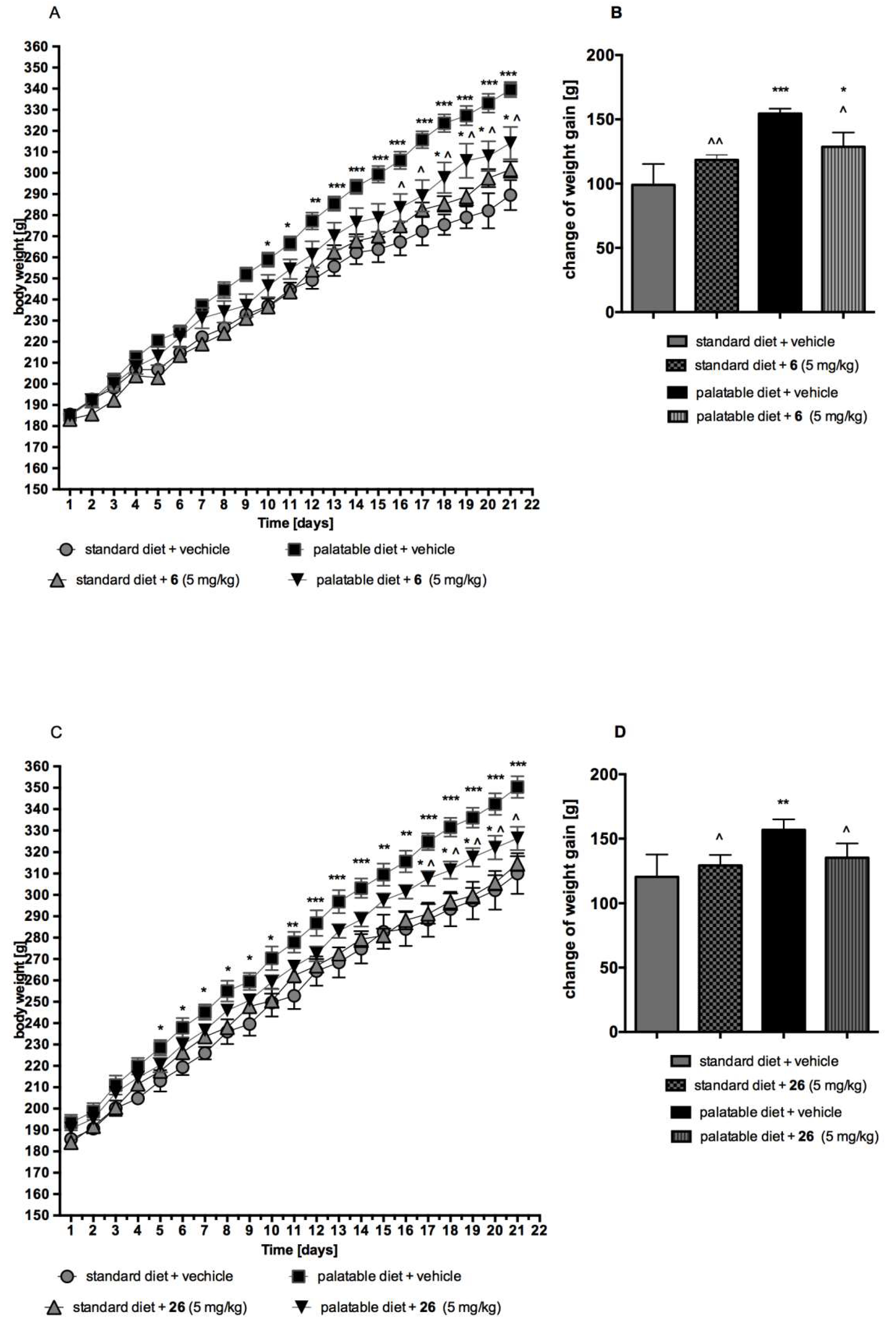
The Effect of compound 6 or 26 on Body Weight by Non-Obese Rats Fed Palatable Diet (Model of Excessive Eating)

In order to determine the anorectic activity of 6 or 26, the model of excessive eating was used [43,44]. Male Wistar rats (170–190 g) were housed in a pair in plastic cages in constant temperature facilities exposed to a light-dark cycle. Water and food were available ad libitum. Two groups of 6 rats were fed with diets consisting of milk chocolate with nuts, cheese, salted peanuts, and 7% condensed milk and also had access to standard feed (Labofeed B, Morawski Manufacturer Feed, Kcynia, Poland) and water ad libitum for 3 weeks. Palatable control group (palatable diet + vehicle) received vehicle (1% Tween 80, intraperitoneally), while palatable test group (palatable diet + 6 or palatable + 26) was injected (intraperitoneally) with 6 or 26 at the dose of 5 mg/kg b.w. dissolved in 1% Tween 80 (1 mL/kg). Body weights were measured daily, immediately prior to administration of drugs.
The palatable diet contained: 100 g peanuts—614 kcal; 100 mL condensed milk—131 kcal; 100 g milk chocolate—529 kcal; 100g cheese (Greek type)—270 kcal. The standard diet contained 100 g feed—280 kcal.

The Effect of compound 6 or 26 on Body Weight by Non-Obese Rats Fed Only with Standard Diet

Male Wistar rats (170–190 g) were housed in a pair in plastic cages in constant temperature facilities exposed to a light-dark cycle. Control group (standard diet + vehicle) received vehicle (1% Tween 80, intraperitoneally), while the test group (standard diet + 6 or standard diet + 26) was injected (intraperitoneally) with 6 or 26 at the dose of 5 mg/kg b.w. dissolved in 1% Tween 80 (1 mL/kg). Body weights were measured daily immediately prior to administration of drugs.

4.6. Statistical Analysis

4.6.1. Lipophilicity

Statistical analysis was performed using Statistica v10 software (StatSoft, Tulsa, OK, USA). The correlation coefficients (r, r2), and the standard errors of the slope, interception, and estimate (Sa, Sb, Se) served as the basis for testing the linearity of regression plots.

4.6.2. Studies In Vitro and In Vivo

Statistical calculations were performed using GraphPad Prism 6 software (GraphPad Software, La Jolla, CA, USA). Results are provided as arithmetic means with a standard error of the mean. The data of behavioral studies were evaluated by an analysis of variance one-way ANOVA followed by Bonferroni’s post hoc test (statistical significance set at p < 0.05). Statistical significance of body weight was calculated using one-way ANOVA with Sidak’s multiple comparison test post-hoc or two-way ANOVA with Sidak’s multiple comparison test post-hoc. Differences were considered statistically significant at: p ≤ 0.05, p ≤ 0.01, p ≤ 0.001.

5. Conclusions

These comprehensive studies, including: Computer-aided design, synthesis, pharmacological screening in vitro and in vivo, “drugability” experiments and an extensive docking-based SAR analysis, have provided an insight into chemical properties, ligand–receptor interactions and potential therapeutic perspectives for the new group of 1,3,5-triazine molecules. Lead structures with two topological variants of the hydantoin linker have been identified, followed by the synthesis of numerous lead modifications. As result, a new series of 18 active 5-HT6 agents, with submicromolar affinities confirmed in the radioligand binding assays, has been obtained (groups A and B). Computer-aided SAR analysis indicates the significance of both, linker and aromatic moiety properties, and their mutual dependences, for the 5-HT6R affinity of the 1,3,5-triazine derived compounds. Thus, the combination of 1-unsubstituted hydantoin (group B) with the β-naphthyl moiety at position 5 (26) was found as the most profitable, followed by either the corresponding α-naphthyl analogue (25) and the tri-substituted combination of 1-(4-chlorobenzyl)-3-((4-amino-6-(4-methylpiperazin-1-yl)-1,3,5-triazin-2-yl)methyl)-5,5-dimethylimidazolidine-2,4-dione (6, group A). The most active agent (26) displayed also antidepressant-like and anxiolytic-like activities in vivo, whereas the best members of both groups (6 and 26) demonstrated anti-obesity properties in animals fed palatable feed, as well as lipophilicity favorable for CNS drugs with a low risk of toxicity in vitro.
The interesting pharmacological properties together with a promising “drugability” designate this group of chemical molecules as a good starting point for further consideration in order to explore molecular mechanisms of the 5-HT6R–ligand interaction, and even to search of new drug candidates for the treatment of depression and obesity. In this context, 3-((4-Amino-6-(4-methylpiperazin-1-yl)-1,3,5-triazin-2-yl)methyl)-5-methyl-5-(naphthalen-2-yl)imidazolidine-2,4-dione (26) can be selected as a new lead structure.

Supplementary Materials

The supplementary materials are available online.

Author Contributions

R.K. and J.H. conceived and designed the article; W.A., D.Ł., M.W. and J.H. performed syntheses; J.H. and K.K.-K. supervised synthesis works; W.A. elaborated synthesis description (paragraph 4.1); R.K. performed molecular modeling studies, G.S. performed radioligand binding assays; M.K. performed metabolic tests in vivo; M.J.W., A.P., and A.W. performed behavioral tests in vivo, A.L. and, G.L. performed toxicity in vitro test; M.S. and M.D. performed lipophilicity experiments, R.K., J.H., W.A., and C.J. wrote the paper.

Funding

This study was financially supported by Polish National Science Centre (NCN) grants: No UMO-2015/17/B/NZ7/02973, UMO-2016/21/N/NZ7/03265 (Toxicity in vitro) and UMO-2014/15/D/NZ7/01782. Authors thank also to the University of Saarland and the Erasmus Plus scheme for financial support of Wesam Ali.

Acknowledgments

Authors thank Andrzej J. Bojarski for the opportunity to conduct RBA and MM studies in his Department. Thanks to our excellent technician, Maria Kaleta and to students: Kinga Półchłopek, Angelika Nowakowska and Raluca Mihaela Șișu, for their contribution in the synthesis work.

Conflicts of Interest

The authors declare no conflict of interest.

References

  1. Barnes, N.M.; Sharp, T. A review of central 5-HT receptors and their function. Neuropharmacology 1999, 38, 1083–1152. [Google Scholar] [CrossRef]
  2. Ruat, M.; Traiffort, E.; Arrang, J.M.; Tardivellacombe, J.; Diaz, J.; Leurs, R.; Schwartz, J.C. A Novel Rat Serotonin (5-HT6) Receptor: Molecular Cloning, Localization and Stimulation of cAMP Accumulation. Biochem. Biophys. Res. Commun. 1993, 193, 268–276. [Google Scholar] [CrossRef] [PubMed]
  3. Kohen, R.; Metcalf, M.A.; Khan, N.; Druck, T.; Huebner, K.; Lachowicz, J.E.; Meltzer, H.Y.; Sibley, D.R.; Roth, B.L.; Hamblin, M.W. Cloning, Characterization, and Chromosomal Localization of a Human 5-HT6 Serotonin Receptor. J. Neurochem. 2002, 66, 47–56. [Google Scholar] [CrossRef]
  4. Liu, K.G.; Robichaud, A.J. 5-HT6 Medicinal Chemistry. Int. Rev. Neurobiol. 2010, 94, 1–34. [Google Scholar] [PubMed]
  5. Benhamú, B.; Martín-Fontecha, M.; Vázquez-Villa, H.; Pardo, L.; López-Rodríguez, M.L. Serotonin 5-HT6 Receptor Antagonists for the Treatment of Cognitive Deficiency in Alzheimer’s Disease. J. Med. Chem. 2014, 57, 7160–7181. [Google Scholar] [CrossRef] [PubMed]
  6. Wesołowska, A. Potential role of the 5-HT6 receptor in depression and anxiety: An overview of preclinical data. Pharmacol. Rep. 2010, 62, 564–577. [Google Scholar] [CrossRef]
  7. Heal, D.; Gosden, J.; Smith, S. The 5-HT6 Receptor as a Target for Developing Novel Antiobesity Drugs. Int. Rev. Neurobiol. 2011, 96, 73–109. [Google Scholar] [PubMed]
  8. Frassetto, A.; Zhang, J.; Lao, J.Z.; White, A.; Metzger, J.M.; Fong, T.M.; Chen, R.Z. Reduced sensitivity to diet-induced obesity in mice carrying a mutant 5-HT6 receptor. Brain Res. 2008, 1236, 140–144. [Google Scholar] [CrossRef] [PubMed]
  9. Dudek, M.; Marcinkowska, M.; Bucki, A.; Olczyk, A.; Kołaczkowski, M. Idalopirdine—A small molecule antagonist of 5-HT6 with therapeutic potential against obesity. Metab. Brain Dis. 2015, 30, 1487–1494. [Google Scholar] [CrossRef] [PubMed]
  10. Krogsgaard-Larsen, N.; Jensen, A.A.; Schrøder, T.J.; Christoffersen, C.T.; Kehler, J. Novel Aza-analogous Ergoline Derived Scaffolds as Potent Serotonin 5-HT6 and Dopamine D2 Receptor Ligands. J. Med. Chem. 2014, 57, 5823–5828. [Google Scholar] [CrossRef] [PubMed]
  11. De la Fuente, T.; Martín-Fontecha, M.; Sallander, J.; Benhamú, B.; Campillo, M.; Medina, R.A.; Pellissier, L.P.; Claeysen, S.; Dumuis, A.; Pardo, L.; et al. Benzimidazole Derivatives as New Serotonin 5-HT6 Receptor Antagonists. Molecular Mechanisms of Receptor Inactivation. J. Med. Chem. 2010, 53, 1357–1369. [Google Scholar] [CrossRef] [PubMed]
  12. Bojarski, A. Pharmacophore Models for Metabotropic 5-HT Receptor Ligands. Curr. Top. Med. Chem. 2006, 6, 2005–2026. [Google Scholar] [CrossRef] [PubMed]
  13. Łażewska, D.; Kurczab, R.; Więcek, M.; Kamińska, K.; Satała, G.; Jastrzębska-Więsek, M.; Partyka, A.; Bojarski, A.J.; Wesołowska, A.; Kieć-Kononowicz, K.; et al. The computer-aided discovery of novel family of the 5-HT6 serotonin receptor ligands among derivatives of 4-benzyl-1,3,5-triazine. Eur. J. Med. Chem. 2017, 135, 117–124. [Google Scholar] [CrossRef] [PubMed]
  14. Latacz, G.; Kechagioglou, P.; Papi, R.; Łażewska, D.; Więcek, M.; Kamińska, K.; Wencel, P.; Karcz, T.; Schwed, J.S.; Stark, H.; et al. The Synthesis of 1,3,5-triazine Derivatives and JNJ7777120 Analogues with Histamine H 4 Receptor Affinity and Their Interaction with PTEN Promoter. Chem. Biol. Drug Des. 2016, 88, 254–263. [Google Scholar] [CrossRef] [PubMed]
  15. Handzlik, J.; Bojarski, A.J.; Satała, G.; Kubacka, M.; Sadek, B.; Ashoor, A.; Siwek, A.; Więcek, M.; Kucwaj, K.; Filipek, B.; et al. SAR-studies on the importance of aromatic ring topologies in search for selective 5-HT7 receptor ligands among phenylpiperazine hydantoin derivatives. Eur. J. Med. Chem. 2014, 78, 324–339. [Google Scholar] [CrossRef] [PubMed]
  16. Łażewska, D.; Więcek, M.; Ner, J.; Kamińska, K.; Kottke, T.; Schwed, J.S.; Zygmunt, M.; Karcz, T.; Olejarz, A.; Kuder, K.; et al. Aryl-1,3,5-triazine derivatives as histamine H4 receptor ligands. Eur. J. Med. Chem. 2014, 83, 534–546. [Google Scholar] [CrossRef] [PubMed]
  17. Grychowska, K.; Kurczab, R.; Śliwa, P.; Satała, G.; Dubiel, K.; Matłoka, M.; Moszczyński-Pętkowski, R.; Pieczykolan, J.; Bojarski, A.J.; Zajdel, P. Pyrroloquinoline scaffold-based 5-HT6R ligands: Synthesis, quantum chemical and molecular dynamic studies, and influence of nitrogen atom position in the scaffold on affinity. Bioorg. Med. Chem. 2018, 26, 3588–3595. [Google Scholar] [CrossRef] [PubMed]
  18. González-Vera, J.A.; Medina, R.A.; Martín-Fontecha, M.; Gonzalez, A.; de la Fuente, T.; Vázquez-Villa, H.; García-Cárceles, J.; Botta, J.; McCormick, P.J.; Benhamú, B.; et al. A new serotonin 5-HT6 receptor antagonist with procognitive activity—Importance of a halogen bond interaction to stabilize the binding. Sci. Rep. 2017, 7, 41293. [Google Scholar] [CrossRef] [PubMed]
  19. Borsini, F.; Meli, A. Is the forced swimming test a suitable model for revealing antidepressant activity? Psychopharmacology (Berlin) 1988, 94, 147–160. [Google Scholar] [CrossRef]
  20. Borsini, F. Role of the serotonergic system in the forced swimming test. Neurosci. Biobehav. Rev. 1995, 19, 377–395. [Google Scholar] [CrossRef]
  21. Wesołowska, A.; Nikiforuk, A.; Stachowicz, K. Anxiolytic-like and antidepressant-like effects produced by the selective 5-HT6 receptor antagonist SB-258585 after intrahippocampal administration to rats. Behav. Pharmacol. 2007, 18, 439–446. [Google Scholar] [CrossRef] [PubMed]
  22. Soczewiński, E.; Wachtmeister, C.A. The relation between the composition of certain ternary two-phase solvent systems and RM values. J. Chromatogr. A 1962, 7, 311–320. [Google Scholar] [CrossRef]
  23. Wager, T.T.; Hou, X.; Verhoest, P.R.; Villalobos, A. Moving beyond rules: The development of a central nervous system multiparameter optimization (CNS MPO) approach to enable alignment of druglike properties. ACS Chem. Neurosci. 2010, 1, 435–449. [Google Scholar] [CrossRef] [PubMed]
  24. Schrödinger Release 2017-3: QikProp; Schrödinger LLC: New York, NY, USA, 2017.
  25. Wilcken, R.; Zimmermann, M.O.; Lange, A.; Zahn, S.; Boeckler, F.M. Using halogen bonds to address the protein backbone: A systematic evaluation. J. Comput. Aided Mol. Des. 2012, 26, 935–945. [Google Scholar] [CrossRef] [PubMed]
  26. Wilcken, R.; Zimmermann, M.O.; Lange, A.; Joerger, A.C.; Boeckler, F.M. Principles and Applications of Halogen Bonding in Medicinal Chemistry and Chemical Biology. J. Med. Chem. 2013, 56, 1363–1388. [Google Scholar] [CrossRef] [PubMed]
  27. Matys, A.; Podlewska, S.; Witek, K.; Witek, J.; Bojarski, A.J.; Schabikowski, J.; Otrębska-Machaj, E.; Latacz, G.; Szymańska, E.; Kieć-Kononowicz, K.; et al. Imidazolidine-4-one derivatives in the search for novel chemosensitizers of Staphylococcus aureus MRSA: Synthesis, biological evaluation and molecular modeling studies. Eur. J. Med. Chem. 2015, 101, 313–325. [Google Scholar] [CrossRef] [PubMed] [Green Version]
  28. Safari, J.; Naeimi, H.; Ghanbari, M.M.; Sabzi Fini, O. Preparation of phenytoin derivatives under solvent-free conditions using microwave irradiation. Russ. J. Org. Chem. 2009, 45, 477–479. [Google Scholar] [CrossRef]
  29. Werbel, L.M.; Elslager, E.F.; Islip, P.J.; Closier, M.D. Antischistosomal effects of 5-(2,4,5-trichlorophenyl)hydantoin and related compounds. J. Med. Chem. 1977, 20, 1569–1572. [Google Scholar] [CrossRef] [PubMed]
  30. Linol, J.; Coquerel, G. Influence of high energy milling on the kinetics of the polymorphic transition from the monoclinic form to the orthorhombic form of (±)5-methyl-5-(4′-methylphenyl)hydantoin. J. Therm. Anal. Calorim. 2007, 90, 367–370. [Google Scholar] [CrossRef]
  31. Keshtov, M.L.; Rusanov, A.L.; Belomoina, N.M.; Mikitaev, A.K. Improved synthesis of bis[p-(phenylethynyl)phenyl]hetarylenes. Russ. Chem. Bull. 1997, 46, 1794–1796. [Google Scholar] [CrossRef]
  32. Safari, J.; Javadian, L. Montmorillonite k-10 as a catalyst in the synthesis of 5,5-disubstituted hydantoins under ultrasound irradiation. J. Chem. Sci. 2013, 125, 981–987. [Google Scholar] [CrossRef]
  33. Sherman, W.; Day, T.; Jacobson, M.P.; Friesner, R.A.; Farid, R. Novel Procedure for Modeling Ligand/Receptor Induced Fit Effects. J. Med. Chem. 2006, 49, 534–553. [Google Scholar] [CrossRef] [PubMed]
  34. Friesner, R.A.; Banks, J.L.; Murphy, R.B.; Halgren, T.A.; Klicic, J.J.; Mainz, D.T.; Repasky, M.P.; Knoll, E.H.; Shelley, M.; Perry, J.K.; et al. Glide: A New Approach for Rapid, Accurate Docking and Scoring. 1. Method and Assessment of Docking Accuracy. J. Med. Chem. 2004, 47, 1739–1749. [Google Scholar] [CrossRef] [PubMed]
  35. Kooistra, A.J.; Kuhne, S.; De Esch, I.J.P.; Leurs, R.; De Graaf, C. A structural chemogenomics analysis of aminergic GPCRs: Lessons for histamine receptor ligand design. Br. J. Pharmacol. 2013, 170, 101–126. [Google Scholar] [CrossRef] [PubMed]
  36. Schrödinger Release 2017-3: LigPrep; Schrödinger LLC: New York, NY, USA, 2017.
  37. Schrödinger Release 2017-3: Epik; Schrödinger LLC: New York, NY, USA, 2017.
  38. Harder, E.; Damm, W.; Maple, J.; Wu, C.; Reboul, M.; Xiang, J.Y.; Wang, L.; Lupyan, D.; Dahlgren, M.K.; Knight, J.L.; et al. OPLS3: A Force Field Providing Broad Coverage of Drug-like Small Molecules and Proteins. J. Chem. Theory Comput. 2016, 12, 281–296. [Google Scholar] [CrossRef] [PubMed]
  39. Schrödinger Release 2017-3: Glide; Schrödinger LLC: New York, NY, USA, 2017.
  40. Cheng, Y.-C.; Prusoff, W.H. Relationship between the inhibition constant (KI) and the concentration of inhibitor which causes 50 per cent inhibition (I50) of an enzymatic reaction. Biochem. Pharmacol. 1973, 22, 3099–3108. [Google Scholar] [PubMed]
  41. Porsolt, R.D.; Bertin, A.; Jalfre, M. “Behavioural despair” in rats and mice: Strain differences and the effects of imipramine. Eur. J. Pharmacol. 1978, 51, 291–294. [Google Scholar] [CrossRef]
  42. Vogel, J.R.; Beer, B.; Clody, D.E. A simple and reliable conflict procedure for testing anti-anxiety agents. Psychopharmacologia 1971, 21, 1–7. [Google Scholar] [CrossRef] [PubMed]
  43. Kotańska, M.; Lustyk, K.; Bucki, A.; Marcinkowska, M.; Śniecikowska, J.; Kołaczkowski, M. Idalopirdine, a selective 5-HT6 receptor antagonist, reduces food intake and body weight in a model of excessive eating. Metab. Brain Dis. 2018, 33, 733–740. [Google Scholar] [CrossRef] [PubMed]
  44. Kotańska, M.; Śniecikowska, J.; Jastrzębska-Więsek, M.; Kołaczkowski, M.; Pytka, K. Metabolic and Cardiovascular Benefits and Risks of EMD386088—A 5-HT6 Receptor Partial Agonist and Dopamine Transporter Inhibitor. Front. Neurosci. 2017, 11, 50. [Google Scholar] [CrossRef] [PubMed]
Sample Availability: Not available.
Figure 1. Modified pharmacophore model based of the 1,3,5-triazine with mapped compound 2, including four key pharmacophore features, namely: A positive ionizable group (PI, red), an aromatic ring (AR, orange), a hydrogen bond acceptor group (HBA, green) and hydrophobic group (HYD, blue) (A). The lead compound 2 mapped onto the binding site of 5-HT6R (B). Examples of the most active 1,3,5-traizne 5-HT6 ligands found recently 13 (C).
Molecules 23 02529 g001
Figure 2. The rationale for the design of novel lead structures of the 5-HT6R ligand among hydantoin 1,3,5-triazine derivatives.
Molecules 23 02529 g002
Scheme 1. Synthesis of compounds 526 (Table 1); (a) 3-arylmethylhydantoin product 5; (b) 1-arylmethylhydantoin derivatives (6, 918); (c) 5-aryl-5-methylhydantoin derivatives (7, 1926); (d) 5,5-diarylhydantoin derivative and triazines (8). Reagents and conditions: (i) RCH2Cl, K2CO3, TEBA, acetone, 3–5 h reflux; (ii) BrCH2COOMe, K2CO3, TEBA, acetone, 5–7 h reflux; (iii) 1-(imino(4-methylpiperazin-1-yl)methyl)guanidine hydrochloride, MeONa, reflux for 3–5 h, then rt, 10–20 h; (iv) KCN, (NH4)2CO3, 50% EtOH, 55 °C; (v) KCN, EtOH, 1 h reflux; (vi) HNO3 conc., 3 h reflux; (vii) EtONa, urea, 2 h reflux, HCl.
Molecules 23 02529 sch001
Figure 3. Influence of compound 26 on the immobility time of rats in the forced swim test. Compound 26 was administered i.p. 60 min before the test. The animals were observed for 5 min. The data are presented as the mean ± SEM of 6–8 rats. The data were statistically evaluated by one-way ANOVA followed by Bonferroni’s post-hoc test, * p < 0.05 vs. vehicle group.
Molecules 23 02529 g003
Figure 4. Compound 26 increases the number of shocks accepted in the Vogel conflict test in rats. Compound 26 was administered i.p. 60 min before the test. The animals were monitored for 5 min. The data are presented as the mean ± SEM of 6–8 rats. The data were statistically evaluated by one-way ANOVA followed by Bonferroni’s post-hoc test, * p < 0.05 vs. vehicle group.
Molecules 23 02529 g004
Figure 5. Influence of long-term administration of 6 or 26 on the body weight in male Wistar rats. The change in body weight in Wistar rats fed palatable diet or standard diet and in Wistar rats fed palatable diet or standard diet treated for 21 days with 6 (A,B) or 26 (C,D). Results are means ± SEM, n = 6. Multiple comparisons were performed by two-way ANOVA, Sidak post-hoc (A,C) or by one-way ANOVA, Sidak post-hoc (B,D); * p < 0.05, ** p < 0.01, *** p < 0.001 significant vs. control rats fed standard diet; ^ p < 0.05, ^^ p < 0.01 significant vs. control rats fed palatable diet.
Molecules 23 02529 g005
Figure 6. Lipophilicity parameters evaluated using R-TLC: RM0 values for compounds 13, 58, 18, 23, 25 and 26.
Molecules 23 02529 g006
Figure 7. The influence of doxorubicin (DX) and 5-HT6R ligands on the viability of cultured HEK-293 cells. Statistical significance was evaluated by one-way ANOVA, followed by Bonferroni’s comparison test (*** p < 0.001 compared with the negative control).
Molecules 23 02529 g007
Figure 8. Molecular docking results of the representative library members for the 5-HT6 receptor. (A) Comparison of the binding mode of compounds 6 (cyan) and 7 (gray). (B) Binding mode of 18 (orange) and 26 (blue). (C) A superposition of the poses of compounds 9 (magenta), 17 (yellow), 6 (cyan), and 10 (violet) against the putative halogen binding pocket interaction spheres for the 5-HT6 receptor. The chlorine-oxygen theoretical interaction spheres illustrate the projected qualities of the formed ligand-receptor halogen bonds. Key residues in the binding site are presented as thick dark-grey sticks. The dotted yellow lines represent hydrogen bonds with polar residues.
Molecules 23 02529 g008
Table 1. Structures and affinities for serotonin/dopamine receptors of hydantoin-triazine compounds 426.
Molecules 23 02529 i001
CpdGrRKi (nM) a
5-HT6 [3H]-LSDD2 [3H]-Raclopride5-HT1A [3H]-8-OH-DPAT5-HT2A [3H]-Ketanserin5-HT7 [3H]-5-CT
4BMethyl42722,3309.99 × 106nt35,790
5--4275500715,430nt1620
6A4-Chlorophenyl127409823,300nt3711
7B4-Chlorophenyl17631,24028,070nt938
8--105868553052nt594
9APhenyl592878342,6301596350
10A3-Chlorophenyl271463113,570328159
11A2,5-dichlorophenyl59728674155809279
12A2,4-dichlorophenyl205681769335891116
13A4-Fluorophenyl663655748,3603548953
14A3-Fluorophenyl447657737,09023988
15A3-Bromophenyl570667712,9301327571
16A3-Methoxyphenyl69215,49023,91020363587
17A3-Methylphenyl895520716,990817420
18A2-Naphthyl182888571819071525
19BPhenyl72641,04031,62012,1501096
20B3-Chlorophenyl41816,78041017396791
21B2,5-dichlorophenyl40311,55026,19011,0501565
22B2,4-dichlorophenyl45715,09013,98010,77010,130
23B2,3,4-trichlorophenyl1954738602834338905
24B4-Methylphenyl66723,30022,32024,410564
25B1-Naphthyl1217811714680803367
26B2-Naphthyl87424714,16017,170514
Ref. 7 b9 b20 c-18 d
a Tested experimentally in the radioligand binding assay, binding affinity, Ki, expressed as the average of at least two independent experiments; nt—not tested. b–d Reference ligands for GPCRs investigated, b olanzapine, c buspirone, d clozapine.
Table 2. The blood-brain barrier permeability parameter (QPlogBB) for compounds 426.
CpdQPlogBBCpdQPlogBBCpdQPlogBB
4−0.9712−0.3320−0.63
5−0.6013−0.7321−0.46
6−0.6014−0.4622−0.47
7−0.8915−0.6523−0.38
8−0.6216−0.9224−0.84
9−0.8417−0.5425−0.98
10−0.4018−0.6326−0.92
11−0.3719−0.75

