Abstract
The degree of hippocampal dentation, a morphologic feature of the inferior surface of the human hippocampus, has been positively associated with episodic memory performance in healthy adults. This study examined hippocampal dentation in healthy children and adolescents. The Cincinnati MR Imaging of NeuroDevelopment (C-MIND) dataset was used to examine the relationship between age and hippocampal dentation in 90 healthy children, age < 1 to 18 years old, using T1-weighted MPRAGE scans. Hippocampal dentation was assessed by counting the number of dentes for the left and right hippocampi. Participants had slightly more left than right hippocampal dentes, on average. Dentation did not differ significantly between males and females. Correlational analyses revealed that the numbers of left, right, and total dentes were positively associated with age in this sample. Interestingly, these data highlight the wide variability of dentation in older age groups. While younger children tended to have absent or few dentes, a range of dentation was present in older children and adolescents (ranging from absent to numerous, bilaterally). This is consistent with previous research in a healthy adult cohort, where a range of dentation was also observed. This study is the first to examine hippocampal dentation in children.
1. Introduction
Human hippocampal anatomy is particularly complex, given its unique shape and layered cellular structure. A large portion of this structural development occurs during gestation, and by 34 weeks gestational age, hippocampal subfields (i.e., subiculum, CA1, CA2, and CA3) are distinguishable and the dentate gyrus has developed an adult-like appearance [,]. Postnatal change includes decreases in cell density [] and myelination of the hippocampal formation, continuing well into adulthood []. Regarding volume, several studies indicate a non-linear relationship between hippocampal volume and age in children [,,], with increases early in childhood followed by relative stability later in childhood [] and during adolescence []. Despite reported changes in volume, no significant morphological changes are known to occur from childhood into adulthood [].
Hippocampal dentation is an aspect of hippocampal morphology that has not been previously examined in children. Hippocampal dentation, present on the inferior surface of the hippocampus (primarily CA1/subiculum layer), has been described in few existing studies in adults [,,,], and has been mentioned only briefly in anatomical references [,,]. This region has only recently been the target of morphological measurement via high-resolution neuroimaging techniques []. Hippocampal dentation has been referred to variously as “folds of CA1” [], and “digitations” or “digitationes hippocampi” [,]; however, we differentiate hippocampal dentation, which is a ridged contour on the inferior aspect of the hippocampus that varies considerably across individuals [], from digitation, which is seen ubiquitously on the anterior superior aspect of the hippocampal head []. Research has shown that healthy adults exhibit wide variability in the quantity and prominence of hippocampal dentation, including the complete absence of any observable dentation in 9% of a healthy adult sample. Hippocampal dentation has also recently been shown to be asymmetrically associated with hippocampal sclerosis in temporal lobe epilepsy (TLE), with 70% of the affected hippocampi showing nearly complete loss of dentes []. Interestingly, loss of dentation was associated with the epileptic hippocampus, even after correcting for volume loss, suggesting that loss of dentation in TLE is part of the pathologic process and not simply a secondary effect of hippocampal volume loss. Additionally, hippocampal dentation was associated with aspects of both verbal and visual memory performance in a healthy adult cohort, highlighting the functional significance of this structural feature []. The present study is the first to examine the development of hippocampal dentation in children and adolescents. Given that (1) postnatal change has been documented within the hippocampus and (2) an increase in cortical gyrification has been observed during childhood and adolescence [], we hypothesized that hippocampal dentation would increase during this developmental period (i.e., birth to 18 years old).
In addition, given that previous research has shown gender differences in the total volume and developmental trajectory of the hippocampus in males and females [,,], and that males had a greater degree of dentation than females in a healthy adult cohort [], this study also examined whether hippocampal dentation differs between males and females in a younger age group.
2. Materials and Methods
2.1. Data Acquisition (C-MIND)
The data presented in this work were obtained from the Cincinnati MR Imaging of NeuroDevelopment (C-MIND) database, provided by the Pediatric Functional Neuroimaging Research Network at https://research.cchmc.org/c-mind/ (accessed on 16 March 2017). This Network and the resulting C-MIND database were supported by a contract from the Eunice Kennedy Shriver National Institute of Child Health and Human Development (HHSN275200900018C). Participants were native English speakers with no history of neurological or psychiatric disease in the child or first-degree relatives. Exclusionary criteria included the following: gestation outside 37 to 42 weeks, birth weight less than the 10th percentile, body mass index outside the 5th to 95th percentiles, chronic illness, a school grade average of D+ or below, special education placement based on ability or behavior, previous head trauma, abnormal neurological examination, head circumference outside the 5th to 95th percentiles, and abnormal MRI findings. Please see the C-MIND User Manual, available online at https://research.cchmc.org/c-mind/manual-project-overview (accessed on 16 March 2017), for more information on the study protocol and data collection, which were approved by the Institutional Review Board at Cincinnati Children’s Hospital Medical Center (CCHMC). Data were used in the current study with permission from the Pediatric Neuroimaging Research Consortium, as well as the Institutional Review Board at the University of Alabama at Birmingham.
2.2. Sample Demographics
Ninety healthy, right-handed children, between birth and 18 years old were randomly selected from the C-MIND database (age rangesample: 0 years, 9 months to 18 years, 10 months). Scan quality was rated 0–3 by researchers at the C-MIND Consortium (0 = perfect and 3 = poor). For this study, only scans of quality 0 (“perfect data”) or 1 (“good image quality and contrast”) were considered for inclusion in order to adequately visualize hippocampal dentation (see Section 8: Quality Assurance Procedures of the C-MIND User Manual at https://research.cchmc.org/c-mind/8-quality-assurance-procedures (accessed on 16 March 2017) for more information).
The sample size was based on feasibility and scan quality, with up to five participants selected at each age, from birth to 18 years old. Scan quantity and/or quality were limited in several age groups, including less than one year of age (2 scans available), 17-year-olds (4 scans), and 18-year-olds (4 scans). For all other age groups, random selection was used to identify five participants for inclusion. The sample included 50 females (56%) and 40 males. Of note, the database uses the term “gender” and does not differentiate gender from biological sex, therefore the data and results are presented using the term gender. Parent race was reported as follows: 66% of mothers in the study were Caucasian, 29% were African American, 1% were Asian, 1% were biracial, and 3% were not reported; fathers’ race included 62% Caucasian, 32% African American, 1% Asian, and 4% not reported. Participant ethnicity included 6% Hispanic or Latino. Annual household income was reported in ranges ($0–5000 to over $150,000) and median household income was $50,000–75,000 in this sample. For more information on sample household income as well as parental education level, see Table A1 and Table A2.
2.3. Neuroimaging
Neuroimaging data included an anatomical MRI scan (acquisition time: approximately 38 min). The protocol included a desensitization period to acclimate participants to the scanning environment, described previously [,] and outlined online at https://research.cchmc.org/c-mind/visitors/preparing (accessed on 16 March 2017). A Philips 3T Achieva scanner with 32-channel head coil was used to acquire 3D T1-weighted anatomical MPRAGE scans (1 × 1 × 1 mm3). Scans were visualized using 3D Slicer software, Version 4.5.0.
2.4. Dentation Assessment
Hippocampal dentation, present in the CA1/subiculum of the human hippocampus, was assessed in this study by counting the number of hippocampal dentes for the left and right hippocampus of each participant individually (Figure 1, Figure 2 and Figure 3). Hippocampal dentation was visualized in the sagittal plane, using all available sagittal slices to determine the total number of dentes for each hippocampus. Hippocampal dentation was described previously in adults using ultra high-resolution structural neuroimaging (HR-MICRA technique with 0.5 × 0.5 × 0.75 mm3 resolution) []. While a previous study described hippocampal dentation in terms of both quantity and prominence, in this study, dentation was measured by quantity only, adapted due to the reduced visibility of dentation and the SRLM layer (stratum radiatum, lacunosum, and moleculare) in this scan resolution. See Beattie and colleagues (2017) for more detailed methodology on the previous study. In the current study, right and left hippocampal dentes were counted for all participants by a researcher trained in hippocampal anatomy (J.F.B.). The main analyses were conducted using these results. Approximately 25% of scans (n = 24) were counted by two additional trained researchers (T.A.C. and R.Q.J.) to assess inter-rater reliability. All raters were blinded to participant variables, including age, while counting dentation.
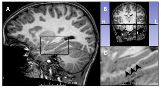
Figure 1.
Hippocampal dentation visible in a 7-year-old participant. (A) Sagittal view, with the left hippocampus shown in the black box. (B) Coronal view, with corresponding sagittal placement of the viewing panes in (A,C). A larger image of the hippocampus is visible in (C), with black arrowheads indicating three dentes visible in this sagittal slice. All available sagittal slices were used to count the dentes for each hippocampus.
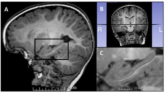
Figure 2.
(A) Sagittal view of the left hippocampus of a 9-month-old participant. The black box highlights the location of the hippocampus in this view. (B) Coronal view indicating the lateral placement of the sagittal plane seen in (A,C). (C) A more detailed view of the left hippocampus for this participant, with no visible dentes and the smooth contour of the inferior aspect of the hippocampus traced.
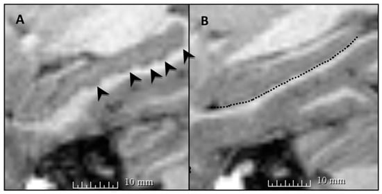
Figure 3.
Both (A,B) show sagittal views of left hippocampi from two different 18-year-old participants. (A) A hippocampus with multiple dentes (indicated by black arrowheads), and (B) a relatively flat hippocampus, with the inferior surface traced by the black dotted line. This highlights the variability in hippocampal dentation observed among healthy individuals of the same age. Hippocampal dentation is pictured here in a single representative sagittal slice for each participant.
2.5. Experimental Design and Statistical Analysis
Visual inspection of bivariate scatter plots revealed a linear trend between the number of dentes and age. Independent and dependent variables had skewness and kurtosis values less than the absolute value of 2.0. No multivariate outliers were found for the relationships between demographic variables (i.e., age, height, and weight) and left, right, or total number of dentes (Mahalonobis’ Distance of less than 15.0 and Cook’s Distance less than 1.0). The independence of residuals was observed, based on a Durbin–Watson test statistic close to 2. Normality of residuals was assessed for the relationship between age and number of left, right, and total dentes using the Kolmogorov–Smirnov test for normality. Normality was violated for the relationship between age and number of left dentes, but the null hypothesis of a normal distribution was retained for the relationship between age and number of right dentes and number of total dentes. Visual inspection of bivariate scatter plots of the residual and predicted data points revealed mild heteroscedasticity in the data. Square root and logarithmic transformations were explored; however, homoscedasticity was not achieved with these transformations, therefore, the data were analyzed in their original form to preserve interpretation. Implications were considered. Statistical analysis was performed in SPSS.
There were no missing data for the primary variables of interest, including hippocampal dentation (right, left, and total) and participant age. Birth weight, current height, and current weight were not available for a small number of individuals (see Table 1); however, the number of individuals with missing data points was small and scattered among age groups (<3% of individuals with missing data; see Table 1); therefore, the impact of these missing data was considered minimal [].

Table 1.
Pearson correlations: hippocampal dentation and demographics.
Reliability of dentation assessment at this age and scan resolution was measured using an intra-class correlation coefficient ICC (3,1) to assess the absolute agreement for two additional raters (T.A.C. and R.Q.J.) with the dentation counts used in the main analyses (J.F.B.), according to methods reported elsewhere []. The analysis included left and right dentes, considered together. Agreement between all three raters was 0.53, with a 95% confidence interval (CI) (0.29, 0.70), and was considered fair. When considered individually, ICCs between rater J.F.B. and raters T.A.C. and R.Q.J. were 0.62 and 0.70, which is considered to be a good reliability [].
3. Results
A range of dentation was observed for left (range: 0–8, = 2.82, s = 1.79), right (range: 0–7, = 2.44, s = 1.69), and total dentes (range: 0–14, = 5.27, s = 3.17). Two-tailed Pearson correlational analyses were conducted to examine the relationship between hippocampal dentation and age in this sample. Analyses revealed a significant positive association between age and number of left (r = 0.34, p = 0.001), right (r = 0.31, p = 0.003), and total dentes (r = 0.36, p = 0.001) (Figure 4 and Figure 5). The means and standard deviations for number of hippocampal dentes by age group are included in Table A3.
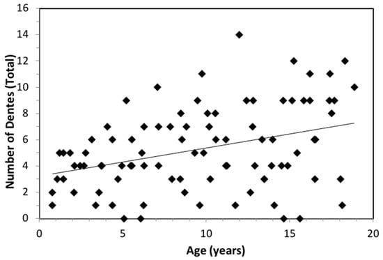
Figure 4.
The relationship between total number of hippocampal dentes and age (n = 90), with a linear trend line (p = 0.001). Of note, the variability in number of dentes is high, particularly in older age groups. Individuals with few or no dentes are also seen across the age range.
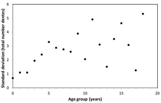
Figure 5.
Scatter plot showing the correlation between standard deviation of the number of total hippocampal dentes and age group. The larger standard deviation among older age groups indicates greater variability in the number of hippocampal dentes among older children/adolescents. A significant positive association was found between age group and standard deviation (p = 0.01). The range in variability for older age groups may be observed due to the small sample size in each age group.
Visual inspection indicated that variability in dentation appeared to increase with age, such that there was more variability in number of dentes in older children and adolescents when compared with infants and young children. Given the increasing variability in the number of dentes with participant age noted in bivariate scatter plots (Figure 4), an additional analysis was conducted to determine whether there was a significant relationship between variability in hippocampal dentation (i.e., standard deviation) and age group. This analysis, consisting of a two-tailed Pearson correlation, revealed a significant positive association between the age group and standard deviation of the number of total hippocampal dentes (r = 0.56, p = 0.01) (Figure 5).
A paired samples t-test was conducted to determine whether there was a significant difference between the number of left and right hippocampal dentes in the sample. Individuals in this sample tended to have more left than right dentes (difference score (L–R): = 0.38, s = 1.44), t(89) = 2.49, p = 0.02 (Figure 6).
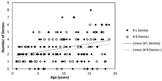
Figure 6.
Association between number of left and right dentes individually with age, displayed with linear trend lines. Paired samples t-test revealed a greater number of left dentes for participants, on average.
Two-tailed Pearson correlations were conducted to examine the relationship between the number of hippocampal dentes and demographic variables, including birth weight, current height, and current weight. Analyses revealed significant associations between the number of left, right, and total dentes with height (p < 0.05), but not with current weight or birth weight (p > 0.05) (Table 1).
Independent samples t-tests were conducted to determine whether hippocampal dentation differed between males and females. No significant differences were observed between groups, including the number of left dentes between males ( = 3.05, s = 1.77) and females ( = 2.64, s = 1.80); t(88) = 1.08, p = 0.28, number of right dentes between males ( = 2.75, s = 1.79) and females ( = 2.20, s = 1.58), t(88) = 1.55, p = 0.13, or number of total dentes between males ( = 5.80, s = 3.17) and females ( = 4.84, s = 3.13) in this sample, t(88) = 1.44, p = 0.15 (see Figure 7).
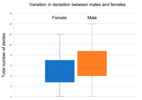
Figure 7.
The range of hippocampal dentation among males and females. No significant gender differences were observed (p > 0.05).
4. Discussion
4.1. Hippocampal Dentation and Age
Based on a sample of healthy children and adolescents (age 9 months to 18 years old), hippocampal dentation showed a small positive correlation with age, such that older children had a larger number of dentes, on average. This does not appear to directly mirror volumetric changes, which broadly exhibit relative stability following early childhood [,]. This differential developmental pattern between hippocampal dentation and volume appears to be generally consistent with past research in adults, which found no association between hippocampal dentation and hippocampal volume []. Overall, hippocampal dentation describes shape of the hippocampus rather than the size, and does not appear to be a redundant or surrogate measure of volume. In addition, this finding of a greater number of hippocampal dentes with age introduces a potential form of morphological change that occurs during childhood and adolescence, previously believed to be minimal in the human hippocampus []; however, conclusions here are limited by a cross-sectional study design. Given the functional significance of hippocampal dentation in adults, it is important to describe the typical developmental trajectory of dentation—that is, describing when dentation first develops and whether this continues throughout the lifespan. At the same time, there seems to be a significant degree of individual variability, such that not all adults show visible dentation.
4.2. Variability of Hippocampal Dentation by Age
While primary analyses revealed a significant positive association between age and number of hippocampal dentes, visual inspection of the data (see Figure 4) indicated that while younger children tend to have fewer hippocampal dentes, older children and adolescents display wide variability in the number of hippocampal dentes. This was supported by a post-hoc analysis, which revealed a positive association between age group and variability in hippocampal dentation (i.e., standard deviation). The observed degree of variability found in our current study is consistent with a previous finding in a healthy adult cohort, where a wide range of hippocampal dentation was observed []. In a study examining volume, substantial variability in hippocampal volume was observed in healthy children aged 4–18 years old; however, the results did not indicate whether the distribution of hippocampal volume varied with age [].
4.3. Hippocampal Dentation and Gender
Previous research varies in both methodology and reporting when analyzing sex and/or gender differences [,,]. No gender differences were observed between males and females in the right, left, or total number of hippocampal dentes in this sample. Gender differences were previously observed in healthy adults, with males having a greater degree of right and left hippocampal dentation []. Observed differences in the two studies could be attributable to age, with gender differences emerging during adulthood. Alternatively, both prominence and quantity were examined in the previous study, thus it is possible that males have a tendency to have more arciform (prominent) dentes, where females have a higher rate of sinusoidal dentes, leading researchers to detect gender differences using one methodology (i.e., the rating system in the previous study) and not the other (i.e., counting dentes used in the present study). More research is needed to elucidate sex and gender differences in hippocampal dentation across the lifespan.
4.4. Hippocampal Dentation and Demographics
In addition to showing a positive relationship with age, the quantity of hippocampal dentes (right, left, and total) was associated with height in this sample. This is not surprising given the strong correlation between age and height in children. No significant correlation was found with weight (current nor birth weight).
Interestingly, prematurity and extremely low birth weight have been associated with memory difficulties during childhood [,], which may persist into adulthood [], justifying an investigation of hippocampal dentation related to birth weight or gestational age. In our study sample, there was no relationship between birth weight and hippocampal dentation; however, due to the study aims of investigating typical development, children with birth weight less than the 10th percentile, as well as those born prematurely (<37 weeks), were excluded from the study. The resulting data were therefore truncated in these parameters. This could lead to limited power to detect an effect related to hippocampal dentation. Future research may examine this relationship in a sample of individuals who were born prematurely and/or at a very low or extremely low birth weight.
4.5. Reliability of Dentation Assessment
Reliability analysis was conducted to determine the consistency of counting dentes at this resolution and in this age range. In a previous study with healthy adults [], reliability was excellent, ICC (3,1) = 0.94, 95% CI (0.89–0.96). Based on non-overlapping confidence intervals, we can deduce that the ICC in the previous adult study was higher than the current study. While the reliability in this study was good, the ICC values may be relatively lower when compared to the previous study due to the decreased scan resolution and contrast, as well as the younger age range. Given that dentation is associated with age, it is possible that dentation “emerges” during childhood. That is, the prominence of dentes may increase over time (although the current study did not address this hypothesis), making it difficult to distinguish when to “count” a dente (i.e., a threshold). This could contribute to less absolute agreement between raters when counting hippocampal dentation. Additionally, a fixed voxel size appears relatively larger compared to overall hippocampal size in a smaller brain (e.g., infants), making visualization of hippocampal dentation more difficult, assuming the size of the dentes is proportional to the overall hippocampal size. Finally, the methodology differed between the two studies (counting dentes vs. rating quantity and prominence). Reliability in this study was classified as “fair” to “good” and was considered acceptable for measuring this construct; however, the lower reliability in this sample provided support for developing more objective techniques to measure hippocampal dentation.
4.6. Limitations
The limitations of this study included a lower scan resolution compared to previous work examining hippocampal dentation. This presented methodological challenges; however, this scan resolution was determined to be acceptable for viewing the quantity of hippocampal dentes and provided significantly more generalizability for studying hippocampal dentation. Scan acquisition time for an ultra-high resolution neuroimaging session may present substantial barriers, particularly in studying a population of infants and young children. Examining dentation in this image resolution also allows for more readily available clinical use and the interpretation of this construct, in addition to larger sample sizes in research.
Although this study had a relatively large sample size and was well-powered to detect a main effect of age, this sample included a relatively small number of individuals in each age group, particularly in very young children (i.e., less than 12 months of age). Due to rapid brain development during the first years of life, it would be interesting to study additional individuals in this age range so as to better describe characteristics of hippocampal dentation in the early years of life. Sample diversity was generally representative of the region; however, increasing the sample diversity would help detect potential differences in development across groups. The cross-sectional design of this study limits conclusions about developmental trajectory over time.
Participant gender was one of the demographic variables collected in the data set utilized in this study, but was not differentiated from biological sex. As sex (biological descriptive term) and gender (socially-defined construct) are not synonymous, we were unable to explore the nuances of these relationships as part of this study. While we saw no significant gender differences in hippocampal dentation, we cannot definitively rule out sex differences within this sample. This should continue to be explored in future research.
4.7. Future Directions
Future work should examine the development of dentation in longitudinal samples to better assess stability and/or development of this construct over time within individuals. Furthermore, the quantity of hippocampal dentation was related to age in children and adolescents, but not in adulthood, when focusing on early to middle adulthood []. It would be interesting to examine this relationship in older adults to determine whether loss of hippocampal dentation occurs during healthy aging, or whether this construct is stable after initial development early in life.
Thus far, hippocampal dentation has been examined primarily in healthy populations to describe the typical development and course of this morphological feature. Important future directions include examining this construct in individuals with neurological conditions, including those where memory impairment is pronounced (e.g., temporal lobe epilepsy, Alzheimer’s disease), such as the work recently established by Ramaniharan and colleagues []. Additionally, the examination of this feature in individuals with neurodevelopmental differences may shed light on whether this feature plays a role in neurodevelopmental disorders.
Future work should also explore genetic and/or environmental contributors to the development of dentation, including factors such as family socioeconomic status, parent educational attainment, and variables exploring quality of education, as well as parental dentation.
Finally, future directions include the development of a more objective and less time-intensive method to assess hippocampal dentation. Ideally, this measure would incorporate both quantity and prominence, and would be available for use in a relatively accessible scan resolution (e.g., MPRAGE). Recent work towards this effort has been reported []. The lower ICCs in this study, while still acceptable, suggests that objective methods would be particularly beneficial for infants and young children, where hippocampal dentation could be more subtle and voxel size appears to be relatively larger.
5. Conclusions
This study provides the first examination of hippocampal dentation in children and adolescents, to the best of our knowledge. The aim was to describe the development of hippocampal dentation in this age range. The results revealed that there is a significant positive association between age and number of hippocampal dentes (left, right, and total). Hippocampal dentation was not related to gender in this sample. Individuals had slightly more left than right dentes. Interestingly, variability in the number of dentes increased significantly with age, such that older children and adolescents displayed a wide range in number of hippocampal dentes, ranging from absent to numerous in both the left and right hippocampi. These results are consistent with a description of hippocampal dentation in a sample of healthy adults, which indicated wide variability in the degree of hippocampal dentation.
Author Contributions
Conceptualization, J.F.B., R.C.M., M.D.T. and R.K.K.; methodology, J.F.B., R.C.M., E.W.C.III, M.D.T. and L.W.V.H.; validation, J.F.B., R.Q.J., T.A.C. and L.W.V.H.; formal analysis, J.F.B., R.C.M., E.W.C.III and L.W.V.H.; investigation, J.F.B., R.C.M. and L.W.V.H.; resources, R.C.M. and L.W.V.H.; data curation, J.F.B., R.Q.J. and T.A.C.; writing—original draft preparation, J.F.B., R.C.M. and L.W.V.H.; writing—review and editing, J.F.B., R.C.M., M.D.T., R.K.K., A.K.R. and L.W.V.H.; visualization, J.F.B., A.K.R. and L.W.V.H.; supervision, R.C.M. and L.W.V.H.; project administration, J.F.B.; funding acquisition, J.F.B., R.C.M. and L.W.V.H. All authors have read and agreed to the published version of the manuscript.
Funding
This research was supported by the Civitan Foundation through a Civitan Emerging Scholars research grant, awarded to the first author.
Institutional Review Board Statement
The study was conducted in accordance with the Declaration of Helsinki, and was approved by the Institutional Review Board of the University of Alabama at Birmingham (protocol number E160205010 (exempt) and date IRB designation issued: 3/24/16).
Informed Consent Statement
Informed consent was obtained from all subjects involved in the study. Informed consent was conducted at the original data collection site.
Data Availability Statement
The CincinnatiMR Imaging of NeuroDevelopment (C-MIND) database is publicly accessible online at http://research.cchmc.org/c-mind (accessed on 16 March 2017).
Acknowledgments
Special thanks to Jerzy Szaflarski, M.D., Ph.D., and Jane Allendorfer, Ph.D., for their consultation and support. We would also like to thank the C-MIND Authorship Consortium at Cincinnati Children’s Hospital Medical Center (CCHMC) for access to this data set and support in obtaining this data for our study.
Conflicts of Interest
The authors declare no conflict of interest. The funders had no role in the design of the study; in the collection, analyses, or interpretation of data; in the writing of the manuscript; or in the decision to publish the results.
Appendix A
Table A1.
Parents’ educational attainment.
Table A1.
Parents’ educational attainment.
| Mothers | Fathers | |
|---|---|---|
| Less than high school | 4% (n = 4) | 7% (n = 6) |
| High school | 17% (n = 15) | 23% (n = 21) |
| Some college | 19% (n = 17) | 24% (n = 22) |
| College | 30% (n = 27) | 27% (n = 24) |
| Some graduate level | 6% (n = 5) | (n = 0) |
| Graduate level | 21% (n = 19) | 13% (n = 12) |
| Not reported | 3% (n = 3) | 6% (n = 5) |
Table A2.
Annual household income.
Table A2.
Annual household income.
| Percent of Sample | |
|---|---|
| $0–$5000 | 12% |
| $5000–$10,000 | 1% |
| $10,000–$15,000 | 6% |
| $15,000–$25,000 | 8% |
| $25,000–$35,000 | 8% |
| $35,000–$50,000 | 10% |
| $50,000–$75,000 | 14% |
| $75,000–$100,000 | 21% |
| $100,000–$150,000 | 11% |
| Over $150,000 | 4% |
| Not reported | 4% |
Table A3.
Descriptive statistics: hippocampal dentation by age group.
Table A3.
Descriptive statistics: hippocampal dentation by age group.
| Number of Dentes | ||||
|---|---|---|---|---|
| Age Group (Years) | Frequency | Left | Right | Total |
| <1 | 2 | 0.50 (0.71) | 1.00 (0.00) | 1.50 (0.71) |
| 1 | 5 | 2.40 (0.55) | 1.80 (0.84) | 4.20 (1.10) |
| 2 | 5 | 2.00 (0.71) | 1.80 (0.84) | 3.80 (1.10) |
| 3 | 5 | 2.00 (1.00) | 1.40 (1.14) | 3.40 (1.95) |
| 4 | 5 | 2.40 (1.82) | 1.80 (0.84) | 4.20 (2.39) |
| 5 | 5 | 2.60 (1.67) | 2.00 (1.87) | 4.60 (3.29) |
| 6 | 5 | 1.80 (1.30) | 1.60 (1.67) | 3.40 (2.88) |
| 7 | 5 | 3.00 (1.22) | 3.20 (1.64) | 6.20 (2.77) |
| 8 | 5 | 2.60 (1.52) | 2.60 (1.95) | 5.20 (2.59) |
| 9 | 5 | 3.20 (2.68) | 3.00 (1.73) | 6.20 (3.90) |
| 10 | 5 | 3.40 (0.89) | 3.00 (1.58) | 6.40 (2.07) |
| 11 | 5 | 3.20 (2.59) | 2.60 (2.51) | 5.80 (4.92) |
| 12 | 5 | 3.40 (1.82) | 2.80 (1.30) | 6.20 (3.11) |
| 13 | 5 | 2.40 (1.14) | 2.00 (0.71) | 4.40 (1.52) |
| 14 | 5 | 1.80 (1.92) | 1.80 (1.79) | 3.60 (3.51) |
| 15 | 5 | 3.60 (3.05) | 3.40 (2.19) | 7.00 (4.64) |
| 16 | 5 | 4.20 (1.30) | 2.80 (2.28) | 7.00 (3.08) |
| 17 | 4 | 4.75 (0.96) | 4.50 (1.73) | 9.25 (1.26) |
| 18 | 4 | 3.50 (3.00) | 3.00 (2.45) | 6.50 (5.32) |
References
- Insausti, R.; Amaral, D.G. Hippocampal formation. Hum. Nerv. Syst. 2004, 2, 871–914. [Google Scholar]
- Ver Hoef, L.; Deshpande, H.; Cure, J.; Selladurai, G.; Beattie, J.; Kennedy, R.E.; Knowlton, R.C.; Szaflarski, J.P. Clear and Consistent Imaging of Hippocampal Internal Architecture with High Resolution Multiple Image Co-registration and Averaging (HR-MICRA). Front. Neurosci. 2021, 15, 546312. [Google Scholar] [CrossRef]
- Benes, F.M.; Turtle, M.; Khan, Y.; Farol, P. Myelination of a key relay zone in the hippocampal formation occurs in the human brain during childhood, adolescence, and adulthood. Arch. Gen. Psychiatry 1994, 51, 477–484. [Google Scholar] [CrossRef]
- Knickmeyer, R.C.; Gouttard, S.; Kang, C.; Evans, D.; Wilber, K.; Smith, J.K.; Hamer, R.M.; Lin, W.; Gerig, G.; Gilmore, J.H. A structural MRI study of human brain development from birth to 2 years. J. Neurosci. 2008, 28, 12176–12182. [Google Scholar] [CrossRef]
- Uematsu, A.; Matsui, M.; Tanaka, C.; Takahashi, T.; Noguchi, K.; Suzuki, M.; Nishijo, H. Developmental trajectories of amygdala and hippocampus from infancy to early adulthood in healthy individuals. PLoS ONE 2012, 7, e46970. [Google Scholar] [CrossRef] [Green Version]
- Ostby, Y.; Tamnes, C.K.; Fjell, A.M.; Westlye, L.T.; Due-Tonnessen, P.; Walhovd, K.B. Heterogeneity in subcortical brain development: A structural magnetic resonance imaging study of brain maturation from 8 to 30 years. J. Neurosci. 2009, 29, 11772–11782. [Google Scholar] [CrossRef]
- Beattie, J.F.; Martin, R.C.; Kana, R.K.; Deshpande, H.; Lee, S.; Curé, J.; Ver Hoef, L. Hippocampal dentation: Structural variation and its association with episodic memory in healthy adults. Neuropsychologia 2017, 101, 65–75. [Google Scholar] [CrossRef]
- Simic, G.; Kostovic, I.; Winblad, B.; Bogdanovic, N. Volume and number of neurons of the human hippocampal formation in normal aging and Alzheimer’s disease. J. Comp. Neurol. 1997, 379, 482–494. [Google Scholar] [CrossRef]
- Ramaniharan, A.K.; Zhang, M.; Martin, R.C.; Parpura, V.; Selladurai, G.; Ver Hoef, L. An objective method to quantify hippocampal dentation and predict the side of seizure onset in temporal lobe epilepsy. In Proceedings of the 2021 IEEE Signal Processing in Medicine and Biology Symposium (SPMB), Philadelphia, PA, USA, 4 December 2021. [Google Scholar]
- Kilpattu Ramaniharan, A.; Zhang, M.W.; Selladurai, G.; Martin, R.; Ver Hoef, L. Loss of hippocampal dentation in hippocampal sclerosis and its relationship to memory dysfunction. Epilepsia 2022. [Google Scholar] [CrossRef]
- Duvernoy, H.M. The Human Hippocampus: Functional Anatomy, Vascularization and Serial Sections with MRI; Springer Science & Business Media: Berlin/Heidelberg, Germany, 2005. [Google Scholar]
- Federau, C.; Gallichan, D. Motion-Correction Enabled Ultra-High Resolution In-Vivo 7T-MRI of the Brain. PLoS ONE 2016, 11, e0154974. [Google Scholar] [CrossRef]
- Isaacson, R. The Limbic System; Springer Science & Business Media: Berlin/Heidelberg, Germany, 2013. [Google Scholar]
- Chang, C.; Huang, C.; Zhou, N.; Li, S.X.; Ver Hoef, L.; Gao, Y. The bumps under the hippocampus. Hum. Brain Mapp. 2018, 39, 472–490. [Google Scholar] [CrossRef] [Green Version]
- Ding, S.L.; Van Hoesen, G.W. Organization and Detailed Parcellation of Human Hippocampal Head and Body Regions Based on a Combined Analysis of Cyto- and Chemoarchitecture. J. Comp. Neurol. 2015, 523, 2233–2253. [Google Scholar] [CrossRef]
- White, T.; Su, S.; Schmidt, M.; Kao, C.-Y.; Sapiro, G. The development of gyrification in childhood and adolescence. Brain Cogn. 2010, 72, 36–45. [Google Scholar] [CrossRef] [Green Version]
- Giedd, J.N.; Snell, J.W.; Lange, N.; Rajapakse, J.C.; Casey, B.J.; Kozuch, P.L.; Vaituzis, A.C.; Vauss, Y.C.; Hamburger, S.D.; Kaysen, D.; et al. Quantitative magnetic resonance imaging of human brain development: Ages 4–18. Cereb. Cortex 1996, 6, 551–560. [Google Scholar] [CrossRef] [Green Version]
- Suzuki, M.; Hagino, H.; Nohara, S.; Zhou, S.Y.; Kawasaki, Y.; Takahashi, T.; Matsui, M.; Seto, H.; Ono, T.; Kurachi, M. Male-specific volume expansion of the human hippocampus during adolescence. Cereb. Cortex 2005, 15, 187–193. [Google Scholar] [CrossRef] [Green Version]
- Byars, A.W.; Holland, S.K.; Strawsburg, R.H.; Bommer, W.; Dunn, R.S.; Schmithorst, V.J.; Plante, E. Practical aspects of conducting large-scale functional magnetic resonance imaging studies in children. J. Child Neurol. 2002, 17, 885–890. [Google Scholar] [CrossRef]
- Vannest, J.; Rajagopal, A.; Cicchino, N.D.; Franks-Henry, J.; Simpson, S.M.; Lee, G.; Altaye, M.; Sroka, C.; Holland, S.K.; the CMIND Authorship Consortium. Factors determining success of awake and asleep magnetic resonance imaging scans in nonsedated children. Neuropediatrics 2014, 45, 370–377. [Google Scholar] [CrossRef]
- Bennett, D.A. How can I deal with missing data in my study? Aust. N. Z. J. Public Health 2001, 25, 464–469. [Google Scholar] [CrossRef]
- Shrout, P.E.; Fleiss, J.L. Intraclass correlations: Uses in assessing rater reliability. Psychol. Bull. 1979, 86, 420–428. [Google Scholar] [CrossRef]
- Cicchetti, D.V.; Sparrow, S.A. Developing criteria for establishing interrater reliability of specific items: Applications to assessment of adaptive behavior. Am. J. Ment. Defic. 1981, 86, 127–137. [Google Scholar]
- Yurgelun-Todd, D.A.; Killgore, W.D.; Cintron, C.B. Cognitive correlates of medial temporal lobe development across adolescence: A magnetic resonance imaging study. Percept. Mot. Ski. 2003, 96, 3–17. [Google Scholar] [CrossRef]
- Isaacs, E.B.; Lucas, A.; Chong, W.K.; Wood, S.J.; Johnson, C.L.; Marshall, C.; Vargha-Khadem, F.; Gadian, D.G. Hippocampal volume and everyday memory in children of very low birth weight. Pediatr. Res. 2000, 47, 713–720. [Google Scholar] [CrossRef]
- Molloy, C.S.; Wilson-Ching, M.; Doyle, L.W.; Anderson, V.A.; Anderson, P.J.; for the Victorian Infant Collaborative Study Group. Visual memory and learning in extremely low-birth-weight/extremely preterm adolescents compared with controls: A geographic study. J. Pediatr. Psychol. 2014, 39, 316–331. [Google Scholar] [CrossRef] [Green Version]
- Aanes, S.; Bjuland, K.J.; Skranes, J.; Lohaugen, G.C. Memory function and hippocampal volumes in preterm born very-low-birth-weight (VLBW) young adults. Neuroimage 2015, 105, 76–83. [Google Scholar] [CrossRef]
Publisher’s Note: MDPI stays neutral with regard to jurisdictional claims in published maps and institutional affiliations. |
© 2022 by the authors. Licensee MDPI, Basel, Switzerland. This article is an open access article distributed under the terms and conditions of the Creative Commons Attribution (CC BY) license (https://creativecommons.org/licenses/by/4.0/).