Abstract
Background: Tissue factor pathway inhibitors (TFPI1 and TFPI2) are ubiquitously distributed in humans and exhibit inhibitory activity against serine proteinases. TFPI1 inhibits the tissue factor (TF)-dependent extrinsic coagulation pathway, while TFPI2 modulates extracellular matrix remodeling. TFPI2 has been reported to be an epigenetically silenced tumor suppressor and independent prognostic factor in various human cancers. However, elevated serum levels of TFPI2 have been observed in ovarian and endometrial cancers compared to healthy controls, with increased levels correlating with poor prognosis in endometrial cancer. This raises the question of why the tumor suppressor TFPI2 is elevated in the blood of patients with gynecological cancers and is associated with adverse outcomes. Methods: A comprehensive literature search was performed in PubMed and Google Scholar without time restriction. Results: TFPI2 gene expression may be influenced by both cancer cell-specific gene expression profiles (e.g., oncogenic signaling pathways) and epigenetic modifications (e.g., DNA methylation, histone modifications, and non-coding RNAs). Although TFPI2 generally exhibits an anti-invasion effect in most human cancers, it has been reported to have a paradoxical pro-invasive effect in certain cancers. TFPI2 facilitates cancer invasion through aberrant alternative splicing or through a pathophysiological process known as angiotropism or vasculogenic mimicry. The overproduction of TFPI2 in the tumor microenvironment may reinforce the extracellular matrix, thereby enhancing tumor cell adhesion and invasion. Conclusion: This review summarizes the current understanding of the seemingly contradictory functions of TFPI2 in human malignancies, primarily focusing on the mechanisms regulating its expression and function, and discusses future prospects for translational research.
1. Introduction
Tissue factor pathway inhibitor 2 (TFPI2) is a widely expressed extracellular matrix-associated Kunitz-type serine proteinase inhibitor [1,2,3,4,5,6,7]. Initially discovered three decades ago, TFPI2 has garnered significant interest due to its multifaceted roles in cancer biology [1,2]. While numerous studies have proposed TFPI2 as a tumor suppressor gene, epigenetically silenced in various malignancies [8,9,10], emerging evidence suggests that it may paradoxically promote tumor invasion and metastasis in certain cellular contexts [11,12]. This apparent dichotomy in TFPI2’s functions has raised intriguing questions about the underlying mechanisms governing its expression and activity in different cancer types. Recent investigations have shed light on potential regulatory mechanisms, including alterations in oncogenic signaling pathways, epigenetic modifications [8,9,10], and dysregulated alternative splicing [12]. Furthermore, the process of angiotropism, or vasculogenic mimicry, has been implicated in TFPI2-mediated tumor progression [11].
Despite these advances, our understanding of TFPI2’s dual role in human cancers remains incomplete, and several key questions remain unanswered. For instance, the factors that determine whether TFPI2 exerts tumor-suppressive or tumor-promoting effects in specific cancer types are not fully elucidated. Additionally, the potential clinical implications of TFPI2 as a diagnostic or prognostic biomarker, or as a therapeutic target, warrant further exploration [13,14,15].
In this review, we critically evaluate the current state of knowledge regarding TFPI2’s opposing functions in human malignancies. We examine the mechanisms regulating its expression and activity, with a particular focus on epigenetic modifications, alternative splicing, and the process of angiotropism. Furthermore, we discuss the potential translational implications of these findings and highlight areas requiring further investigation to advance our understanding of this multifaceted molecule’s role in cancer biology.
2. Materials and Methods
Search Strategy and Selection Criteria
In this narrative review, a literature search without time restriction was conducted in PubMed and Google Scholar to identify relevant studies, using the keywords described in Table 1. The search terms were combined with the Boolean operators AND and OR. Included were original studies published in the English language and reference lists in review articles. Duplicated studies, any literature unrelated to the research topic, and non-English publications were excluded. The flowchart depicted in Figure 1 outlines the study selection process, detailing both inclusion and exclusion criteria. The initial phase involves identifying records through electronic database searches and the reference lists of relevant articles and reviews. Additionally, a manual reference search of published articles was conducted. Titles and abstracts underwent a preliminary screening. After duplicates were removed, these titles and abstracts were reviewed to discard non-relevant studies. The final phase of eligibility involved analyzing the full-text articles, excluding any from which detailed data could not be obtained. Authors independently evaluated the articles to determine their suitability for inclusion or exclusion.

Table 1.
The keyword and search term combinations.
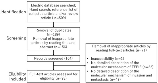
Figure 1.
The flowchart outlines the study selection process.
3. Results
3.1. Molecular Structure and Function of TFPI2
Tissue factor pathway inhibitor 2 (TFPI2) is a 32-kDa extracellular matrix-associated Kunitz-type serine proteinase inhibitor [1,2]. The discovery of TFPI2 dates back exactly three decades. In 1994, Sprecher et al. first reported the gene cloning, purification, expression, and biochemical characterization of TFPI2 [1]. Concurrently, Miyagi et al. demonstrated that a serine proteinase inhibitor purified from the conditioned medium of the human glioblastoma cell line T98G was identical to placental protein 5 (PP5), which shares the same amino acid sequence as TFPI2 [2]. Although TFPI2 is structurally homologous to TFPI1, they are encoded by distinct genes [16,17]. TFPI1, as its name implies, inhibits the extrinsic pathway of coagulation through tissue factor (TF) [16,17]. TF binds to factor VII (FVII), forming the TF–FVII complex and activates FVII, FIX, and FX, thereby catalyzing the proteolytic cleavage of prothrombin to thrombin, resulting in the conversion of fibrinogen to fibrin [18]. Elevated TF expression is not only linked to hypercoagulability in cancer patients but also plays a crucial role in cancer-related processes such as tumor growth, angiogenesis, and metastasis [16]. TFPI1 is an anticoagulant protein that inhibits the TF–FVIIa complex and the prothrombinase complex (FXa, FVa, calcium, and phospholipid) [16]. The mature TFPI1 protein contains three Kunitz domains (KDs), with the first, second, and third KD binding to FVIIa, FXa, and cell surface lipoproteins, respectively, thereby blocking the activation of the blood coagulation cascade [16].
Conversely, accumulating evidence suggests that TFPI2 is produced and secreted by various normal cells (https://www.ncbi.nlm.nih.gov/gene/7980 (accessed on 19 May 2024)). TFPI2 comprises three KDs that differ functionally from TFPI1 in substrate specificities [3,4]. The first KD1 domain of TFPI2 inhibits plasmin, trypsin [5,6], and kallikrein (e.g., kallikrein-related peptidase 5 (KLK5) and KLK12) [7]. Given that these proteinases contribute to the activation of pro-matrix metalloproteinase 1 (proMMP1) and proMMP-3 [7,19], TFPI2 may regulate extracellular matrix (ECM) remodeling to maintain tissue structure, function, and homeostasis in normal cells (e.g., vascular endothelial cells, macrophages, and trophoblasts) [7,11,20]. Additionally, plasmin promotes fibrin clot lysis via activating proMMPs and inactivates several coagulation factors (such as FV, FVIII, FIX, and FX) [21]. Therefore, unlike TFPI1, TFPI2 is unlikely to significantly inhibit TF-initiated thrombin generation and is thought to regulate matrix homeostasis by inhibiting plasmin-dependent fibrinolysis or further promote coagulation. Consequently, TFPI2 has also been considered a marker for an increased thrombosis risk [22,23]. Furthermore, complement C1q binding protein (C1QBP, also known as gC1qR) [24] and prosaposin [25] have been identified as proteins specifically binding to the KD2 domain of TFPI2. gC1qR plays a pivotal role in modulating fibrin formation, bradykinin generation, and intravascular inflammation [24,26], as well as tumor cell proliferation, invasion, and metastasis [27]. Prosaposin promotes cancer cell survival and inhibits apoptosis via immune evasion [26,28]. Considering these facts, it is plausible to assume that TFPI2 is involved in inhibiting tumor progression [8,29,30]. Indeed, numerous studies from 1994 to the present have reported that the downregulation of TFPI2 promotes tumor growth, invasion, and metastasis in vitro and in vivo, and that TFPI2 is an independent prognostic factor in various human cancers [8,9,10]. TFPI2 may have an essential role in controlling cancer progression through regulating pathological ECM remodeling. Based on these findings, TFPI2 has been proposed as a putative tumor suppressor. However, it has become evident that in some cancers, TFPI2 exhibits a paradoxical pro-invasive effect and is an indicator of poor prognosis [11,12].
3.2. TFPI2 as a Potential Inhibitor to Suppress Tumor Invasion and Metastasis
In this section, we elucidate how TFPI2 regulates tumorigenesis, focusing on the molecular mechanism underlying matrix degradation, invasion, epithelial–mesenchymal transition (EMT), apoptosis, and angiogenesis (Figure 2A).
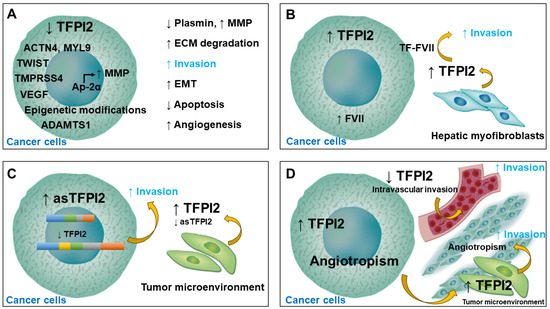
Figure 2.
The mechanistic role of TFPI2 in tumorigenesis and invasion. (A) Downregulation of TFPI2 enhances invasion and metastasis and inhibits apoptosis in various cancer cells by modulating the expression or activity of proteinases involved in tumor microenvironment remodeling. (B) TFPI2 promotes hepatocellular carcinoma invasion by binding to the TF–FVIIa complex. (C) Aberrantly spliced TFPI2 is significantly upregulated in various cancers. (D) Highly invasive cancer cells enhance angiotropism by upregulating TFPI2.
3.2.1. ECM Degradation
TFPI2 predominantly localizes to pericellular matrices on the surface of endothelial cells and within the ECM via heparin, with some presence in the cytoplasm and nucleus [31,32]. TFPI2 effectively suppresses the activation of MMPs capable of degrading ECM components by inhibiting plasmin [9,10]. Thus, TFPI2 is considered to play an inhibitory role in tumor progression across various malignancies by regulating plasmin-mediated matrix remodeling [10,33]. Additionally, TFPI2-mediated ECM remodeling is regulated by ADAMTS1 (A Disintegrin and Metalloproteinase with Thrombospondin 1) [34], which is involved in diverse pathophysiological processes such as tumor invasion and inflammation.
3.2.2. Tumor Growth and Invasion
Efforts, including the identification of novel proteins that bind to TFPI2, have been made to elucidate its physiological function. Co-immunoprecipitation revealed direct interactions of TFPI2 with actinin-4 (ACTN4) and myosin light chain 9 (MYL9) [35]. ACTN4 is implicated in chromatin remodeling, signaling transduction, hypermotility, and the tumorigenesis of breast cancer cells [36]. Additionally, cancer gene expression datasets indicate that MYL9 plays a crucial role in immune cell infiltration and tumor metastasis, predicting poor prognosis in several cancers [37]. ACTN4 required full-length TFPI2 for stable binding, while MYL9 preferentially binds to the N-terminus and KD1 of TFPI2 [35]. Thus, TFPI2 may regulate cancer cell proliferation and invasion through interactions with ACTN4 and/or MYL9. Moreover, TFPI2 has been shown to inhibit cancer cell proliferation by downregulating ERK1/2 activation [35], which promotes tumor invasion and angiogenesis through the upregulation of MMP expression [38].
3.2.3. Epithelial–Mesenchymal Transition
EMT confers phenotypic plasticity to epithelial cells, crucial for tumor stemness, initiation, invasion, and metastatic seeding. EMT is driven by transcriptional factors such as Twist-related protein 1 (TWIST1) [39,40], which induces breast cancer progression via integrin α5 induction [39,40]. TWIST expression is downregulated by TFPI2 or upregulated by TFPI2 silencing [40], with TWIST1 expression negatively correlated with TFPI2 in breast cancer patients [40]. In vitro and in vivo studies demonstrate that TFPI2 suppressed breast cancer progression by inhibiting the TWIST-integrin α5 pathway [40]. Additionally, the transmembrane protease serine 4 (TMPRSS4), highly expressed on the cell surface of various cancers such as pancreatic, thyroid, and prostate cancers [41,42], induces EMT by upregulating TWIST expression and promoting tumor cell migration [42]. The downregulation of TFPI2 may lead to TMPRSS4 overexpression, contributing to tumorigenesis in non-small-cell lung carcinoma [41].
3.2.4. Apoptosis
TFPI2 induces apoptosis in various cancers (e.g., glioblastoma [43,44], pancreatic cancer [45], laryngeal squamous cell carcinoma [46], nasopharyngeal cancer [47], esophageal cancer [48], thyroid cancer [49], small cell lung cancer [50], hepatocellular carcinoma [51], gallbladder cancer [52], renal cell cancer [53], bladder cancer [54], cervical cancer [55], and fibrosarcoma [56]). TFPI2 induces apoptosis in human choriocarcinoma cell lines BeWo and JEG-3 [57] while inhibiting apoptosis in immortalized human first-trimester extravillous trophoblast HTR8/SVneo cells [58]. Little is known about how TFPI2 induces distinct and opposite effects on normal and malignant trophoblast cells. The distinct effects of TFPI2 on normal and malignant trophoblast cells suggest that malignancy levels may influence TFPI2 function.
3.2.5. Angiogenesis
TFPI2 inhibits angiogenesis in both normal vascular endothelial cells [59] and cancer cells, including cervical cancer [55], esophageal cancer [60], and fibrosarcoma [29]. TFPI2 expression is upregulated by vascular endothelial growth factor (VEGF) in microvascular and umbilical vein endothelial cells, but TFPI2 impairs VEGF-induced vascular endothelial cell migration and proliferation [61,62], suggesting its role as a regulator of ECM remodeling homeostasis.
3.2.6. Transcriptional Activity
The transcription factor Ap-2α (Adaptor Protein complex 2, alpha subunit) directly binds to the MMP-2 gene promoter, inducing MMP-2 transcriptional activity and promoting ECM protein degradation [63]. TFPI2, localized in the nucleus, interacts with Ap-2α and diminishes its specific DNA binding, thereby inhibiting breast cancer cell proliferation and invasion [63]. Thus, TFPI2 not only serves as a protease inhibitor but also inhibits tumor invasion and proliferation by reducing MMP-2 expression via the Ap-2α.
3.2.7. Tumor Suppressor
Epigenetic alterations, including DNA methylation, histone modifications, and non-coding RNAs, regulate the TFPI2 gene [64]. TFPI2 mRNA is induced by 5-aza-2′-deoxycytidine, a DNA methyltransferase inhibitor, or trichostatin A, a histone deacetylase inhibitor [65,66,67,68]. Bisulfite sequencing analysis reveals the hypermethylation of the CpG island within the TFPI2 promoter region [69], leading to transcriptional silencing in various tumors [65,66,67,69]. Consequently, TFPI2 is often downregulated in numerous cancers and recognized as a tumor-suppressor gene [27,70,71,72]. Moreover, the methylation of the TFPI2 gene has been reported to be a leading panel for the detection of various cancers, including glioblastoma [65,73], oral squamous cell carcinoma [74,75], esophageal squamous cell carcinoma [76,77], gastric carcinoma [78], pancreatic ductal adenocarcinoma [66], colorectal cancer [79,80,81,82,83], prostate cancer [84], cervical cancer [85], vulvar cancer [86], malignant melanoma [87]. TFPI2 DNA methylation in the serum of melanoma patients was more closely associated with metastatic disease than with primary disease [71]. The methylation of the TFPI2 gene may be a potential biomarker for early cancer detection, patient prognosis, and cancer surveillance [64].
3.3. TFPI2 as a Potential Inducer to Promote Tumor Invasion and Metastasis
This section synthesizes the extant literature on the potential mechanisms underlying TFPI2-mediated tumor progression.
3.3.1. Tumor Invasion via Binding to the TF-FVIIa Complex
In most cancers, including hepatocellular carcinoma (HCC), TFPI2 functions as an anti-tumor protein that stabilizes the ECM and inhibits its remodeling by enzymatically inhibiting MMP activity. Moreover, TFPI2 inhibits the proliferation, adhesion, and invasion of HCC cells, possibly by downregulating MMP via the NF-κB signaling pathway [88] and also induces cell differentiation [51]. However, in 2000, Neaud et al. reported a paradoxical pro-invasive role of TFPI2 on human HCC cells (Figure 2B) [89]. Specifically, increased invasive potential was observed in HCC cells stably transfected with TFPI2. Mechanistically, TFPI2 secreted by liver myofibroblasts facilitates HCC invasion by directly binding to the TF–FVIIa complex on the cancer cell surface [89]. This is corroborated by the finding that the blockade of the TFPI2–FVII interaction with an antibody against FVII abolished the pro-invasive capacity of TFPI2 [89], and TFPI2 inhibited the invasion of FVII-deficient HT1080 cells [90]. Therefore, TFPI2 may trigger tumor progression only under specific cellular contexts, such as FVII-producing HCC.
3.3.2. Upregulation of Aberrantly Spliced TFPI2 in Cancers
Several spliced variants generated by alternative splicing or RNA editing are aberrantly expressed in many cancers [91]. Aberrant alternative splicing yields multiple protein isoforms from the same gene locus. In 2007, a 289-nucleotide splice variant of the TFPI2 transcript (i.e., aberrantly spliced TFPI2, asTFPI2) was identified in both human normal and tumor cells (Figure 2C) [12]. asTFPI-2 contains complete exons II and V and part of exons III and IV. The expression levels of asTFPI2 were significantly upregulated in various tumor tissues and several tumor cell lines but were very low and often undetectable in normal tissues. Additionally, Kempaiah et al. showed that immunoblotting using a polyclonal rabbit antibody against recombinant human α-TFPI2 demonstrated no band corresponding to TFPI2 fragments [27]. This suggests that either asTFPI2 is not translated or its fragments are not recognized by the rabbit polyclonal antibody. Although the biological significance of asTFPI2 remains to be proven, its overproduction may reflect the downregulation of TFPI2 expression in tumor cells.
Recently, preoperative serum TFPI2 levels were found to be significantly higher in patients with ovarian cancer [13,14] and endometrial cancer [15] compared to controls. Two anti-TFPI2 monoclonal antibodies were generated to quantify the TFPI2 concentration in serum samples using a sandwich-type, one-step immunofluorometric assay on an automated immunoassay analyzer (AIA) system (TOSOH, Tokyo, Japan) [92]. A single-center retrospective cohort study showed for the first time that serum TFPI2 levels were independent prognostic factors and predicted poor overall survival in patients with endometrial cancer [15]. Given that TFPI2 expression is downregulated in endometrial cancer tissues [93], circulating TFPI2 may be produced by vascular endothelial cells and fibroblasts constituting the tumor microenvironment rather than by cancer cells themselves. Alternatively, the TFPI2 protein detected in the blood might not be the full-length protein but fragmented proteins corresponding to asTFPI2. However, it remains to be determined whether the serum TFPI2 protein found in endometrial cancer patients is full-length or short fragments. Circulating asTFPI2 protein was not detected with an anti-TFPI2 polyclonal antibody; however, it is worth exploring whether asTFPI2 protein can be quantified using the AIA system.
3.3.3. Angiotropism or Vasculogenic Mimicry in Cancers
In general, the intravascular invasion of tumor cells is a crucial step in cancer metastasis [94]. Cancer cells invade surrounding tissues, effectively intravasate into vessel lumina, enter peripheral circulation, survive in the circulation, extravasate into target organs, seed distant organs, and eventually form metastases. However, it has been found that highly invasive cancer cells may not directly invade blood vessels but preferentially migrate along outside the vessel wall, supporting the formation of vasculature, disseminating to distant sites, and forming secondary tumors (Figure 2D) [11]. This process, known as angiotropism (i.e., vasculogenic mimicry or perivascular migration), involves the differential expression of key genes and pathways [11]. Enrichment analysis of differentially expressed genes identified TFPI2 as a potential biomarker for angiotropism [11]. Indeed, the downregulation of TFPI2 attenuated the perivascular migration of highly invasive melanoma cells [11]. Angiotropism is also observed in other cancers besides malignant melanoma [94]. Given that TFPI2 is downregulated in highly invasive cancer cells, it is plausible that TFPI2 is produced by cell populations distinct from cancer cells. For instance, TFPI2 protein is reported to be produced in vascular endothelial cells within the tumor microenvironment [95]. The coordinated upregulation of TFPI2 within the tumor microenvironment may play a significant role in angiotropism.
3.4. Key Regulators Controlling TFPI2 Expression
3.4.1. Key Regulators Downregulating TFPI2 Expression
The principal molecules that downregulate TFPI2 expression include activated RAS oncogene [70], E2F transcription factor 5 (E2F5) [96], CD24 [97,98,99], c-Src [97], lysine-specific demethylase 2 (LSD2) [100], ten-eleven translocation 1 (TET1) [101], methyl-CpG binding domain protein 3 (MBD3) [73], N6-methyladenosine (m6A) RNA demethylase fat mass and obesity-related protein (FTO) [102], microRNA-616 (miR-616) [103], maternally expressed gene 8 (MEG8) [59], miR-195 [43], lncRNA AGAP2-AS1 [104], and miR-23a [45] (Figure 3, green pie chart). These genes are involved in oncogenic signaling pathways and epigenetic modifications such as DNA methylation, histone modifications, and non-coding RNAs. Their characteristics and biological functions are summarized below. DNA methylation patterns and levels are known to be highly dynamic during cancer development. LSD1 and LSD2 demethylate histone (H3) lysine 4 and 9 (H3K4 and H3K9, respectively) and are required for both repression and activation of gene expression. LSD2 reduces TFPI2 expression and promotes small-cell lung cancer cell growth either by mediating DNA methyltransferase 3B (DNMT3B) expression or by regulating H3K4me1 demethylation in the TFPI2 gene [100]. TET1, an epigenetic regulator, is a 5-methylcytosine hydroxylase promoting DNA demethylation [105]. The mono-ADP ribosylation of arginine at position 117 of histone H3 (H3R117) inhibits TFPI2 demethylation by preventing interaction with TET1 [101]. MBD3 inhibits TFPI2 transcription via histone deacetylation, activates the Phosphoinositide 3-kinase (PI3K)/AKT signaling pathway, and enhances MMP activation, leading to HCC growth and metastasis [73]. FTO inhibits TFPI2 expression, accelerating pancreatic cancer cell proliferation, migration, and invasion [102].
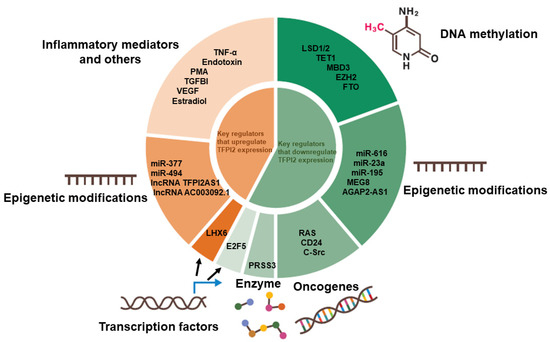
Figure 3.
Key regulators modulating TFPI2 expression. Green and orange pie charts indicate key regulators that downregulate or upregulate TFPI2 expression, respectively.
Moreover, microRNAs (miRNAs/miRs) induce the post-transcriptional regulation of diverse target genes, especially in cancer development [45]. MiR-616, miR-23a, miR-195, and lncRNAs MEG8 and AGAP2-AS1 can regulate TFPI2 gene expression via translational repression. MiR-616 induces prostate cancer cell proliferation by downregulating TFPI2 expression [103]. MiR-616 also promotes glioma cell proliferation and inhibits apoptosis by suppressing the Wnt-SOX7 signaling pathway [106]. MiR-195 is identified as a tumor suppressor leading to decreased TFPI2 expression [43]. MiR-23a promotes proliferation, migration and invasion while inhibiting the apoptosis of pancreatic cancer cells by downregulating TFPI2 [45]. Furthermore, MEG8, a long non-coding RNA located at the 14q32 locus, increases histone modification on histone H3 lysine 27 (H3K27me3) at the TFPI2 promoter, downregulating TFPI2 expression in vascular endothelial cells [59]. Indeed, MEG8 promotes several malignancies, including lung, pancreatic and liver cancer [107]. LncRNA AGAP2-AS1 downregulates TFPI2 gene expression through interaction with an enhancer of zeste 2 polycomb repressive complex 2 subunit (EZH2) and LSD1 in glioblastoma [104].
Additionally, oncogenic RAS promotes cancer cell proliferation by triggering exit from the G0/G1-phase and entry into the S-phase, leading to TFPI2 downregulation [70]. CD24 enhances tumor metastasis by binding to integrin-β1, P-selectin, and Siglec-10, preventing the immunological elimination of cancer cells through inhibiting natural killer cell-mediated cytotoxicity and macrophage-mediated phagocytosis [97,98,99]. CD24 also enhances tumor cell invasion through c-Src-mediated TFPI2 downregulation [97]. Serine protease 3 (PRSS3, also known as mesotrypsin), a member of the trypsin family of serine proteases, promotes tumor progression via the PRSS3-dependent proteolytic inactivation of TFPI2 [62,108]. E2F5, a transcription factor, inhibits cell cycle progression via forming a complex with retinoblastoma proteins, promoting prostate cancer cell migration and invasion through upregulating MMP-2 and MMP-9 and downregulating TFPI2 [96]. Collectively, the downregulation of the TFPI2 gene is mainly caused by epigenetic modifications and oncogene activation.
3.4.2. Key Regulators Upregulating TFPI2 Expression
Next, we summarize mechanisms controlling the upregulation of the TFPI2 gene (Figure 3, orange pie chart).
TFPI2 mRNA and protein expression are increased by inflammatory mediators (e.g., TNF-α, endotoxin, or phorbol 12-myristate 13-acetate (PMA)) in normal cells (e.g., human umbilical vein endothelial cells) [3] and malignant cells (e.g., glioblastoma cell Hs683) [109]. Transforming growth factor-beta-induced (TGFBI) protein, also known as keratoepithelin or Beta ig h3 protein (BIGH3), has important roles in cell adhesion and ECM formation, and an imbalance in the immune inflammatory response restores TFPI2 expression and reduces the proliferation and invasion of human neuroblastoma [110]. VEGF upregulates TFPI2 expression via the mitogen-activated protein kinase (MEK) pathway in microvascular endothelial cells and umbilical vein endothelial cells [61,62]. Estradiol (E2) induces TFPI2 upregulation via estrogen receptor-alpha (ERα) in MCF7 breast cancer cells [111].
Furthermore, miR-377, miR-494, lncRNA TFPI2AS1, and lncRNA AC003092.1 promote TFPI2 expression. TFPI2 mRNA is upregulated by miR-494 in breast cancer cell line MCF-7 [112]. MiR-494 inhibits ovarian cancer growth [113]. MiR-377 inhibits proliferation and induces the apoptosis of pancreatic cancer cells by upregulating TFPI2 expression via DNMT1 downregulation [114]. LncRNA TFPI2AS1 inhibits non-small-cell lung cancer cell proliferation and migration by upregulating TFPI2 via the suppression of the G1/S transition and the upregulation of cyclin D1 and cyclin-dependent kinase 2 (CDK2) [115]. LncRNA AC003092.1 inhibits proliferation and induces apoptosis in glioblastoma and gallbladder cancer cells by upregulating TFPI2 expression via miR-195 inhibition. The use of agents that upregulate TFPI2, such as demethylating agents (e.g., 5-aza 2′-deoxycytidine) and histone deacetylase inhibitors (e.g., depsipeptide FK228), might be novel cancer therapeutic strategies [68]. For example, the transcriptional regulator LIM homeobox domain 6 (LHX6) suppresses human pancreatic cancer cell proliferation through TFPI2 upregulation [116]. LHX6 is downregulated in pancreatic cancer cell lines and surgical specimens [116].
In addition, all-trans-retinoic acid (ATRA), an active metabolite of vitamin A, exerts immunomodulatory and antitumor effects [117]. ATRA inhibits HCC cell invasion through TFPI2 upregulation [117]. Curcumin, a natural polyphenolic compound derived from turmeric with diverse pharmacologic effects, including anti-cancer characteristics, inhibits the invasion and migration of pancreatic cancer cells by upregulating TFPI2 mRNA and protein and inhibiting ERK (extracellular regulated MAP kinase)- and JNK (c-Jun NH2-terminal kinase)-mediated epithelial–mesenchymal transition [118]. Collectively, TFPI2 is upregulated by molecules that primarily function in epigenetic modifications and inflammation (Figure 3, orange pie chart).
3.5. TFPI2 Expression in Cancer Tissues and Its Biological Significance
The TFPI2 transcript is ubiquitously expressed in various tissues from healthy individuals, including placenta, liver, lung, skeletal muscle, heart, kidney, and pancreas (https://www.ncbi.nlm.nih.gov/gene/7980 (accessed on 19 May 2024)) [2,17]. TFPI2 is synthesized and secreted by a variety of cell types of diverse origins, such as epithelial, endothelial, and mesenchymal cells [4,5]. The majority of the TFPI2 secreted by endothelial cells was shown to accumulate on the extracellular matrix via binding to heparin or proteoglycans [3,32]. Thus, TFPI2 may be involved in maintaining cell structure, function, and tissue integrity [3,63,70]. Researchers have extensively evaluated the potential role of TFPI2 in a variety of neoplasms. This section summarizes the latest information on TFPI2 present in cancer cell lines, human cancer tissues, and circulating blood to explore the mechanisms underlying its diverse functions by cancer type (Figure 4). The left and right halves of Figure 4 depict human cancers in which TFPI2 has been implicated to play pro- and anti-invasive roles, respectively. Initially, we will outline the types of cancer where TFPI2 demonstrates anti-invasive properties. The Supplementary Material offers additional insights into the role of TFPI2 in human cancers. Here we have summarized the following cancers: glioblastoma [2,5,33,44,109,119,120,121], leukemia [9], malignant sinonasal inverted papilloma [122], oral cancer, pharyngeal cancer [46,47,74,75,123], esophageal squamous cell carcinoma [48,60,76,77], thyroid cancer [49], breast cancer [35,69,124], lung cancer [41,50,67,100,125,126,127], gastric cancer [78,128,129,130], hepatocellular carcinoma [51,88,89,131], gallbladder cancer [52,132], pancreatic cancer [66,133,134,135], colorectal cancer [136], renal cell carcinoma [53], prostate cancer [137,138], bladder cancer [54,139], cervical cancer [55,85,140,141,142], endometrial cancer [15,99,143], ovarian cancer [2,13,14,22,23,31,92,144,145,146], vulvar cancer [86], choriocarcinoma [147,148], malignant melanoma [71,87,149,150,151], and fibrosarcoma [25,29,56,90] (See the Supplementary Material). In vitro and in vivo experiments using various cancer cells demonstrated that TFPI2 inhibits tumor growth, invasion, and metastasis through remodeling the tumor microenvironment [8,9,10]. TFPI2 expression was inversely correlated with the progression of various tumors [133] and frequently downregulated in aggressive cancers [8,9]. Indeed, restoration of the TFPI2 gene by recombinant adeno-associated virus in a human glioblastoma cell line [152,153] significantly reduced migration, invasion, proliferation, and angiogenesis and triggered apoptosis in in vitro and in vivo animal models. Furthermore, the overexpression of TFPI2 protein via stable vector transfections inhibited malignant meningioma cell invasion, growth, and angiogenesis and induced apoptosis [33]. Conversely, the knockdown or gene silencing of TFPI2 promoted the proliferation, migration, and invasion abilities of non-small-cell lung cancer cells [9,10] and glioblastoma cells [119]. TFPI2 knockdown resulted in the induction of the expression of MMP-1 [9,10,119], MMP-2 [119], MMP-3 [10], or MMP-7 [10] depending on the type of tumor through tumor–stroma interactions.
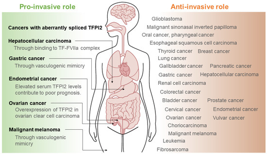
Figure 4.
Human cancers exhibiting anti- or pro-invasive effects of TFPI2. The right side illustrates cancers where TFPI2 has an anti-invasive impact, while the left side depicts cancers where TFPI2 has a pro-invasive effect.
Thus, TFPI2 demonstrates anti-invasive properties in most cancers. Next, we will summarize some cancers where TFPI2 demonstrates a paradoxical dual role (i.e., pro-invasive and anti-invasive properties).
Gastric cancer: TFPI2 promoter hypermethylation was significantly higher in patients with gastric cancer compared to their paired non-tumor tissues [154]. The hypermethylation of TFPI2 was associated with gastric carcinogenesis [128] and clinicopathological features such as tumor size and stage [129], indicating that TFPI2 hypermethylation is an unfavorable prognostic marker [78]. Until now, TFPI2 has been considered a typical tumor suppressor epigenetically inactivated by CpG methylation in human gastric cancer [78]. However, in 2022, Wang et al. reported that TFPI2 overexpression is associated with a poor prognosis (i.e., worse overall survival and progression-free survival) in gastric cancer patients with a pathological feature called vasculogenic mimicry, frequently activated during cancer progression (See Figure 2D) [130]. The authors found a potential association between TFPI2 overexpression, tumor-infiltrating immune cells, and prognosis [130].
Hepatocellular carcinoma: TFPI2 has been reported to be frequently silenced in HCC via epigenetic alterations, including histone modification, DNA methylation, and non-coding RNA modulation [131]. Overexpressed TFPI2 repressed the proliferation and invasion of HCC cells [131]. Furthermore, TFPI2 overexpression not only suppressed cell proliferation and induced apoptosis but also decreased the proportion of CD133-positive cancer stem cells in HCC, leading to a reduced tumorigenicity [51]. TFPI2 silencing in the HCC cell line MHCC97-L exhibited the opposite by upregulating MMP-1/3, CD44, and intercellular adhesion molecule 1 (ICAM-1) via the nuclear factor-kappaB (NF-κB)/AKT-mediated signaling pathway [88]. Meanwhile, as shown in Figure 2B, cancer-associated hepatic myofibroblast-derived TFPI2 promoted cell invasion only in the FVII-producing HCC [89]. TFPI2 has been shown to exhibit a dual role, possibly according to the experimental conditions. TFPI2 might have opposing effects depending on the expression levels of inherent genes (e.g., FVII, AKT, NF-κB, MMP-1/3, CD44, or ICAM-1) within individual cancer cells.
Endometrial cancer: The expression level of TFPI2 in endometrial cancer tissues was decreased with an increasing degree of malignancy [93]. Recently, Kawaguchi et al. reported that TFPI2 levels are elevated in the preoperative serum of patients with endometrial cancer, and this elevation may contribute to poor prognosis [15]. In multivariate analyses, a TFPI2 level ≥ 177 pg/mL (HR, 2.42; p = 0.043) was an independent prognostic factor of overall survival in endometrial cancer patients [15]. Furthermore, immunohistochemistry revealed that TFPI2 is a promising biomarker for the diagnosis of endometrial clear cell carcinoma and allows for the exclusion of other histological types such as endometrioid carcinoma and serous carcinoma [143]. The question is why, in advanced endometrial cancer, the protein levels of TFPI2 diminish within the cancerous tissue yet increase in the peripheral blood, and why TFPI2 expression is exclusive to clear cell carcinoma. This could be due to the production of TFPI2 protein by surrounding non-cancerous tissues and its systemic release. Another possibility is that asTFPI2, detectable by the AIA system, may be present in the blood of patients with endometrial cancer. Further studies are needed to evaluate epigenetic modifications, including DNA methylation, histone modification, chromatin remodeling, and miRNAs, and their consequential changes to TFPI2 gene expression in clear cell carcinoma.
Ovarian cancer: It was already shown 30 years ago that TFPI2 is abundantly expressed in some ovarian cancer cells [2]. Arakawa et al. presented the first evidence for the potential of serum TFPI2 as a biomarker for ovarian clear cell carcinoma [92]. Immunohistochemistry identified TFPI2 expression in at least one of the nuclear, cytoplasmic, and extracellular matrix fractions of 52 (67.5%) of 77 ovarian clear cell carcinoma tissues, but TFPI2 was not detected in the other subtypes, including serous ovarian cancers (n = 65) [31]. The overexpression of TFPI2 in ovarian clear cell carcinoma tissues has enabled its clinical application as a serodiagnostic marker [144]. After several retrospective and prospective multicenter studies, TFPI2 became covered by the National Health Insurance in Japan in 2021 as a serodiagnostic marker for ovarian cancer [13,14]. Furthermore, TFPI2 has been reported to be useful as a diagnostic index equivalent to the conventional Risk of Malignancy Algorithm (ROMA) value [145]. In 2015, Wang et al. reported the diagnostic performance of TFPI2 methylation in ovarian cancer tissue and blood [146]. Serum levels of free TFPI2 DNA methylation were higher in the ovarian cancer group than those in the benign and healthy control groups [146]. TFPI2 was frequently methylated even at an early ovarian cancer stage [146]. Thus, serum levels of TFPI2 methylation may reflect the DNA methylation spectrum of ovarian cancer tissues. Unfortunately, out of a total of 71 ovarian cancer patients, only 7 patients had clear cell carcinoma, so we were unable to draw conclusive findings regarding TFPI2 DNA methylation in ovarian clear cell carcinoma.
Malignant melanoma: TFPI2 is the most frequent target of aberrant methylation in melanoma patients and was identified as one of the tumor-suppressor genes [87,149]. Konduri et al. demonstrated for the first time that TFPI2 overexpression in stably transfected amelanotic melanoma cell line C-32 led to a decrease in the invasive capacity [150]. The silencing effects of TFPI2 triggered invasive and metastatic potentials of melanoma [149]. The blood-based DNA methylation of TFPI2 has been reported to be a biomarker to predict melanoma metastasis [71]. On the other hand, blood coagulation-related genes (e.g., TF, TFPI1, TFPI2) are also known to be upregulated as hub genes in aggressive melanoma [151]. TFPI2, synthesized by melanoma cells and adjacent noncancerous tissues, is deposited in the extracellular matrix, inhibiting matrix degradation and structurally reinforcing the scaffold [151]. Additionally, the process of vasculogenic mimicry may require TFPI1-mediated anticoagulant activity to maintain blood flow [151]. TFPI2 not only exerts its tumor-suppressor function but also confers vasculogenic-like properties to malignant melanoma, but the mechanisms underlying these opposing processes remain unclear.
4. Discussion
The findings presented in this review underscore the complex and multifaceted role of TFPI2 in human cancers. While initially characterized as a tumor-suppressor gene [4,40,55,70,72,73,75,79,82,87,93,97,104,120,121,122,123,124] (Figure 2A), accumulating evidence suggests that TFPI2 may paradoxically promote tumor invasion and metastasis under specific cellular contexts [3,11,12,13,14,15,87,89,93,127,151] (Figure 2B–D). This apparent dichotomy in TFPI2’s functions has sparked intense research interest, aimed at elucidating the underlying mechanisms governing its expression and activity. One key mechanism that has emerged is the dysregulation of TFPI2 expression through epigenetic modifications, such as DNA methylation and histone acetylation [64] (Figure 3). Numerous studies have demonstrated that TFPI2 is frequently silenced by promoter hypermethylation in various cancer types, supporting its role as a tumor suppressor. However, the factors that determine the extent and specificity of these epigenetic alterations remain poorly understood, warranting further investigation into the interplay between TFPI2 and epigenetic regulatory mechanisms.
Another intriguing aspect of TFPI2’s dual role in cancer is the phenomenon of alternative splicing (Figure 2C, Figure 4 left). Emerging data suggest that aberrantly spliced TFPI2 isoforms may contribute to tumor progression, potentially by exerting distinct or opposing functions compared to the full-length protein [12]. Unraveling the intricate patterns of TFPI2 alternative splicing and their functional consequences in different cancer types could provide valuable insights into its paradoxical effects. Furthermore, the process of angiotropism, or vasculogenic mimicry, has been implicated in TFPI2-mediated tumor invasion and metastasis (Figure 2D. Figure 4, left). While the exact mechanisms underlying this phenomenon remain unclear, it is hypothesized that TFPI2 overexpression in the tumor microenvironment may promote tumor cell adhesion and invasion by strengthening the extracellular matrix [3,11,89,151]. Tumor cell invasion and metastasis require scaffold construction, cell-matrix adhesion, matrix degradation, proper ECM remodeling, and tumor cell migration (Figure 5). Tumor-associated fibroblasts and vascular endothelial cells may adapt to the needs of TFPI2-negative tumor cells (e.g., construction of sufficient ECM) by facilitating the generation of TFPI2 possibly through epigenetic modifications, such as DNA methylation, histone modification, and non-coding RNAs. The overexpression of TFPI2 may trigger tumor cell adhesion by accumulating more ECM at the invasive front and promote subsequent invasion and metastasis. The constant interactions between tumor cells and the tumor microenvironment may influence TFPI2 generation and thereby tumor progression. Further work is needed to verify whether this mechanism is specific to gynecological cancers such as ovarian and endometrial cancers. Further exploration of the interplay between TFPI2, angiotropism, and the tumor microenvironment yields important insights into this intriguing aspect of cancer biology.
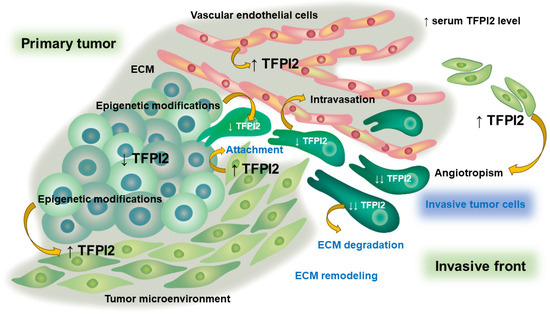
Figure 5.
Role of TFPI2 at the cancer invasive front in a coordinated tumor microenvironment. The interplay between the tumor and its microenvironment may render tumor progression by upregulating TFPI2 at the tumor invasion front.
Despite these advances, several limitations and challenges remain in our understanding of TFPI2’s dual role in cancer. One significant limitation of the current literature is the lack of a cohesive structure and logical flow in presenting and integrating the various mechanisms and findings. Many studies have focused on specific aspects of TFPI2’s functions or regulatory mechanisms, but a comprehensive and well-organized synthesis of these findings is lacking. This lack of cohesion may hinder a holistic understanding of TFPI2’s multifaceted roles and the interplay between different regulatory mechanisms. To address these limitations, future research efforts should prioritize a more integrated and systematic approach to investigating TFPI2’s functions in cancer. This could involve comprehensive multi-omics studies, combining genomic, epigenomic, transcriptomic, and proteomic analyses to unravel the complex interplay between TFPI2 expression, alternative splicing, and epigenetic regulation across diverse cancer types. Additionally, in-depth mechanistic studies, employing advanced in vitro and in vivo models, could elucidate the precise molecular pathways and cellular contexts that govern TFPI2’s tumor-suppressive or tumor-promoting activities. Moreover, translational research efforts should focus on exploring the potential clinical applications of TFPI2 as a diagnostic or prognostic biomarker, or as a therapeutic target. While some studies have evaluated TFPI2 methylation as a potential cancer biomarker [70,93,120], more extensive validation in larger patient cohorts is needed. Similarly, the therapeutic potential of targeting TFPI2 expression or activity, either through epigenetic modulators or specific inhibitors, warrants further investigation in preclinical and clinical studies.
In conclusion, TFPI2 represents a fascinating and multifaceted molecule with seemingly opposing roles in human cancers. While significant progress has been made in understanding the mechanisms underlying its dual functions, several key questions and challenges remain. By addressing these limitations through integrated, multi-disciplinary approaches and translational research efforts, we can further unravel the complexities of TFPI2’s involvement in cancer biology and potentially unlock new avenues for diagnostic, prognostic, and therapeutic applications.
Supplementary Materials
The following supporting information can be downloaded at: https://www.mdpi.com/article/10.3390/ijtm4030028/s1, File S1: The role of TFPI2 in human cancers.
Author Contributions
Conception and design, H.K.; Acquisition of data, S.M., C.Y. and H.S.; Analysis and Interpretation of data, S.I.; Drafting of the manuscript, H.K.; Critical revision of the manuscript for important intellectual content, S.M., C.Y., H.S. and S.I.; Administrative technical or material support, H.K.; Supervision, H.K. All authors have read and agreed to the published version of the manuscript.
Funding
This research received no external funding.
Institutional Review Board Statement
Not applicable.
Informed Consent Statement
Not applicable.
Data Availability Statement
No new data were created or analyzed in this study.
Acknowledgments
All figures were created by Toyomi Kobayashi (Ms.Clinic MayOne, Nara, Japan); https://www.mscl-mayone.com/ (accessed on 5 May 2024).
Conflicts of Interest
The authors declare no conflicts of interest.
References
- Sprecher, C.A.; Kisiel, W.; Mathewes, S.; Foster, D.C. Molecular cloning, expression, and partial characterization of a second human tissue-factor-pathway inhibitor. Proc. Natl. Acad. Sci. USA 1994, 91, 3353–3357. [Google Scholar] [CrossRef] [PubMed]
- Miyagi, Y.; Koshikawa, N.; Yasumitsu, H.; Miyagi, E.; Hirahara, F.; Aoki, I.; Misugi, K.; Umeda, M.; Miyazaki, K. cDNA cloning and mRNA expression of a serine proteinase inhibitor secreted by cancer cells: Identification as placental protein 5 and tissue factor pathway inhibitor-2. J. Biochem. 1994, 116, 939–942. [Google Scholar] [CrossRef] [PubMed]
- Iino, M.; Foster, D.C.; Kisiel, W. Quantification and Characterization of Human Endothelial Cell-Derived Tissue Factor Pathway Inhibitor-2. Arterioscler. Thromb. Vasc. Biol. 1998, 18, 40–46. [Google Scholar] [CrossRef] [PubMed]
- Chand, H.S.; Foster, D.C.; Kisiel, W. Structure, function and biology of tissue factor pathway inhibitor-2. Thromb. Haemost. 2005, 94, 1122–1130. [Google Scholar] [CrossRef]
- Konduri, S.D.; Osman, F.A.; Rao, C.N.; Srinivas, H.; Yanamandra, N.; Tasiou, A.; Dinh, D.H.; Olivero, W.C.; Gujrati, M.; Foster, D.C.; et al. Minimal and inducible regulation of tissue factor pathway inhibitor-2 in human gliomas. Oncogene 2002, 21, 921–928. [Google Scholar] [CrossRef] [PubMed]
- Schmidt, A.E.; Chand, H.S.; Cascio, D.; Kisiel, W.; Bajaj, S.P. Crystal structure of Kunitz domain 1 (KD1) of tissue factor pathway inhibitor-2 in complex with trypsin. Implications for KD1 specificity of inhibition. J. Biol. Chem. 2005, 280, 27832–27838. [Google Scholar] [CrossRef] [PubMed]
- Lavergne, M.; Guillon-Munos, A.; Lenga Ma Bonda, W.; Attucci, S.; Kryza, T.; Barascu, A.; Moreau, T.; Petit-Courty, A.; Sizaret, D.; Courty, Y.; et al. Tissue factor pathway inhibitor 2 is a potent kallikrein-related protease 12 inhibitor. Biol. Chem. 2021, 402, 1257–1268. [Google Scholar] [CrossRef] [PubMed]
- Sierko, E.; Wojtukiewicz, M.Z.; Kisiel, W. The role of tissue factor pathway inhibitor-2 in cancer biology. Semin. Thromb. Hemost. 2007, 33, 653–659. [Google Scholar] [CrossRef] [PubMed]
- Iochmann, S.; Bléchet, C.; Chabot, V.; Saulnier, A.; Amini, A.; Gaud, G.; Gruel, Y.; Reverdiau, P. Transient RNA silencing of tissue factor pathway inhibitor-2 modulates lung cancer cell invasion. Clin. Exp. Metastasis 2009, 26, 457–467. [Google Scholar] [CrossRef]
- Gaud, G.; Iochmann, S.; Guillon-Munos, A.; Brillet, B.; Petiot, S.; Seigneuret, F.; Touzé, A.; Heuzé-Vourc’h, N.; Courty, Y.; Lerondel, S.; et al. TFPI-2 silencing increases tumour progression and promotes metalloproteinase 1 and 3 induction through tumour-stromal cell interactions. J. Cell. Mol. Med. 2011, 15, 196–208. [Google Scholar] [CrossRef]
- Mo, J.; Zhao, X.; Wang, W.; Zhao, N.; Dong, X.; Zhang, Y.; Cheng, R.; Sun, B. TFPI2 Promotes Perivascular Migration in an Angiotropism Model of Melanoma. Front. Oncol. 2021, 11, 662434. [Google Scholar] [CrossRef] [PubMed]
- Kempaiah, P.; Chand, H.S.; Kisiel, W. Identification of a human TFPI-2 splice variant that is upregulated in human tumor tissues. Mol. Cancer 2007, 6, 20. [Google Scholar] [CrossRef] [PubMed]
- Arakawa, N.; Kobayashi, H.; Yonemoto, N.; Masuishi, Y.; Ino, Y.; Shigetomi, H.; Furukawa, N.; Ohtake, N.; Miyagi, Y.; Hirahara, F.; et al. Clinical Significance of Tissue Factor Pathway Inhibitor 2, a Serum Biomarker Candidate for Ovarian Clear Cell Carcinoma. PLoS ONE 2016, 11, e0165609. [Google Scholar] [CrossRef]
- Miyagi, E.; Arakawa, N.; Sakamaki, K.; Yokota, N.R.; Yamanaka, T.; Yamada, Y.; Yamaguchi, S.; Nagao, S.; Hirashima, Y.; Kasamatsu, Y.; et al. Validation of tissue factor pathway inhibitor 2 as a specific biomarker for preoperative prediction of clear cell carcinoma of the ovary. Int. J. Clin. Oncol. 2021, 26, 1336–1344. [Google Scholar] [CrossRef] [PubMed]
- Kawaguchi, R.; Maehana, T.; Yamanaka, S.; Miyake, R.; Kawahara, N.; Iwai, K.; Yamada, Y.; Kimura, F. Preoperative serum tissue factor pathway inhibitor-2 level as a prognostic marker for endometrial cancer. Oncol. Lett. 2023, 26, 463. [Google Scholar] [CrossRef] [PubMed]
- Bajaj, M.S.; Birktoft, J.J.; Steer, S.A.; Bajaj, S.P. Structure and biology of tissue factor pathway inhibitor. Thromb. Haemost. 2001, 86, 959–972. [Google Scholar] [PubMed]
- Miyagi, Y.; Yasumitsu, H.; Eki, T.; Miyata, S.; Kkawa, N.; Hirahara, F.; Aoki, I.; Misugi, K.; Miyazaki, K. Assignment of the human PP5/TFPI-2 gene to 7q22 by FISH and PCR-based human/rodent cell hybrid mapping panel analysis. Genomics 1996, 35, 267–268. [Google Scholar] [CrossRef] [PubMed]
- Komiyama, Y.; Pedersen, A.H.; Kisiel, W. Proteolytic activation of human factors IX and X by recombinant human factor VIIa: Effects of calcium, phospholipids, and tissue factor. Biochemistry 1990, 29, 9418–9425. [Google Scholar] [CrossRef]
- Rao, C.N.; Mohanam, S.; Puppala, A.; Rao, J.S. Regulation of ProMMP-1 and ProMMP-3 activation by tissue factor pathway inhibitor-2/matrix-associated serine protease inhibitor. Biochem. Biophys. Res. Commun. 1999, 255, 94–98. [Google Scholar] [CrossRef]
- Rollin, J.; Régina, S.; Vourc’h, P.; Iochmann, S.; Bléchet, C.; Reverdiau, P.; Gruel, Y. Influence of MMP-2 and MMP-9 promoter polymorphisms on gene expression and clinical outcome of non-small cell lung cancer. Lung Cancer 2007, 56, 273–280. [Google Scholar] [CrossRef]
- Kobayashi, H.; Matsubara, S.; Imanaka, S. The role of tissue factor pathway inhibitor 2 in the coagulation and fibrinolysis system. J. Obstet. Gynaecol. Res. 2023, 49, 1677–1683. [Google Scholar] [CrossRef] [PubMed]
- Yamanaka, S.; Miyake, R.; Yamada, Y.; Kawaguchi, R.; Ootake, N.; Myoba, S.; Kobayashi, H. Tissue Factor Pathway Inhibitor 2: A Novel Biomarker for Predicting Asymptomatic Venous Thromboembolism in Patients with Epithelial Ovarian Cancer. Gynecol. Obstet. Investig. 2022, 87, 133–140. [Google Scholar] [CrossRef] [PubMed]
- Miyake, R.; Yamada, Y.; Yamanaka, S.; Kawaguchi, R.; Ootake, N.; Myoba, S.; Kobayashi, H. Tissue factor pathway inhibitor 2 as a serum marker for diagnosing asymptomatic venous thromboembolism in patients with epithelial ovarian cancer and positive D-dimer results. Mol. Clin. Oncol. 2022, 16, 46. [Google Scholar] [CrossRef] [PubMed]
- Peerschke, E.I.; Petrovan, R.J.; Ghebrehiwet, B.; Ruf, W. Tissue factor pathway inhibitor-2 (TFPI-2) recognizes the complement and kininogen binding protein gC1qR/p33 (gC1qR): Implications for vascular inflammation. Thromb. Haemost. 2004, 92, 811–819. [Google Scholar] [CrossRef] [PubMed]
- Xu, C.; Deng, F.; Mao, Z.; Zhang, J.; Wang, H.; Wang, J.; Mu, J.; Deng, S.; Ma, D. The interaction of the second Kunitz-type domain (KD2) of TFPI-2 with a novel interaction partner, prosaposin, mediates the inhibition of the invasion and migration of human fibrosarcoma cells. Biochem. J. 2012, 441, 665–674. [Google Scholar] [CrossRef]
- Peerschke, E.I.; Ghebrehiwet, B. Human blood platelet gC1qR/p33. Immunol. Rev. 2001, 180, 56–64. [Google Scholar] [CrossRef] [PubMed]
- Lei, Y.; Li, X.; Qin, D.; Zhang, Y.; Wang, Y. gC1qR: A New Target for Cancer Immunotherapy. Front. Immunol. 2023, 14, 1095943. [Google Scholar] [CrossRef]
- Wu, Y.; Sun, L.; Zou, W.; Xu, J.; Liu, H.; Wang, W.; Yun, X.; Gu, J. Prosaposin, a regulator of estrogen receptor alpha, promotes breast cancer growth. Cancer Sci. 2012, 103, 1820–1825. [Google Scholar] [CrossRef]
- Chand, H.S.; Du, X.; Ma, D.; Inzunza, H.D.; Kamei, S.; Foster, D.; Brodie, S.; Kisiel, W. The effect of human tissue factor pathway inhibitor-2 on the growth and metastasis of fibrosarcoma tumors in athymic mice. Blood 2004, 103, 1069–1077. [Google Scholar] [CrossRef]
- Mino, K.; Nishimura, S.; Ninomiya, S.; Tujii, H.; Matsumori, Y.; Tsuchida, M.; Hosoi, M.; Koseki, K.; Wada, S.; Hasegawa, M.; et al. Regulation of tissue factor pathway inhibitor-2 (TFPI-2) expression by lysine-specific demethylase 1 and 2 (LSD1 and LSD2). Biosci. Biotechnol. Biochem. 2014, 78, 1010–1017. [Google Scholar] [CrossRef]
- Ota, Y.; Koizume, S.; Nakamura, Y.; Yoshihara, M.; Takahashi, T.; Sato, S.; Myoba, S.; Ohtake, N.; Kato, H.; Yokose, T.; et al. Tissue factor pathway inhibitor-2 is specifically expressed in ovarian clear cell carcinoma tissues in the nucleus, cytoplasm and extracellular matrix. Oncol. Rep. 2021, 45, 1023–1032. [Google Scholar] [CrossRef]
- Liu, Y.; Stack, S.M.; Lakka, S.S.; Khan, A.J.; Woodley, D.T.; Rao, J.S.; Rao, C.N. Matrix localization of tissue factor pathway inhibitor-2/matrix-associated serine protease inhibitor (TFPI-2/MSPI) involves arginine-mediated ionic interactions with heparin and dermatan sulfate: Heparin accelerates the activity of TFPI-2/MSPI toward plasmin. Arch. Biochem. Biophys. 1999, 370, 112–118. [Google Scholar] [CrossRef][Green Version]
- Kondraganti, S.; Gondi, C.S.; Gujrati, M.; McCutcheon, I.; Dinh, D.H.; Rao, J.S.; Olivero, W.C. Restoration of tissue factor pathway inhibitor inhibits invasion and tumor growth in vitro and in vivo in a malignant meningioma cell line. Int. J. Oncol. 2006, 29, 25–32. [Google Scholar] [CrossRef] [PubMed]
- Torres-Collado, A.X.; Kisiel, W.; Iruela-Arispe, M.L.; Rodríguez-Manzaneque, J.C. ADAMTS1 interacts with, cleaves, and modifies the extracellular location of the matrix inhibitor tissue factor pathway inhibitor-2. J. Biol. Chem. 2006, 281, 17827–17837. [Google Scholar] [CrossRef]
- Wang, G.; Huang, W.; Li, W.; Chen, S.; Chen, W.; Zhou, Y.; Peng, P.; Gu, W. TFPI-2 suppresses breast cancer cell proliferation and invasion through regulation of ERK signaling and interaction with actinin-4 and myosin-9. Sci. Rep. 2018, 8, 14402. [Google Scholar] [CrossRef] [PubMed]
- Hsu, K.S.; Kao, H.Y. Alpha-actinin 4 and tumorigenesis of breast cancer. Vitam. Horm. 2013, 93, 323–351. [Google Scholar] [CrossRef]
- Lv, M.; Luo, L.; Chen, X. The landscape of prognostic and immunological role of myosin light chain 9 (MYL9) in human tumors. Immun. Inflamm. Dis. 2022, 10, 241–254. [Google Scholar] [CrossRef] [PubMed]
- Wang, Y.; Wang, L.; Li, D.; Wang, H.B.; Chen, Q.F. Mesothelin promotes invasion and metastasis in breast cancer cells. J. Int. Med. Res. 2012, 40, 2109–2116. [Google Scholar] [CrossRef]
- Pei, H.; Li, Y.; Liu, M.; Chen, Y. Targeting Twist expression with small molecules. MedChemComm 2016, 8, 268–275. [Google Scholar] [CrossRef]
- Zhao, D.; Qiao, J.; He, H.; Song, J.; Zhao, S.; Yu, J. TFPI2 suppresses breast cancer progression through inhibiting TWIST-integrin alpha5 pathway. Mol. Med. 2020, 26, 27. [Google Scholar] [CrossRef]
- Hamamoto, J.; Soejima, K.; Naoki, K.; Yasuda, H.; Hayashi, Y.; Yoda, S.; Nakayama, S.; Satomi, R.; Terai, H.; Ikemura, S.; et al. Methylation-induced downregulation of TFPI-2 causes TMPRSS4 overexpression and contributes to oncogenesis in a subset of non-small-cell lung carcinoma. Cancer Sci. 2015, 106, 34–42. [Google Scholar] [CrossRef] [PubMed]
- Jianwei, Z.; Qi, L.; Quanquan, X.; Tianen, W.; Qingwei, W. TMPRSS4 Upregulates TWIST1 Expression through STAT3 Activation to Induce Prostate Cancer Cell Migration. Pathol. Oncol. Res. 2018, 24, 251–257. [Google Scholar] [CrossRef] [PubMed]
- Xu, N.; Liu, B.; Lian, C.; Doycheva, D.M.; Fu, Z.; Liu, Y.; Zhou, J.; He, Z.; Yang, Z.; Huang, Q.; et al. Long noncoding RNA AC003092.1 promotes temozolomide chemosensitivity through miR-195/TFPI-2 signaling modulation in glioblastoma. Cell Death Dis. 2018, 9, 1139. [Google Scholar] [CrossRef] [PubMed]
- Tasiou, A.; Konduri, S.D.; Yanamandra, N.; Dinh, D.H.; Olivero, W.C.; Gujrati, M.; Obeyesekere, M.; Rao, J.S. A novel role of tissue factor pathway inhibitor-2 in apoptosis of malignant human gliomas. Int. J. Oncol. 2001, 19, 591–597. [Google Scholar] [CrossRef] [PubMed]
- Wang, W.; Ning, J.Z.; Tang, Z.G.; He, Y.; Yao, L.C.; Ye, L.; Wu, L. MicroRNA-23a acts as an oncogene in pancreatic carcinoma by targeting TFPI-2. Exp. Ther. Med. 2020, 20, 53. [Google Scholar] [CrossRef] [PubMed]
- Sun, Y.; Xie, M.; Liu, M.; Jin, D.; Li, P. Growth suppression of human laryngeal squamous cell carcinoma by adenovirus-mediated tissue factor pathway inhibitor gene 2. Laryngoscope 2006, 116, 596–601. [Google Scholar] [CrossRef]
- Wang, S.; Xiao, X.; Zhou, X.; Huang, T.; Du, C.; Yu, N.; Mo, Y.; Lin, L.; Zhang, J.; Ma, N.; et al. TFPI-2 is a putative tumor suppressor gene frequently inactivated by promoter hypermethylation in nasopharyngeal carcinoma. BMC Cancer 2010, 10, 617. [Google Scholar] [CrossRef]
- Jia, Y.; Yang, Y.; Brock, M.V.; Cao, B.; Zhan, Q.; Li, Y.; Yu, Y.; Herman, J.G.; Guo, M. Methylation of TFPI-2 is an early event of esophageal carcinogenesis. Epigenomics 2012, 4, 135–146. [Google Scholar] [CrossRef]
- Yang, Y.; Zhang, C.; Li, S.; Liu, J.; Qin, Y.; Ge, A. Tissue factor pathway inhibitor 2 suppresses the growth of thyroid cancer cells through by induction of apoptosis. Asia Pac. J. Clin. Oncol. 2021, 17, e48–e56. [Google Scholar] [CrossRef]
- Lavergne, M.; Jourdan, M.L.; Blechet, C.; Guyetant, S.; Pape, A.L.; Heuze-Vourc’h, N.; Courty, Y.; Lerondel, S.; Sobilo, J.; Iochmann, S.; et al. Beneficial role of overexpression of TFPI-2 on tumour progression in human small cell lung cancer. FEBS Open Bio 2013, 3, 291–301. [Google Scholar] [CrossRef]
- Li, Z.; Xu, Y.; Wang, Q.; Xie, C.; Liu, Y.; Tu, Z. Tissue factor pathway inhibitor-2 induced hepatocellular carcinoma cell differentiation. Saudi J. Biol. Sci. 2017, 24, 95–102. [Google Scholar] [CrossRef]
- Qin, Y.; Zhang, S.; Gong, W.; Li, J.; Jia, J.; Quan, Z. Adenovirus-mediated gene transfer of tissue factor pathway inhibitor-2 inhibits gallbladder carcinoma growth in vitro and in vivo. Cancer Sci. 2012, 103, 723–730. [Google Scholar] [CrossRef] [PubMed]
- Gu, B.; Ding, Q.; Xia, G.; Fang, Z. EGCG inhibits growth and induces apoptosis in renal cell carcinoma through TFPI-2 overexpression. Oncol. Rep. 2009, 21, 635–640. [Google Scholar] [PubMed]
- Feng, C.; Ho, Y.; Sun, C.; Xia, G.; Ding, Q.; Gu, B. TFPI-2 expression is decreased in bladder cancer and is related to apoptosis. J. BUON 2016, 21, 1518–1523. [Google Scholar] [PubMed]
- Zhang, Q.; Zhang, Y.; Wang, S.Z.; Wang, N.; Jiang, W.G.; Ji, Y.H.; Zhang, S.L. Reduced expression of tissue factor pathway inhibitor-2 contributes to apoptosis and angiogenesis in cervical cancer. J. Exp. Clin. Cancer Res. 2012, 31, 1. [Google Scholar] [CrossRef] [PubMed]
- Kempaiah, P.; Kisiel, W. Human tissue factor pathway inhibitor-2 induces caspase-mediated apoptosis in a human fibrosarcoma cell line. Apoptosis 2008, 13, 702–715. [Google Scholar] [CrossRef] [PubMed]
- Zhou, Q.; Xiong, Y.; Chen, Y.; Du, Y.; Zhang, J.; Mu, J.; Guo, Q.; Wang, H.; Ma, D.; Li, X. Effects of tissue factor pathway inhibitor-2 expression on biological behavior of BeWo and JEG-3 cell lines. Clin. Appl. Thromb. Hemost. 2012, 18, 526–533. [Google Scholar] [CrossRef] [PubMed]
- Zheng, L.; Huang, J.; Su, Y.; Wang, F.; Kong, H.; Xin, H. Overexpression of tissue factor pathway inhibitor 2 attenuates trophoblast proliferation and invasion in preeclampsia. Hum. Cell. 2020, 33, 512–520. [Google Scholar] [CrossRef] [PubMed]
- Kremer, V.; Bink, D.I.; Stanicek, L.; van Ingen, E.; Gimbel, T.; Hilderink, S.; Günther, S.; Nossent, A.Y.; Boon, R.A. MEG8 regulates Tissue Factor Pathway Inhibitor 2 (TFPI2) expression in the endothelium. Sci. Rep. 2022, 12, 843. [Google Scholar] [CrossRef]
- Ran, Y.; Pan, J.; Hu, H.; Zhou, Z.; Sun, L.; Peng, L.; Yu, L.; Sun, L.; Liu, J.; Yang, Z. A novel role for tissue factor pathway inhibitor-2 in the therapy of human esophageal carcinoma. Hum. Gene Ther. 2009, 20, 41–49. [Google Scholar] [CrossRef]
- Xu, Z.; Maiti, D.; Kisiel, W.; Duh, E.J. Tissue factor pathway inhibitor-2 is upregulated by vascular endothelial growth factor and suppresses growth factor-induced proliferation of endothelial cells. Arterioscler. Thromb. Vasc. Biol. 2006, 26, 2819–2825. [Google Scholar] [CrossRef] [PubMed]
- Ghilardi, C.; Anastasia, A.; Avigni, R.; Lupi, M.; Giavazzi, R.; Bani, M.R. PO-44—Tissue factor pathway inhibitor-2 (TFPI-2) is cleaved by PRSS3: Implication for tumor endothelial cells migration. Thromb. Res. 2016, 140 (Suppl. 1), S192–S193. [Google Scholar] [CrossRef]
- Wang, G.; Zeng, Y.; Chen, S.; Li, D.; Li, W.; Zhou, Y.; Singer, R.H.; Gu, W. Localization of TFPI-2 in the nucleus modulates MMP-2 gene expression in breast cancer cells. Sci. Rep. 2017, 7, 13575. [Google Scholar] [CrossRef] [PubMed]
- Fukushige, S.; Horii, A. DNA methylation in cancer: A gene silencing mechanism and the clinical potential of its biomarkers. Tohoku J. Exp. Med. 2013, 229, 173–185. [Google Scholar] [CrossRef]
- Konduri, S.D.; Srivenugopal, K.S.; Yanamandra, N.; Dinh, D.H.; Olivero, W.C.; Gujrati, M.; Foster, D.C.; Kisiel, W.; Ali-Osman, F.; Kondraganti, S.; et al. Promoter methylation and silencing of the tissue factor pathway inhibitor-2 (TFPI-2), a gene encoding an inhibitor of matrix metalloproteinases in human glioma cells. Oncogene 2003, 22, 4509–4516. [Google Scholar] [CrossRef]
- Sato, N.; Parker, A.R.; Fukushima, N.; Miyagi, Y.; Iacobuzio-Donahue, C.A.; Eshleman, J.R.; Goggins, M. Epigenetic inactivation of TFPI-2 as a common mechanism associated with growth and invasion of pancreatic ductal adenocarcinoma. Oncogene 2005, 24, 850–858. [Google Scholar] [CrossRef]
- Rollin, J.; Iochmann, S.; Bléchet, C.; Hubé, F.; Régina, S.; Guyétant, S.; Lemarié, E.; Reverdiau, P.; Gruel, Y. Expression and methylation status of tissue factor pathway inhibitor-2 gene in non-small-cell lung cancer. Br. J. Cancer 2005, 92, 775–783. [Google Scholar] [CrossRef] [PubMed]
- Steiner, F.A.; Hong, J.A.; Fischette, M.R.; Beer, D.G.; Guo, Z.S.; Chen, G.A.; Weiser, T.S.; Kassis, E.S.; Nguyen, D.M.; Lee, S.; et al. Sequential 5-Aza 2′-deoxycytidine/depsipeptide FK228 treatment induces tissue factor pathway inhibitor 2 (TFPI-2) expression in cancer cells. Oncogene 2005, 24, 2386–2397. [Google Scholar] [CrossRef]
- Guo, H.; Lin, Y.; Zhang, H.; Liu, J.; Zhang, N.; Li, Y.; Kong, D.; Tang, Q.; Ma, D. Tissue factor pathway inhibitor-2 was repressed by CpG hypermethylation through inhibition of KLF6 binding in highly invasive breast cancer cells. BMC Mol. Biol. 2007, 8, 110. [Google Scholar] [CrossRef][Green Version]
- Izumi, H.; Takahashi, C.; Oh, J.; Noda, M. Tissue factor pathway inhibitor-2 suppresses the production of active matrix metalloproteinase-2 and is down-regulated in cells harboring activated ras oncogenes. FEBS Lett. 2000, 481, 31–36. [Google Scholar] [CrossRef][Green Version]
- Lo Nigro, C.; Wang, H.; McHugh, A.; Lattanzio, L.; Matin, R.; Harwood, C.; Syed, N.; Hatzimichael, E.; Briasoulis, E.; Merlano, M.; et al. Methylated tissue factor pathway inhibitor 2 (TFPI2) DNA in serum is a biomarker of metastatic melanoma. J. Investig. Dermatol. 2013, 133, 1278–1285. [Google Scholar] [CrossRef] [PubMed]
- Sun, F.K.; Sun, Q.; Fan, Y.C.; Gao, S.; Zhao, J.; Li, F.; Jia, Y.B.; Liu, C.; Wang, L.Y.; Li, X.Y.; et al. Methylation of tissue factor pathway inhibitor 2 as a prognostic biomarker for hepatocellular carcinoma after hepatectomy. J. Gastroenterol. Hepatol. 2016, 31, 484–492. [Google Scholar] [CrossRef] [PubMed]
- Yan, W.; Han, Q.; Gong, L.; Zhan, X.; Li, W.; Guo, Z.; Zhao, J.; Li, T.; Bai, Z.; Wu, J.; et al. MBD3 promotes hepatocellular carcinoma progression and metastasis through negative regulation of tumour suppressor TFPI2. Br. J. Cancer 2022, 127, 612–623. [Google Scholar] [CrossRef] [PubMed]
- Li, Y.F.; Hsiao, Y.H.; Lai, Y.H.; Chen, Y.C.; Chen, Y.J.; Chou, J.L.; Chan, M.W.; Lin, Y.H.; Tsou, Y.A.; Tsai, M.H.; et al. DNA methylation profiles and biomarkers of oral squamous cell carcinoma. Epigenetics 2015, 10, 229–236. [Google Scholar] [CrossRef] [PubMed]
- Kim, S.Y.; Han, Y.K.; Song, J.M.; Lee, C.H.; Kang, K.; Yi, J.M.; Park, H.R. Aberrantly hypermethylated tumor suppressor genes were identified in oral squamous cell carcinoma (OSCC). Clin. Epigenetics 2019, 11, 116. [Google Scholar] [CrossRef] [PubMed]
- Tsunoda, S.; Smith, E.; De Young, N.J.; Wang, X.; Tian, Z.Q.; Liu, J.F.; Jamieson, G.G.; Drew, P.A. Methylation of CLDN6, FBN2, RBP1, RBP4, TFPI2, and TMEFF2 in esophageal squamous cell carcinoma. Oncol. Rep. 2009, 21, 1067–1073. [Google Scholar] [CrossRef]
- de Klerk, L.K.; Goedegebuure, R.S.A.; van Grieken, N.C.T.; van Sandick, J.W.; Cats, A.; Stiekema, J.; van der Kaaij, R.T.; Farina Sarasqueta, A.; van Engeland, M.; Jacobs, M.A.J.M.; et al. Molecular profiles of response to neoadjuvant chemoradiotherapy in oesophageal cancers to develop personalized treatment strategies. Mol. Oncol. 2021, 15, 901–914. [Google Scholar] [CrossRef]
- Jee, C.D.; Kim, M.A.; Jung, E.J.; Kim, J.; Kim, W.H. Identification of genes epigenetically silenced by CpG methylation in human gastric carcinoma. Eur. J. Cancer 2009, 45, 1282–1293. [Google Scholar] [CrossRef]
- Ahlquist, D.A.; Zou, H.; Domanico, M.; Mahoney, D.W.; Yab, T.C.; Taylor, W.R.; Butz, M.L.; Thibodeau, S.N.; Rabeneck, L.; Paszat, L.F.; et al. Next-generation stool DNA test accurately detects colorectal cancer and large adenomas. Gastroenterology 2012, 142, 248–256; quiz e25–e26. [Google Scholar] [CrossRef]
- Gerecke, C.; Scholtka, B.; Löwenstein, Y.; Fait, I.; Gottschalk, U.; Rogoll, D.; Melcher, R.; Kleuser, B. Hypermethylation of ITGA4, TFPI2 and VIMENTIN promoters is increased in inflamed colon tissue: Putative risk markers for colitis-associated cancer. J. Cancer Res. Clin. Oncol. 2015, 141, 2097–2107. [Google Scholar] [CrossRef]
- Li, M.; Gao, F.; Xia, Y.; Tang, Y.; Zhao, W.; Jin, C.; Luo, H.; Wang, J.; Li, Q.; Wang, Y. Filtrating colorectal cancer associated genes by integrated analyses of global DNA methylation and hydroxymethylation in cancer and normal tissue. Sci. Rep. 2016, 6, 31826. [Google Scholar] [CrossRef] [PubMed]
- Kang, B.; Lee, H.S.; Jeon, S.W.; Park, S.Y.; Choi, G.S.; Lee, W.K.; Heo, S.; Lee, D.H.; Kim, D.S. Progressive alteration of DNA methylation of Alu, MGMT, MINT2, and TFPI2 genes in colonic mucosa during colorectal cancer development. Cancer Biomark. 2021, 32, 231–236. [Google Scholar] [CrossRef] [PubMed]
- Chen, J.J.; Wang, A.Q.; Chen, Q.Q. DNA methylation assay for colorectal carcinoma. Cancer Biol. Med. 2017, 14, 42–49. [Google Scholar] [CrossRef] [PubMed]
- Röbeck, P.; Franzén, B.; Cantera-Ahlman, R.; Dragomir, A.; Auer, G.; Jorulf, H.; Jacobsson, S.P.; Viktorsson, K.; Lewensohn, R.; Häggman, M.; et al. Multiplex protein analysis and ensemble machine learning methods of fine needle aspirates from prostate cancer patients reveal potential diagnostic signatures associated with tumour grade. Cytopathology 2023, 34, 286–294. [Google Scholar] [CrossRef] [PubMed]
- Santin, A.D.; Zhan, F.; Bignotti, E.; Siegel, E.R.; Cané, S.; Bellone, S.; Palmieri, M.; Anfossi, S.; Thomas, M.; Burnett, A.; et al. Gene expression profiles of primary HPV16- and HPV18-infected early stage cervical cancers and normal cervical epithelium: Identification of novel candidate molecular markers for cervical cancer diagnosis and therapy. Virology 2005, 331, 269–291. [Google Scholar] [CrossRef] [PubMed]
- Oonk, M.H.; Eijsink, J.J.; Volders, H.H.; Hollema, H.; Wisman, G.B.; Schuuring, E.; van der Zee, A.G. Identification of inguinofemoral lymph node metastases by methylation markers in vulvar cancer. Gynecol. Oncol. 2012, 125, 352–357. [Google Scholar] [CrossRef] [PubMed]
- Yamamoto, Y.; Matsusaka, K.; Fukuyo, M.; Rahmutulla, B.; Matsue, H.; Kaneda, A. Higher methylation subtype of malignant melanoma and its correlation with thicker progression and worse prognosis. Cancer Med. 2020, 9, 7194–7204. [Google Scholar] [CrossRef] [PubMed]
- Zhu, B.; Zhang, P.; Zeng, P.; Huang, Z.; Dong, T.F.; Gui, Y.K.; Zhang, G.W. Tissue factor pathway inhibitor-2 silencing promotes hepatocellular carcinoma cell invasion in vitro. Anat. Rec. 2013, 296, 1708–1716. [Google Scholar] [CrossRef]
- Neaud, V.; Hisaka, T.; Monvoisin, A.; Bedin, C.; Balabaud, C.; Foster, D.C.; Desmoulière, A.; Kisiel, W.; Rosenbaum, J. Paradoxical pro-invasive effect of the serine proteinase inhibitor tissue factor pathway inhibitor-2 on human hepatocellular carcinoma cells. J. Biol. Chem. 2000, 275, 35565–35569. [Google Scholar] [CrossRef]
- Rao, C.N.; Cook, B.; Liu, Y.; Chilukuri, K.; Stack, M.S.; Foster, D.C.; Kisiel, W.; Woodley, D.T. HT-1080 fibrosarcoma cell matrix degradation and invasion are inhibited by the matrix-associated serine protease inhibitor TFPI-2/33 kDa MSPI. Int. J. Cancer 1998, 76, 749–756. [Google Scholar] [CrossRef]
- Keren, H.; Lev-Maor, G.; Ast, G. Alternative splicing and evolution: Diversification, exon definition and function. Nat. Rev. Genet. 2010, 11, 345–355. [Google Scholar] [CrossRef] [PubMed]
- Arakawa, N.; Miyagi, E.; Nomura, A.; Morita, E.; Ino, Y.; Ohtake, N.; Miyagi, Y.; Hirahara, F.; Hirano, H. Secretome-based identification of TFPI2, a novel serum biomarker for detection of ovarian clear cell adenocarcinoma. J. Proteome Res. 2013, 12, 4340–4350. [Google Scholar] [CrossRef]
- Wojtukiewicz, M.Z.; Sierko, E.; Zimnoch, L.; Kozlowski, L.; Kisiel, W. Immunohistochemical localization of tissue factor pathway inhibitor-2 in human tumor tissue. Thromb. Haemost. 2003, 90, 140–146. [Google Scholar]
- Lugassy, C.; Kleinman, H.K.; Vermeulen, P.B.; Barnhill, R.L. Angiotropism, pericytic mimicry and extravascular migratory metastasis: An embryogenesis-derived program of tumor spread. Angiogenesis 2020, 23, 27–41. [Google Scholar] [CrossRef] [PubMed]
- Wojtukiewicz, M.Z.; Mysliwiec, M.; Matuszewska, E.; Sulkowski, S.; Zimnoch, L.; Politynska, B.; Wojtukiewicz, A.M.; Tucker, S.C.; Honn, K.V. Imbalance in Coagulation/Fibrinolysis Inhibitors Resulting in Extravascular Thrombin Generation in Gliomas of Varying Levels of Malignancy. Biomolecules 2021, 11, 663. [Google Scholar] [CrossRef]
- Karmakar, D.; Maity, J.; Mondal, P.; Shyam Chowdhury, P.; Sikdar, N.; Karmakar, P.; Das, C.; Sengupta, S. E2F5 promotes prostate cancer cell migration and invasion through regulation of TFPI2, MMP-2 and MMP-9. Carcinogenesis 2020, 41, 1767–1780. [Google Scholar] [CrossRef] [PubMed]
- Bretz, N.; Noske, A.; Keller, S.; Erbe-Hofmann, N.; Schlange, T.; Salnikov, A.V.; Moldenhauer, G.; Kristiansen, G.; Altevogt, P. CD24 promotes tumor cell invasion by suppressing tissue factor pathway inhibitor-2 (TFPI-2) in a c-Src-dependent fashion. Clin. Exp. Metastasis 2012, 29, 27–38. [Google Scholar] [CrossRef]
- Barkal, A.A.; Brewer, R.E.; Markovic, M.; Kowarsky, M.; Barkal, S.A.; Zaro, B.W.; Krishnan, V.; Hatakeyama, J.; Dorigo, O.; Barkal, L.J.; et al. CD24 signalling through macrophage Siglec-10 is a target for cancer immunotherapy. Nature 2019, 572, 392–396. [Google Scholar] [CrossRef]
- Gu, Y.; Zhou, G.; Tang, X.; Shen, F.; Ding, J.; Hua, K. The biological roles of CD24 in ovarian cancer: Old story, but new tales. Front. Immunol. 2023, 14, 1183285. [Google Scholar] [CrossRef]
- Cao, Y.; Guo, C.; Yin, Y.; Li, X.; Zhou, L. Lysine-specific demethylase 2 contributes to the proliferation of small cell lung cancer by regulating the expression of TFPI-2. Mol. Med. Rep. 2018, 18, 733–740. [Google Scholar] [CrossRef]
- Li, M.; Tang, Y.; Li, Q.; Xiao, M.; Yang, Y.; Wang, Y. Mono-ADP-ribosylation of H3R117 traps 5mC hydroxylase TET1 to impair demethylation of tumor suppressor gene TFPI2. Oncogene 2019, 38, 3488–3503. [Google Scholar] [CrossRef] [PubMed]
- Wang, W.; He, Y.; Zhai, L.L.; Chen, L.J.; Yao, L.C.; Wu, L.; Tang, Z.G.; Ning, J.Z. m(6)A RNA demethylase FTO promotes the growth, migration and invasion of pancreatic cancer cells through inhibiting TFPI-2. Epigenetics 2022, 17, 1738–1752. [Google Scholar] [CrossRef] [PubMed]
- Ma, S.; Chan, Y.P.; Kwan, P.S.; Lee, T.K.; Yan, M.; Tang, K.H.; Ling, M.T.; Vielkind, J.R.; Guan, X.Y.; Chan, K.W. MicroRNA-616 induces androgen-independent growth of prostate cancer cells by suppressing expression of tissue factor pathway inhibitor TFPI-2. Cancer Res. 2011, 71, 583–592. [Google Scholar] [CrossRef] [PubMed]
- Luo, W.; Li, X.; Song, Z.; Zhu, X.; Zhao, S. Long non-coding RNA AGAP2-AS1 exerts oncogenic properties in glioblastoma by epigenetically silencing TFPI2 through EZH2 and LSD1. Aging 2019, 11, 3811–3823. [Google Scholar] [CrossRef] [PubMed]
- Rawłuszko-Wieczorek, A.A.; Siera, A.; Jagodziński, P.P. TET proteins in cancer: Current ‘state of the art’. Crit. Rev. Oncol. Hematol. 2015, 96, 425–436. [Google Scholar] [CrossRef] [PubMed]
- Bai, Q.L.; Hu, C.W.; Wang, X.R.; Shang, J.X.; Yin, G.F. MiR-616 promotes proliferation and inhibits apoptosis in glioma cells by suppressing expression of SOX7 via the Wnt signaling pathway. Eur. Rev. Med. Pharmacol. Sci. 2017, 21, 5630–5637. [Google Scholar] [CrossRef] [PubMed]
- Ghafouri-Fard, S.; Khoshbakht, T.; Hussen, B.M.; Taheri, M.; Shojaei, S. A review on the role of MEG8 lncRNA in human disorders. Cancer Cell Int. 2022, 22, 285. [Google Scholar] [CrossRef] [PubMed]
- Hockla, A.; Miller, E.; Salameh, M.A.; Copland, J.A.; Radisky, D.C.; Radisky, E.S. PRSS3/mesotrypsin is a therapeutic target for metastatic prostate cancer. Mol. Cancer Res. 2012, 10, 1555–1566. [Google Scholar] [CrossRef]
- Konduri, S.D.; Yanamandra, N.; Dinh, D.H.; Olivero, W.C.; Gujrati, M.; Foster, D.C.; Kisiel, W.; Rao, J.S. Physiological and chemical inducers of tissue factor pathway inhibitor-2 in human glioma cells. Int. J. Oncol. 2003, 22, 1277–1283. [Google Scholar] [CrossRef]
- Becker, J.; Volland, S.; Noskova, I.; Schramm, A.; Schweigerer, L.L.; Wilting, J. Keratoepithelin reverts the suppression of tissue factor pathway inhibitor 2 by MYCN in human neuroblastoma: A mechanism to inhibit invasion. Int. J. Oncol. 2008, 32, 235–240. [Google Scholar] [CrossRef]
- Andresen, M.S.; Ali, H.O.; Myklebust, C.F.; Sandset, P.M.; Stavik, B.; Iversen, N.; Skretting, G. Estrogen induced expression of tissue factor pathway inhibitor-2 in MCF7 cells involves lysine-specific demethylase 1. Mol. Cell Endocrinol. 2017, 443, 80–88. [Google Scholar] [CrossRef] [PubMed]
- Andresen, M.S.; Stavik, B.; Sletten, M.; Tinholt, M.; Sandset, P.M.; Iversen, N.; Skretting, G. Indirect regulation of TFPI-2 expression by miR-494 in breast cancer cells. Sci. Rep. 2020, 10, 4036. [Google Scholar] [CrossRef] [PubMed]
- Yuan, J.; Wang, K.; Xi, M. MiR-494 Inhibits Epithelial Ovarian Cancer Growth by Targeting c-Myc. Med. Sci. Monit. 2016, 22, 617–624. [Google Scholar] [CrossRef]
- Azizi, M.; Fard-Esfahani, P.; Mahmoodzadeh, H.; Fazeli, M.S.; Azadmanesh, K.; Zeinali, S.; Teimoori-Toolabi, L. MiR-377 reverses cancerous phenotypes of pancreatic cells via suppressing DNMT1 and demethylating tumor suppressor genes. Epigenomics 2017, 9, 1059–1075. [Google Scholar] [CrossRef] [PubMed]
- Gao, S.; Lin, Z.; Li, C.; Wang, Y.; Yang, L.; Zou, B.; Chen, J.; Li, J.; Song, Z.; Liu, G. TFPI2AS1, a novel lncRNA that inhibits cell proliferation and migration in lung cancer. Cell Cycle. 2017, 16, 2249–2258. [Google Scholar] [CrossRef] [PubMed]
- Abudurexiti, Y.; Gu, Z.; Chakma, K.; Hata, T.; Motoi, F.; Unno, M.; Horii, A.; Fukushige, S. Methylation-mediated silencing of the LIM homeobox 6 (LHX6) gene promotes cell proliferation in human pancreatic cancer. Biochem. Biophys. Res. Commun. 2020, 526, 626–632. [Google Scholar] [CrossRef] [PubMed]
- Tsuchiya, H.; Oura, S. Involvement of MAFB and MAFF in Retinoid-Mediated Suppression of Hepatocellular Carcinoma Invasion. Int. J. Mol. Sci. 2018, 19, 1450. [Google Scholar] [CrossRef] [PubMed]
- Zhai, L.L.; Li, W.B.; Chen, L.J.; Wang, W.; Ju, T.F.; Yin, D.L. Curcumin inhibits the invasion and migration of pancreatic cancer cells by upregulating TFPI-2 to regulate ERK- and JNK-mediated epithelial-mesenchymal transition. Eur. J. Nutr. 2024, 63, 639–651. [Google Scholar] [CrossRef] [PubMed]
- Gessler, F.; Voss, V.; Seifert, V.; Gerlach, R.; Kögel, D. Knockdown of TFPI-2 promotes migration and invasion of glioma cells. Neurosci. Lett. 2011, 497, 49–54. [Google Scholar] [CrossRef]
- Rao, C.N.; Lakka, S.S.; Kin, Y.; Konduri, S.D.; Fuller, G.N.; Mohanam, S.; Rao, J.S. Expression of tissue factor pathway inhibitor 2 inversely correlates during the progression of human gliomas. Clin. Cancer Res. 2001, 7, 570–576. [Google Scholar]
- Konduri, S.D.; Rao, C.N.; Chandrasekar, N.; Tasiou, A.; Mohanam, S.; Kin, Y.; Lakka, S.S.; Dinh, D.; Olivero, W.C.; Gujrati, M.; et al. A novel function of tissue factor pathway inhibitor-2 (TFPI-2) in human glioma invasion. Oncogene 2001, 20, 6938–6945. [Google Scholar] [CrossRef] [PubMed]
- Yu, H.; Liu, Q.; Wang, H.; Wang, D.; Hu, L.; Sun, X.; Liu, J. The role of tissue factor pathway inhibitor-2 in malignant transformation of sinonasal inverted papilloma. Eur. Arch. Otorhinolaryngol. 2014, 271, 2191–2196. [Google Scholar] [CrossRef]
- Lai, Y.H.; He, R.Y.; Chou, J.L.; Chan, M.W.; Li, Y.F.; Tai, C.K. Promoter hypermethylation and silencing of tissue factor pathway inhibitor-2 in oral squamous cell carcinoma. J. Transl. Med. 2014, 12, 237. [Google Scholar] [CrossRef] [PubMed]
- Xu, C.; Wang, H.; He, H.; Zheng, F.; Chen, Y.; Zhang, J.; Lin, X.; Ma, D.; Zhang, H. Low expression of TFPI-2 associated with poor survival outcome in patients with breast cancer. BMC Cancer 2013, 13, 118. [Google Scholar] [CrossRef]
- Dong, Y.Q.; Liang, J.S.; Zhu, S.B.; Zhang, X.M.; Ji, T.; Xu, J.H.; Yin, G.L. Effect of 5-aza-2’-deoxycytidine on cell proliferation of non- small cell lung cancer cell line A549 cells and expression of the TFPI-2 gene. Asian Pac. J. Cancer Prev. 2013, 14, 4421–4426. [Google Scholar] [CrossRef]
- Wu, D.; Xiong, L.; Wu, S.; Jiang, M.; Lian, G.; Wang, M. TFPI-2 methylation predicts poor prognosis in non-small cell lung cancer. Lung Cancer 2012, 76, 106–111. [Google Scholar] [CrossRef] [PubMed]
- Lakka, S.S.; Konduri, S.D.; Mohanam, S.; Nicolson, G.L.; Rao, J.S. In vitro modulation of human lung cancer cell line invasiveness by antisense cDNA of tissue factor pathway inhibitor-2. Clin. Exp. Metastasis 2000, 18, 239–244. [Google Scholar] [CrossRef] [PubMed]
- Takada, H.; Wakabayashi, N.; Dohi, O.; Yasui, K.; Sakakura, C.; Mitsufuji, S.; Taniwaki, M.; Yoshikawa, T. Tissue factor pathway inhibitor 2 (TFPI2) is frequently silenced by aberrant promoter hypermethylation in gastric cancer. Cancer Genet. Cytogenet. 2010, 197, 16–24. [Google Scholar] [CrossRef] [PubMed]
- Hibi, K.; Goto, T.; Kitamura, Y.H.; Sakuraba, K.; Shirahata, A.; Mizukami, H.; Saito, M.; Ishibashi, K.; Kigawa, G.; Nemoto, H.; et al. Methylation of the TFPI2 gene is frequently detected in advanced gastric carcinoma. Anticancer Res. 2010, 30, 4131–4133. [Google Scholar]
- Wang, J.; Xia, W.; Huang, Y.; Li, H.; Tang, Y.; Li, Y.; Yi, B.; Zhang, Z.; Yang, J.; Cao, Z.; et al. A vasculogenic mimicry prognostic signature associated with immune signature in human gastric cancer. Front. Immunol. 2022, 13, 1016612. [Google Scholar] [CrossRef]
- Wong, C.M.; Ng, Y.L.; Lee, J.M.; Wong, C.C.; Cheung, O.F.; Chan, C.Y.; Tung, E.K.; Ching, Y.P.; Ng, I.O. Tissue factor pathway inhibitor-2 as a frequently silenced tumor suppressor gene in hepatocellular carcinoma. Hepatology 2007, 45, 1129–1138. [Google Scholar] [CrossRef] [PubMed]
- Chu, X.; Zhao, P.; Lv, Y.; Liu, L. Decreased expression of TFPI-2 correlated with increased expression of CD133 in cholangiocarcinoma. Int. J. Clin. Exp. Pathol. 2015, 8, 328–336. [Google Scholar] [PubMed]
- Tang, Z.; Geng, G.; Huang, Q.; Xu, G.; Hu, H.; Chen, J.; Li, J. Prognostic significance of tissue factor pathway inhibitor-2 in pancreatic carcinoma and its effect on tumor invasion and metastasis. Med. Oncol. 2010, 27, 867–875. [Google Scholar] [CrossRef]
- Tang, Z.; Geng, G.; Huang, Q.; Xu, G.; Hu, H.; Chen, J.; Li, J. Expression of tissue factor pathway inhibitor 2 in human pancreatic carcinoma and its effect on tumor growth, invasion, and migration in vitro and in vivo. J. Surg. Res. 2011, 167, 62–69. [Google Scholar] [CrossRef] [PubMed]
- Zhai, L.L.; Cai, C.Y.; Wu, Y.; Tang, Z.G. Correlation and prognostic significance of MMP-2 and TFPI-2 differential expression in pancreatic carcinoma. Int. J. Clin. Exp. Pathol. 2015, 8, 682–691. [Google Scholar]
- Hibi, K.; Goto, T.; Kitamura, Y.H.; Yokomizo, K.; Sakuraba, K.; Shirahata, A.; Mizukami, H.; Saito, M.; Ishibashi, K.; Kigawa, G.; et al. Methylation of TFPI2 gene is frequently detected in advanced well-differentiated colorectal cancer. Anticancer Res. 2010, 30, 1205–1207. [Google Scholar]
- Ribarska, T.; Ingenwerth, M.; Goering, W.; Engers, R.; Schulz, W.A. Epigenetic inactivation of the placentally imprinted tumor suppressor gene TFPI2 in prostate carcinoma. Cancer Genom. Proteom. 2010, 7, 51–60. [Google Scholar]
- Konduri, S.D.; Tasiou, A.; Chandrasekar, N.; Rao, J.S. Overexpression of tissue factor pathway inhibitor-2 (TFPI-2), decreases the invasiveness of prostate cancer cells in vitro. Int. J. Oncol. 2001, 18, 127–131. [Google Scholar] [CrossRef]
- Liu, J.; Xie, J.; Huang, Y.; Xie, J.; Yan, X. TFPI-2 inhibits the invasion and metastasis of bladder cancer cells. Prog. Urol. 2021, 31, 71–77. [Google Scholar] [CrossRef]
- Fullár, A.; Karászi, K.; Hollósi, P.; Lendvai, G.; Oláh, L.; Reszegi, A.; Papp, Z.; Sobel, G.; Dudás, J.; Kovalszky, I. Two ways of epigenetic silencing of TFPI2 in cervical cancer. PLoS ONE 2020, 15, e0234873. [Google Scholar] [CrossRef]
- Lim, E.H.; Ng, S.L.; Li, J.L.; Chang, A.R.; Ng, J.; Ilancheran, A.; Low, J.; Quek, S.C.; Tay, E.H. Cervical dysplasia: Assessing methylation status (Methylight) of CCNA1, DAPK1, HS3ST2, PAX1 and TFPI2 to improve diagnostic accuracy. Gynecol. Oncol. 2010, 119, 225–231. [Google Scholar] [CrossRef] [PubMed]
- Dong, Y.; Tan, Q.; Tao, L.; Pan, X.; Pang, L.; Liang, W.; Liu, W.; Zhang, W.; Li, F.; Jia, W. Hypermethylation of TFPI2 correlates with cervical cancer incidence in the Uygur and Han populations of Xinjiang, China. Int. J. Clin. Exp. Pathol. 2015, 8, 1844–1854. [Google Scholar] [PubMed]
- Kawaguchi, R.; Maehana, T.; Sugimoto, S.; Kawahara, N.; Iwai, K.; Yamada, Y.; Kimura, F. Immunohistochemical Analysis of the Tissue Factor Pathway Inhibitor-2 in Endometrial Clear Cell Carcinoma: A Single-center Retrospective Study. Int. J. Gynecol. Pathol. 2024, 43, 25–32. [Google Scholar] [CrossRef] [PubMed]
- Kobayashi, H.; Imanaka, S. Toward an understanding of tissue factor pathway inhibitor-2 as a novel serodiagnostic marker for clear cell carcinoma of the ovary. J. Obstet. Gynaecol. Res. 2021, 47, 2978–2989. [Google Scholar] [CrossRef] [PubMed]
- Kobayashi, H.; Yamada, Y.; Kawaguchi, R.; Ootake, N.; Myoba, S.; Kimura, F. Tissue factor pathway inhibitor 2: A potential diagnostic marker for discriminating benign from malignant ovarian tumors. J. Obstet. Gynaecol. Res. 2022, 48, 2442–2451. [Google Scholar] [CrossRef] [PubMed]
- Wang, B.; Yu, L.; Yang, G.Z.; Luo, X.; Huang, L. Application of multiplex nested methylated specific PCR in early diagnosis of epithelial ovarian cancer. Asian Pac. J. Cancer Prev. 2015, 16, 3003–3007. [Google Scholar] [CrossRef][Green Version]
- Jin, M.; Udagawa, K.; Miyagi, E.; Nakazawa, T.; Hirahara, F.; Yasumitsu, H.; Miyazaki, K.; Nagashima, Y.; Aoki, I.; Miyagi, Y. Expression of serine proteinase inhibitor PP5/TFPI-2/MSPI decreases the invasive potential of human choriocarcinoma cells in vitro and in vivo. Gynecol. Oncol. 2001, 83, 325–333. [Google Scholar] [CrossRef]
- Hubé, F.; Reverdiau, P.; Iochmann, S.; Rollin, J.; Cherpi-Antar, C.; Gruel, Y. Transcriptional silencing of the TFPI-2 gene by promoter hypermethylation in choriocarcinoma cells. Biol. Chem. 2003, 384, 1029–1034. [Google Scholar] [CrossRef]
- Nobeyama, Y.; Okochi-Takada, E.; Furuta, J.; Miyagi, Y.; Kikuchi, K.; Yamamoto, A.; Nakanishi, Y.; Nakagawa, H.; Ushijima, T. Silencing of tissue factor pathway inhibitor-2 gene in malignant melanomas. Int. J. Cancer 2007, 121, 301–307. [Google Scholar] [CrossRef]
- Konduri, S.D.; Tasiou, A.; Chandrasekar, N.; Nicolson, G.L.; Rao, J.S. Role of tissue factor pathway inhibitor-2 (TFPI-2) in amelanotic melanoma (C-32) invasion. Clin. Exp. Metastasis 2000, 18, 303–308. [Google Scholar] [CrossRef]
- Ruf, W.; Seftor, E.A.; Petrovan, R.J.; Weiss, R.M.; Gruman, L.M.; Margaryan, N.V.; Seftor, R.E.; Miyagi, Y.; Hendrix, M.J. Differential role of tissue factor pathway inhibitors 1 and 2 in melanoma vasculogenic mimicry. Cancer Res. 2003, 63, 5381–5389. [Google Scholar] [PubMed]
- Yanamandra, N.; Kondraganti, S.; Gondi, C.S.; Gujrati, M.; Olivero, W.C.; Dinh, D.H.; Rao, J.S. Recombinant adeno-associated virus (rAAV) expressing TFPI-2 inhibits invasion, angiogenesis and tumor growth in a human glioblastoma cell line. Int. J. Cancer 2005, 115, 998–1005. [Google Scholar] [CrossRef] [PubMed]
- George, J.; Gondi, C.S.; Dinh, D.H.; Gujrati, M.; Rao, J.S. Restoration of tissue factor pathway inhibitor-2 in a human glioblastoma cell line triggers caspase-mediated pathway and apoptosis. Clin. Cancer Res. 2007, 13, 3507–3517. [Google Scholar] [CrossRef] [PubMed]
- Hu, H.; Chen, X.; Wang, C.; Jiang, Y.; Li, J.; Ying, X.; Yang, Y.; Li, B.; Zhou, C.; Zhong, J.; et al. The role of TFPI2 hypermethylation in the detection of gastric and colorectal cancer. Oncotarget 2017, 8, 84054–84065. [Google Scholar] [CrossRef] [PubMed]
Disclaimer/Publisher’s Note: The statements, opinions and data contained in all publications are solely those of the individual author(s) and contributor(s) and not of MDPI and/or the editor(s). MDPI and/or the editor(s) disclaim responsibility for any injury to people or property resulting from any ideas, methods, instructions or products referred to in the content. |
© 2024 by the authors. Licensee MDPI, Basel, Switzerland. This article is an open access article distributed under the terms and conditions of the Creative Commons Attribution (CC BY) license (https://creativecommons.org/licenses/by/4.0/).