Review

Future Therapeutics: Targeting the NLRP3 Inflammasome Pathway to Manage Diabetic Retinopathy Development and Progression

1
Buchanan Ocular Therapeutics Unit, Department of Ophthalmology, Aotearoa-New Zealand National Eye Centre, The University of Auckland, Auckland 1023, New Zealand
2
Department of Medicine, Faculty of Medical and Health Sciences, The University of Auckland, Auckland 1023, New Zealand
*
Author to whom correspondence should be addressed.
Int. J. Transl. Med. 2024, 4(3), 402-418; https://doi.org/10.3390/ijtm4030027
Submission received: 1 May 2024 / Revised: 14 June 2024 / Accepted: 17 June 2024 / Published: 24 June 2024

Abstract

:
While existing local therapies partially restore vision loss from diabetic retinopathy (DR), there is currently no reliable treatment to prevent the onset or stop the progression of the disease. This review seeks to explore the inflammatory molecular mechanisms underpinning DR pathogenesis, which have not been targeted by current interventions. Specifically, this review explores the role of the nucleotide-binding oligomerization domain (NOD)-like receptor (NLR) protein 3 (NLRP3) inflammasome in DR onset and progression. Evidence through clinical trials has begun to note that specific drugs (fenofibrate, metformin) appear effective in slowing DR progression independent of lipid or glucose-lowering, respectively, suggesting that other mechanisms are at play. Novel therapeutics that inhibit the activation of the NLRP3 inflammasome pathway may provide a novel treatment for halting DR progression.

1. Introduction

Diabetic retinopathy (DR) is a sight-threatening microvascular complication of diabetes mellitus, exerting a significant impact on global public health [1]. In 2020, DR was estimated to affect 103.12 million individuals worldwide and this number is expected to project to 160.50 million by 2045 [1]. DR is found in 22% of patients with diabetes, and its prevalence continues to grow in parallel with the increasing global diabetic population [1]. A systematic literature review and meta-analysis that included population-based surveys of eye diseases from 1980 to 2018 demonstrated DR as the fifth leading cause of blindness globally in adults aged 50 years and above [2]. Unlike other primary causes of blindness, including cataracts, glaucoma, under-corrected refractive error, and age-related macular degeneration, DR emerged as the sole ocular disease that increased in age-standardized prevalence between 1990 and 2020 [2]. This is likely influenced by the aging global population, extended lifespan in patients with diabetes, and shifts in lifestyle patterns that predispose individuals to type 2 diabetes mellitus (T2DM), which is rapidly increasing in incidence [1]. Furthermore, compared to the other top causes of global blindness, DR requires multidisciplinary care from ophthalmologists specialized in retinal laser and surgery, as well as collaborative care with endocrinologists and general practitioners. As DR targets predominantly the working-age population, it also poses a significant burden on the economy due to loss of productivity. Given that vision loss from DR is more commonly found in patients with a long duration of poorly controlled diabetes, initiating early DR monitoring and preventative care to manage risk factors for DR progression such as hyperglycemia, hypertension, dyslipidemia, and smoking is paramount to prevent retinal damage and preserve vision.
As a progressive and sight-threatening microvascular disorder, DR encompasses a spectrum of retinal alterations, ranging from non-proliferative early stages to proliferative manifestations characterized by neovascularization. Since the disease often remains asymptomatic until it has reached sight-threatening stages, there is a need for regular retinal screening. Current clinical management of diabetes is used to reduce the incidence and slow down the progression of DR while it is still at an early stage and intraocular surgeries are available to stop active retinal vascular leakage causing vision loss; however, more potent treatments to stop its onset or progression to a sight-threatening severity level are needed.
Historically, DR has been considered a local microvascular disease; however, with advancements in technology, changes in neurovascular retinal functions prior to the manifestation of DR signs have become increasingly recognized [3]. More recently, the benefits of anti-inflammatory therapeutics on DR and evidence from molecular studies have highlighted that changes in the innate immune system are closely associated with DR [3]. The nucleotide-binding oligomerization domain (NOD)-like receptor (NLR) protein 3 (NLRP3) inflammasome pathway, part of the innate immune system, becomes overactivated and contributes to chronic inflammation during metabolic diseases. It has been implicated in several metabolic and inflammatory diseases, including rheumatoid arthritis, gout, Alzheimer’s disease, and diabetes [4,5,6,7]. Furthermore, studies have suggested that inhibition of the NLRP3 inflammatory pathway contributes to the beneficial outcome of anti-diabetic medications such as metformin [8,9,10]. As such, the aim of this review is to reflect on limitations of current DR treatments and explore the potential of targeting the NLRP3 inflammasome pathway to prevent DR progression.

2. Methods

A comprehensive literature search was conducted using Google Scholar and included studies between 2000 and 2022. Clinical cases and research involving human participants with T2DM and DR were included. Preclinical studies relating to the NLRP3 inflammasome were also considered to investigate the biomolecular pathway involved in DR pathogenesis. This study focused only on T2DM and excluded studies involving type 1 diabetes mellitus (T1DM), other diabetes mellitus (DM) subtypes, and studies involving children under the age of 18 years. Non-English studies were also excluded. To visualize the trend in research activity, key terms such as “NLRP3 inflammasome in ocular diseases” and “NLRP3 inflammasome in diabetic retinopathy” were input in Google Scholar and the number of studies found each year from 2000 to 2022 was recorded and plotted against the year. The search was updated as of 22 May 2024 (Supplementary File S1). Novel treatments for DR currently in clinical trials were identified using the clinicaltrials.gov database. For each identified clinical trial, the following information was extracted: the route of administration, drug name, action of mechanism, study phase, activity status, registration number, and sponsors (Table 1).

3. Clinical Risk Factors of DR

This review focuses on the main clinically modifiable risk factors of DR including hyperglycemia, hypertension, and dyslipidemia; non-modifiable, other risk factors that could affect DR progression include the duration of diabetes and age of onset. The recommended levels of the modifiable risk factors by the American Diabetes Association are clinically used as guidelines to manage the overall control of diabetes. The discussion below will explore the benefits of controlling hyperglycemia, hypertension, and dyslipidemia through clinical evidence and highlight the gaps in current strategies for managing the progression of DR.

3.1. Hyperglycemia

Hyperglycemia is one of the most important modifiable clinical risk factors for DR. Glycated hemoglobin (HbA1c) is considered the gold standard for monitoring glycemic control in the management of both type 1 and type 2 diabetes mellitus (T1DM and T2DM) [11]. The target glycemic goal recommended by the American Diabetes Association for nonpregnant adults is an HbA1c < 7% (53 mmol/mol) without inducing significant hypoglycemia, while a less stringent goal, such as < 8%, may be appropriate for patients with limited life expectancy or where adverse effects, such as hypoglycemia, outweigh the benefit of the treatment [12]. Overall, elevation in HbA1c is closely related to an increased risk of macrovascular and microvascular diseases, including DR. This may also include monitoring of granular daily fluctuations in glycemic values through capillary glucose testing or the use of subcutaneous continuous glucose sensor devices [11].
The United Kingdom Prospective Diabetes Study (UKPDS) [13], which recruited individuals newly diagnosed with T2DM, demonstrated a lower risk for progression in the group with lower HbA1c at study entry (relative risk [RR] = 1.4 for patients with HbA1c 6.2–7.4% vs. RR = 2.5 for patients with HbA1c ≥ 7.5%). The study randomized patients to a conventional (diet) or relatively intensive glucose-lowering treatment (insulin or sulfonylurea). Results showed that the group with intensive glycemic control (median HbA1c = 7%) had a lower progression of retinopathy than those with conventional diet treatment (median HbA1c = 7.9%). A 10-year follow-up study demonstrated that after the study had ended, the HbA1c values between the two groups equalized but the intensive treatment group still had a significantly lower risk for microvascular disease, myocardial infarction, and death from any cause compared to the conventional therapy group [14]. This highlights the “legacy effect” or “metabolic memory”, which describes the long-lasting benefit from previous intense glycemic control; however, at 12 years [15], the proportion of patients who were blind was not significantly different between the groups (6/734 or 0.8% in the intensive vs. 5/263 or 1.9% in the conventional treatment group, p = 0.15). Furthermore, the median HbA1c continued to rise in both groups, reaching 8.1% and 8.7% at the 15-year follow-up in the intensive and conventional treatment groups, respectively, which is outside the recommended HbA1c levels suggested by the American Diabetes Association.
The benefits of glycemic control on DR onset and progression were also shown in patients with a long duration of T2DM and existing cardiovascular risks, as highlighted in the Action to Control Cardiovascular Risk in Diabetes (ACCORD) Follow-On Eye Study (ACCORDION-Eye) [16], which randomly assigned patients between standard therapy (targeting an HbA1c of 7.0–7.9%) and intensive therapy (targeting an HbA1c of < 6.0%). Compared to UKPDS, ACCORDION participants at study entry were older by 8 years, had diabetes diagnosed for 10 years, had a larger percentage of non- or ex-smokers and a smaller percentage of current smokers, had a larger body mass index, higher HbA1c, with a larger proportion having moderate non-proliferative diabetic retinopathy (NPDR) or worse. In agreement with the UKPDS, the 8-year follow-up in ACCORDION-Eye showed more progression in the standard treatment group (12.7%) compared to the intensive treatment group (5.8%) (p < 0.0001). Similar to the UKPDS, ACCORDION-Eye demonstrated a slower rate of progression in DR over time but a similar proportion of patients still ended up with moderate vision loss at 8 years (31.7% in the standard vs. 29.6% in the intensive treatment group, p = 0.67). Overall, both UKPDS and ACCORDION-Eye showed a slower rate of progression achieved by intensive glycemic control; however, patients in both groups experienced similar levels of vision loss over time.
Interestingly, the Action in Diabetes and Vascular Disease: Preterax and Diamicron Modified Release Controlled Evaluation (ADVANCE) study [17], which recruited 11,140 T2DM patients with a mean age of 66 years and previous major macrovascular or microvascular diseases, found no significant difference (p = 0.50) in the proportion of patients with new or worsening retinopathy between the intensive (332/5571 or 6.0%) and standard glycemic control groups (349/5569 or 6.3%). This was a 5-year longitudinal study and both intensive and standard glycemic control groups had a similar duration of diabetes (7.9 years in the intensive vs. 8.1 years in the standard control group). The lack of DR onset and progression was attributed to the fact that HbA1c was well controlled at the start of the study, and also improved in both groups at the end of follow-up (HbA1c was 7.48% in both groups at baseline, 6.49% in the intensive group, and 7.24% in the standard control group at the end of the follow-up).
Another 5-year study, the Veterans Affairs Diabetes Trial (VADT), [18] found that intensive glycemic control reduces the risk of DR incidence in patients younger than 55 years of age but increases the risk of new DR in those older than 70 years of age. This study purposely recruited patients with poorly controlled diabetes (n = 858). At baseline, the average age of patients was 60 years, the average diabetes duration was 11 years, and the average HbA1c level was 9.4%. Within these patients, 36.0% had cardiovascular disease, 71.3% had hypertension, and 69.3% had existing DR. Compared to ADVANCE, participants in the VADT were younger but had a longer diabetes duration and a less-controlled HbA1c level. ADVANCE found no effect of glycemic control on DR while VADT showed a positive outcome in the younger group. These studies showed that the success of glycemic control on DR progression is dependent on other risk factors, such as age, duration of diabetes, and the initial hyperglycemic control before intensive hyperglycemia treatment commences.
These large randomized clinical trials have demonstrated that intensive glycemic control at an early stage is beneficial for DR management, with better outcomes found in those without existing DR and with good diabetes control at study entry; however, except for ADVANCE, in which participants had good HbA1c control throughout the study, all other studies demonstrated that HbA1c continues to increase over time in both intensive and standard glycemic treatment groups, exceeding recommended levels. Furthermore, similar proportions of patients in both standard and intensive glycemic control groups eventually have the same level of vision loss, indicating the limited effect of glycemic control in DR progression. Additionally, other studies have also shown that acute intensive control of glycemic levels can induce early worsening of DR transiently within 3 and 6 months [19,20]. This has been reported in patients after pancreas transplantation and bariatric surgery [19]. In a systematic literature review, Feldman-Billard et al. [19] concluded that prolonged diabetes duration, inadequate glycemic control prior to undergoing intensive therapy, and the initial severity of DR are pivotal factors contributing to the emergence of early worsening of DR; however, clinically, it is difficult to determine the pace of glucose reduction as each patient responds to treatment differently. More importantly, the UKPDS [15], ADVANCE [17], and ACCORD-Eye [21] studies showed a significantly higher rate of hypoglycemia in the group that received intensive glycemic treatment compared to standard treatment, demonstrating that intensive hyperglycemic control is not without risks. Findings from these large longitudinal clinical trials demonstrate that intensive glycemic control does not stop DR progression, highlighting the need to explore complementary treatment avenues to prevent DR-induced vision loss in the long term.

3.2. Hypertension

Studies have shown conflicting findings regarding the effect of hypertension control on DR progression. In 1148 patients with T2DM and hypertension, the UKPDS [22] demonstrated a 37% reduction in the risk ratio (RR) (p = 0.009) of microvascular diseases such as vitreous hemorrhage, retinal photocoagulation, or renal failure. This is due to the reduced risk of retinal photocoagulation in the group subjected to tight blood pressure control (aiming for a blood pressure < 150/85 mmHg), compared to the group with less tight control (aiming for a blood pressure < 180/105 mmHg). On the other hand, ACCORD [21] showed no significant difference in the rate of DR progression in those subjected to tight blood pressure control (systolic blood pressure < 120 mmHg) compared to those with poorer blood pressure control (systolic blood pressure < 140 mmHg). Four years after the ACCORD study was terminated, ACCORDION-Eye [16] found that intensive blood pressure control had no significant effect on the rate of DR progression (7.5% in the intensive group (n = 262), 6.0% in the standard group (n = 226), p = 0.59). Furthermore, a recent Cochrane systematic literature review [23] demonstrated that there are insufficient studies to support the notion that blood pressure control can slow DR progression. In contrast to this, another meta-analysis—which included data from 21 randomized clinical trials and a total of 13,823 individuals—found that renin–angiotensin system inhibitors prescribed for hypertension reduce the risk for developing DR and may also have an effect on slowing DR progression [24]. Taken together, the literature on hypertension control and DR progression remains inconclusive, suggesting that any effect of hypertension on DR onset and progression may be minimal; therefore, alternative and more effective avenues are needed to halt DR progression.

3.3. Dyslipidemia

Several studies have investigated the effect of dyslipidemia on DR but the relationship between serum lipid levels and DR progression remains inconclusive. Dyslipidemia is assessed by measuring circulating levels of total cholesterol (TC), low-density lipoprotein cholesterol (LDL-C), and high-density lipoprotein cholesterol (HDL-C). Increased cardiovascular risks in T2DM patients are closely related to elevated LDL-C and TC, as well as reduced HDL-C. A sub-study of the Wisconsin Epidemiologic Study of Diabetic Retinopathy (WESDR) [25] demonstrated that the ratio of TC/HDL-C correlated significantly with the risk of progression to PDR, as well as the incidence of diabetic macular edema (DME) and worsening of hard exudates; however, these correlations were not statistically significant when factors such as duration of diabetes, HbA1c level, diastolic blood pressure, proteinuria, and body mass index were accounted for. In a meta-analysis, Zhou et al. [26] also did not find obvious differences in triglycerides (TG), TC, and HDL-C levels between patients with and without DR. Cikamatana et al. [27] and Morisaki et al. [28] observed no significant changes in serum lipids levels between DR progressors and controls over 5 years. In contrast to these studies, the use of fenofibrate—a medication for controlling blood lipid levels—has been shown to reduce the rate of DR progression, although the associated mechanism of action appears to be more complex than the serum lipid-lowering effect alone [21,29]. This was first found in the Fenofibrate Intervention and Event Lowering in Diabetes (FIELD) study [29], a multinational randomized trial of 9795 patients aged 50–75 years with T2DM. It showed that patients taking 200 mg of fenofibrate daily had a reduced need for laser photocoagulation compared to the placebo group (HR 0.69, 95%CI 0.56–0.84, p = 0.0002). The beneficial effect of fenofibrate was again shown in ACCORDION-Eye [16], in which the group treated with fenofibrate and simvastatin demonstrated a significantly lower risk of progression compared to the group with placebo and simvastatin (5.1% progressed in the fenofibrate group vs. 9.0% in the placebo group, p = 0.002). In patients with existing DR, vision worsened in significantly fewer patients taking fenofibrate compared to patients on the placebo (3.1% vs. 14.6%, p = 0.004); however, when fenofibrate was stopped after the study ended, the difference in serum TG eventually equalized and the risk of DR progression became similar in the two groups (11.8% in the fenofibrate vs. 10.2% in the placebo group, p = 0.60) [16]. This suggests that continued fenofibrate is required to maintain a beneficial effect on DR progression. As such, the effect of treating dyslipidemia is inconsistent between studies, and in the case of fenofibrate, a mechanism beyond just controlling lipid levels may be associated with DR progression. Studies suggest that the beneficial effect of fenofibrate is due to its anti-inflammatory properties because as a peroxisome proliferator-activated receptor-alpha (PPAR-α) agonist, it is able to reduce TG, enhance HDL-C, and reduce LDL-C [30].
Clinically, managing hyperglycemia, hypertension, and dyslipidemia alone appears insufficient to halt DR progression over time, highlighting unresolved aspects of DR pathogenesis. Interestingly, the anti-inflammatory effect of fenofibrate has shown benefits for DR progression, suggesting that inflammation—independent of hyperglycemia, hypertension, and dyslipidemia—plays a crucial role in the disease. Furthermore, fenofibrate has been suggested to mitigate DR by inhibiting the inflammasome pathway in diabetic mice [31,32]. This helps explain why treatments targeting the modifiable risk factors alone have been ineffective in halting DR progression.

4. The NLRP3 Inflammasome

Over the past two decades, there has been a remarkable surge in research studies dedicated to examining the role of the NLRP3 inflammasome in eye diseases (Figure 1A). Notably, this exponential growth in research also extends to investigations specifically related to DR (Figure 1B). This exponential increase in research activity strongly implies that the inflammasome pathway holds significant promise as a key player in various ocular disorders, with particular emphasis on its involvement in the development of DR.

4.1. The NLRP3 Inflammasome

To ensure survival in various environments, our innate immune system has evolved to recognize and remove danger signals from either microbial pathogens and their secretion, also known as pathogen-associated molecular patterns (PAMPs), or cellular debris and accumulation of endogenous compounds, otherwise known as danger-associated molecular patterns (DAMPs) [5,6,33]. Pattern recognition receptors (PRRs), which are expressed on the cell membranes of immune and inflammatory cells, recognize the antigens from PAMPs and DAMPs and subsequently present them to the adaptive immune system to ensure long-term protection [5,6,33].
Nucleotide-binding domain (NOD)-like receptors (NLRs) are part of the PRR family and are able to form large molecular complexes called inflammasomes that orchestrate inflammation induced by innate immunity [34]. Activation of the inflammasome elicits proteolytic cleavage of caspase-1 in the canonical pathway and caspase-4 in humans in the non-canonical pathway, which subsequently cleaves the membrane-bound precursors of IL-1β and IL-18 into their active forms. The release of IL-1β and IL-18 consequently triggers further inflammatory cascades in order to remove the detected PAMPs and DAMPs. Furthermore, activation of inflammasomes can result in pyroptosis, an inflammatory cell death program in which pathological pores develop on cell membranes, leading to cell rupture through lysis [34,35,36]. A number of inflammasomes containing different NLRs have been identified, including NLRP1, NLRP2, NLRP3, double-stranded DNA (dsDNA) sensors absent in melanoma 2 (AIM2), and NLR family caspase recruitment domain (CARD) domain-containing protein 4 (NLRC4) (for a full review see [37]). Of these, the NLR protein 3 (NLRP3) inflammasome is the most widely characterized. It stands out from other inflammasomes due to the fact that it can be primed and activated by a diverse range of stimuli, including adenosine triphosphate (ATP), cholesterol crystals, amyloid-β, alum, silica, as well as bacterial, viral, and fungal PAMPs (for a detailed review see [7]). While the NLRP3 inflammasome is an exceptional sensor of potential hazards, it also has a higher risk for inappropriate activation of the NLRP3 inflammasome. This is evidenced by the fact that chronic activation of the NLRP3 inflammasome is associated with numerous pathologies, including cryopyrin-associated periodic syndrome (CAPS), gout, atherosclerosis, T2DM, non-alcoholic fatty liver diseases, colitis, as well as neurodegenerative diseases such as Alzheimer’s and Parkinson’s disease [5,6,33,38].

4.2. Structure of the NLRP3 Inflammasome

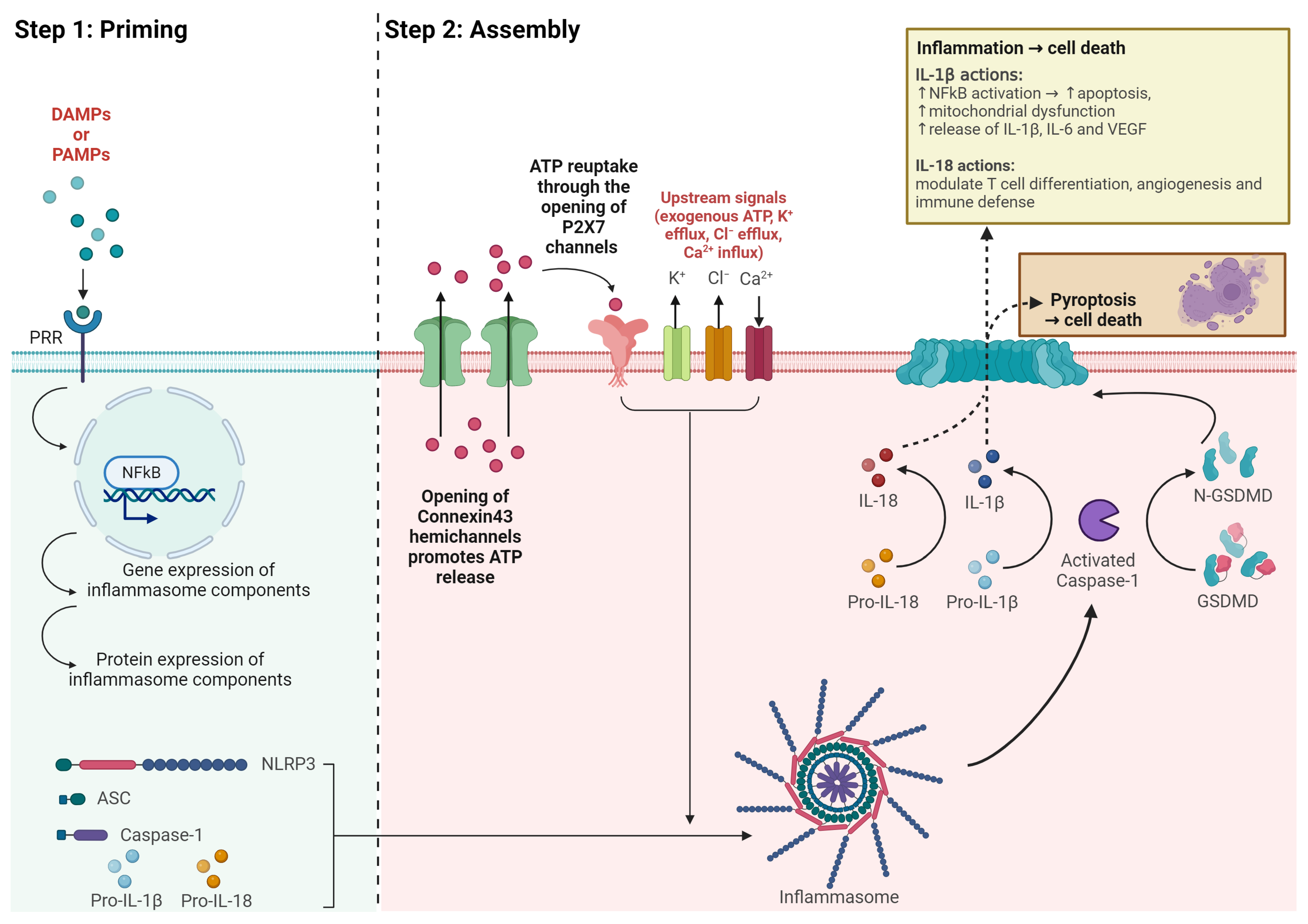
Inflammasomes are large protein complexes formed from the assembly of several copies of a monomer containing a sensor, an adaptor, and an effector [7]. For the NLRP3 inflammasome, NLRP3 is the sensor and it consists of three protein domains: a pyrin domain (PYD), a nucleotide oligomerization domain (NOD), which has adenosine triphosphatase (ATPase) activity that allows it to self-associate when assembling to form the large complex, and a leucine-rich domain (LRR) that plays a role in autoinhibition. The adaptor is the apoptosis-associated speck-like protein containing a caspase recruitment domain (CARD) (ASC), previously known as PYCARD. As its name suggests, it is made from two domains: a PYD and a CARD. The effector, caspase-1, includes a central catalytic domain (p20) and a small catalytic subunit (p10) [7,37]. Upon stimulation, NLRP3 oligomerizes through homotypic interactions within its NOD domains. The resulting molecule subsequently recruits ASC through PYD–PYD interactions, giving rise to a concentrated cluster of ASC filaments (ASC specks) [7,37]. Subsequently, ASC specks recruit caspase-1 through CARD–CARD interactions, prompting proximity-induced self-cleavage of caspase-1 into its active form. The activated caspase-1, in turn, plays a pivotal role in catalyzing the cleavage of membrane-bound precursors of IL-1β and IL-18 into their active forms, thereby instigating downstream inflammatory cascades. Activated caspase-1 also cleaves and activates gasdermin D, resulting in the formation of plasma membrane pores by amino-terminal fragments of gasdermin D, which facilitates the secretion of IL-1β and IL-18 and results in pyroptosis (Figure 2) [39].

4.3. Clinical Evidence of the NLRP3 Inflammasome in DR Progression

Clinical studies have shown that the protein and mRNA levels of NLRP3-inflammasome components, NLRP3, and caspase-1, as well as inflammatory biomarkers, IL-1β and IL-18, in the vitreous increase with DR severity levels [40,41]. A systematic literature review showed that DR progression correlates with increasing levels of inflammasome biomarkers in both the vitreous and serum, suggesting serum inflammasome levels can potentially be used to predict DR progression [42]. Moreover, a clinical pilot study showed that in a cohort of patients who have undergone bariatric surgery, the group that progressed in DR 1 year post-surgery had a significant increase in serum IL-18, one of the activated inflammasome biomarkers, compared to those who regressed or remained stable [43]. Collectively, these studies strongly suggest that the NLRP3 inflammasome becomes increasingly activated as DR becomes more severe.

4.4. NLRP3 Inflammasome Activation

As the activation of the NLRP3 inflammasome is capable of instigating downstream inflammation, strict regulation of the activation process is imperative to prevent excessive inflammation. This regulation is accomplished through a two-step activation process. The initial “priming” step ensures that there is adequate cellular expression of inflammasome-associated proteins (NLRP3, ASC, pro-caspase-1, pro-IL-1β, and pro-IL-18). NLRP3 and pro-IL-1β are typically expressed at low levels in cells; therefore, their transcription is increased during the priming step. This is promoted via detection of PAMPs and DAMPs, activation of NF-κB, and subsequent gene transcription by cytokines such as tumor necrosis factor (TNF)-α and IL-1β [7]. While pro-IL-18 is constitutively expressed in cells, its transcription level is also elevated [44]. ASC and pro-caspase-1 are sufficiently expressed regardless of the cell’s inflammatory state [45]. The priming stage also encompasses the post-translational modification of NLRP3, maintaining it in an inactive state but still responsive to signals.
The subsequent “assembly” step involves the oligomerization of NLRP3 monomers, with the recruitment of ASC and pro-caspase-1, leading to the aggregation of the large inflammasome complex. This occurs when a primed cell is exposed to an activator signal. Numerous activators capable of triggering NLRP3 inflammasome activation have been identified but the exact mechanisms involved remain poorly understood. A common feature shared by many activators is the dependency on K+ efflux to drive inflammasome activation [45]. These include activators that form pores or ion channels on the cell surface membrane, such as ionophores, nigericin, and gramicidin. Mugisho et al. [46] showed that in DR, the opening of connexin43 hemichannels promotes the release of ATP and subsequently drives the reuptake of extracellular ATP by P2X7 channels, activating the inflammasome [45,46]. While blocking connexin43 hemichannels prevented inflammasome activation, replacing the extracellular ATP resulted in the reactivation of the inflammasome pathway [46]. Disrupted lysosomal integrity caused by phagocytosis of crystalline materials (uric acid, cholesterol, silica, asbestos) and proteinaceous aggregates (amyloid-beta or amylin), also activate the NLRP3 inflammasome via K+ efflux [5,45]. So far, the only known canonical inflammasome activators independent of K+ efflux are glycolysis inhibition, mitochondrial NADH oxidase inhibition, and the displacement of hexokinase 2 from the mitochondria, which are associated with metabolic dysfunction [45]. In addition, mitochondrial reactive oxygen species, lipid cardiolipin, and mitophagy—which are related to mitochondrial dysfunction—have been suggested as potential inflammasome activators. However, additional evidence is required to determine whether they act upstream or downstream of NLRP3 inflammasome activation [45].
K+ efflux is also a requirement in the NLRP3 inflammasome pathways that involve other caspases instead of caspase-1. In the non-canonical inflammasome pathway triggered by lipopolysaccharide (LPS) from Gram-negative bacteria, activation of caspase-4 in humans (or caspase-11 in mice) leads to the opening of pannexin-1 channels, promoting ATP release and K+ efflux essential for NLRP3 inflammasome activation. The increased extracellular ATP engages with P2X7 receptors, forming P2X7 pores that promote pyroptosis [5]. Active caspase-4 also promotes pyroptosis via cleavage of gasdermin D and the subsequent formation of cell surface pores. Necroptosis, a form of cell death initiated by RIP3 and pseudokinase mixed-lineage kinase domain-like protein (MLKL) that is triggered by caspase-8 inhibition, forms pores on the plasma membrane and also relies on K+ efflux to activate the NLRP3 inflammasome. In contrast, alternative NLRP3 inflammasome activation is independent of K+ efflux. This pathway is specific to monocytes that release active IL-1β in response to LPS and require receptor-interacting serine/threonine-protein kinase 1 (RIPK1), Fas-associated death domain protein (FADD), and caspase-8 to activate NLRP3 [5].

4.5. Cytokine Release

IL-1β and IL-18 are the first two pro-inflammatory cytokines released directly as a result of NLRP3 inflammasome activation. They are both released by their precursor forms via proteolytic cleavage by cleaved caspase-1 as a result of inflammasome activation. Physiologically, IL-1β is elevated post-prandially to upregulate insulin levels; however, a chronic increase in IL-1β levels enhances glucose uptake into macrophages, causing insulin resistance in immune cells [47]. In patients with diabetes, IL-1β has been shown to mediate insulin resistance and glucotoxicity in pancreatic β cells [48,49]. Although IL-1β-induced inflammatory pathways in DR have not been clarified, preclinical studies suggest that IL-1β may contribute to DR capillary degeneration by increasing mitochondrial dysfunction and increasing apoptosis of retinal endothelial cells through the upregulation of transcription factor NF-ĸB [50,51,52]. Furthermore, IL-1β can induce its own expression in retinal endothelial cells [53] and stimulate the release of other pro-inflammatory cytokines, including IL-6 and VEGF, perpetrating inflammation [54]. Inhibition of the caspase-1-IL-1β pathway using minocycline, a caspase-1 inhibitor, has also been shown to ameliorate capillary degeneration in DR [55]. IL-18 is a pleiotropic cytokine and its interaction with various cytokines helps shape the immune response to different challenges. It works with IL-12 or IL-15 to induce the production of interferon-gamma (IFN)-γ leading to differentiation of T cells to type 1 T helper cells (Th1) [56]. During tissue injury, IL-12 and IL-18 can stimulate natural killer cells to secrete IFN-γ to enhance immune defense and modulate tissue regeneration [57]. Furthermore, IL-18 and IL-5 play a major role in eosinophilic immune disorders [58]. As the function of IL-18 appears to change in the presence of different cytokines, it is difficult to conclude its role; there is a heated debate on whether IL-18 is pro- or anti-angiogenic in choroidal neovascularization found in age-related macular degeneration [59,60,61,62], and its function in DR also remains to be determined.

4.6. Inflammasome-Targeting Novel Therapeutics

Currently, there are no clinical DR interventions designed to target the NLRP3 inflammasome; however, statins [63], fenofibrate [31], and metformin [8]—which are used routinely to manage DR-associated risk factors—have been shown to affect NLRP3 inflammasome activation in patients with T2DM. Fenofibrate, as mentioned previously, has shown beneficial effects in reducing the DR progression rate as well as the need for PRP. While it is generally used for dyslipidemia due to its ability to remarkably reduce TG and increase HDL-C, a different mechanism is involved in its anti-angiogenic effect. Studies suggest that as a peroxisome proliferator-activated receptor-alpha (PPAR-α) agonist, fenofibrate may ameliorate DR through suppression of pro-inflammatory transcription factors NF-ĸB and activator protein-1, inhibition of vascular endothelial growth factor (VEGF) production, prevention of apoptosis by inhibiting activation of adenosine monophosphate (AMP)-activated protein kinase, and reduction in oxidative stress by upregulating superoxide dismutase [64,65,66,67]. It may also benefit DR through suppressing angiogenesis via inhibiting cytochrome P450 epoxygenase 2C activity [68]. A phase 3 clinical trial is currently recruiting to investigate fenofibrate’s effect on preventing DR progression (NCT04661358).
Metformin has been used to control hyperglycemia in T2DM for many years, however, its mechanism of action is still not fully elucidated [69]. Fan et al. [10] who enrolled 10,044 patients with newly diagnosed T2DM aged 20 years and above, found a 24% reduced risk of developing NPDR in patients treated with metformin compared to the non-metformin group. Among those with NPDR, the risk of developing sight-threatening DR was also lower in the metformin group (43/860) compared to the non-metformin group (14/182) but only borderline statistically significant (adjusted hazard ratio = 0.54, p = 0.0545); however, the size of the participant group was much smaller in the non-metformin group. Furthermore, Shao et al. [70] demonstrated that metformin enhances the outcome of anti-VEGF injections (10 mg/0.2 mL ranibizumab, 10 mg/0.2 mL conbercept, or 40 mg/0.1 mL aflibercept) in patients with DME. Compared to the non-metformin group, vision improved in a larger proportion of patients in the metformin group at the three-month (69.2% in the metformin group vs. 41.7% in the non-metformin group gained more than 15 letters from baseline, p = 0.0498) and six-month follow-up (73.1% % in the metformin group vs. 45.8% in the non-metformin group gained more than 15 letters from baseline, p = 0.0495). Central macular thickness was also reduced significantly more in the metformin-treated group compared to the non-metformin group (p < 0.05). An early phase 1 trial (NCT02587741) is currently in the recruitment stage to compare DR progression among T2DM patients treated with metformin and insulin. Although no study has directly investigated the effect of metformin on DR progression, Lee et al. [8] found that metformin reduces the secretion of IL-1β and IL-18 by macrophages derived from peripheral blood monoclonal cells (PBMC) from patients with diabetes. Bullon et al. [9] also observed significantly reduced levels of NLRP3 protein expression in PBMC of individuals without diabetes. Preclinical studies have also indicated the potential use of metformin in ameliorating diabetic cardiomyopathy [71], psoriasis [72], and acute respiratory distress syndrome [73] by inhibiting the NLRP3 inflammasome pathway. As such, the anti-inflammatory effect of metformin may be associated with NLRP3 inflammasome activation. Moreover, metformin has been shown to inhibit inflammation, retinal cell death, and choroidal neovascularization in neovascular age-related macular degeneration, which, similar to PDR, also involves retinal vascular leakage [74]. Nevertheless, more research is required to investigate the effect of metformin on DR progression [8,9].
Several clinical trials are investigating NLRP3 inflammasome inhibitors as potential DR treatment (Table 1). Minocycline, a broad-spectrum antibiotic that has been shown to inhibit NLRP3 inflammasome activation at transcriptional and posttranscriptional levels [75], is also currently in phase 2 clinical trials (NCT01120899). Tonabersat is a connexin43 hemichannel blocker that prevents ATP efflux through opened connexin43 hemichannels during DR. As ATP efflux can stimulate the assembly stage of inflammasome activation, tonabersat could potentially be an upstream blocker of the activated NLRP3 inflammasome pathway, and it is currently being evaluated for its effect in DME in a phase 2 clinical trial (NCT05727891).

5. Conclusions

Evidence through clinical trials has begun to note that specific drugs (such as fenofibrate and metformin) appear effective in slowing DR progression independent of lipid-lowering and hyperglycemic control, respectively. Although the exact pathways remain to be clarified, they are able to ameliorate vascular lesions in DR through multiple routes such as inhibition of pro-inflammatory cytokine release and pro-inflammatory transcription factor activation, reduction in oxidative damage, enhancement in protective T regulatory cells, or a combination of these. Novel therapeutics that inhibit the activation of the NLRP3 inflammasome pathway may provide more effective treatments for controlling DR progression in the future. There are a few limitations in this review that should be acknowledged. Firstly, the role of the inflammasome in DR progression was not investigated in other types of diabetes besides T2DM. We did not address T1DM or other subtypes such as gestational diabetes, as their disease mechanism is different from T2DM, which primarily involves metabolic dysregulation. Secondly, our focus was restricted to DR but not DME, which was due to the current paucity of research specifically linking the inflammasome to DME. As more studies emerge, future reviews should aim to include this condition for a more comprehensive understanding of diabetic retinal complications.
Thirdly, we concentrated on the NLRP3 inflammasome—the most widely characterized inflammasome—but did not discuss the association between DR and other inflammasome family members, including NLRP1, NLRP2, AIM2, and (NLRC4) (for a full review see [28]). Despite these limitations, our review shows that the NLRP3 inflammasome is a potential target in novel therapeutics to stop DR progression.

Supplementary Materials

The following supporting information can be downloaded at https://www.mdpi.com/article/10.3390/ijtm4030027/s1, File S1: Google Scholar Data.

Author Contributions

Conceptualization, C.Y.J.K. and O.O.M.; writing—original draft preparation, C.Y.J.K.; writing—review and editing, O.O.M., R.M. and I.D.R.; supervision, O.O.M., R.M. and I.D.R.; project administration, O.O.M.; funding acquisition, O.O.M. All authors have read and agreed to the published version of the manuscript.

Funding

C.Y.J.K. is funded by the New Zealand Association of Optometrists Education and Research Fund, the New Zealand Optometric Vision Research Foundation Research Grant, and the Vernon Tews Education Trust. O.O.M. is funded by an Auckland Medical Research Foundation (AMRF) Postdoctoral Fellowship (grant number: 1323001), an AMRF Project Grant (grant number: 1121013), and a Health Research Council Emerging Researcher First Grant (grant number: 22/546). I.D.R.’s directorship is supported by the Buchanan Charitable Foundation.

Institutional Review Board Statement

Not applicable.

Informed Consent Statement

Not applicable.

Data Availability Statement

Sources of data reported have been included in the text.

Conflicts of Interest

O.O.M. is an inventor of patents relating to cytokine modulation in chronic diseases. C.Y.J.K., I.D.R. and R.M. declare that there are no conflicts of interest. The funders had no role in the design of this study; in the collection, analyses, or interpretation of data; in the writing of the manuscript; or in the decision to publish the results.

References

  1. Teo, Z.L.; Tham, Y.-C.; Yu, M.; Chee, M.L.; Rim, T.H.; Cheung, N.; Bikbov, M.M.; Wang, Y.X.; Tang, Y.; Lu, Y. Global prevalence of diabetic retinopathy and projection of burden through 2045: Systematic review and meta-analysis. Ophthalmology 2021, 128, 1580–1591. [Google Scholar] [CrossRef]
  2. Steinmetz, J.D.; Bourne, R.R.; Briant, P.S.; Flaxman, S.R.; Taylor, H.R.; Jonas, J.B.; Abdoli, A.A.; Abrha, W.A.; Abualhasan, A.; Abu-Gharbieh, E.G. Causes of blindness and vision impairment in 2020 and trends over 30 years, and prevalence of avoidable blindness in relation to VISION 2020: The Right to Sight: An analysis for the Global Burden of Disease Study. Lancet Glob. Health 2021, 9, e144–e160. [Google Scholar] [CrossRef]
  3. Pan, W.W.; Lin, F.; Fort, P.E. The innate immune system in diabetic retinopathy. Prog. Retin. Eye Res. 2021, 84, 100940. [Google Scholar] [CrossRef]
  4. Abderrazak, A.; Syrovets, T.; Couchie, D.; El Hadri, K.; Friguet, B.; Simmet, T.; Rouis, M. NLRP3 inflammasome: From a danger signal sensor to a regulatory node of oxidative stress and inflammatory diseases. Redox Biol. 2015, 4, 296–307. [Google Scholar] [CrossRef] [PubMed]
  5. He, Y.; Hara, H.; Núñez, G. Mechanism and regulation of NLRP3 inflammasome activation. Trends Biochem. Sci. 2016, 41, 1012–1021. [Google Scholar] [CrossRef]
  6. Shao, B.-Z.; Xu, Z.-Q.; Han, B.-Z.; Su, D.-F.; Liu, C. NLRP3 inflammasome and its inhibitors: A review. Front. Pharmacol. 2015, 6, 262. [Google Scholar] [CrossRef] [PubMed]
  7. Swanson, K.V.; Deng, M.; Ting, J.P.-Y. The NLRP3 inflammasome: Molecular activation and regulation to therapeutics. Nat. Rev. Immunol. 2019, 19, 477–489. [Google Scholar] [CrossRef] [PubMed]
  8. Lee, H.-M.; Kim, J.-J.; Kim, H.J.; Shong, M.; Ku, B.J.; Jo, E.-K. Upregulated NLRP3 inflammasome activation in patients with type 2 diabetes. Diabetes 2013, 62, 194–204. [Google Scholar] [CrossRef]
  9. Bullón, P.; Alcocer-Gómez, E.; Carrión, A.M.; Marín-Aguilar, F.; Garrido-Maraver, J.; Román-Malo, L.; Ruiz-Cabello, J.; Culic, O.; Ryffel, B.; Apetoh, L. AMPK phosphorylation modulates pain by activation of NLRP3 inflammasome. Antioxid. Redox. Signal. 2016, 24, 157–170. [Google Scholar] [CrossRef]
  10. Fan, Y.-P.; Wu, C.-T.; Lin, J.-L.; Hsiung, C.A.; Liu, H.Y.; Lai, J.-N.; Yang, C.-C. Metformin treatment is associated with a decreased risk of nonproliferative diabetic retinopathy in patients with type 2 diabetes mellitus: A population-based cohort study. J. Diabetes Res. 2020, 2020, 9161039. [Google Scholar] [CrossRef]
  11. American Diabetes Association Professional Practice Committee. 3. Prevention or delay of diabetes and associated comorbidities: Standards of care in diabetes—2024. Diabetes Care 2024, 47, S43–S51. [Google Scholar] [CrossRef] [PubMed]
  12. Assessment, G. 6. Glycemic targets: Standards of medical care in diabetes—2022. Diabetes Care 2022, 45, S83. [Google Scholar]
  13. Stratton, I.; Kohner, E.; Aldington, S.; Turner, R.; Holman, R.; Manley, S.; Matthews, D. UKPDS 50: Risk factors for incidence and progression of retinopathy in Type II diabetes over 6 years from diagnosis. Diabetologia 2001, 44, 156–163. [Google Scholar] [CrossRef] [PubMed]
  14. Klonoff, D.C. United Kingdom prospective diabetes study follow-up studies establish a legacy effect of therapy for hyperglycemia but not hypertension. J. Diabetes Sci. Technol. 2008, 2, 922–924. [Google Scholar] [CrossRef] [PubMed]
  15. The United Kingdom Prospective Diabetes Study (UKPDS) Group. Intensive blood-glucose control with sulphonylureas or insulin compared with conventional treatment and risk of complications in patients with type 2 diabetes (UKPDS 33). Lancet 1998, 352, 837–853. [Google Scholar] [CrossRef]
  16. Action to Control Cardiovascular Risk in Diabetes Follow-On (ACCORDION) Eye Study Group. Persistent effects of intensive glycemic control on retinopathy in type 2 diabetes in the action to control cardiovascular risk in diabetes (ACCORD) follow-on study. Diabetes Care 2016, 39, 1089–1100. [Google Scholar] [CrossRef] [PubMed]
  17. ADVANCE Collaborative Group. Intensive blood glucose control and vascular outcomes in patients with type 2 diabetes. N. Engl. J. Med. 2008, 358, 2560–2572. [Google Scholar] [CrossRef]
  18. Azad, N.; Agrawal, L.; Emanuele, N.V.; Klein, R.; Bahn, G.D.; Reaven, P. Association of blood glucose control and pancreatic reserve with diabetic retinopathy in the Veterans Affairs Diabetes Trial (VADT). Diabetologia 2014, 57, 1124–1131. [Google Scholar] [CrossRef] [PubMed]
  19. Feldman-Billard, S.; Larger, É.; Massin, P. Early worsening of diabetic retinopathy after rapid improvement of blood glucose control in patients with diabetes. Diabetes Metab. 2018, 44, 4–14. [Google Scholar] [CrossRef]
  20. Lim, S.W.; van Wijngaarden, P.; Harper, C.A.; Al-Qureshi, S.H. Early worsening of diabetic retinopathy due to intensive glycaemic control. Clin. Exp. Ophthalmol. 2019, 47, 265–273. [Google Scholar] [CrossRef]
  21. Chew, E.Y.; Davis, M.D.; Danis, R.P.; Lovato, J.F.; Perdue, L.H.; Greven, C.; Genuth, S.; Goff, D.C.; Leiter, L.A.; Ismail-Beigi, F. The effects of medical management on the progression of diabetic retinopathy in persons with type 2 diabetes: The Action to Control Cardiovascular Risk in Diabetes (ACCORD) Eye Study. Ophthalmology 2014, 121, 2443–2451. [Google Scholar] [CrossRef] [PubMed]
  22. The United Kingdom Prospective Diabetes Study (UKPDS) Group. Tight blood pressure control and risk of macrovascular and microvascular complications in type 2 diabetes: UKPDS 38. Br. Med. J. 1998, 317, 703–713. [Google Scholar] [CrossRef]
  23. Do, D.V.; Han, G.; Abariga, S.A.; Sleilati, G.; Vedula, S.S.; Hawkins, B.S. Blood pressure control for diabetic retinopathy. Cochrane Database Syst. Rev. 2023, 3, CD006127. [Google Scholar]
  24. Wang, B.; Wang, F.; Zhang, Y.; Zhao, S.-H.; Zhao, W.-J.; Yan, S.-L.; Wang, Y.-G. Effects of RAS inhibitors on diabetic retinopathy: A systematic review and meta-analysis. Lancet Diabetes Endocrinol. 2015, 3, 263–274. [Google Scholar] [CrossRef] [PubMed]
  25. Klein, B.E.; Klein, R.; Moss, S.E. Is serum cholesterol associated with progression of diabetic retinopathy or macular edema in persons with younger-onset diabetes of long duration? Am. J. Ophthalmol. 1999, 128, 652–654. [Google Scholar] [CrossRef]
  26. Zhou, Y.; Wang, C.; Shi, K.; Yin, X. Relationship between dyslipidemia and diabetic retinopathy: A systematic review and meta-analysis. Medicine 2018, 97, e12283. [Google Scholar] [CrossRef]
  27. Cikamatana, L.; Mitchell, P.; Rochtchina, E.; Foran, S.; Wang, J. Five-year incidence and progression of diabetic retinopathy in a defined older population: The Blue Mountains Eye Study. Eye 2007, 21, 465–471. [Google Scholar] [CrossRef]
  28. Morisaki, N.; Watanabe, S.; Kobayashi, J.; Kanzaki, T.; Takahashi, K.; Yokote, K.; Tezuka, M.; Tashiro, J.; Inadera, H.; Saito, Y. Diabetic control and progression of retinopathy in elderly patients: Five-year follow-up study. J. Am. Geriatr. Soc. 1994, 42, 142–145. [Google Scholar] [CrossRef] [PubMed]
  29. Keech, A.C.; Mitchell, P.; Summanen, P.; O’Day, J.; Davis, T.M.; Moffitt, M.; Taskinen, M.-R.; Simes, R.J.; Tse, D.; Williamson, E. Effect of fenofibrate on the need for laser treatment for diabetic retinopathy (FIELD study): A randomised controlled trial. Lancet 2007, 370, 1687–1697. [Google Scholar] [CrossRef]
  30. Chandra, A.; Kaur, P.; Sahu, S.K.; Mittal, A. A new insight into the treatment of diabetes by means of pan PPAR agonists. Chem. Biol. Drug Des. 2022, 100, 947–967. [Google Scholar] [CrossRef]
  31. Liu, Q.; Zhang, F.; Zhang, X.; Cheng, R.; Ma, J.-x.; Yi, J.; Li, J. Fenofibrate ameliorates diabetic retinopathy by modulating Nrf2 signaling and NLRP3 inflammasome activation. Mol. Cell. Biochem. 2018, 445, 105–115. [Google Scholar] [CrossRef] [PubMed]
  32. Deng, Y.; Han, X.; Yao, Z.; Sun, Y.; Yu, J.; Cai, J.; Ren, G.; Jiang, G.; Han, F.; Hospital, Z.X. PPARα agonist stimulated angiogenesis by improving endothelial precursor cell function via a NLRP3 inflammasome pathway. Cell. Physiol. Biochem. 2017, 42, 2255–2266. [Google Scholar] [CrossRef] [PubMed]
  33. Paik, S.; Kim, J.K.; Silwal, P.; Sasakawa, C.; Jo, E.-K. An update on the regulatory mechanisms of NLRP3 inflammasome activation. Cell. Mol. Immunol. 2021, 18, 1141–1160. [Google Scholar] [CrossRef]
  34. Jacobs, S.R.; Damania, B. NLRs, inflammasomes, and viral infection. J. Leukoc. Biol. 2012, 92, 469–477. [Google Scholar] [CrossRef] [PubMed]
  35. Martinon, F.; Burns, K.; Tschopp, J. The inflammasome: A molecular platform triggering activation of inflammatory caspases and processing of proIL-β. Mol. Cell 2002, 10, 417–426. [Google Scholar] [CrossRef]
  36. Dinarello, C.A. A clinical perspective of IL-1β as the gatekeeper of inflammation. Eur. J. Immunol. 2011, 41, 1203–1217. [Google Scholar] [CrossRef] [PubMed]
  37. Velloso, F.J.; Trombetta-Lima, M.; Anschau, V.; Sogayar, M.C.; Correa, R.G. NOD-like receptors: Major players (and targets) in the interface between innate immunity and cancer. Biosci. Rep. 2019, 39, BSR20181709. [Google Scholar] [CrossRef]
  38. Duez, H.; Pourcet, B. Nuclear receptors in the control of the NLRP3 inflammasome pathway. Front. Endocrinol. 2021, 12, 630536. [Google Scholar] [CrossRef]
  39. Wang, C.; Yang, T.; Xiao, J.; Xu, C.; Alippe, Y.; Sun, K.; Kanneganti, T.-D.; Monahan, J.B.; Abu-Amer, Y.; Lieberman, J. NLRP3 inflammasome activation triggers gasdermin D–independent inflammation. Sci. Immunol. 2021, 6, eabj3859. [Google Scholar] [CrossRef]
  40. Loukovaara, S.; Piippo, N.; Kinnunen, K.; Hytti, M.; Kaarniranta, K.; Kauppinen, A. NLRP3 inflammasome activation is associated with proliferative diabetic retinopathy. Acta Ophthalmol. 2017, 95, 803–808. [Google Scholar] [CrossRef]
  41. Chen, H.; Zhang, X.; Liao, N.; Mi, L.; Peng, Y.; Liu, B.; Zhang, S.; Wen, F. Enhanced Expression of NLRP3 Inflammasome-Related Inflammation in Diabetic Retinopathy. Investig. Ophthalmol. Vis. Sci. 2018, 59, 978–985. [Google Scholar] [CrossRef] [PubMed]
  42. Kuo, C.Y.J.; Murphy, R.; Rupenthal, I.D.; Mugisho, O.O. Correlation between the progression of diabetic retinopathy and inflammasome biomarkers in vitreous and serum—A systematic review. BMC Ophthalmol. 2022, 22, 238. [Google Scholar] [CrossRef] [PubMed]
  43. Kuo, C.Y.-J.; Rupenthal, I.D.; Booth, M.; Murphy, R.; Mugisho, O.O. Systemic Inflammasome Biomarkers as Predictors of Diabetic Retinopathy Progression: Evidence from a Pilot Study. Future Pharm. 2023, 3, 612–624. [Google Scholar] [CrossRef]
  44. Zhu, Q.; Kanneganti, T.-D. Cutting edge: Distinct regulatory mechanisms control proinflammatory cytokines IL-18 and IL-1β. J. Immunol. 2017, 198, 4210–4215. [Google Scholar] [CrossRef] [PubMed]
  45. Mangan, M.S.; Olhava, E.J.; Roush, W.R.; Seidel, H.M.; Glick, G.D.; Latz, E. Targeting the NLRP3 inflammasome in inflammatory diseases. Nat. Rev. Drug Discov. 2018, 17, 588–606. [Google Scholar] [CrossRef] [PubMed]
  46. Mugisho, O.O.; Green, C.R.; Kho, D.T.; Zhang, J.; Graham, E.S.; Acosta, M.L.; Rupenthal, I.D. The inflammasome pathway is amplified and perpetuated in an autocrine manner through connexin43 hemichannel mediated ATP release. Biochim. Biophys. Acta 2018, 1862, 385–393. [Google Scholar] [CrossRef] [PubMed]
  47. Dror, E.; Dalmas, E.; Meier, D.T.; Wueest, S.; Thévenet, J.; Thienel, C.; Timper, K.; Nordmann, T.M.; Traub, S.; Schulze, F. Postprandial macrophage-derived IL-1β stimulates insulin, and both synergistically promote glucose disposal and inflammation. Nat. Immunol. 2017, 18, 283–292. [Google Scholar] [CrossRef] [PubMed]
  48. Vandanmagsar, B.; Youm, Y.-H.; Ravussin, A.; Galgani, J.E.; Stadler, K.; Mynatt, R.L.; Ravussin, E.; Stephens, J.M.; Dixit, V.D. The NLRP3 inflammasome instigates obesity-induced inflammation and insulin resistance. Nat. Med. 2011, 17, 179–188. [Google Scholar] [CrossRef]
  49. Maedler, K.; Sergeev, P.; Ris, F.; Oberholzer, J.; Joller-Jemelka, H.I.; Spinas, G.A.; Kaiser, N.; Halban, P.A.; Donath, M.Y. Glucose-induced β cell production of IL-1β contributes to glucotoxicity in human pancreatic islets. J. Clin. Investig. 2002, 110, 851–860. [Google Scholar] [CrossRef]
  50. Kowluru, R.; Odenbach, S. Role of interleukin-1β in the pathogenesis of diabetic retinopathy. Br. J. Ophthalmol. 2004, 88, 1343–1347. [Google Scholar] [CrossRef]
  51. Vallejo, S.; Palacios, E.; Romacho, T.; Villalobos, L.; Peiró, C.; Sánchez-Ferrer, C.F. The interleukin-1 receptor antagonist anakinra improves endothelial dysfunction in streptozotocin-induced diabetic rats. Cardiovasc. Diabetol. 2014, 13, 158. [Google Scholar] [CrossRef]
  52. Kowluru, R.A.; Mohammad, G.; Santos, J.M.; Tewari, S.; Zhong, Q. Interleukin-1β and mitochondria damage, and the development of diabetic retinopathy. J. Ocul. Biol. Dis. Infor. 2011, 4, 3–9. [Google Scholar] [CrossRef]
  53. Liu, Y.; Costa, M.B.; Gerhardinger, C. IL-1β is upregulated in the diabetic retina and retinal vessels: Cell-specific effect of high glucose and IL-1β autostimulation. PLoS ONE 2012, 7, e36949. [Google Scholar] [CrossRef] [PubMed]
  54. Mesquida, M.; Drawnel, F.; Fauser, S. The role of inflammation in diabetic eye disease. Semin. Immunopathol. 2019, 41, 427–445. [Google Scholar] [CrossRef]
  55. Vincent, J.A.; Mohr, S. Inhibition of caspase-1/interleukin-1β signaling prevents degeneration of retinal capillaries in diabetes and galactosemia. Diabetes 2007, 56, 224–230. [Google Scholar] [CrossRef]
  56. Keyel, P.A. How is inflammation initiated? Individual influences of IL-1, IL-18 and HMGB1. Cytokine 2014, 69, 136–145. [Google Scholar] [CrossRef] [PubMed]
  57. Thomas, H.; Jäger, M.; Mauel, K.; Brandau, S.; Lask, S.; Flohé, S.B. Interaction with mesenchymal stem cells provokes natural killer cells for enhanced IL-12/IL-18-induced interferon-gamma secretion. Mediat. Inflamm. 2014, 2014, 143463. [Google Scholar] [CrossRef] [PubMed]
  58. Kandikattu, H.K.; Venkateshaiah, S.U.; Mishra, A. Synergy of Interleukin (IL)-5 and IL-18 in eosinophil mediated pathogenesis of allergic diseases. Cytokine Growth Factor Rev. 2019, 47, 83–98. [Google Scholar] [CrossRef]
  59. Hirano, Y.; Yasuma, T.; Mizutani, T.; Fowler, B.J.; Tarallo, V.; Yasuma, R.; Kim, Y.; Bastos-Carvalho, A.; Kerur, N.; Gelfand, B.D. IL-18 is not therapeutic for neovascular age-related macular degeneration. Nat. Med. 2014, 20, 1372–1375. [Google Scholar] [CrossRef]
  60. Doyle, S.L.; Campbell, M.; Ozaki, E.; Salomon, R.G.; Mori, A.; Kenna, P.F.; Farrar, G.J.; Kiang, A.-S.; Humphries, M.M.; Lavelle, E.C. NLRP3 has a protective role in age-related macular degeneration through the induction of IL-18 by drusen components. Nat. Med. 2012, 18, 791–798. [Google Scholar] [CrossRef]
  61. Doyle, S.L.; López, F.J.; Celkova, L.; Brennan, K.; Mulfaul, K.; Ozaki, E.; Kenna, P.F.; Kurali, E.; Hudson, N.; Doggett, T. IL-18 immunotherapy for neovascular AMD: Tolerability and efficacy in nonhuman primates. Investig. Ophthalmol. Vis. Sci. 2015, 56, 5424–5430. [Google Scholar] [CrossRef] [PubMed]
  62. Doyle, S.L.; Ozaki, E.; Brennan, K.; Humphries, M.M.; Mulfaul, K.; Keaney, J.; Kenna, P.F.; Maminishkis, A.; Kiang, A.-S.; Saunders, S.P. IL-18 attenuates experimental choroidal neovascularization as a potential therapy for wet age-related macular degeneration. Sci. Transl. Med. 2014, 6, 230ra244. [Google Scholar] [CrossRef] [PubMed]
  63. Satoh, M.; Tabuchi, T.; Itoh, T.; Nakamura, M. NLRP3 inflammasome activation in coronary artery disease: Results from prospective and randomized study of treatment with atorvastatin or rosuvastatin. Clin. Sci. 2014, 126, 233–241. [Google Scholar] [CrossRef] [PubMed]
  64. Noonan, J.E.; Jenkins, A.J.; Ma, J.-X.; Keech, A.C.; Wang, J.J.; Lamoureux, E.L. An update on the molecular actions of fenofibrate and its clinical effects on diabetic retinopathy and other microvascular end points in patients with diabetes. Diabetes 2013, 62, 3968–3975. [Google Scholar] [CrossRef]
  65. Inoue, K.; Kataoka, S.Y.; Kawano, S.; Furukawa, T.A.; Lois, N.; Watanabe, N. Fenofibrate for diabetic retinopathy. Cochrane Database Syst. Rev. 2019, 16, 90. [Google Scholar] [CrossRef]
  66. Abcouwer, S.F. Direct effects of PPARα agonists on retinal inflammation and angiogenesis may explain how fenofibrate lowers risk of severe proliferative diabetic retinopathy. Diabetes 2013, 62, 36. [Google Scholar] [CrossRef] [PubMed]
  67. Jin, L.; Hua, H.; Ji, Y.; Jia, Z.; Peng, M.; Huang, S. Anti-inflammatory role of fenofibrate in treating diseases. Biomol. Biomed. 2023, 23, 376. [Google Scholar] [CrossRef] [PubMed]
  68. Gong, Y.; Shao, Z.; Fu, Z.; Edin, M.L.; Sun, Y.; Liegl, R.G.; Wang, Z.; Liu, C.-H.; Burnim, S.B.; Meng, S.S. Fenofibrate inhibits cytochrome P450 epoxygenase 2C activity to suppress pathological ocular angiogenesis. EBioMedicine 2016, 13, 201–211. [Google Scholar] [CrossRef] [PubMed]
  69. LaMoia, T.E.; Shulman, G.I. Cellular and molecular mechanisms of metformin action. Endocr. Rev. 2021, 42, 77–96. [Google Scholar] [CrossRef]
  70. Shao, Y.; Wang, M.; Zhu, Y.; Li, X.; Liu, J. Association of metformin treatment with enhanced effect of anti-VEGF agents in diabetic macular edema patients. Acta Diabetol. 2022, 59, 553–559. [Google Scholar] [CrossRef]
  71. Yang, F.; Qin, Y.; Wang, Y.; Meng, S.; Xian, H.; Che, H.; Lv, J.; Li, Y.; Yu, Y.; Bai, Y. Metformin inhibits the NLRP3 inflammasome via AMPK/mTOR-dependent effects in diabetic cardiomyopathy. Int. J. Biol. Sci. 2019, 15, 1010. [Google Scholar] [CrossRef] [PubMed]
  72. Tsuji, G.; Hashimoto-Hachiya, A.; Yen, V.H.; Takemura, M.; Yumine, A.; Furue, K.; Furue, M.; Nakahara, T. Metformin inhibits IL-1β secretion via impairment of NLRP3 inflammasome in keratinocytes: Implications for preventing the development of psoriasis. Cell Death Discov. 2020, 6, 11. [Google Scholar] [CrossRef] [PubMed]
  73. Xian, H.; Liu, Y.; Nilsson, A.R.; Gatchalian, R.; Crother, T.R.; Tourtellotte, W.G.; Zhang, Y.; Aleman-Muench, G.R.; Lewis, G.; Chen, W. Metformin inhibition of mitochondrial ATP and DNA synthesis abrogates NLRP3 inflammasome activation and pulmonary inflammation. Immunity 2021, 54, 1463–1477.e11. [Google Scholar] [CrossRef] [PubMed]
  74. Dang, K.-R.; Wu, T.; Hui, Y.-N.; Du, H.-J. Newly-found functions of metformin for the prevention and treatment of age-related macular degeneration. Int. J. Ophthalmol. 2021, 14, 1274. [Google Scholar] [CrossRef]
  75. Chen, W.; Zhao, M.; Zhao, S.; Lu, Q.; Ni, L.; Zou, C.; Lu, L.; Xu, X.; Guan, H.; Zheng, Z. Activation of the TXNIP/NLRP3 inflammasome pathway contributes to inflammation in diabetic retinopathy: A novel inhibitory effect of minocycline. Inflamm. Res. 2017, 66, 157–166. [Google Scholar] [CrossRef]
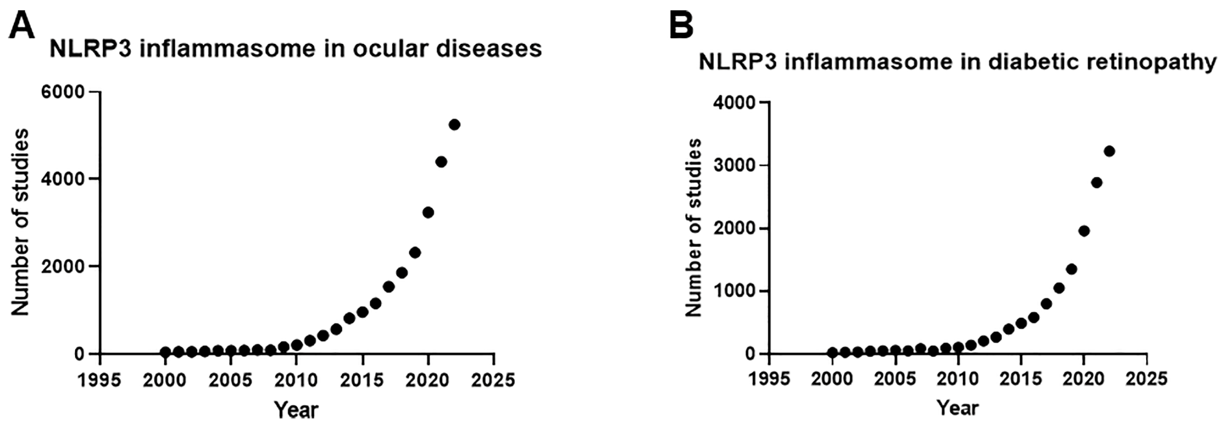
Figure 1. Rapid increase in inflammasome research over the past two decades: (A) number of studies on eye diseases associated with inflammasomes increased exponentially from year 2000 to 2022; (B) number of studies on DR associated with inflammasomes also demonstrated exponential growth from year 2000 to 2022.
Ijtm 04 00027 g001
Figure 2. Schematic illustrating the two-step inflammasome activation process: The first (priming) stage involves the transcription and translation of non-activated components of the inflammasome complex. Upon detecting danger signals from DAMPs or PAMPs, a signal is received by the nuclear transcription factor (NF-ĸB), which increases the transcription of inactive NLRP3 and precursors of interleukin (IL)-1β (pro-IL-1β) and IL-18 (pro-IL18). The opening of connexin43 hemichannels promotes the release of ATP and subsequently drives the reuptake of extracellular ATP by P2X purinoceptor 7 (P2X7) channels, activating the inflammasome. The second (assembly) stage is initiated following an upstream signal such as opening of connexin43 hemichannels, an increase in exogenous ATP concentration, K+ efflux, Cl efflux, and Ca2+ influx, which triggers the oligomerization and activation of the inflammasome complex. Subsequently, caspase-1 is cleaved into its active form, which in turn, cleaves pro-IL-1β and pro-IL-18 to IL-1β and IL-18, respectively. Subsequently, IL-1β and IL-18 are released from the cell, leading to downstream inflammation. Activated caspase-1 also cleaves and activates gasdermin D, resulting in the formation of plasma membrane pores by amino-terminal fragments of gasdermin D that facilitate the secretion of IL-1β and IL-18, resulting in pyroptosis. Illustration created with BioRender.com.
Ijtm 04 00027 g002
Table 1. Novel treatments in clinical trials for DR.
Administration RouteDrugMechanism of ActionPhasesStudy StatusClinical Trial RegistrationSponsor
Intravitreal injectionOCU200Fusion of human transferrin and human tumstatin1Not yet recruitingNCT05802329Ocugen (Malvern, PA, USA)
IBI324Anti-VEGF-A and anti-Angiotensin-2 bispecific antibody1Not yet recruitingNCT05489718Innovent Biologics (Suzhou) Co., Ltd. (Suzhou, China)
Foselutoclax
(UBX1325)
Potent small-molecule inhibitor of Bcl-xL, member of the Bcl-2 family of apoptosis-regulating proteins2RecruitingNCT06011798Unity Biotechnology, Inc. (South San Francisco, CA, USA)
EYE103
(Restoret™)
Wnt signaling pathway agonist1|2RecruitingNCT05919693EyeBiotech Ltd. (London, UK)
Infliximab
(Remicade™)
TNF-α inhibitor1|2UnknownNCT00959725Icahn School of Medicine at Mount Sinai (New York, NY, USA)
iCo-007Anti-sense drug targeting c-Raf Kinase2Terminated due to disease progression and vision reductionNCT01565148Johns Hopkins University (Baltimore, MD, USA)
REGN910-3
(Nesvacumab)
Immunoglobulin G1 (IgG1) monoclonal antibody targeting Ang22CompletedNCT02712008Regeneron Pharmaceuticals (Tarrytown, NY, USA)
Ocriplasmin
(also known as microplasmin)
Truncated recombinant form of human plasmin, used to treat vitreomacular traction with or without full-thickness macular hole2CompletedNCT00412451ThromboGenics (Louvain, Belgium)
Risuteganib
(Luminate®®)
Integrin inhibitor2CompletedNCT02348918Allegro Ophthalmics, LLC (San Juan Capistrano, CA, USA)
GB-102
(sunitinib malate)
Multiple intracellular tyrosine kinases inhibitor2CompletedNCT04085341Graybug Vision (La Jolla, CA, USA)
KVD001Targets plasma kallikrein2CompletedNCT03466099KalVista Pharmaceuticals, Ltd. (Cambridge, MA, USA)
BI 765128Undisclosed1|2CompletedNCT04919499Boehringer Ingelheim (Ingelheim, Germany)
Subcutaneous
injection
AKB-9778Activator or Tie-2, a receptor tyrosine kinase (RTK)2CompletedNCT03197870EyePoint Pharmaceuticals, Inc. (Watertown, MA, USA)
Periocular injectionAIV007Broad-spectrum tyrosine kinase inhibitor1RecruitingNCT05698329AiViva BioPharma, Inc. (Costa Mesa, CA, USA)
subconjunctival injectionSirolimusInhibition of rapamycin (mTOR), protein kinase that specifically governs cell growth, replication, and viability1|2CompletedNCT00711490National Eye Institute (NEI) (Bethesda, MD, USA)
OralMS-553Selective PKC-β inhibitor1RecruitingNCT04187443MingSight Pharmaceuticals Co., Ltd. (San Diego, CA, USA)
TonabersatConnexin43 hemichannel blocker, upstream inhibitor of the NLRP3 inflammasome pathway2RecruitingNCT05727891Jaeb Center for Health
Research (Tampa, FL, USA)
RZ402Small-molecule selective and potent plasma kallikrein inhibitor2RecruitingNCT05712720, NCT04527107Rezolute (Redwood City, CA, USA)
CU06-1004
(also known as Sac-1004)
Enhances endothelial cell survival and prevents endothelial barrier disruption; inhibits ICAM-1 and VCAM-1 expression by inhibiting NF-κB activation2Active, not recruitingNCT05573100Curacle Co., Ltd. (Seongnam-si, Republic of Korea)
RuboxistaurinActive β-selective Phosphate Kinase C inhibitor3CompletedNCT00266695Chromaderm, Inc. (Cambridge, MA, USA)
CompletedNCT00604383
CompletedNCT00090519
CompletedNCT00133952
PF-04634817Chemokine receptor (CCR2/5) antagonist2Terminated due to changes in drug development prioritizationNCT01994291Pfizer (New York, NY, USA)
BI 1467335
(formerly PXS-4728A)
Inhibits neutrophil tethering and rolling, reduces inflammation2CompletedNCT03238963Boehringer Ingelheim (Ingelheim, Germany)
MinocyclineAntibiotic binding to the bacterial 30S ribosomal subunit and inhibiting protein synthesis1|2CompletedNCT01120899National Eye Institute (NEI) (Bethesda, MD, USA)
Calcium dobesilateReduces capillary permeability, inhibits platelet aggression and blood viscosity, inhibits apoptosis of vascular endothelial cells, inhibits the expression of inflammatory and upstream VEGF regulator, ICAM-1, and protects against reactive oxygen species4UnknownNCT04283162Zhongda Hospital (Nanjing, China)
FenofibrateActivates PPARα3RecruitingNCT04661358Jaeb Center for Health
Research (Tampa, FL, USA)
YD312
(imatinib)
Inhibits excessive vascular angiogenesis observed in oxygen-induced retinopathy2UnknownNCT03635814YD Global Life Science Co., Ltd. (Seongnam, Republic of Korea)
Finerenone
(BAY94-8862)
Selective mineralocorticoid receptor antagonistNACompletedNCT04477707, NCT04795726Bayer (Leverkusen, Germany)
Intravenous injectionMethotrexateInhibits production of nucleic acid synthesis by inhibiting the enzyme dihydrofolate reductaseNATerminated as Lucentis to be an effective treatmentNCT00779142Wake Forest University (Winston-Salem, NC, USA)
Ophthalmic solution (eyedrop)OTT166Small-molecule arginylglycylaspartic acid integrin inhibitor2CompletedNCT05409235OcuTerra Therapeutics, Inc. (Boston, MA, USA)
Clinical trial information obtained from https://www.clinicaltrials.gov/ accessed on 22 May 2024.
Disclaimer/Publisher’s Note: The statements, opinions and data contained in all publications are solely those of the individual author(s) and contributor(s) and not of MDPI and/or the editor(s). MDPI and/or the editor(s) disclaim responsibility for any injury to people or property resulting from any ideas, methods, instructions or products referred to in the content.