Article

Antiproliferative Potential of Eugenia uniflora L. Leaf Essential Oil in Normal and Tumoral Human Colon Cells

by
Ana G. Sánchez
1,*,
Macarena Menoni
1,
Pamela Lombardo
2,
Eduardo Dellacassa
3,
María Angélica Severi
4,
Gabriela Ferragut
4,
Beatriz Vignale
5,
Juan Cedano
6,
María José Zuluaga
1 and
Deborah J. Keszenman
1,7,*
1
Grupo de Biofisicoquímica, Departamento de Ciencias Biológicas, CENUR Litoral Norte, Universidad de la República, Salto 50000, Uruguay
2
Fitopatología, Departamento de Ciencias Biológicas, CENUR Litoral Norte, Universidad de la República, Salto 50000, Uruguay
3
Laboratorio de Biotecnología de Aromas, Departamento de Química Orgánica, Facultad de Química, Universidad de la República, Montevideo 11800, Uruguay
4
Laboratorio de Inmunología “Dr. Alberto Nieto”, Departamento de Ciencias Biológicas, CENUR Litoral Norte, Universidad de la República, Salto 50000, Uruguay
5
Departamento de Producción Vegetal, Estación Experimental de Facultad de Agronomía, Salto 50000, Uruguay
6
Departament de Bioquímica i Biologia Molecular, Universitat Autonoma de Barcelona, 08193 Bellaterra, Spain
7
Citizen Science Program, Biology Department, Bard College, Annandale-on-Hudson, NY 12504, USA
*
Authors to whom correspondence should be addressed.
Biologics 2025, 5(3), 19; https://doi.org/10.3390/biologics5030019
Submission received: 26 May 2025 / Revised: 26 June 2025 / Accepted: 1 July 2025 / Published: 3 July 2025
(This article belongs to the Section Natural Products)

Abstract

Background/Objectives: Natural products are important in healthcare due to their accessibility and linkage to a healthy lifestyle. However, their effectiveness is uncertain due to insufficient scientific data. Cancer patients are frequent users of natural products to relieve symptoms or for chemoprevention. Eugenia uniflora leaf essential oil (EO), traditionally used for digestive disorders, emerges as a potential antineoplastic agent. We investigated the cytotoxic and antiproliferative effects of E. uniflora EO in human normal CCD 841 CoN and tumoral Caco-2 colonic cell lines. Methods: CCD 841 CoN and Caco-2 cells were exposed to different concentrations of E. uniflora EO, and the cytotoxicity was determined by MTT and Trypan Blue assays. Cell proliferation kinetics were analyzed at a low EO concentration, and the induction of DNA damage and oxidative stress was assessed by Comet and Cellular ROS assays. Results: Both cell lines exhibited cytotoxicity produced by the EO and decreased cell viability of the exposed cells and their progeny. CCD 841 CoN proliferation was impaired by low EO concentration, while the proliferation kinetics of the Caco-2 cells was modified. EO treatment induced variable DNA damage and oxidative stress depending on the cell line. Conclusions: Our results suggest that E. uniflora EO may prevent the proliferation of normal cells, inducing loss of viability. The EO produced cytotoxic and antiproliferative effects in tumoral cells by inducing DNA damage and increased oxidative stress. These effects support the consideration of E. uniflora EO (or its bioactive compounds) as a potential agent for the chemoprevention and treatment of colorectal cancer.

Graphical Abstract

1. Introduction

Natural products have played a vital role in healthcare across different cultures throughout history. Today, nearly 80% of the global population depends on traditional medicine and natural products to meet their health needs. These resources are often preferred in developing countries because of their accessibility, affordability, and connection to local cultural practices. In contrast, in developed countries, their use is linked to a healthy lifestyle and a greater concern for environmental sustainability [1,2,3,4].
Nowadays, a wide variety of natural products and herbal medicines are available over-the-counter in many countries, giving rise to a growing global market for these products. Some are used as potential therapeutic agents for diverse diseases, and others are consumed in preventive medicine [5]. However, many commercialized products are unregulated, and their effectiveness is uncertain due to insufficient scientific data. Patients with certain pathological conditions, such as cancer, more frequently use herbal medicines to relieve symptoms, mitigate adverse effects of radiotherapy or chemotherapy, improve quality of life, and serve as chemopreventive agents to help prevent recurrence or metastasis. Nonetheless, observational studies have indicated that the concomitant use of natural products with cancer therapies may negatively interact, producing adverse side effects by lowering or neutralizing the benefits of conventional treatments [1,2,6,7,8,9]. The amount of scientific data related to the components and biological effects of many natural products, as well as the clinical uses and outcomes of herbal medicine, has increased notably over the last 20 years. However, more research is still needed to identify and characterize the bioactive compounds, their benefits, and risks in disease prevention and treatment.
Cancer is one of the ten leading causes of morbidity and mortality worldwide [10,11]. It constitutes a complex pathological and often multisystemic condition affecting any part of the human body. In 2022, almost 20 million new cases were diagnosed (including non-melanoma skin cancer), and 9.7 million deaths due to cancer were recorded globally [11]. Colorectal cancer (CRC), which includes colon, rectum, and anus, is the third most common cancer and ranks second in terms of mortality. Although stabilizing or declining trends in the incidence rates for all ages combined have been observed in high-income countries, a rise in CRC among younger adults has been reported [12,13]. Additionally, there is a striking and steady increase in the incidence rate in certain regions of the world, including Eastern Europe and South America [14,15]. Behavioral and dietary changes in lifestyle, especially those occurring during early life or young adulthood, lead to an increase in the influence of risk factors such as increased consumption of red or processed meat, less fiber in the diet, and sedentarism [11,16].
In South America, numerous native plant species have been traditionally used for medicinal purposes [17,18,19,20]. Among them, those belonging to the Myrtaceae family are particularly noteworthy due to their use in traditional medicine for their antidiarrheal, antimicrobial, antioxidant, antirheumatic, and anti-inflammatory properties [17,21]. Within this family, Eugenia uniflora L., commonly known as Pitanga, is widely distributed in tropical and subtropical regions of Northeastern Argentina, Southern Brazil, and Uruguay [22,23]. Its fruits are globular berries, varying in color, with a bright appearance and a distinctive, intense flavor that ranges from sweet to slightly acidic. Pitanga fruits are rich in vitamins A and C, phosphorus, calcium, and iron [24,25]. Infusions, decoctions, and alcoholic extracts of the leaves are used in traditional medicine because of their diuretic, anti-inflammatory, carminative, and antidiarrheal effects [17]. The leaf essential oil (EO) has been reported as possessing antifungal, antibacterial, cytotoxic, and antiparasitic activities [24,26,27,28,29,30,31,32,33,34,35,36].
E. uniflora EO has been reported to contain bioactive compounds with potential antitumor properties, including antioxidant capacity, apoptotic activity, and anti-inflammatory effects [32,37,38]. SiHa (HPV 16-positive) human cervical cancer cells treated with E. uniflora leaf aqueous crude extract exhibit decreased viability, apoptotic cell death, and reduced migration. However, the same treatment does not modify the viability of normal human lymphocytes [39]. In addition, other studies have reported cytotoxic effects of the EO in various cancer cell lines [27]. Since E. uniflora EO is frequently used as herbal medicine for digestive disorders and could emerge as a natural product candidate to provide bioactive compounds for colon cancer therapy and prevention, we studied antineoplastic properties of E. uniflora leaf EO in human normal CCD 841 CoN and tumoral Caco-2 colonic cells lines by determining the effect on cell proliferation kinetics, cell viability, and induction of DNA damage and oxidative stress. Our main results present evidence of the cytotoxic and antiproliferative effects of E. uniflora EO in both human normal CCD 841 CoN and tumoral Caco-2 cell lines, as shown by the increased lethality, loss of viability, and altered proliferation kinetics. In addition, variable levels of DNA damage and oxidative stress were observed, depending on the cell line. These findings support the recognition of E. uniflora as a promising plant species with potential for chemoprevention and the treatment of CRC. This contribution aims to be particularly relevant in the ongoing research of bioactive compounds with antineoplastic properties, offering new possibilities for biotechnological applications in cancer therapy and preventive medicine.

2. Materials and Methods

2.1. Herbal Material

Leaves of E. uniflora were collected from the plant specimen VIII-7, located in the Introduction Garden of the Experimental Station of the Faculty of Agronomy (Universidad de la República) in Salto, Uruguay (31°22′ S 57°56′ W). This specimen was selected to represent the plant population to extract the essential oil from leaves. Botanical identification was carried out by the Botany Department of the Faculty of Agronomy, CENUR Litoral Norte, Salto. A voucher specimen was deposited in the herbarium of the “Museo Jardín Botánico Prof. Atilio Lombardo” in Montevideo, Uruguay, E. uniflora MVJB 29158.
Four independent collections of herbal material were carried out during Spring 2018 (October–November) and Summer 2019 (February–March). It is worth mentioning that, in each collection, just enough leaves to yield the appropriate amount of EO were harvested to preserve the plant. This environmental consideration should be taken into account when using native plants as sources of potential nutraceutical or pharmacological agents, as the uncontrolled extraction of any plant species’ components may lead to their depletion or disappearance. After collection, the leaves were dispersed on a flat surface and allowed to dry at room temperature under shaded conditions for one week. After grinding, the plant material was weighed, and 5600 g was used.

2.2. Essential Oil Extraction and Solutions

The EO was extracted by hydrodistillation using a Clevenger-type apparatus. The obtained EO was stored in amber glass bottles at 4 °C for later use and analysis. The EO stock solution was prepared as follows: 0.76 g of the original extracted EO was diluted in a final volume of 5.75 mL of 10% DMSO (DMSO, Sigma-Aldrich, St. Louis, MO, USA). The final concentration of this stock solution was 132 mg/mL. The 10% DMSO solution was prepared in phosphate-buffered saline (PBS, Sigma-Aldrich, Milwaukee, WI, USA). Working solutions were individually prepared by appropriately diluting the stock solution to obtain the following concentrations: 0.66, 1.32, 2.64, 6.60, and 13.2 mg/mL.

2.3. Essential Oil Analysis: Gas Chromatography Coupled with Mass Spectrometry (GC-MS)

The GC-MS analyses were conducted using a Shimadzu QP 2020 mass spectrometer (Shimadzu Corporation, Kyoto, Japan) equipped with reference libraries [40,41,42] and a BP 20 bonded fused-silica capillary column (25 m × 0.25 mm i.d.), coated with a 0.25 µm thick layer of poly(ethylene glycol) (SGE, Ringwood, VIC, Australia). The experimental conditions were the same as those reported by Dellacassa et al. [43,44]. The temperature program was as follows: 40 °C (6 min), 40–180 °C at 3 °C/min and then to 220 °C at 10 °C/min, 220 °C (20 min); injector temperature, 250 °C; carrier gas, helium at 122.2 kPa (51.6 cm/s); injection mode, split; split ratio, 1:40; the sample volume injected was 1 μL of a 1:10 solution in dichloromethane; interface temperature, 250 °C; energy, 70 eV; and acquisition mass range, 40–400 a.m.u.
The EO components were tentatively identified by comparing the mass spectra of the samples with the data system [40,41,42] and based on the linear retention index (LRI) of either reference substances or literature values. Compounds were considered tentatively identified when the experimental and reported LRI did not differ by more than 10 units and when the similarity between the mass spectrum of each chromatographic peak and spectrum of the mass spectral libraries was at least 80%. Whenever possible, MS identification was confirmed with authentic references. The relative amounts of components present in the EO were expressed as percentages of the peak area of total chromatograms. Values less than 0.01 were considered statistically insignificant.

2.4. Cell Culture

The human normal colon cell line CCD 841 CoN was purchased directly from ATCC, Manassas, VA, USA (ATCC®® CRL1790™), and the cells were cultured in α-Minimum Essential Medium (α-MEM, Gibco, Grand Island, NY, USA) supplemented with 10% fetal bovine serum (FBS, CAPRICORN Scientific, Ebsdorfergrund, Germany) in T25 flasks (Cellstar®, Kremsmünster, Austria). Culture passages of CCD 841 CoN between +19 and +25 were used in the experiments. The human colon carcinoma cell line Caco-2 (ATCC®® HTB-37™) was kindly provided by Andrés Lizasoain (Laboratory of Virology, CENUR Litoral Norte, Universidad de la República, Salto, Uruguay). Cells were cultured in T25 flasks containing nutrient medium Dulbecco’s Modified Eagle’s Medium (DMEM, Gibco, Grand Island, NY, USA) supplemented with 10% FBS. No antibiotic or antifungal supplements were added to the culture media. Both cell lines were maintained in a humidified incubator at 37 °C, 5% CO2, and a stable pH of 7.40. Subconfluent cultures (60–80% confluency) were used in all experiments.
Caco-2 cells were subcultured twice a week and CCD 841 CoN cells every 6–8 days. Culture medium was replaced with fresh medium every 48 h for the Caco-2 and every 96 h for the CCD 841 CoN cells. Adherent cells were washed twice with 1X PBS at pH 7.40. Cell detachment was performed using a 0.25% Trypsin–EDTA solution (CAPRICORN Scientific, Ebsdorfergrund, Germany) at 37 °C for 3–5 min. Microscopic assessment of viable and non-viable cells was performed using a 0.4% Trypan Blue solution (TB, Sigma-Aldrich, Milwaukee, WI, USA) diluted in 1X PBS, and cell counts were recorded by a hemocytometer.

2.5. MTT Assay

CCD 841 CoN and Caco-2 cell cultures that showed greater than 95% cell viability were used. In 96-well plates, 3.0 × 10⁴ cells/well of CCD 841 CoN and 1.6 × 10⁴ cells/well of Caco-2 were seeded in a final volume of 100 μL of culture medium in triplicate, and plates were incubated at 37 °C, 5% CO2. After 24 h, E. uniflora EO was added at different concentrations, and cells were incubated for 14 h at 37 °C and 5% CO2. The cell seeding densities were optimized in preliminary experiments to ensure logarithmic growth during the 14 h exposure period without reaching confluence. Cells in culture media without EO and cells treated with 1% DMSO were used as untreated negative controls and solvent negative controls, respectively. A final concentration of 1% DMSO was used as a solvent control, as higher concentrations have been shown to alter cell behavior in certain studies [45,46]. No specific positive control was employed, as the MTT assay inherently measures mitochondrial activity, and untreated control cells were used to represent maximal viability. Blanks of culture medium, DMSO, and E. uniflora extract were included in the 96-well plates. Three repeats of controls, blanks, and treatments were performed.
After treatment, cells were washed with 1X PBS, and 100 μL of MTT (3-(4,5-dimethylthiazol-2-yl)-2,5-diphenyltetrazolium bromide) stock solution (5 mg/mL in 1X PBS and 1.1 M glucose) was added to allow for the formation of formazan crystals by viable cells, as described by Mosmann et al. [47]. After 4 h of incubation at 37 °C and 5% CO2 in the dark, the optical density (O.D.) was measured using an ELISA plate reader at a 540 nm wavelength. The survival fraction was calculated using the following equation:
S u r v i v a l   f r a c t i o n   %   =   O . D .   t r e a t e d   s a m p l e s O . D .   c o n t r o l   s a m p l e s   ×   100

2.6. Trypan Blue Viability Assay

In 6-well plates, 5.0 × 10⁴ cells/well were seeded in α-MEM + 10% FBS (CCD 841 CoN) or DMEM + 10% FBS (Caco-2) and incubated in a humidified atmosphere of 5% CO2 at 37 °C. One 6-well plate was prepared for each time point. After 24 h, E. uniflora EO was added to each well at the following concentrations: 0, 0.66, 1.32, 2.64, 6.60, and 13.2 mg/mL. After 0, 4, 8, 12, 16, 20, 24, 28, and 32 h of incubation, cells were harvested by trypsinization and centrifuged. Cell pellets were resuspended in 1X PBS, and 100 μL of the cell suspension was mixed with 10 μL of 0.4% TB solution for counting in a hemocytometer [48]. At least three independent experiments were performed. The unstained cells were counted as viable cells, while the blue-stained cells were counted as dead. The percentage of cell viability was calculated according to the following equation:
V i a b i l i t y   %   =   N u m b e r   o f   v i a b l e   c e l l s T o t a l   c e l l   n u m b e r   ×   100

2.7. Assessment of Cell Proliferation Kinetics

To determine the proliferation kinetics of the CCD 841 CoN cell line, 2.0 × 10⁵ cells/plate in α-MEM + 10% FBS were seeded in 60 mm Petri dishes (2 dishes per time point). Due to the slow growth of this cell line, the culture medium was replaced every 96 h. To study Caco-2 cells, 60 mm Petri dishes (2 dishes per time point) were seeded with 6.4 × 10⁴ cells/plate in DMEM + FBS and incubated in a humidified environment at 5% CO2 and 37 °C for the duration of the study. The culture medium was replaced every 48 h with fresh medium.
CCD 841 CoN cell proliferation was monitored for 17 days, and Caco-2 proliferation was observed for up to 14 days by tracking the increase in cell number as a function of time. At each time point, cells were harvested by washing and trypsinization. Cell pellets were resuspended in 3 mL of 1X PBS, and 100 μL of this cell suspension was mixed with 10 μL of 0.4% TB solution for counting viable cells. The duration of the proliferation kinetics study was adjusted according to the growth characteristics of each cell line. The faster-growing tumoral Caco-2 cells were monitored for 14 days, while the slower-growing normal CCD 841 CoN cells required up to 17 days to reach the stationary phase.
To assess the effect of E. uniflora, the proliferation kinetics of both cell lines—CCD 841 CoN and Caco-2—treated with the EO were studied at a concentration of 0.66 mg/mL. The selection of the EO concentration was based on the results obtained from the MTT assay, which indicated the concentration that yields the lowest lethality.
The analysis of the cell proliferation kinetics was performed by applying the following logistic model of cell population growth:
N ( t )   =   N m a x 1   +   e ln 2 D T t t 1 / 2
where N(t) corresponds to the number of cells/mL quantified at time t; Nmax corresponds to the number of cells/mL observed in the stationary phase of growth; DT corresponds to the cell doubling time; and t1/2 corresponds to the time it takes for the cell population to reach half of Nmax. The lag phase time (tlag) represents the time required for the cell population to initiate proliferation [49].

2.8. DNA Damage Assay

DNA damage was evaluated in cultured cells of both cell lines using the single-cell gel electrophoresis, or Comet assay [50]. Briefly, the control and treated samples containing 1.5 × 10⁵ cells were embedded in 0.75% low-melting point agarose (Invitrogen, Thermo Fischer Scientific, Carlsbad, CA, USA) and then plated onto a slide coated with 1% running agarose (SeaKem Agarose, Lonza, Cambridge, MA, USA). Cell samples treated with 40 µg/mL Bleomycin for 2 h were used as a positive control. Once solidified, the slides were treated with detergents and high-ionic-strength solutions to remove lipids, carbohydrates, and proteins, leaving only DNA molecules whose strands were separated due to the alkaline lysis buffer treatment (lysis buffer: 2.5 M NaCl, 100 mM EDTA, 10 mM Tris-HCl, NH2C(CH2OH)3 • HCl and NaOH to adjust the pH to 10, then 1% Triton-X-100 and 10% DMSO were added; Sigma, USA). The slides were placed in a horizontal electrophoresis tank system (COMPAC-50, Cleaver Scientific, Rugby, Warwickshire, UK) with alkaline running buffer (0.3 M NaOH, 1 mM EDTA) at 4 °C. The running parameters were 15 V and 280–300 mA (voltage: 1 V/cm) for 20 min. Once the electrophoresis was completed, the slides were placed in the neutralization buffer (0.4 M Tris, pH 7.50) and stained for 3 min with 6 mg/mL DAPI (Sigma, USA). Images were obtained using a Nikon Eclipse Ts2R (Yokohama, Kanagawa, Japan) inverted epifluorescence microscope with a 361-389/435 nm UV filter at 400× magnification. To evaluate DNA damage in the Comet assay, we visually classified comets into five categories based on tail length and intensity, as described in [51].

2.9. Reactive Oxygen Species Detection Assay

CCD 841 CoN and Caco-2 cells were cultured in 96-well culture plates by adding 200 μL/well of a 1.5 × 10⁴ cell suspension in α-MEM + 10% FBS or DMEM + 10% FBS, pH 7.40, respectively. The cells were incubated at 37 °C with 5% CO2 for 24 h to allow for adhesion. To detect reactive oxygen species (ROS) in both cell lines, the Cellular ROS Assay Kit (ab186027, Abcam, Waltham, MA, USA) was used according to the manufacturer’s recommended instructions. Treated cells were exposed to different concentrations of E. uniflora EO (0 ≤ [EO] ≤ 13.2 mg/mL) for 2 h. Hydrogen peroxide (200 μM) was used as a positive control for ROS production. The presence of ROS in the control and treated samples was quantified by measuring the increase in fluorescence at 540 nm using a microplate reader.

2.10. Statistical Analysis

Data from at least three independent experiments are presented as the mean ± standard error of the mean (S.E.M.). Graphs were obtained using the statistical program SigmaPlot v.15.0, and where error bars are not shown, they were smaller than the size of the symbol. Significance calculations using the t-test were made using SigmaPlot. In all cases, statistically significant differences were considered when p < 0.05. The IC50 for the survival response corresponds to the EO concentration that leads to 50% cell survival. The IC50 values for both cell lines, CCD 841 CoN and Caco-2, were derived using a linear regression model for the exponential component of the curve, yielding an R2 of 0.93 and 0.99, respectively.

3. Results

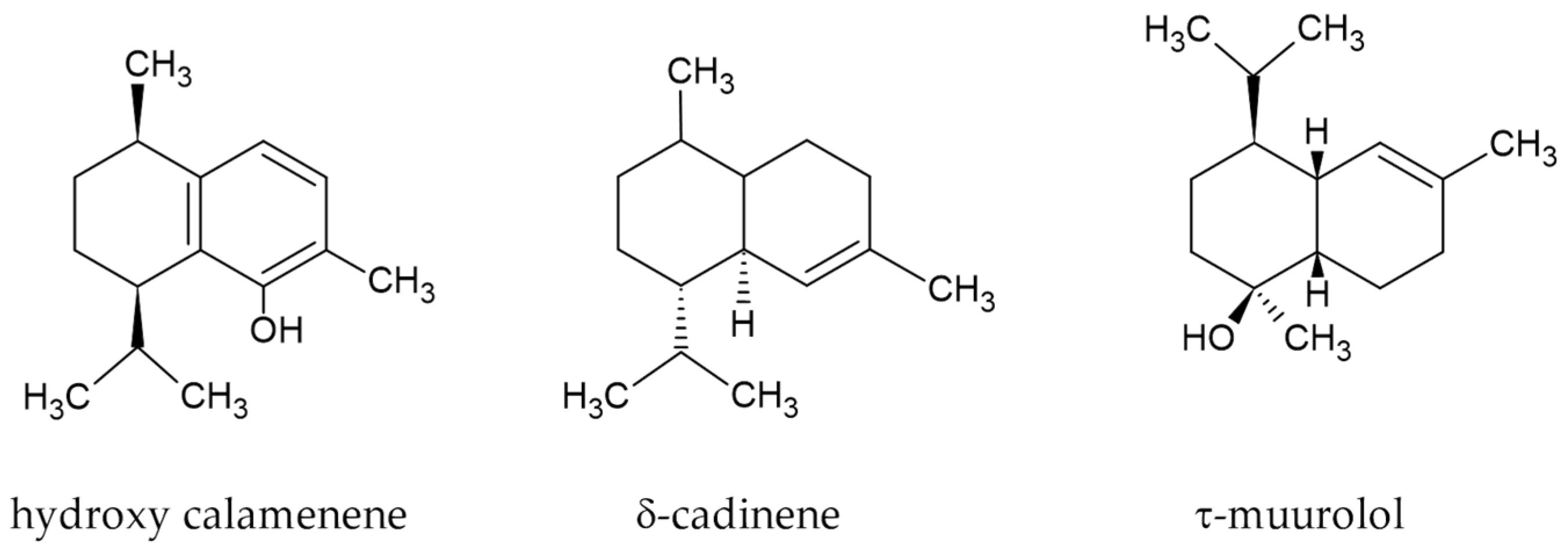
3.1. Chemical Characterization of E. uniflora EO

The E. uniflora EO extracted by hydrodistillation was chemically analyzed by GC-MS. The average EO yield of the hydrodistillation process was 0.98% (v/w). Forty-nine compounds were identified, representing 95.51% of the total, of which twelve were monoterpenes (4.21%) and thirty-three sesquiterpenes (91.18%) (Table 1). The main sesquiterpenes were hydroxy calamenene (41.04%), δ-cadinene (18.73%), and τ-muurolol (12.72%) (Figure 1).

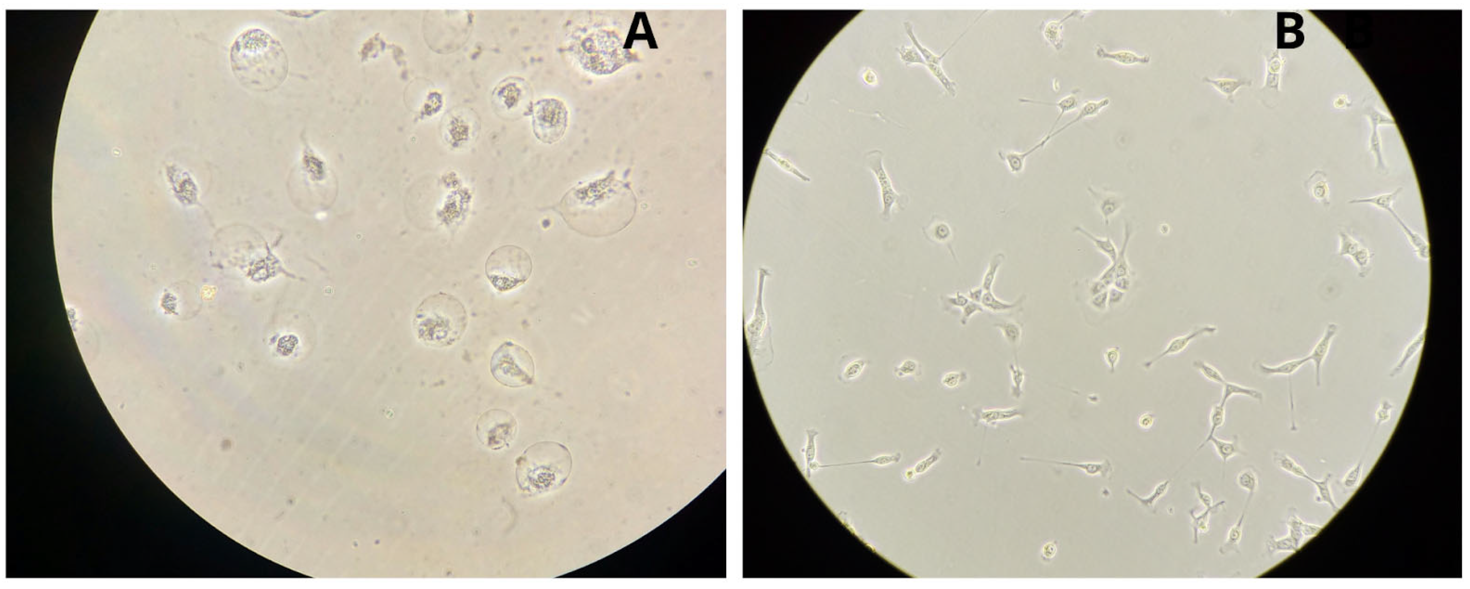
3.2. Microscopic Morphology of Colonic Cell Lines

Microscopic observation of the normal CCD 841 CoN cell culture under optimal conditions showed elongated cells that grew disorganized, resulting in a monolayer 7 days after seeding (168 h) (Figure 2A). Tumoral Caco-2 cells, on the other hand, exhibited a polygonal appearance, and the culture formed an organized monolayer 144 h after seeding (Figure 2B).

3.3. Evaluation of the Cytotoxic Effect of E. uniflora EO on Cell Lines

To study the cytotoxic effect of E. uniflora EO on the normal CCD 841 CoN and tumor Caco-2 colonic cell lines, the survival fraction to different concentrations of EO (0, 0.66, 1.32, 2.64, 6.60, and 13.2 mg/mL) was determined by the MTT assay (Figure 3).
The dose–response curve for the normal colon cell line CCD 841 CoN showed that the lowest concentration of EO tested ([EO] = 0.66 mg/mL) did not significantly modify the survival compared to the control. Cell lethality increased exponentially at higher concentrations, reaching about a 1% survival fraction at [EO] = 2.64 mg/mL, and remained unchanged thereafter. It should be noted that cell survival decreased 40-fold when the EO concentration doubled from 1.32 mg/mL to 2.64 mg/mL. The survival curve for the tumoral cell line Caco-2 exhibited an exponential decrease with two components. The initial exponential component, characterized by the steepest slope, was observed up to [EO] = 2.64 mg/mL, where cell survival was 4.5%. At higher EO concentrations, the slope decreased, with cell survival dropping to 1% at the highest tested dose, [EO] = 13.2 mg/mL. Based on the dose–response curves presented in Figure 3, the IC50 (half-maximal inhibitory concentration) values calculated for CCD 841 CoN was 1.56 mg/mL and Caco-2 IC50 was 1.47 mg/mL.
The MTT assay provided a snapshot of the cell lethality induced by a continuous 14 h exposure to EO at the end of the treatment. However, it did not determine whether surviving cells might exhibit delayed lethality after treatment. To investigate the time course of the cytotoxic effects of the EO, cell viability was assessed over the exposure period to [EO] = 0.66 mg/mL, the lowest tested concentration. Briefly, cells were seeded in 6-well culture plates and exposed to this concentration for 32 h. The numbers of viable and non-viable cells were recorded at regular intervals, and cell viability was calculated.
Figure 4A shows that the total cell count of the EO-treated Caco-2 culture decreased up to 14 h, followed by a transient two-fold increase at 24 h, and then decreased again until the end of the assay. The cell viability, calculated at the same time points (Figure 4B), decreased as a function of time, reaching a minimum at 20 h, followed by an increase at 24 h, which coincided with the peak in cell number. After this time point, both cell viability and cell count decreased.
To test whether the decrease in cell viability observed in Caco-2 tumor cell cultures continuously exposed to [EO] = 0.66 mg/mL was due to effects on the cell progeny, the proliferation capability was evaluated after treatment removal by scoring the total number of cells. Cells were counted immediately after 24 h or 48 h EO treatments. Also, cell counts were scored after removing the EO treatments and incubating the cells under optimal culture conditions for 72 h. Figure 5 shows that the number of cells in the culture decreased 25% when the duration of the treatment increased from 24 to 48 h (p = 0.03). Cell counts after the post-treatment incubation without EO for 72 h showed a 2.6-fold increase for the 24 h EO treatment (p = 0.006) and 1.8-fold for the 48 h one (p = 0.006). The difference in proliferation observed with increasing exposure time to the EO was significant (p = 0.008). Therefore, the EO treatment duration affected the progeny of treated cells, decreasing their proliferation capacity.
After 48 h of EO treatment (0.66 mg/mL), the microscopic observation of both cell lines showed striking differences (Figure 6). While the CCD 841 CoN image shows cells with clearly altered morphology (compare Figure 2A and Figure 6A), the Caco-2 showed both long adherent and rounded cells in suspension (compare Figure 2B and Figure 6B).

3.4. Cell Proliferation Kinetics of CCD 841 CoN and Caco-2 Cell Lines

The potential antiproliferative effect of E. uniflora EO was investigated by assessing the proliferation kinetics of CCD 841 CoN normal colon cells and Caco-2 tumor cells without and with EO treatment.
As shown in Figure 7A,B, the proliferation kinetics of CCD 841 CoN and Caco-2 were studied under optimal conditions for 17 and 14 days, respectively, and the calculated parameters are presented in Table 2. The observed differences in the proliferation kinetics between the CCD 841 CoN and Caco-2 cell lines are consistent with the normal and tumoral characteristics, respectively. The kinetics of the Caco-2 tumor cell line, compared with the normal CCD 841 CoN, presented a steeper slope in the exponential phase, indicating faster proliferation. The analysis of the proliferation kinetics parameters showed that the Caco-2 tumor cell line reached the stationary phase with a cell number 8.2 times higher than the normal cell line CCD 841 CoN. The half-time (t1/2) values for Caco-2 and CCD 841 CoN were 168 h and 309.5 h, respectively. Regarding the tlag, it was 5.5 times higher in the normal cell line than in the tumor cell line.
Cell viability was monitored throughout the proliferation study using the TB staining method. Under optimal culture conditions, the cell viability remained above 85% throughout the observation period in both cell lines. However, the Caco-2 tumor cell line exhibited higher cell viability (greater than 95%) than the normal cell line CCD 841 CoN (Figure 7C).
To study the effect of E. uniflora EO on cell proliferation and based on the previous survival results, the lowest concentration of EO (0.66 mg/mL) that allowed for the highest cell survival was selected to assess the proliferation kinetics. Treated Caco-2 cells showed proliferation up to 11 days, and afterward, the number of cells declined progressively (Figure 8A). A slower kinetics was evidenced by a lower maximum number of cells (Nmax) reached before the decline phase, a 3.5-fold increase in DT, and an increase in t½ from 168.0 h to 225.0 h. It was striking that, despite a decrease in proliferation, the lag phase (tlag) decreased to 0 (Table 2). Simultaneously with the quantification of the proliferation kinetics, Caco-2 cell viability was evaluated as a function of time (Figure 8B). Cell viability was not notably affected until 200 h of the proliferation study, after which it began to decline with a steep slope.
For CCD 841 CoN cells, a proliferation curve could not be obtained, as the cells displayed non-viable characteristics immediately following EO exposure, with pronounced cell death observed after 48 h of incubation, preventing further cell count measurements (Figure 6B).

3.5. DNA Damage Produced by the E. uniflora EO

DNA damage induced by E. uniflora EO in CCD 841 CoN and Caco-2 cells was assessed using the Comet assay. Figure 9 shows DAPI-stained images of control and EO-treated cells of CCD 841 CoN (A and B, respectively) and Caco-2 (C and D, respectively). Cells treated with Bleomycin, a radiomimetic antitumor antibiotic that produces single- and double-strand DNA breaks and base loss, thus inducing DNA fragmentation [52], were used as positive controls (Figure 9E). An image of the treated CCD 841 CoN (Figure 9B) shows amorphous cell structures and inhomogeneous staining, characteristic of extensive cell destruction. In comparison, Figure 9D shows an image of comets formed by the migration of DNA fragments from the nucleus due to strand breaks produced by 0.66 mg/mL of EO in Caco-2 cells. The visual evaluation of the comets showed remarkable migration all the way to the edge of the comet tail, corresponding to Class 3 comets.

3.6. Oxidative Stress Produced by E. uniflora EO

To investigate whether E. uniflora EO produces modifications at the level of oxidative stress that may be a possible molecular mechanism underlying the lethal and antiproliferative effects, the levels of ROS induced by treatment with different concentrations of EO were determined in CCD 841 CoN and Caco-2 cells at 2 h after treatment using the Cellular ROS Assay Kit. In the normal colon cell line CCD 841 CoN (Figure 10, white bars), the ROS levels increased significantly at the highest concentration of EO (p = 0.0002). At lower EO concentrations, no significant ROS level variation was observed. In the Caco-2 cell line, the ROS levels increased with the EO concentration (Figure 10, striped bars). Significantly higher fluorescence values were observed in the tumoral cell line compared with the CCD 841 CoN at all EO concentrations tested. Interestingly, the spontaneous levels of ROS ([EO] = 0 mg/mL) were similar in both normal and tumor cell lines (p = 0.574).

4. Discussion

Cancer—in particular colorectal cancer—represents a significant global public health concern, contributing to high mortality rates, substantial morbidity, and considerable socioeconomic burdens [11,13]. This worldwide health challenge demands innovative research to develop new tools for prevention, rapid diagnosis, and effective treatment of the disease. From a scientific point of view, natural products are well-recognized sources of bioactive compounds with various biomedical applications [53,54,55]. The native plant E. uniflora emerges as a good candidate to identify biological compounds with potential chemopreventive and chemotherapeutic properties. To investigate the antineoplastic potential of E. uniflora leaf essential oil in CRC, the immediate and delayed cytotoxicity and the effect on cell proliferation were studied in normal CCD 841 CoN and tumoral Caco-2 colonic cell lines.
To evaluate the cytotoxic effects of the E. uniflora EO, the cell survival and viability were assessed by MTT assay and TB staining in both CCD 841 CoN and Caco-2 cell lines upon treatment with different concentrations of the EO. The dose–response survival curve (Figure 3) showed [56] that the E. uniflora EO produced lethal effects on both normal and tumoral cells in a concentration-dependent manner. At low EO concentration, the Caco-2 cell line showed a more pronounced and faster immediate cytotoxic response, indicating higher sensitivity of the tumoral cells compared to the normal CCD 841 CoN cells. This result suggests that, at low concentrations, the EO may preferentially target rapidly dividing cells—such as cancer cells—while sparing quiescent or slowly proliferating normal cells. In addition, the calculated IC50 values for CCD 841 CoN and Caco-2 (1.56 and 1.47 mg/mL, respectively) suggest a slightly higher sensitivity of the tumor cell line, although the difference is relatively small. In other cell lines, IC50 values for the cytotoxic effect of various compounds present in E. uniflora EO are reported [27,57,58]. However, to our knowledge, no data on the EO are available for comparison. In the case of the normal CCD 841 CoN cells, no significant lethality was observed at the lowest concentration ([EO] = 0.66 mg/mL). However, the cell viability of the cell culture was severely compromised, as observed upon microscopic inspection. A comparison of the microscopic image of CCD 841 CoN cells incubated under optimal conditions to that of EO-treated cells clearly showed drastic cell morphological changes, including variable cell shape modifications, cell disruption, and nuclear condensation, which are typical indicators of apoptosis or necrosis and, therefore, of non-viable cells (compare Figure 2A and Figure 6A). This high cytotoxic effect of the EO in the normal human colonic cell line was unexpected, as the MTT results showed no lethality. It should be noted that the MTT assessment of cell health constitutes a snapshot of functioning mitochondria after 14 h of EO exposure. However, microscopic observation was performed after the cells were incubated with EO for 48 h. In vitro and in vivo studies have shown that E. uniflora EO does not affect normal cells, as demonstrated by the lack of viability loss in normal human lymphocytes [39] and also the absence of animal lethality after oral administration to rodents [27,32,38,59]. A possible explanation for the selective delayed cytotoxicity observed in the normal colonic CCD 841 CoN cells in the present study could be related to the culture conditions, as the EO treatment was performed in proliferating, non-confluent cultures, where cells were actively cycling. Considering this interpretation, the result indicates that the possible underlying mechanism may involve cell cycle interference rather than immediate, direct necrotic, or apoptotic death.
To confirm the lethality produced by low EO concentrations in the Caco-2 tumor cell line and to assess the time course of the cytotoxic effect, the cell viability was determined as a function of exposure time to [EO] = 0.66 mg/mL, using the TB assay (Figure 4). An initial decrease in the cell count (Figure 4A) and viability (Figure 4B) was observed. It should be noted that the viability fell below the value at t = 0 after a brief increase. Based on this result, the effect of the EO treatment on the cell progeny was evaluated, assessing the proliferation capability of cells after 72 h in optimal conditions post-treatment removal. Figure 5 shows a significant decrease in the number of cells after 48 h of EO treatment compared with the 24 h one, evidencing that longer exposures decrease the proliferation capacity of the progeny. The microscopic observation of Caco-2 cells incubated under optimal conditions showed a polygonal appearance (Figure 2B). However, after 48 h of the low-EO-concentration treatment, the culture appearance changed, exhibiting both long adherent cells and rounded suspended cells (Figure 6B). Our results are in agreement with other studies, where E. uniflora EO cytotoxicity was observed in various tumoral cell lines, including MCF-7 (breast), HCT-116 (colon), AGP-01 (malignant gastric ascites), and SKMEL-19 (melanoma), even at low concentrations [27,57,58].
Dysregulated cell proliferation is one of the hallmarks of carcinogenesis [60]. As a step toward determining the potential chemopreventive and/or therapeutic properties of E. uniflora EO in CRC, we studied the effect of this EO on cell proliferation. The analysis of the proliferation kinetics under optimal culture conditions of normal CCD 841 CoN and Caco-2 cell lines (Figure 7A,B, Table 2) showed that, as expected, the tumoral Caco-2 cell proliferation was faster and reached a higher number of cells in the stationary phase compared with normal CCD 841 CoN cells. Also, the cell viability determined during cell proliferation (Figure 7C) evidenced the tumoral characteristic of the Caco-2 cell line, showing higher viability than the normal CCD 841 CoN throughout the proliferation study.
To study the potential antiproliferative effect of E. uniflora EO, the lowest concentration of EO (0.66 mg/mL) that ensures maximum cell survival was selected. Interestingly, although the CCD 841 CoN cell line exhibited 100% survival upon treatment with this concentration, the proliferation was impaired, and no viable cells were observed (Figure 6B). In the case of the tumoral Caco-2 cells, and unlike the normal CCD 841 CoN, the analysis of the proliferation kinetics and cell viability upon E. uniflora EO treatment (Figure 8A,B, Table 2) evidenced that the prolonged exposure to a low concentration of EO produced the progressive loss of cell viability, contributing to a slower proliferation kinetics and the decline phase observed after 11 days. This finding confirmed the modification of the proliferation capability of the progeny shown in Figure 5.
Several studies have reported the antiproliferative effects of Eugenia sp. extracts or their chemical components in cancer cells, as assessed by cytotoxicity assays, such as the MTT assay and apoptosis determinations [27,61,62,63]. To our knowledge, studies on cell proliferation kinetics using normal and tumoral human colon cell lines, as well as their comparison, have not been reported. Our findings demonstrate that the E. uniflora leaf EO affects cell proliferation, producing lethal events and probably altering the cell cycle. In the chemical characterization of the Pitanga EO, 49 compounds were identified (Table 1), of which 12 were monoterpenes and 33 sesquiterpenes, mainly hydroxy calamenene, δ-cadinene, and τ-muurolol. Various monoterpenes and sesquiterpenes with anti-inflammatory, antioxidant, and antiproliferative properties were confirmed. Myrcene combined with other monoterpenes, such as β-pinene, has been shown to inhibit colon carcinoma tumorigenesis in in vivo and in vitro assays [64]. Other monoterpenes detected, such as terpinolene and limonene, have been proposed as potential anticarcinogenic agents, both as chemopreventives and chemotherapeutics [65,66,67,68]. Among the sesquiterpenes, calamenene inhibits the cell proliferation of various human tumor lines, including lung (A549), breast (MCF7), liver (HepG2), cervical (HeLa), and prostate (PC-3) cancer cells [69]. The compound hydroxy calamenene exhibits antifungal activity in in vivo testing for the protection of plants [70]. Additionally, this compound exhibits radical scavenging properties, as demonstrated by significant antioxidant activity in the DPPH assay [56]. Also, δ-cadinene acts by inhibiting ovarian cancer cell proliferation through cell cycle arrest and induction of apoptosis [71]. Little information is available on the biological effects of τ-muurolol, although anti-inflammatory and antioxidant properties have been reported in EOs containing it [72].
The main chemical compounds identified in the E. uniflora EO sample, shown in Figure 1, share some common chemical structures that may influence their interaction and effect on biological targets. Their structure of a hydrocarbon-rich skeleton imparts lipophilic properties facilitating passage across the cell membrane [73]. Also, the bicyclic three-dimensional structure of the two fused rings and the functional groups, such as the hydroxyl group, may determine the affinity, selectivity, and redox properties of these compounds, which are likely involved in the cytotoxic effect [56,70,71,74,75]. The presence of these sesquiterpenes may contribute to the overall bioactivity of the EO, probably through synergistic interactions. Although ongoing research is being conducted into the potential anticancer properties of sesquiterpenes, including those identified as the main components in this study, further studies using isolated compounds and their combinations are necessary to fully understand their effects.
To explore further the possible mechanisms underlying the cytotoxic and antiproliferative effects of E. uniflora EO, the induction of DNA damage and oxidative stress was assessed by Comet assay and Cellular ROS assay, respectively. Our results show genomic damage in the normal CCD 841 CoN and Caco-2 cell lines, as revealed by the observation of DNA fragmentation, even at low doses (Figure 9). In agreement with the CCD 841 CoN cell viability results, extensive cell destruction was observed in the EO-treated samples (compare Figure 9A,B). In the treated Caco-2 cells, DNA damage was also observed, as shown by the presence of comets evaluated as Class 3 according to the visual scoring system reported in Møller et al. [51] (Figure 9C,D); however, the overall DNA fragmentation was less dramatic than that of normal cells.
It is known that in normal cells the concentration of ROS remains low and is regulated by antioxidant defenses. In tumor cells, an imbalance occurs that leads to an increased demand for ROS during proliferation. However, increased oxidative stress can also generate genomic alterations, resulting in apoptosis and cell death. Therefore, increasing oxidative stress may constitute an antineoplastic mechanism [76]. The determination of the ROS levels as a function of the concentration of E. uniflora EO in CCD 841 CoN cells showed a significant increase only at the highest concentration tested ([EO] = 13.2 mg/mL) (Figure 10, white bars). This result indicates that the detrimental effect of the E. uniflora EO on normal CCD 841 CoN cells’ cytotoxicity and viability is unlikely to be related to an increase in oxidative stress. In contrast, Caco-2 cells exhibited a significant increase in oxidative stress as a function of the EO concentration (Figure 10, striped bars), thus contributing to the detrimental effect on cell viability. Research on Drosophila melanogaster indicates that exposure to E. uniflora EO can increase ROS production, lipid peroxidation, and alterations in antioxidant enzyme activity [77,78]. Ascari et al. [57] reported pro-oxidative effects and cytotoxicity of two compounds isolated from E. uniflora leaf extract in A549 lung adenocarcinoma epithelial cells and IMR90 human lung fibroblasts. In the same line of evidence about the role of the EO in ROS induction, several studies have demonstrated that E. uniflora EO increases the antioxidant cellular defenses such as glutathione and superoxide dismutase [27,38], suggesting that the EO triggers ROS production, leading to the induction of an antioxidant cellular response. Also, our findings of DNA damage and increased ROS levels in EO-treated Caco-2 cells align with previous studies, which have shown that sesquiterpenes—such as calamenene and cadinene derivatives—can induce oxidative stress and DNA strand breaks in cancer cells [79]. Notably, in addition to the pro-oxidative effects of E. uniflora leaf extract, genotoxicity in lung cancer cells was reported [57], although at higher concentrations than those used in our study. In our case, even low EO concentrations (0.66 mg/mL) induced detectable DNA damage, suggesting a potent mechanism of action that may involve ROS-mediated pathways.

5. Conclusions

In conclusion, the present study provides evidence of the cytotoxic and antiproliferative effects of E. uniflora EO in human normal CCD 841 CoN and tumoral Caco-2 cell lines. Interestingly, the cytotoxic effects and impaired proliferation responses induced by EO exposure exhibited distinct characteristics between the two cell lines. Selective delayed toxicity was observed in the normal CCD 841 CoN cells since the EO did not induce immediate toxicity but impaired long-term cell survival or proliferation capacity. This response suggests that the EO may contribute to the prevention of abnormal cell proliferation. Thus, the E. uniflora EO may have chemopreventive potential, for example, in high-risk populations, where moderate suppression of cell proliferation in pre-neoplastic lesions could be beneficial without inducing widespread toxicity. On the other hand, tumoral Caco-2 cells evidenced a faster response to E. uniflora EO treatment. Even exposure to low concentrations resulted in lethality and loss of viability in the Caco-2-treated cells and their progeny, indicating high sensitivity of the tumoral cells. Correspondingly, the detrimental effects on cell proliferation, DNA damage, and oxidative stress production in both cell lines led to the consideration of this EO as a potential antineoplastic agent. These findings support the need for further investigation to consider the E. uniflora EO as a whole or their isolated bioactive compounds as antineoplastic therapeutic agents.

Author Contributions

Conceptualization: A.G.S. and D.J.K.; methodology: M.M., P.L., E.D., M.J.Z. and D.J.K.; formal analysis: A.G.S., P.L., E.D. and D.J.K.; investigation: A.G.S., M.M., P.L., E.D., M.A.S., G.F., B.V., J.C. and D.J.K.; writing—original draft preparation: A.G.S., M.M. and D.J.K.; writing—review and editing: A.G.S. and D.J.K.; supervision: D.J.K.; project administration: A.G.S., M.A.S. and D.J.K.; funding acquisition: D.J.K. All authors have read and agreed to the published version of the manuscript.

Funding

This research was funded by the “United Nations Development Programme”, grant number: 5381—Proyecto Global ABS UND/GEF “Fortalecimiento de recursos humanos, marco legal y capacidades institucionales para la implementación del Protocolo de Nagoya”.

Institutional Review Board Statement

Not applicable.

Informed Consent Statement

Not applicable.

Data Availability Statement

The original contributions presented in this study are included in the article. Further inquiries can be directed to the corresponding authors.

Acknowledgments

The authors (A.G.S., M.M., and D.J.K.) acknowledge R. Daniel Peluffo (Biophysical Chemistry Group, Biological Sciences Department, CENUR Litoral Norte, Universidad de la República) for access to resources and support. The authors thank Natalia Ibargoyen for technical support.

Conflicts of Interest

The authors declare no conflicts of interest.

Abbreviations

The following abbreviations are used in this manuscript:
EOEssential Oil
TBTrypan Blue
DTCell Doubling Time
FBSFetal Bovine Serum
PBSPhosphate Buffer saline
CRCColorectal Cancer

References

  1. Dzobo, K. 2.20—The Role of Natural Products as Sources of Therapeutic Agents for Innovative Drug Discovery. In Comprehensive Pharmacology; Kenakin, T., Ed.; Elsevier: Oxford, UK, 2022; pp. 408–422. [Google Scholar]
  2. Ekor, M. The growing use of herbal medicines: Issues relating to adverse reactions and challenges in monitoring safety. Front. Pharmacol. 2014, 4, 2013. [Google Scholar] [CrossRef] [PubMed]
  3. Keszenman, D.J.; German, P. Traditional medicine usage of pitanga’s leaves for medicinally valuable infusions is directing the search for potential chemotherapeutic agents against colon cancer. In ABS Is Genetic Resources for Sustainable Development; UNDP and Global Environment Facility: Montreal, QC, Canada, 2018; pp. 212–219. [Google Scholar]
  4. WHO. WHO Global Report on Traditional and Complementary Medicine 2019; World Health Organization: Geneva, Switzerland, 2019.
  5. Deligiannidou, G.-E.; Kyrgou, C.; Nena, E.; Manolopoulos, V.G.; Bezirtzoglou, E.; Kontogiorgis, C.A.; Constantinidis, T.C. Use of Edible, Medicinal, and Aromatic Plants in Various Health Disorders: A Cross-Sectional Evaluation among Inhabitants in the Area of Thrace, North-Eastern Greece. Int. J. Environ. Res. Public Health 2022, 19, 12576. [Google Scholar] [CrossRef] [PubMed]
  6. Allegra, S.; De Francia, S.; Turco, F.; Bertaggia, I.; Chiara, F.; Armando, T.; Storto, S.; Mussa, M.V. Phytotherapy and Drugs: Can Their Interactions Increase Side Effects in Cancer Patients? J. Xenobiotics 2023, 13, 75–89. [Google Scholar] [CrossRef]
  7. Asiimwe, J.B.; Nagendrappa, P.B.; Atukunda, E.C.; Kamatenesi, M.M.; Nambozi, G.; Tolo, C.U.; Ogwang, P.E.; Sarki, A.M. Prevalence of the Use of Herbal Medicines among Patients with Cancer: A Systematic Review and Meta-Analysis. Evid. Based Complement. Altern. Med. 2021, 2021, 9963038. [Google Scholar] [CrossRef] [PubMed]
  8. Tayeb, B.A.; Kusuma, I.Y.; Osman, A.A.M.; Minorics, R. Herbal compounds as promising therapeutic agents in precision medicine strategies for cancer: A systematic review. J. Integr. Med. 2024, 22, 137–162. [Google Scholar] [CrossRef]
  9. Youn, B.-Y.; Kim, J.-H.; Jo, Y.-K.; Yoon, S.; Im, J.-Y.; Kim, H.-J.; Lee, J.-D.; Ko, S.-G. Current Characteristics of Herbal Medicine Interventions for Cancer on Clinical Databases: A Cross-Sectional Study. Integr. Cancer Ther. 2023, 22, 15347354231218255. [Google Scholar] [CrossRef]
  10. Wu, Z.; Xia, F.; Lin, R. Global burden of cancer and associated risk factors in 204 countries and territories, 1980–2021: A systematic analysis for the GBD 2021. J. Hematol. Oncol. 2024, 17, 119. [Google Scholar] [CrossRef]
  11. Bray, F.; Laversanne, M.; Sung, H.; Ferlay, J.; Siegel, R.L.; Soerjomataram, I.; Jemal, A. Global cancer statistics 2022: GLOBOCAN estimates of incidence and mortality worldwide for 36 cancers in 185 countries. CA Cancer J. Clin. 2024, 74, 229–263. [Google Scholar] [CrossRef]
  12. Chambers, A.C.; Dixon, S.W.; White, P.; Williams, A.C.; Thomas, M.G.; Messenger, D.E. Demographic trends in the incidence of young-onset colorectal cancer: A population-based study. Br. J. Surg. 2020, 107, 595–605. [Google Scholar] [CrossRef]
  13. Howren, A.; Sayre, E.C.; Loree, J.M.; Gill, S.; Brown, C.J.; Raval, M.J.; Farooq, A.; De Vera, M.A. Trends in the Incidence of Young-Onset Colorectal Cancer with a Focus on Years Approaching Screening Age: A Population-Based Longitudinal Study. JNCI J. Natl. Cancer Inst. 2021, 113, 863–868. [Google Scholar] [CrossRef]
  14. Musetti, C.; Garau, M.; Alonso, R.; Piñeros, M.; Soerjomataram, I.; Barrios, E. Colorectal Cancer in Young and Older Adults in Uruguay: Changes in Recent Incidence and Mortality Trends. Int. J. Environ. Res. Public Health 2021, 18, 8232. [Google Scholar] [CrossRef]
  15. Piñeros, M.; Laversanne, M.; Barrios, E.; Cancela, M.d.C.; de Vries, E.; Pardo, C.; Bray, F. An updated profile of the cancer burden, patterns and trends in Latin America and the Caribbean. Lancet Reg. Health Am. 2022, 13, 100294. [Google Scholar] [CrossRef]
  16. Clinton, S.K.; Giovannucci, E.L.; Hursting, S.D. The World Cancer Research Fund/American Institute for Cancer Research Third Expert Report on Diet, Nutrition, Physical Activity, and Cancer: Impact and Future Directions. J. Nutr. 2020, 150, 663–671. [Google Scholar] [CrossRef] [PubMed]
  17. Arrillaga, B. Plantas Usadas en Medicina Natural; Editorial Everest: La Paz, Bolivia, 1997. [Google Scholar]
  18. Davies, P. Fichas Técnicas de Cultivo. In Estudios de Domesticación y Cultivo de Especies Medicinales y Aromáticas Nativas; INIA: Montevideo, Uruguay, 2004; pp. 37–120. [Google Scholar]
  19. Paz, E.A.; Bassagoda, M.J.; Ferreira, F. Yuyos: Uso Racional de las Plantas Medicinales; Editorial Fin de Siglo: Montevideo, Uruguay, 1992. [Google Scholar]
  20. Tabakian, G. Estudio comparativo de plantas medicinales vinculadas a tradiciones indígenas y europeas en Uruguay. Bonplandia 2019, 28, 135–158. [Google Scholar] [CrossRef]
  21. Stefanello, M.É.A.; Pascoal, A.C.R.F.; Salvador, M.J. Essential Oils from Neotropical Myrtaceae: Chemical Diversity and Biological Properties. Chem. Biodivers. 2011, 8, 73–94. [Google Scholar] [CrossRef] [PubMed]
  22. Brussa, C.A.; Grela González, A. Flora Arbórea del Uruguay: Con Énfasis en las Especies de Rivera y Tacuarembó; Editorial Fin de Siglo: Montevideo, Uruguay, 2007; p. 544. [Google Scholar]
  23. Jolochín Manorani, G. Estudios Biogeográficos en Poblaciones Uruguayas de Eugenia uniflora L. Master’s Thesis, Facultad de Ciencias, Universidad de la República, Montevideo, Uruguay, 2016. [Google Scholar]
  24. Bagetti, M.; Facco, E.M.P.; Piccolo, J.; Hirsch, G.E.; Rodriguez-Amaya, D.; Kobori, C.N.; Vizzotto, M.; Emanuelli, T. Physicochemical characterization and antioxidant capacity of pitanga fruits (Eugenia uniflora L.). Food Sci. Technol. 2011, 31, 147–154. [Google Scholar] [CrossRef]
  25. Vizzotto, M. Fitoquímicos em pitanga (Eugenia uniflora L.): Seu potencial na prevencao e comabte a doencas. In Proceedings of the III Simpósio Nacional do Morango II Encontro Sobre Pequenas Frutas e Frutas Nativas do Mercosul; Embrapa Clima Temperado: Pelotas, Brazil, 2006; pp. 29–34. [Google Scholar]
  26. Ferragut, G.; Lombardo, P.; Severi, M.A.; Vignale, B.; Cedano, J.; Dellacassa, E.; Pérez, E. Estudios de bioactividad de extractos de plantas nativas uruguayas y su papel inmunológico. In Proceedings of the XV Jornadas de la Sociedad Uruguaya de Biociencias, Piriápolis, Uruguay, 5–7 September 2014. [Google Scholar]
  27. Fidelis, E.M.; Savall, A.S.P.; de Oliveira Pereira, F.; Quines, C.B.; Ávila, D.S.; Pinton, S. Pitanga (Eugenia uniflora L.) as a source of bioactive compounds for health benefits: A review. Arab. J. Chem. 2022, 15, 103691. [Google Scholar] [CrossRef]
  28. Lombardo, P. Caracterización Química y Bioactividad de Aceites Esenciales Contra Patógenos de los Cítricos. Master’s Thesis, Universidad de la República, Montevideo, Uruguay, 2015. [Google Scholar]
  29. Lombardo, P.; Guimaraens, A.; Franco, J.; Dellacassa, E.; Pérez Faggiani, E. Effectiveness of essential oils for postharvest control of Phyllosticta citricarpa (citrus black spot) on citrus fruit. Postharvest Biol. Technol. 2016, 121, 1–8. [Google Scholar] [CrossRef]
  30. Rodrigues, K.A.d.F.; Amorim, L.V.; Oliveira, J.M.G.d.; Dias, C.N.; Moraes, D.F.C.; Andrade, E.H.d.A.; Maia, J.G.S.; Carneiro, S.M.P.; Carvalho, F.A.d.A. Eugenia uniflora L. Essential Oil as a Potential Anti-Leishmania Agent: Effects on Leishmania amazonensis and Possible Mechanisms of Action. Evid. Based Complement. Altern. Med. 2013, 2013, 279726. [Google Scholar] [CrossRef]
  31. Sánchez, A.G.; Ferragut, G.; Lombardo, P.; Severi, M.A.; Cedano, J.; Vignale, B.; Vázquez, A.; Dellacassa, E.; Keszenman, D.J. Efectos celulares de extractos de Pitanga. In Proceedings of the 8vo Encuentro Nacional de Frutos Nativos, Rocha, Uruguay, 30–31 March 2017. [Google Scholar]
  32. Victoria, F.N.; Lenardão, E.J.; Savegnago, L.; Perin, G.; Jacob, R.G.; Alves, D.; Silva, W.P.d.; Motta, A.d.S.d.; Nascente, P.d.S. Essential oil of the leaves of Eugenia uniflora L.: Antioxidant and antimicrobial properties. Food Chem. Toxicol. 2012, 50, 2668–2674. [Google Scholar] [CrossRef]
  33. Vignale, B.; Cabrera, D.; Rodríguez, P.; Machado, G. Selección de frutales nativos en Uruguay. Hortic. Argent. 2016, 35, 19–29. [Google Scholar]
  34. Weyerstahl, P.; Marschall-Weyerstahl, H.; Christiansen, C.; Oguntimein, B.O.; Adeoye, A.O. Volatile Constituents of Eugenia uniflora Leaf Oil. Planta Med. 1988, 54, 546–549. [Google Scholar] [CrossRef] [PubMed]
  35. Benfatti, C.S.; Cordova, S.M.d.; Guedes, A.; Magina, M.D.A.; Cordova, C.M.M.d. Atividade antibacteriana in vitro de extratos brutos de espécies de Eugenia sp. frente a cepas de molicutes. Rev. Pan. Amaz. Saúde 2010, 1, 33–39. [Google Scholar] [CrossRef]
  36. de Souza Prestes, L.; Damé Schuch, L.F.; Hörnke Alves, G.; Ziemann dos Santos, M.A.; Alves Rodrigues, M.R.; Araújo Meireles, M.C. Evaluación de la actividad bactericida de aceites esenciales de hojas de guayabo, pitango y arazá. Rev. Cuba. Plantas Med. 2011, 16, 324–330. [Google Scholar]
  37. Borsoi, F.T.; Rosa, B.B.d.S.; Filomena, M.; Oliveira, F.D.L.d.; Dulce, B.M.; Kempka, A.P. Eugenia uniflora L. seed and pulp extracts: Phytochemical profile, cytotoxic potential, antitumoral activity, and α-amylase and α-glucosidase inhibition capacity. Nat. Prod. Res. 2023, 37, 3862–3867. [Google Scholar] [CrossRef]
  38. Falcão, T.R.; de Araújo, A.A.; Soares, L.A.L.; de Moraes Ramos, R.T.; Bezerra, I.C.F.; Ferreira, M.R.A.; de Souza Neto, M.A.; Melo, M.C.N.; de Araújo, R.F.; de Aguiar Guerra, A.C.V.; et al. Crude extract and fractions from Eugenia uniflora Linn leaves showed anti-inflammatory, antioxidant, and antibacterial activities. BMC Complement. Altern. Med. 2018, 18, 84. [Google Scholar] [CrossRef]
  39. Núñez, J.G.; Pinheiro, J.d.S.; Silveira, G.F.; Beckenkamp, A.; Buffon, A.; Bruno, A.N. Antineoplastic potential of the aqueous crude extract of Eugenia uniflora L. in human cervical cancer. Braz. J. Pharm. Sci. 2018, 54, e17267. [Google Scholar] [CrossRef]
  40. Adams, R. Identification of Essential Oil Components by Gas Chromatography/Quadrupole Mass Spectroscopy. Carol. Stream 2005, 16, 65–120. [Google Scholar]
  41. McLafferty, F.W.; Stauffer, D.B.; Loh, S.Y. Comparative evaluations of mass spectral data bases. J. Am. Soc. Mass. Spectrom. 1991, 2, 438–440. [Google Scholar] [CrossRef]
  42. Mondello, L. Mass Spectra of Flavors and Fragrances of Natural and Synthetic Compounds; Wiley Blackwell: Hoboken, NJ, USA, 2015; Volume 10. [Google Scholar]
  43. Dellacassa, E.; Lorenzo, D.; Paz, D. Procesos de extracción aplicados a la obtención de productos aromáticos de origen vegetal. In Estudios en Domesticación y Cultivos de Especies Medicinales y Aromáticas Nativas; Davies, P., Ed.; INIA: Montevideo, Uruguay, 2004; pp. 155–160. [Google Scholar]
  44. Dellacassa, E.; Lorenzo, D.; Paz, D. Caracterización fisicoquímica de los aceites esenciales. In Estudios en Domesticación y Cultivos de Especies Medicinales y Aromáticas Nativas; Davies, P., Ed.; INIA: Montevideo, Uruguay, 2004; pp. 161–169. [Google Scholar]
  45. de Abreu Costa, L.; Henrique Fernandes Ottoni, M.; Dos Santos, M.G.; Meireles, A.B.; Gomes de Almeida, V.; de Fátima Pereira, W.; Alves de Avelar-Freitas, B.; Eustáquio Alvim Brito-Melo, G. Dimethyl Sulfoxide (DMSO) Decreases Cell Proliferation and TNF-α, IFN-γ, and IL-2 Cytokines Production in Cultures of Peripheral Blood Lymphocytes. Molecules 2017, 22, 1789. [Google Scholar] [CrossRef]
  46. Chen, X.; Thibeault, S. Effect of DMSO concentration, cell density and needle gauge on the viability of cryopreserved cells in three dimensional hyaluronan hydrogel. Annu. Int. Conf. IEEE Eng. Med. Biol. Soc. 2013, 2013, 6228–6231. [Google Scholar] [CrossRef] [PubMed]
  47. Mosmann, T. Rapid colorimetric assay for cellular growth and survival: Application to proliferation and cytotoxicity assays. J. Immunol. Methods 1983, 65, 55–63. [Google Scholar] [CrossRef]
  48. Avelar-Freitas, B.A.; Almeida, V.G.; Pinto, M.C.; Mourao, F.A.; Massensini, A.R.; Martins-Filho, O.A.; Rocha-Vieira, E.; Brito-Melo, G.E. Trypan blue exclusion assay by flow cytometry. Braz. J. Med. Biol. Res. 2014, 47, 307–315. [Google Scholar] [CrossRef]
  49. Keszenman, D.J.; Salvo, V.A.; Nunes, E. Effects of bleomycin on growth kinetics and survival of Saccharomyces cerevisiae: A model of repair pathways. J. Bacteriol. 1992, 174, 3125–3132. [Google Scholar] [CrossRef] [PubMed]
  50. Benitez-Bribiesca, L.; Sanchez-Suarez, P. Oxidative damage, bleomycin, and gamma radiation induce different types of DNA strand breaks in normal lymphocytes and thymocytes. A comet assay study. Ann. N. Y. Acad. Sci. 1999, 887, 133–149. [Google Scholar] [CrossRef] [PubMed]
  51. Møller, P.; Azqueta, A.; Sanz-Serrano, J.; Bakuradze, T.; Richling, E.; Eyluel Bankoglu, E.; Stopper, H.; Claudino Bastos, V.; Langie, S.A.S.; Jensen, A.; et al. Visual comet scoring revisited: A guide to scoring comet assay slides and obtaining reliable results. Mutagenesis 2023, 38, 253–263. [Google Scholar] [CrossRef]
  52. Keszenman, D.J.; Carmen Candreva, E.; Nunes, E. Cellular and molecular effects of bleomycin are modulated by heat shock in Saccharomyces cerevisiae. Mutat. Res. 2000, 459, 29–41. [Google Scholar] [CrossRef]
  53. Atanasov, A.G.; Zotchev, S.B.; Dirsch, V.M.; Orhan, I.E.; Banach, M.; Rollinger, J.M.; Barreca, D.; Weckwerth, W.; Bauer, R.; Bayer, E.A.; et al. Natural products in drug discovery: Advances and opportunities. Nat. Rev. Drug Discov. 2021, 20, 200–216. [Google Scholar] [CrossRef]
  54. Chaachouay, N.; Zidane, L. Plant-Derived Natural Products: A Source for Drug Discovery and Development. Drugs Drug Candidates 2024, 3, 184–207. [Google Scholar] [CrossRef]
  55. Costa-Font, J.; Sato, A.; Saenz-de-Miera, B. Cultural persistence of self-assessed health: A study of first- and second-generation migrants. J. Migr. Health 2025, 11, 100280. [Google Scholar] [CrossRef]
  56. Da Silva, J.K.R.; Andrade, E.H.A.; Barreto, L.H.; Da Silva, N.C.F.; Ribeiro, A.F.; Montenegro, R.C.; Maia, J.G.S. Chemical Composition of Four Essential Oils of Eugenia from the Brazilian Amazon and Their Cytotoxic and Antioxidant Activity. Medicines 2017, 4, 51. [Google Scholar] [CrossRef] [PubMed]
  57. Ascari, J.; Felipe Maciel Pereira, M.; Schaffka, V.M.; Nunes, D.S.; Magalhães, C.G.; Santos, J.S.; Granato, D.; Carmo, M.; Azevedo, L.; Archilha, M.; et al. Selina-1,3,7(11)-trien-8-one and Oxidoselina-1,3,7(11)-trien-8-one from Eugenia uniflora Leaf Essential Oil and Their Cytotoxic Effects on Human Cell Lines. Molecules 2021, 26, 740. [Google Scholar] [CrossRef] [PubMed]
  58. Figueiredo, P.L.B.; Pinto, L.C.; da Costa, J.S.; da Silva, A.R.C.; Mourão, R.H.V.; Montenegro, R.C.; da Silva, J.K.R.; Maia, J.G.S. Composition, antioxidant capacity and cytotoxic activity of Eugenia uniflora L. chemotype-oils from the Amazon. J. Ethnopharmacol. 2019, 232, 30–38. [Google Scholar] [CrossRef]
  59. Schmeda-Hirschmann, G.; Theoduloz, C.; Franco, L.; Ferro, E.; de Arias, A.R. Preliminary pharmacological studies on Eugenia uniflora leaves: Xanthine oxidase inhibitory activity. J. Ethnopharmacol. 1987, 21, 183–186. [Google Scholar] [CrossRef]
  60. Hanahan, D.; Weinberg, R.A. Hallmarks of cancer: The next generation. Cell 2011, 144, 646–674. [Google Scholar] [CrossRef]
  61. Aranha, E.S.P.; de Azevedo, S.G.; dos Reis, G.G.; Silva Lima, E.; Machado, M.B.; de Vasconcellos, M.C. Essential oils from Eugenia spp.: In vitro antiproliferative potential with inhibitory action of metalloproteinases. Ind. Crops Prod. 2019, 141, 111736. [Google Scholar] [CrossRef]
  62. de Oliveira, G.G.F.V.; Longue, M.F.; Pescinelli, L.M.R.; Charret, T.S.; Nogueira, T.S.R.; Pereira, M.T.M.; Vieira, I.J.C.; Abreu, L.S.; Pascoal, V.D.B.; Pascoal, A.C.R.F. Eugenia brasiliensis: Analysis of the Chemical Profile and Evaluation of Cytotoxic Potential. Chem. Biodivers. 2025, e202500429. [Google Scholar] [CrossRef]
  63. Ismiyati, N.; Putri, D.; Kusumastuti, S.A.; Febriansyah, R. Antiproliferative Effect of Ethanolic Extract Eugenia uniflora Lam. Leaves on T47D Cells. Indones. J. Cancer Chemoprev. 2012, 3, 370. [Google Scholar] [CrossRef]
  64. Spyridopoulou, K.; Tiptiri-Kourpeti, A.; Lampri, E.; Fitsiou, E.; Vasileiadis, S.; Vamvakias, M.; Bardouki, H.; Goussia, A.; Malamou-Mitsi, V.; Panayiotidis, M.I.; et al. Dietary mastic oil extracted from Pistacia lentiscus var. chia suppresses tumor growth in experimental colon cancer models. Sci. Rep. 2017, 7, 3782. [Google Scholar] [CrossRef]
  65. Agus, H.H.; Sarp, C.; Cemiloglu, M. Oxidative stress and mitochondrial impairment mediated apoptotic cell death induced by terpinolene in Schizosaccharomyces pombe. Toxicol. Res. 2018, 7, 848–858. [Google Scholar] [CrossRef]
  66. Jia, S.S.; Xi, G.P.; Zhang, M.; Chen, Y.B.; Lei, B.; Dong, X.S.; Yang, Y.M. Induction of apoptosis by D-limonene is mediated by inactivation of Akt in LS174T human colon cancer cells. Oncol. Rep. 2013, 29, 349–354. [Google Scholar] [CrossRef] [PubMed]
  67. Okumura, N.; Yoshida, H.; Nishimura, Y.; Kitagishi, Y.; Matsuda, S. Terpinolene, a component of herbal sage, downregulates AKT1 expression in K562 cells. Oncol. Lett. 2012, 3, 321–324. [Google Scholar] [CrossRef]
  68. Menezes, I.O.; Scherf, J.R.; Martins, A.O.B.P.B.; Ramos, A.G.B.; Quintans, J.d.S.S.; Coutinho, H.D.M.; Ribeiro-Filho, J.; de Menezes, I.R.A. Biological properties of terpinolene evidenced by in silico, in vitro and in vivo studies: A systematic review. Phytomedicine 2021, 93, 153768. [Google Scholar] [CrossRef]
  69. Zhong, Z.-C.; Zhao, D.-D.; Liu, Z.-D.; Jiang, S.; Zhang, Y.-L. A New Human Cancer Cell Proliferation Inhibition Sesquiterpene, Dryofraterpene A, from Medicinal Plant Dryopteris fragrans (L.) Schott. Molecules 2017, 22, 180. [Google Scholar] [CrossRef] [PubMed]
  70. Scher, J.M.; Speakman, J.B.; Zapp, J.; Becker, H. Bioactivity guided isolation of antifungal compounds from the liverwort Bazzania trilobata (L.) S.F. Gray. Phytochemistry 2004, 65, 2583–2588. [Google Scholar] [CrossRef]
  71. Hui, L.M.; Zhao, G.D.; Zhao, J.J. δ-Cadinene inhibits the growth of ovarian cancer cells via caspase-dependent apoptosis and cell cycle arrest. Int. J. Clin. Exp. Pathol. 2015, 8, 6046–6056. [Google Scholar] [PubMed]
  72. Chien, T.C.; Lo, S.F.; Ho, C.L. Chemical composition and anti-inflammatory activity of Chamaecyparis obtusa f. formosana wood essential oil from Taiwan. Nat. Prod. Commun. 2014, 9, 723–726. [Google Scholar]
  73. Zhu, Q.; Wang, S.; Fu, G.; Guo, F.; Huang, W.; Zhang, T.; Dong, H.; Jin, Z.; Zhang, D. Highly flexible cell membranes are the key to efficient production of lipophilic compounds. J. Lipid Res. 2024, 65, 100597. [Google Scholar] [CrossRef]
  74. Kundu, A.; Supradip, S.; Suresh, W.; Vivek, A.; Kaur, C. Antioxidant potential of essential oil and cadinene sesquiterpenes of Eupatorium adenophorum. Toxicol. Environ. Chem. 2013, 95, 127–137. [Google Scholar] [CrossRef]
  75. Olano, C.; Méndez, C.; Salas, J.A. Antitumor compounds from marine actinomycetes. Mar. Drugs 2009, 7, 210–248. [Google Scholar] [CrossRef]
  76. Sosa, V.; Moliné, T.; Somoza, R.; Paciucci, R.; Kondoh, H.; ME, L.L. Oxidative stress and cancer: An overview. Ageing Res. Rev. 2013, 12, 376–390. [Google Scholar] [CrossRef] [PubMed]
  77. da Cunha, F.A.B.; Wallau, G.L.; Pinho, A.I.; Nunes, M.E.M.; Leite, N.F.; Tintino, S.R.; da Costa, G.M.; Athayde, M.L.; Boligon, A.A.; Coutinho, H.D.M.; et al. Eugenia uniflora leaves essential oil induces toxicity in Drosophila melanogaster: Involvement of oxidative stress mechanisms. Toxicol. Res. 2015, 4, 634–644. [Google Scholar] [CrossRef]
  78. de Carvalho, N.R.; Rodrigues, N.R.; Macedo, G.E.; Bristot, I.J.; Boligon, A.A.; de Campos, M.M.; Cunha, F.A.B.; Coutinho, H.D.; Klamt, F.; Merritt, T.J.S.; et al. Eugenia uniflora leaf essential oil promotes mitochondrial dysfunction in Drosophila melanogaster through the inhibition of oxidative phosphorylation. Toxicol. Res. 2017, 6, 526–534. [Google Scholar] [CrossRef] [PubMed]
  79. Wang, J.; Zhang, Y.-S.; Thakur, K.; Hussain, S.S.; Zhang, J.-G.; Xiao, G.-R.; Wei, Z.-J. Licochalcone A from licorice root, an inhibitor of human hepatoma cell growth via induction of cell apoptosis and cell cycle arrest. Food Chem. Toxicol. 2018, 120, 407–417. [Google Scholar] [CrossRef]
Figure 1. Structures of the major identified compounds in E. uniflora EO. Chemical structures were drawn with ChemSketch (Freeware) 2023.2.0 from ACD/Labs.
Biologics 05 00019 g001
Figure 2. Microphotographs of normal CCD 841 CoN (A) and tumoral Caco-2 (B) cell cultures, grown and maintained under optimal conditions, captured at 20× magnification.
Biologics 05 00019 g002
Figure 3. Cell survival of colonic cell lines as a function of the concentration of E. uniflora EO. Dose–response survival curve of normal CCD 841 CoN (⚪) and tumor Caco-2 (▽) cells exposed for 14 h to different concentrations of EO in nutrient medium. The S.E.M. intervals are included in the figure.
Biologics 05 00019 g003
Figure 4. Cell viability time course of the Caco-2 tumor cell line treated with 0.66 mg/mL of EO: (A) total number of cells (viable and non-viable) as a function of time; (B) percentage of cell viability calculated using the data presented in (A) at the same time points.
Biologics 05 00019 g004
Figure 5. Effect of the E. uniflora EO on the progeny of Caco-2 cells treated with [EO] = 0.66 mg/mL during 24 or 48 h. Cells were scored immediately after EO treatment (black bars) or after incubation for 72 h under optimal conditions without EO (white bars). Data from at least 3 independent experiments are included. Asterisks indicate significant differences between samples and treatments: (*) between immediate and delayed scoring; (**) between samples scored at 72 h; (***) between samples scored immediately). p < 0.05.
Biologics 05 00019 g005
Figure 6. Microphotographs of CCD 841 CoN (A) and Caco-2 (B) cells treated with 0.66 mg/mL of E. uniflora EO for 48 h (40× magnification).
Biologics 05 00019 g006
Figure 7. Proliferation kinetics of the CCD 841 CoN (A) and the Caco-2 (B) under optimal growth conditions and the corresponding cell viability determined at each time point (C). The number of cells per mL (N) as a function of time for CCD 841 CoN was recorded up to 400 h due to slow growth. The grey lines in (A,B) correspond to the curve fitting based on the logistic growth model indicated in Section 2. The dotted lines in (C) indicate 95% confidence intervals for the normal colon cell line CCD 841 CoN (◯) and the tumor cell line Caco-2 (▽). The mean values ± S.E.M. are included in the figures.
Biologics 05 00019 g007
Figure 8. Proliferation kinetics (A) and cell viability (B) of the Caco-2 tumor colon cell line treated with 0.66 mg/mL of EO added to the culture medium. The mean values ± S.E.M. are included in the figures.
Biologics 05 00019 g008
Figure 9. DNA damage produced by E. uniflora EO in the CCD 841 CoN and Caco-2 cell lines determined using the Comet assay. Microphotographs of CCD 841 CoN cells untreated (A) and treated with 0.66 mg/mL of E. uniflora EO for 14 h (B); Caco-2 cells untreated (C) and treated with 0.66 mg/mL of EO for 14 h (D). (E) Positive control: Caco-2 cells treated with 40 µg/mL of Bleomycin.
Biologics 05 00019 g009
Figure 10. ROS levels induced in normal colonic cells CCD 841 CoN (white bars) and tumoral Caco-2 cells (striped bars) by treatment with different concentrations of E. uniflora EO. Measurements were made 2 h post-treatment with EO. (*)In each cell line the asterisk indicate significant differences between untreated and treated samples; (**) indicate significant differences in ROS levels between CCD 841 CoN and Caco-2 cell lines (p < 0.05).
Biologics 05 00019 g010
Table 1. E. uniflora EO chemical compounds identified by GC-MS.
LRI 1Compound 2%LRICompound%
1082Hexanal0.011666α-Humulene0.07
1110β-Pinene0.011668γ-Gurjunene0.06
1122Sabinene0.011699Bicyclogermacrene0.02
1162α-Terpinene0.011682β-Humulene0.13
1167α-Phellandrene0.011684trans-β-Guaiene0.01
1169Myrcene1.101686β-Chamigrene3.71
1198Limonene0.251688Zonarene0.48
1209β-Phellandrene0.261689γ-Muurolene5.22
12392-Pentylfuran0.011708Germacrene D0.22
1250trans-β-Ocimene0.711720α-Cadinene0.41
1234cis-β-Ocimene1.821722Viridiflorene0.71
1270p-Cymene0.031723α-Muurolene0.55
1282Terpinolene0.031746cis-Muurola-3,5-diene0.41
13511-Hexanol0.011755d-Cadinene18.73
1351allo-Ocimene0.011806Germacrene B0.55
1380(Z)-3-Hexen-1-ol0.071808trans-Calamenene0.02
1460α-Cubebene0.061818cis-Calamenene0.27
1523β-Bourbonene0.031918β-Calacorene0.06
1600Guaia-6,9-diene0.031901α-Colacorene0.05
1626β-Copaene0.591930Palustrol0.01
1636trans-Caryophyllene0.232082Globulol0.54
1637Aromadendrene0.172089Viridiflorol3.85
1642trans-Muurola-3,5-diene0.172185τ-Muurolol12.72
1652α-Guaiene0.012325Hydroxy calamenene41.04
1658γ-Elemene0.05
Total95.51
1 LRI based on a series of n-hydrocarbons reported according to their elution order on Carbowax 20M. 2 Identity was tentatively assigned by comparing the acquired mass spectra with those reported in the literature [40,41,42]
Table 2. Proliferation kinetics parameters of the CCD 841 CoN and the Caco-2 cell lines without and with treatment with E. uniflora EO ([EO] = 0.66 mg/mL). The means of at least 3 experiments ± S.E.M. are presented.
Nmax (Cells/mL)DT (h)t1/2 (h)tlag (h)
Control CCD 841 CoN1.0 × 106 ± 1.5 × 10557.9 ± 8.9309.5 ± 30.866.0 ± 3.0
Control Caco-28.2 × 10⁶ ± 2.8 × 10517.1 ± 3.0168.0 ± 5.112.0 ± 0.6
Treated Caco-25.2 × 10⁶ ± 2.6 × 10560.0 ± 3.0225.0 ± 11.30
Disclaimer/Publisher’s Note: The statements, opinions and data contained in all publications are solely those of the individual author(s) and contributor(s) and not of MDPI and/or the editor(s). MDPI and/or the editor(s) disclaim responsibility for any injury to people or property resulting from any ideas, methods, instructions or products referred to in the content.

