Article

Unraveling Potential Compounds of Uncaria gambir (W.Hunter) Roxb. as Antikeloid Agent: In Silico, In Vitro and Ex Vivo Experimental Validation

by
Sri Suciati Ningsih
1,2,
Sri Widia A. Jusman
3,4,
Rahimi Syaidah
1,5,
Muhamad Arif Budiman
2,
Alfi Khatib
6 and
Fadilah Fadilah
1,7,*
1
Doctoral Program in Biomedical Sciences, Faculty of Medicine, Universitas Indonesia, Jakarta 10430, Indonesia
2
Faculty of Medicine, Universitas Muhammadiyah Prof Dr Hamka, Jakarta 12130, Indonesia
3
Department of Biochemistry and Molecular Biology, Faculty of Medicine, Universitas Indonesia, Jakarta 10430, Indonesia
4
Center of Hypoxia and Oxidative Stress Studies, Faculty of Medicine, Universitas Indonesia, Jakarta 10430, Indonesia
5
Department of Histology, Faculty of Medicine, Universitas Indonesia, Jakarta 10430, Indonesia
6
Department of Pharmaceutical Chemistry, Kulliyyah of Pharmacy, International Islamic University Malaysia, Kuantan 25200, Malaysia
7
Department of Medical Chemistry, Faculty of Medicine, Universitas Indonesia, Jakarta 10430, Indonesia
*
Author to whom correspondence should be addressed.
Biologics 2025, 5(3), 18; https://doi.org/10.3390/biologics5030018
Submission received: 23 April 2025 / Revised: 25 May 2025 / Accepted: 18 June 2025 / Published: 27 June 2025
(This article belongs to the Section Natural Products)

Abstract

Background/Objectives: Keloid treatment remains challenging due to limited effectiveness and patient dissatisfaction. Herbal-based therapy offers promising alternatives that require further investigation. Uncaria gambir (W.Hunter) Roxb., an original plant from Indonesia, possesses an antifibrotic effect. However, its potential as an antifibrotic agent in keloid management remains unclear. This study aims to bridge this gap by evaluating the bioactive compound from gambir and its effects on keloid fibroblast primary culture. Methods: The bioactive compounds of gambir extract and fractions (ethanol, hexane, and ethyl acetate fractions) were identified by using liquid chromatography–mass spectrometry (LCMS/MS) analysis. The mechanism of gambir bioactive compounds for keloid was predicted using the compound–protein interaction network and enrichment analysis, and validated using molecular docking and dynamic simulation. The experimental study results, including cytotoxic and bioactivity effects, were represented as IC50 and selectivity index (SI) values, and the ex vivo analysis of keloid tissue explants. Results: Uncariagambiriine was identified as the most potent compound with the lowest binding energy and high stability to the core protein targets: AKT1 and TGFB1. The ethanol fraction was determined to have the highest abundance of gambir’s typical bioactive compounds, with the lowest IC50 (128.76 ± 0.24 µg/mL) and the highest SI (6.32) value. Furthermore, the results of the ex vivo analysis indicated the significant inhibition of keloid fibroblast proliferation and migration by the gambir ethanolic fraction. Conclusions: This study underlines the potential of the gambir ethanolic fraction as an antifibrotic agent in keloid, warranting further investigation and development for clinical applications.

1. Introduction

Keloid is characterized by an abnormal fibroproliferative wound-healing process beyond the boundaries of the original wound. This fibroproliferative disorder may emerge after any kind of dermal injury, leading to cosmetic and physiological issues that affect a patient’s quality of life. The current treatment approaches for keloids are still facing formidable challenges due to high recurrence rates and dissatisfaction [1]. Natural compounds derived from medicinal plants offer a wide range of benefits, including minimal side effects and a diversity of bioactive compounds. The diversity of bioactive compounds may be found in the plant extract or fractions attributed to the complex interactions, which perform synergistic therapeutic effects. The use of these synergistic effects can maximize the therapeutic efficacy, with minimal adverse effects, through multiple compounds and multiple target properties [2,3]. Therefore, exploring novel therapeutic agents derived from natural sources is required to unravel their potential therapeutic benefits against keloid.
Uncaria gambir (W.Hunter) Roxb., a plant species belonging to the Rubiaceae family, has garnered attention for its diverse pharmacological properties, including anti-inflammatory, antioxidant, anticancer, and antifibrotic activities [4,5,6]. This plant is originally from Indonesia and is widely found in West Sumatera. Gambir has long been employed as a traditional medicine for various ailments [7]. Munggari et al. [8] revealed that gambir contains secondary metabolites belonging to the flavonoid, phenolic, and alkaloid groups. Catechin and its derivatives have been recognized as the major flavonoids in gambir, exhibiting various biological effects, including antioxidant activity through the inhibition of lipid peroxidation [9] and analgesic effects in a rat model of osteoarthritis [10]. Unfortunately, the information regarding the antifibrotic properties of Gambir’s metabolites remains limited and fragmented. Previously, in vivo studies by Desdiani et al. [11] and Sriningsih [12] demonstrated the potential of gambir in attenuating pulmonary and hepatic fibrosis via an in vivo study. Therefore, gambir is proposed to have the same effect on skin fibrosis, particularly keloid. Supporting this hypothesis, Jusman et al. [13] reported that gambir extract inhibited the growth of KF with Gambiriin A1, procyanidin B2, and neooxogambirtannine, identifying it as a potent inhibitor of PDGF via an in silico study. Those groups of bioactive compounds are dissolved well in polar solvents like ethanol or methanol. Furthermore, the separation of bioactive compounds by fractionation is required to obtain a group of target compounds that have a high efficiency against fibrosis.
The pathomechanism of keloid relies on cell-to-cell interaction mediated by growth factors. The growth factors contribute to excessive keloid fibroblast proliferation and the overproduction of extracellular matrix components. Notably, keloid fibroblasts exhibit increased expression of growth factors and their receptors, leading to heightened cell proliferation, enhanced cell survival, and excessive synthesis of extracellular matrix (ECM) proteins, particularly collagen [14]. Our previous study identified gambiriin C, isogambirine, and procyanidin B1 as promising compounds targeting transforming growth factor beta 1 (TGFβ1), AKT serine/threonine kinase 1 (AKT1), and matrix metallopeptidase 1 (MMP1). Further gene ontology enrichment and pathway analyses predicted that gambir bioactive compounds may regulate cell proliferation, migration, transcription, and signal transduction via profibrotic cytokine and growth factor signaling pathways [15].
Experimental validation of the potential effects of gambir bioactive compounds on keloid remains unclear. To address this gap, this study investigates the effects of gambir ethanolic extract and its fractions on keloid-derived fibroblasts obtained from explant primary culture. The primary cell culture systems offer a valuable model for closely mimicking the in vivo microenvironment [16]. Keloid fibroblasts are recognized as the key drivers of keloid pathogenesis due to their elevated proliferative activity and increased synthesis of ECM proteins, particularly collagen [17]. To comprehensively evaluate the effects of gambir’s bioactive compounds on keloid fibroblasts, this research conducted fractionation of the gambir ethanolic extract using solvents of varying polarity. The fractions exhibit simpler compound profiles, which facilitate the identification of the most potent active compounds with inhibitory activity against keloid fibroblasts [18]. We focus on the bioactivity, cytotoxicity, and inhibitory effects of keloid-derived fibroblast primary culture. By elucidating the effects of Gambir-derived compounds on keloid fibroblasts, this study aims to advance understanding of their therapeutic potential in keloid management.

2. Materials and Methods

2.1. Extraction and Fractionation Procedure

Dry plant powder derived from the whole part of the gambir plant was used as the sample for this research. The sample was purchased from a local farmer from Pesisir Selatan, West Sumatera, Indonesia, and identified as Uncaria gambir (W.Hunter) Roxb. by herbarium dekopensis, Department of Biology, Universitas Indonesia (JI25-P-66). The sample was extracted by using the maceration technique with 96% ethanol (Merck, 107017, Darmstadt, Germany) as the solvent. 950 mg of the dry plant powder of gambir was weighed and then extracted using 96% ethanol (1:1) at room temperature for 24 h. Subsequently, the crude extract was fractionated using liquid–liquid separation method using solvents with different polarities. Ten grams of gambir ethanol extract were dissolved in 100 mL of ethanol (Merck, 107017, Darmstadt, Germany) and transferred to a separatory funnel. An equal volume of hexane (Merck, 100795, Darmstadt, Germany) was added, and the mixture was shaken for 5 min and left to stand for 30–60 min until phase separation occurred. The upper hexane layer was collected, and 100 mL of ethyl acetate (Merck, 100789, Darmstadt, Germany) was added to the remaining solution. After repeating the mixing and settling steps, the ethyl acetate (lower) and ethanol (upper) fractions were separated. Each fraction was then concentrated, followed by drying and weighing to calculate the percentage of the yields using the following formula:
Y i e l d   % =   W e i g h t   o f   e x t r a c t   o r   f r a c t i o n   o b t a i n e d   W e i g h t   o f s t a r t i n g   m a t e r i a l × 10

2.2. Phytochemical Analysis

Phytochemical analysis was performed by subjecting the gambir extract and fraction to total phenol and flavonoid content. The total phenolic content was determined by following the Folin–Ciocalteu method. The absorbance was assessed through the standard curve 0, 50, 100, 150, 200, 250, and 300 ppm of gallic acid for phenolic and quercetin for flavonoid. The absorbance was examined using a GENESYS™ 150 UV-Vis Spectrophotometer (Thermo Scientific, 840-309400, Waltham, MA, USA) at 765 nm for phenolic [19] and 376 nm for flavonoid [20].

2.3. Gambir Bioactive Compound Screening Process

An ultra-performance liquid chromatography (UPLC) system (LC: ACQUITY UPLC® H-Class System, Waters, Milford, MA, USA) coupled to a mass spectrometer (Xevo G2-S QTof, Waters, Milford, MA, USA) was used for LCMS/MS analysis. Chromatographic separation was performed using a C18 column (1.8 μm 2.1 × 100 mm, ACQUITY UPLC® HSS, Waters, Milford, MA, USA) at a flow rate of 0.2 mL/min, and the column oven temperature was set at 50 °C. The mobile phase consisted of water +5 mM ammonium formic acid and acetonitrile +0.05% formic acid. The sample injection volume was 5 μL (initially filtered through a 0.2 μm syringe filter), with a total run time per sample was 23 min. The liquid chromatography (LC) analysis employed a mobile phase with a specific flow rate, while the mass spectrometry (MS) analysis utilized electrospray ionization (ESI) in positive mode within a defined mass range of 50–1200 m/z and source and desolvation temperatures of 100 and 350 °C, respectively. Correspondingly, cone and desolvation gas flow rates were set at 0 L/h and 793 L/h, along with variable collision energy varied between 4 and 60 eV. Data acquisition, analysis, and instrument control were facilitated using Masslynx software version 4.1 [21].

2.4. In Silico Analysis

The chemical compounds and their canonical Simplified Molecular Input Line Entry System (SMILE) of gambir ethanolic extract and fractions from LCMS/MS data were collected from https://pubchem.ncbi.nlm.nih.gov (accessed between 6 and 8 October 2024). The SMILES numbers were used as keywords to identify potential targets for keloid in the SwissTargetPrediction database http://swisstargetprediction.ch (accessed on 20 October 2024), with human genes set as data retrieval. The keloid target proteins were sourced from our previous study [15]. The Gambir compounds and keloid target proteins interaction network was constructed, visualized, and analyzed using Cytoscape 3.9.1 software on 22 October 2024. The core proteins from the previous steps were imported into the Database for Annotation, Visualization, and Integrated Discovery (DAVID) https://david.ncifcrf.gov/ (accessed on 27 October 2024) for functional and pathway analysis. The analysis was conducted with an adjusted p value less than 0.05 as the cut-off value for indicating statistical significance.
The protein structure of the target protein for molecular docking analysis was retrieved from the Protein Data Bank (PDB) database https://www.rcsb.org/ (accessed on 5 November 2024), while the structures of active compounds as the ligand were sourced from PubChem (accessed on 5 November 2024). Molecular docking of gambir bioactive compounds with target proteins was performed using AutoDockTools 1.5.6 on 9 November 2024. The molecular docking simulation utilized the Lamarckian Genetic Algorithm. The docking score’s absolute value was used to determine the binding affinity and stability between the compounds and their targets. Visualization of the molecular docking results was carried out using Discovery Studio 4.5 on 15 November 2024. The stability of compound-target binding was simulated by molecular dynamic analysis using YASARA software v. 25.12.1 and run with the md_run.mcr file. The molecular dynamics simulation was conducted with a pH of 7.4, using Na and Cl ions, at a temperature of 298 K, with a duration of 30 ns (30,000 ps) and a save interval of 10 ns (10,000 ps). The Amber14 force field was applied as specified in the md_run.mcr file. The radius of gyration (Rg), root mean square deviation (RMSD) of protein, and root mean square fluctuation (RMSF) of amino acid residues were used to evaluate the compound–protein interaction [22].

2.5. Cytotoxic and Bioactivity Effect

Cytotoxic and bioactivity effects were performed using keloid and normal fibroblast primary cultures from fresh keloid tissue based on our previous study [23]. The sample derived from surgical excision was donated for research purposes only with written informed consent. The procedure was approved by the ethics committee of the Faculty of Medicine, Universitas Indonesia—Cipto Mangunkusumo Hospital (No: KET-1206/UN.F1/ETIK/PPM.00.02/2023). The cytotoxic evaluation was performed by using the CCK-8 cell counting kit (Dojindo, CK04, Kumamoto, Japan) assay procedure in various doses (µg/mL) and represented as the IC50 value (the sample concentration that causes 50% inhibition or cell death). In brief, 5000 fibroblast cells derived from passage 2 were cultured in 96 well-culture plates, then incubated in a CO2 incubator at 37 °C and 5% CO2 for 24 h. The fibroblast cell will adhere to the bottom of the dish after 24 h. Afterwards, the media was replaced with 100 µL of treatment media with duplicates for each treatment. The treatment media were made from gambir crude extract, ethanol, ethyl acetate, and hexane fractions with various concentrations. Dimethyl sulfoxide (DMSO) has been employed as the solvent and optimized at a non-cytotoxic concentration, ensuring the effects were due to the gambir treatments. 100 µL of complete media (DMEM supplemented with 10% FBS and 1% antibiotic-antimycotic) was added for the negative control and the blank well. The plate was then incubated in an incubator at 37 °C and 5% CO2 for 24 h. The old media was discarded after 24 h, then rinse each well with 100 µL of sterile Ca and Mg-free Phosphate-Buffered Saline (PBS) (Gibco, 18912-014, Grand Island, NY, USA). Next, the complete media was added to each well, and continued with 10 µL of CCK-8 solution was added to each well. The plate was covered with aluminum foil and placed into a CO2 incubator at 37 °C and 5% CO2 for four hours. The absorbance was read at wavelength 450 nm using a microplate reader, Multiscan FC (Thermo, 357, Waltham, MA, USA), then used for determining the cell viability. The percentage of cell viability was calculated using the following formula:
%   c e l l   v i a b i l i t y = O D   c o n t r o l b l a n k O D   ( t r e a t e d b l a n k ) O D   ( c o n t r o l b l a n k )
The absorbance of treated refers to the optical density (OD) measured in wells treated with the test compounds, while the absorbance of control refers to wells containing untreated cells (negative control). The viability value was employed for measuring IC50 values. This value was obtained from the graphs of percentage inhibition versus concentration of Gambir extract and fraction. Furthermore, the selectivity index (SI) was determined as a simple ratio between the IC50 of normal and keloid fibroblasts, defined as follows. The treatment with the lowest IC50 value and the highest SI will be selected for the ex vivo assay.
S I = I C 50   n o r m a l   f i b r o b l a s t   I C 50   k e l o i d   f i b r o b l a s t  

2.6. Ex Vivo Assay Analysis

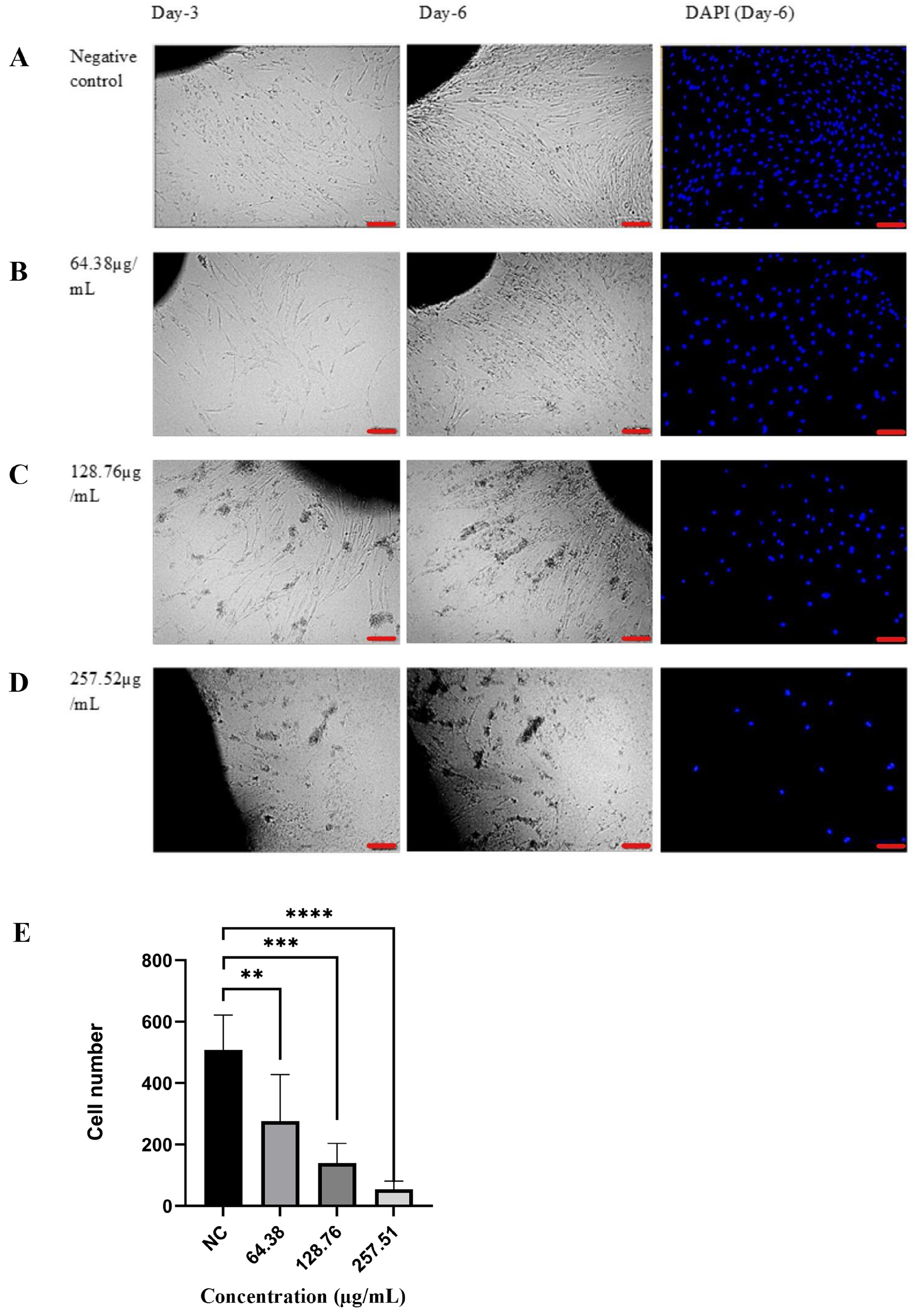
The dermal fragment of keloid tissue was placed into the bottom of 24 well culture plates. Leave the plate semi-opened for 10–15 min in a type 2 biosafety cabinet (BSC) to facilitate the fragment adhering. 200 µL of complete media was added into each well by placing the plate into a CO2 incubator at 37 °C and 5% CO2 for 24 h. After 24 h of incubation, the media were replaced with 200 µL of treatment media. The treatment media was carried out as the cytotoxicity and bioactivity assay with the dose based on the IC50 value, ½× IC50, 1× IC50, and 2× IC50 value. In this study, positive control was not included due to the currently limited understanding of Gambir’s mechanism of action in keloids. Therefore, it is not feasible to select a positive control with a comparable mode of action. Accordingly, the effects of treatments were assessed relative to untreated keloid tissue explant as a negative control to establish a baseline response. All treatments and negative control (complete media only) were plated out in quadruplets. A representative figure was captured at the same location on days 3 and 6. At the end of incubation (day 6), the cell was counted by using a fluorescent imaging assay using 4′,6-diamidino-2-phenylindole (DAPI)-aqueous fluoroshield (Abcam, ab104139, Waltham, MA, USA). The fluorescence image was acquired using an inverted fluorescence microscope (ZOE Fluorescent Cell Imager-Bio-Rad, Bio-Rad, Hercules, CA, USA) under standardized settings. Cell quantification was performed by analyzing the images with ImageJ 1.54g apps, which allowed for accurate counting of DAPI-stained nuclei to estimate cell numbers.

3. Results

3.1. Phytochemical and Bioactive Compounds Analysis of Gambir

The brown solid masses were yielded from dry plant powder of gambir extraction using ethanol as the solvent. The crude extract was then fractionated and yielded three fractions with different structures and colors. The pale brown solid mass was yielded from the ethanol fraction, the hexane fraction was solid and deep brown in color, while a reddish-brown semi-solid mass was obtained from the ethyl acetate fraction. Either the crude extract or fractions of Gambir positively contained phenolic and flavonoid properties in this study (Table 1). The ethanol fraction has the highest total phenol and flavonoid content compared to the other fractions, although the total flavonoid content of the crude extract remains the highest value compared to the fractions. In parallel with the result, gambir’s typical bioactive compounds were highly abundant in this fraction. Table 2 represents the typical bioactive compounds that have been identified in gambir extract and fractions (Supplementary Materials). This study identified procyanidin B1, gambiriin A1, dihydrogambirtannine, uncariagambiriine, and oxogambirtannine as gambir’s typical bioactive compounds found in gambir extract and fractions. Those bioactive compounds have been previously reported as major constituents of gambir extracted using ethanol or ethyl acetate as the solvent [8].

3.2. In Silico Analysis

The interaction between keloid targets and identified gambir’s bioactive compounds from previous analysis is illustrated in Figure 1a. The edges connecting the nodes highlight the potential multi-target effects of Gambir’s compounds on keloid targets. The intensity of the element’s color correlates with the number of interactions between compounds and target proteins, the darker shades indicating a higher degree of interaction. Figure 1b complements the interaction network by illustrating the result of gene ontology (GO) enrichment analysis, highlighting the biological processes (BP), molecular functions (MF), and cellular components (CC) associated with the biological targets. The size of the dots represents the count of genes involved in each category, while the color gradient from green to red corresponds to the significance level (-log10(p-value)), with red indicating higher statistical significance.
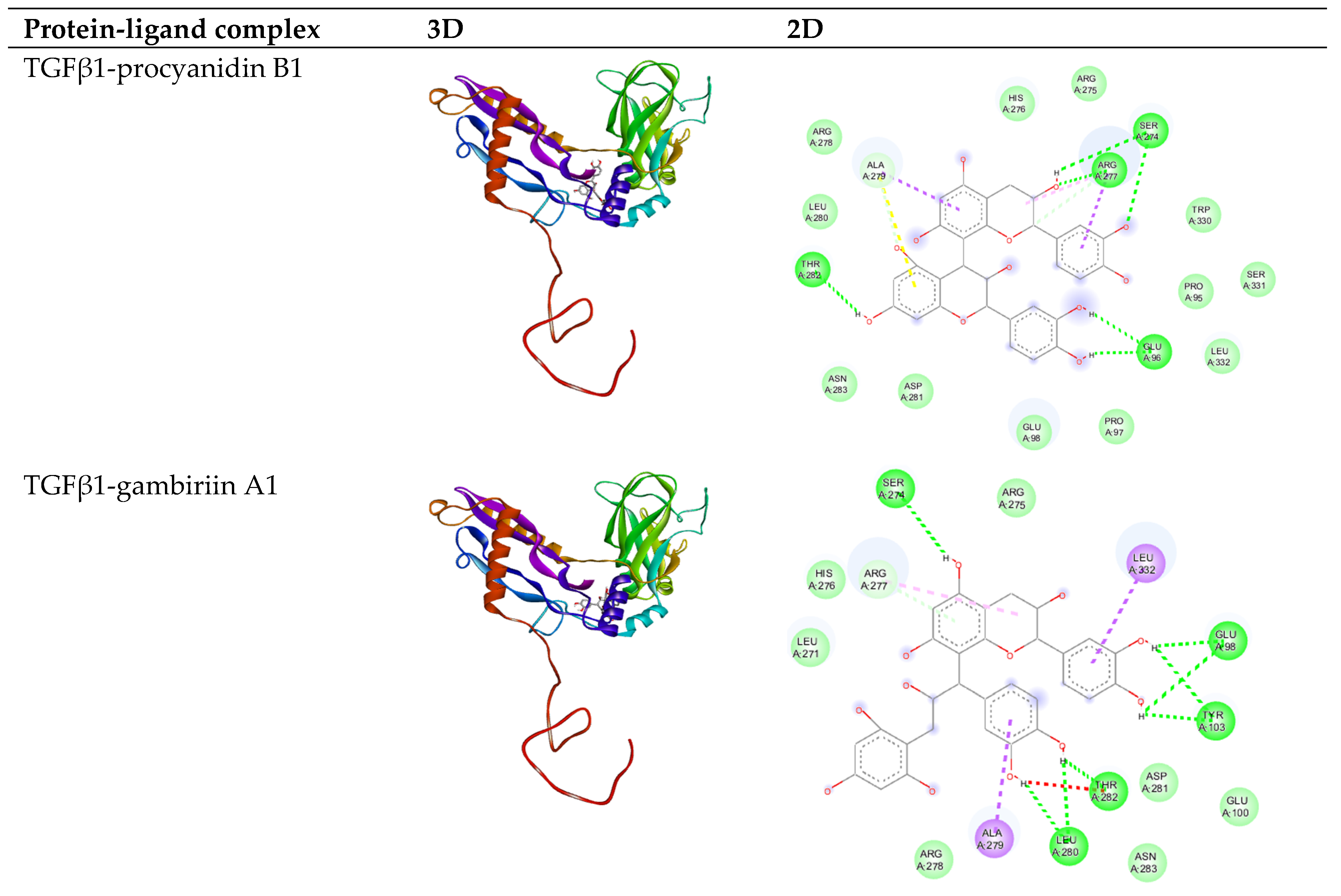
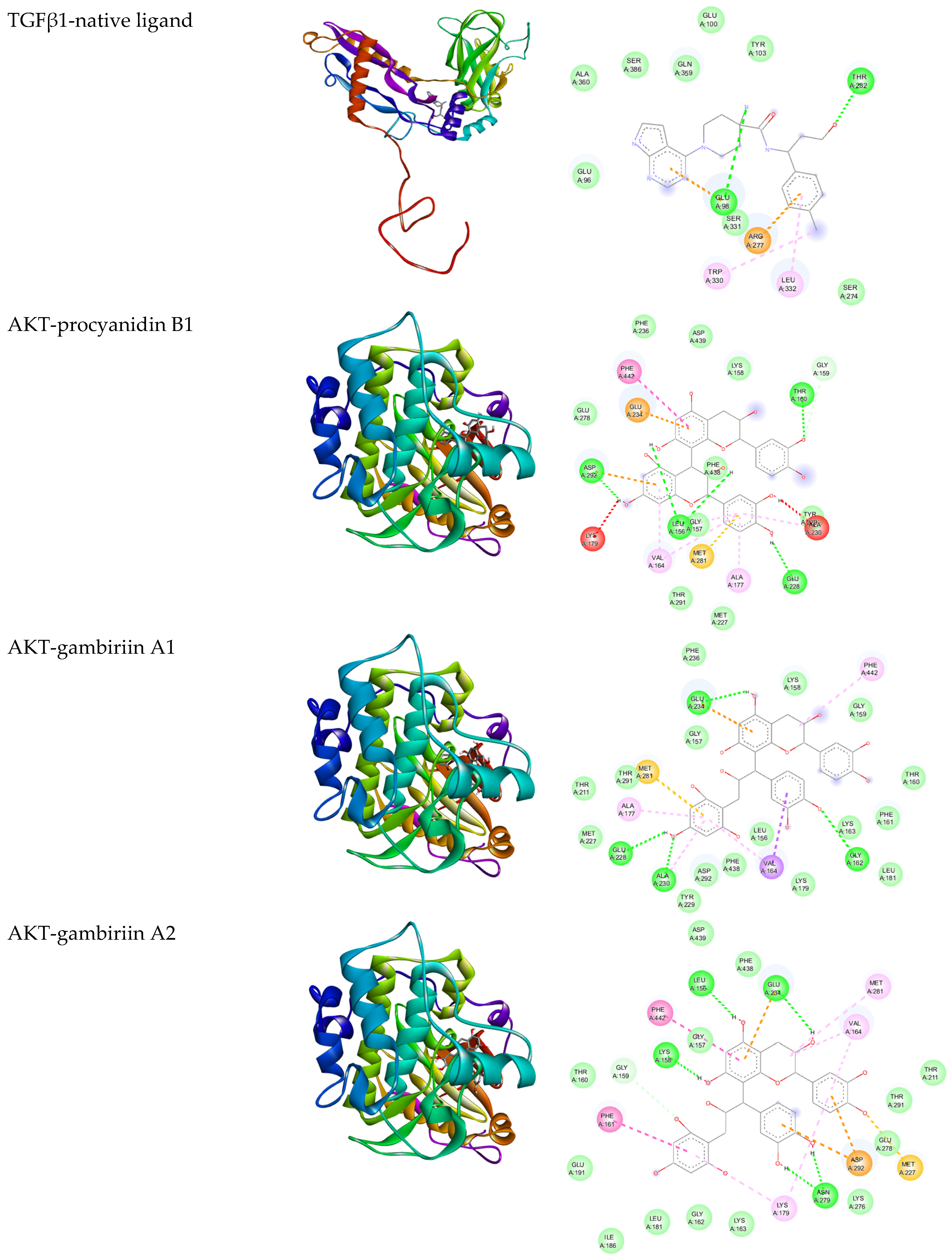
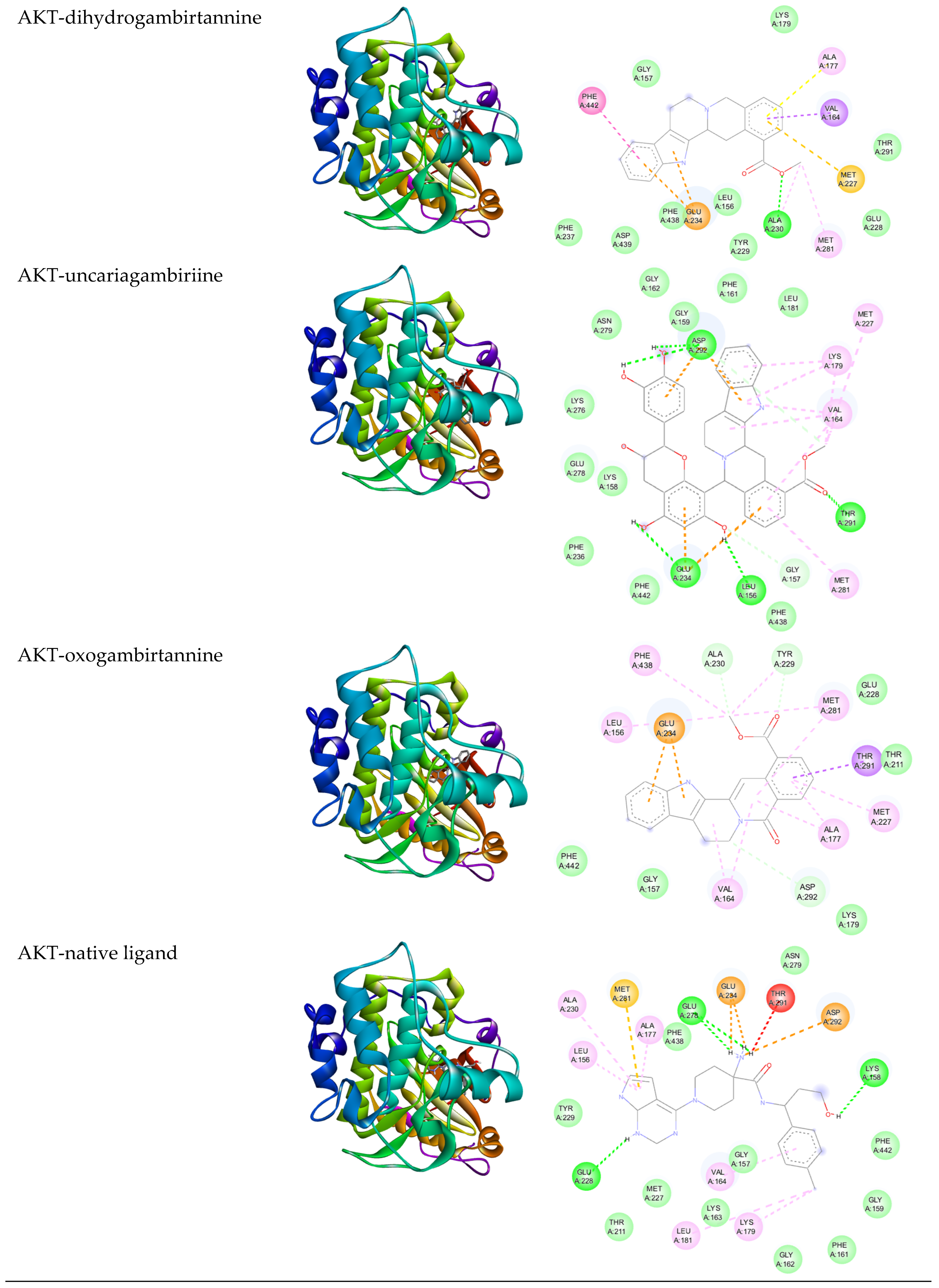
The molecular docking analysis was conducted to validate the interaction between the active compounds of gambir (Table 2) and the keloid target proteins. This study focused on two target proteins: TGFβ1 (3KFD) and Akt1 (4GV1); these proteins belonged to the key targets with the highest interaction scores (based on our previous study [15]) and were involved in the same signaling pathway, which is the non-canonical TGFβ pathway. This molecular docking test is shown in Table 3 and visualized in Figure 2. A lower binding affinity of energy suggests a more stable connection between the protein and its ligand [24]. The binding affinities of the test compound were then compared to the binding affinities of the target proteins and their native ligands. Each target protein possesses a native ligand that is positioned within the active site of the protein, 0XZ501 defined as the native ligand for TGFβ1 (3KFD) and L01EX27 for Akt1 (4GV1).
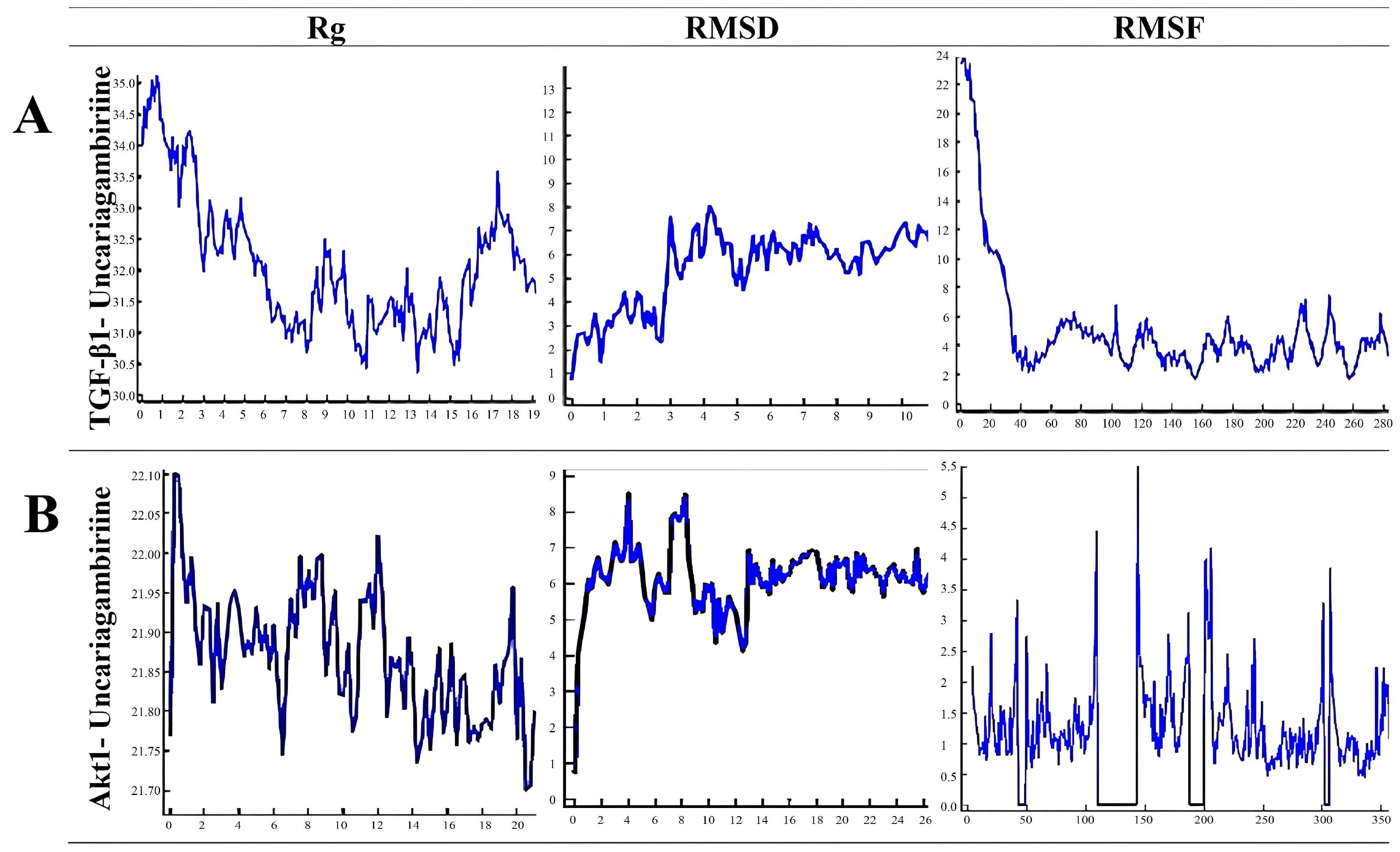
The molecular dynamics simulation data indicates that the solute achieves structural stability after an initial equilibration phase (Figure 3). The radius of gyration (Rg) stabilizes around 33.5–34.0 Å in TGFβ1 and 21.7–22.0 Å in Akt1 after slight compaction during the first 8 ns. The ligand movement RMSD relative to the receptor steadily increases in the first 3 ns and is relatively stable during 4–15 ns. The value starts to fluctuate after 15 ns until the end of the simulation, suggesting significant positional adjustments or weakened binding interactions over time. The ligand conformation RMSD indicated dynamic conformational flexibility after 8 ns. The solute residue RMSF highlights distinct regions of flexibility and rigidity, reflecting functional or interaction-related dynamics. These results suggest stable solute behavior alongside flexible ligand dynamics, which may impact ligand-receptor interactions.

3.3. Cytotoxic and Bioactivity Effect of Gambir Extract and Fraction Towards Keloid Fibroblast

Both bioactivity and toxicity effects are represented as IC50 and SI values in Table 4. The lower IC50 value indicates the higher effectiveness of the treatment in inhibiting half biological activity of keloid fibroblasts [25]. The ethanol fraction possessed the lowest IC50 value, 128.76 ± 0.24 µg/mL, compared with the other samples. It means that the ethanol fraction is more effective at a lower concentration to achieve the same level of action. In addition, gambir extract and fraction possessed an SI higher than 2, indicating high selectivity to keloid fibroblasts. Interestingly, the ethanol fraction had the highest desirable selectivity as well. Therefore, the IC50 value of the ethanol fraction was employed to be the reference for the dose of treatments. The treatments were based on ½× IC50 (64.38 µg/mL), 1× IC50 (128.76 µg/mL), and 2× IC50 (257.52 µg/mL) values.

3.4. Ex Vivo Analysis of Gambir Ethanol Fraction Effect Towards Keloid Fibroblast

This current study also demonstrated that the gambir ethanol fraction possesses significant inhibitor effects, as evidenced by our ex vivo analysis. The administration of gambir ethanol fraction significantly reduced fibroblast proliferation and migration around the edge of the explant, along with the increase in concentration. The inhibition observed in our ex vivo model suggests a potential mechanism of gambir against keloid disease (Figure 4).

4. Discussion

Keloids are pathological scars characterized by an excessive aggregation of fibroblasts and collagen within the reticular dermis [26]. Current therapeutic approaches for keloids, such as corticosteroid injections, surgical excision, and radiation therapy, frequently yield unsatisfactory outcomes and significant side effects. Consequently, there is an urgent need for safer and more effective therapeutic strategies [27]. In this context, herbal-based therapies, which have shown promise in controlling fibrotic diseases, warrant further investigation as a prospective keloid alternative therapy. This study explores the antifibrotic potential of Uncaria gambir (W.Hunter) Roxb. (gambir) as an anti-keloid agent. This research identified typical bioactive compounds from gambir extract dan fractions, as summarized in Table 2. Some of them were classified into flavonoids (procyanidin B1, gambiriin A1, and gambiriin A2) and alkaloids (dihydrogambirtannine, uncariagambiriine, and oxogambirtannine) [8]. Notably, the majority of these compounds were detected in the ethanol and ethyl acetate fractions, consistent with the high solubility of both flavonoid and alkaloid compounds in polar and semi-polar solvents, such as ethanol and ethyl acetate. However, the presence of phenolic compounds in the hexane fraction might be due to the limited solvent selectivity of the liquid–liquid fractionation method, which can cause overlapping of compounds across the fractions [28].
Drug–protein network analysis identified nine critical proteins related to keloid pathogenesis that interact with bioactive compounds from gambir, they were AKT1, STAT3, TGFB1, VEGFA, EGFR, IGFR, TIMP, MMP1, and PDGFA. Enrichment analysis indicates that gambir modulates pathways associated with cell proliferation, migration, protein kinase B, and the MAP signaling pathway. These findings are consistent with the hallmark features of keloids, including enhanced fibroblast proliferation and migration driven by dysregulated cytokines and growth factors [29]. These observations align with previous research indicating gambir’s bioactive compounds target profibrotic inflammatory cytokines and growth factors to regulate cellular proliferation, migration, and signal transduction activity [15]. Most of the growth factors’ signaling activity is primarily mediated by the homodimerization of receptor kinase proteins, including the TGFβ1 signaling pathway [30]. The TGFβ1 signaling pathway plays a central role in keloid pathogenesis through both canonical (via Smad) and noncanonical (non-Smad) mechanisms. Protein kinase B (Akt) is one of the key components of the downstream signaling cascades in the noncanonical pathway [31,32]. Regarding this, AKT1 and TGFβ1 were selected as target proteins for further analysis using molecular docking and dynamic simulation.
Uncariagambirine exhibited the strongest binding affinity to AKT1 and TGFβ1. The binding energy between protein and ligand represents the strength of interaction. The smaller the binding energy, the stronger the interaction [22]. The molecular dynamics simulation was conducted to evaluate the stability of the compound protein in fluid conditions resembling the human body environment [33]. The result suggests stable solute behavior, dynamic ligand flexibility, and localized solute fluctuations that influence ligand-receptor interactions. Further studies, such as free energy calculations, could clarify the binding mechanisms and stability of the complex.
The cytotoxic and bioactivity effects of gambir extract and its fraction towards keloid fibroblasts validate the antifibrotic potential of gambir. Among the tested fractions, the ethanol fraction exhibited the highest abundance of bioactive compounds and the strongest cytotoxic effect on keloid fibroblasts, with an IC50 value of 128.76 ± 0.24 µg/mL. In addition, the ethanol fraction showed the highest selectivity index against the keloid fibroblast, indicating the low cytotoxicity of the normal fibroblast [34]. Ex vivo analysis further highlighted the potential of the ethanolic fraction of gambir against keloid. Treatment with the ethanol fraction significantly inhibited fibroblast proliferation and migration in keloid explants (Figure 4). The inhibition of fibroblast migration is particularly noteworthy, as it suggests that gambir not only reduces existing fibroblast activity but may also prevent the spread of pathological fibroblasts to surrounding tissues. Further experimental studies are required to investigate the mechanisms of action of gambir bioactive compounds in the keloid pathomechanism, such as using keloid-derived fibroblasts for an in vitro approach.
This study reveals the promising potential of gambir’s ethanol fraction as a novel antifibrotic candidate, with uncariagambiriine identified as the primary bioactive compound. The combined in silico and experimental results demonstrated that gambir bioactive compounds (procyanidin B1, gambiriin A1, dihydrogambirtannine, uncariagambiriine, and oxogambirtannine) may have significant potential in inhibiting fibroblast proliferation and migration. These findings emphasize Gambir’s ability to modulate keloid cell proliferation, migration, and extracellular matrix synthesis via multiple targets and pathways associated with keloid pathogenesis [35]. Understanding the underlying mechanism of these effects is essential. Therefore, further research is required to elucidate the pathway mechanism of gambir in keloid, including analysis of the key protein expression involved in keloid pathogenesis. Additionally, the low selectivity partition of liquid–liquid fractionation is acknowledged as the limitation of this study. Therefore, further separation using more effective techniques such as column chromatography is recommended for future studies. Exploring the synergistic effects of gambir’s bioactive compounds with existing keloid therapies could enhance its clinical applicability. Hence, further experimental study needs to be conducted for future research to enhance the potential of gambir as an effective anti-keloid agent.

5. Conclusions

In conclusion, procyanidin B1, gambiriin A1, dihydrogambirtannine, uncariagambiriine, and oxogambirtannin have been identified as the primary typical bioactive compounds in Gambir with potential antikeloid properties. Uncariagambiriine demonstrated the most potent compound, characterized by its high binding affinity to key target proteins, AKT1 and TGFβ1. The ethanol fraction exhibited the lowest IC50 value (128.76 ± 0.24 µg/mL) and highest SI (6.32) to keloid fibroblasts, highlighting its superior potential as an antikeloid candidate. Ex vivo analysis further confirmed that the ethanolic fraction of Gambir significantly inhibited keloid fibroblast proliferation and migration. These findings underscore the potential of the Gambir ethanolic fraction as a promising antifibrotic agent for keloid treatment, warranting further investigation and development for clinical applications.

Supplementary Materials

The following supporting information can be downloaded at https://www.mdpi.com/article/10.3390/biologics5030018/s1, Table S1: LC-MS analysis of Gambir extract and fraction.

Author Contributions

Conceptualization, S.S.N., F.F. and S.W.A.J.; methodology, S.S.N., F.F. and M.A.B.; software, S.S.N. and M.A.B.; validation, F.F., S.W.A.J. and A.K.; formal analysis, S.S.N. and F.F.; investigation, S.S.N.; writing—original draft preparation, S.S.N.; writing—review and editing, S.S.N., F.F., R.S. and S.W.A.J.; visualization, S.S.N.; supervision, F.F., R.S., A.K. and S.W.A.J.; funding acquisition, S.S.N., F.F. and A.K. All authors have read and agreed to the published version of the manuscript.

Funding

This study was supported by Universitas Indonesia (PUTI 2024 grant; grant number: NKB-260/UN2.RST/HKP.05.00/2024).

Institutional Review Board Statement

The study was conducted in accordance with the Declaration of Helsinki and approved by the ethics committee of the Faculty of Medicine, Universitas Indonesia—Cipto Mangunkusumo Hospital (No: KET-1206/UN.F1/ETIK/PPM.00.02/2023, approval date: 21 September 2023).

Informed Consent Statement

Informed consent was obtained from all subjects involved in the study.

Data Availability Statement

All data were available in this manuscript.

Acknowledgments

We extend our gratitude to Universitas Indonesia for funding this research and to the Forensic Laboratory Centre of Indonesian National Police Headquarters for their support in facilitating the LCM/MS analysis.

Conflicts of Interest

The authors declare no conflicts of interest in this study.

Abbreviations

The following abbreviations are used in this manuscript:
LCMS/MSliquid chromatography–mass spectrometry
ECM extracellular matrix
TGFβ1transforming growth factor beta 1
AKT1AKT serine/threonine kinase 1
MMP1matrix metallopeptidase 1
UPLCultra-performance liquid chromatography
ESIelectrospray ionization
SMILEcanonical simplified molecular-input line-entry
DAVIDdatabase for annotation, visualization, and integrated discovery
PDBprotein data bank
Rgradius of gyration
RMSDroot mean square deviation
RMSFroot mean square fluctuation
PBSphosphate-buffered saline
BSCbiosafety cabinet
DAPI4′,6-diamidino-2-phenylindole
GOgene ontology
BPbiological processes
MFmolecular functions
CCcellular components
SIselectivity index

References

  1. Ekstein, S.F.; Wyles, S.P.; Moran, S.L.; Meves, A. Keloids: A review of therapeutic management. Int. J. Dermatol. 2021, 60, 661–671. [Google Scholar] [CrossRef] [PubMed]
  2. Atanasov, A.G.; Zotchev, S.B.; Dirsch, V.M.; International Natural Product Sciences Taskforce; Supuran, C.T. Natural products in drug discovery: Advances and opportunities. Nat. Rev. Drug Discov. 2021, 20, 200–216. [Google Scholar] [CrossRef]
  3. Pezzani, R.; Salehi, B.; Vitalini, S.; Iriti, M.; Zuñiga, F.A.; Sharifi-Rad, J.; Martorell, M.; Martins, N. Synergistic effects of plant derivatives and conventional chemotherapeutic agents: An update on the cancer perspective. Medicina 2019, 55, 110. [Google Scholar] [CrossRef]
  4. Yimam, M.; Lee, Y.C.; Kim, T.W.; Moore, B.; Jiao, P.; Hong, M.; Kim, H.-J.; Nam, J.-B.; Kim, M.-R.; Oh, J.-S.; et al. UP3005, a botanical composition containing two standardized extracts of Uncaria gambir and Morus alba, improves pain sensitivity and cartilage degradations in monosodium iodoacetate-induced rat OA disease model. Evid. Based Complement. Altern. Med. 2015, 2015, 785638. [Google Scholar] [CrossRef]
  5. Syarifah, S.; Widyawati, T.; Rita Anggraini, D.; Sari Wahyuni, A.; Indah Sari, M. Anticancer activity of Uncaria gambir roxb on T47D breast cancer cells. J. Phys. Conf. Ser. 2019, 1317, 012106. [Google Scholar] [CrossRef]
  6. Musdja, M.Y.; Rahman, H.A.; Hasan, D. Antioxidant Activity of Catechins Isolate of Uncaria gambir Roxb in Male Rats. LIFE Int. J. Health Life-Sci. 2018, 4, 34–46. [Google Scholar] [CrossRef]
  7. Mat Saad, M.F.; Goh, H.H.; Rajikan, R.; Tuan Yusof, T.R.; Baharum, S.N.; Bunawan, H. From phytochemical composition to pharmacological importance. Trop. J. Pharm. Res. 2020, 19, 1767–1773. [Google Scholar] [CrossRef]
  8. Munggari, I.P.; Kurnia, D.; Deawati, Y.; Julaeha, E. Current Research of Phytochemical, Medicinal and Non-Medicinal Uses of Uncaria gambir Roxb.: A Review. Molecules 2022, 27, 6551. [Google Scholar] [CrossRef]
  9. Nur Sazwi, N.; Nalina, T.; Rahim, Z.H.A. Antioxidant and cytoprotective activities of Piper betle, Areca catechu, Uncaria gambir and betel quid with and without calcium hydroxide. BMC Complement. Altern. Med. 2013, 13, 1–12. [Google Scholar] [CrossRef]
  10. Yimam, M.; Lee, Y.C.; Kim, T.W.; Moore, B.; Jiao, P.; Hong, M.; Kim, H.-J.; Nam, J.-B.; Kim, M.-R.; Oh, J.-S.; et al. Analgesic and anti-inflammatory effect of UP3005, a botanical composition Containing two standardized extracts of Uncaria gambir and Morus alba. Pharmacogn. Res. 2015, 7, S39–S46. [Google Scholar] [CrossRef]
  11. Desdiani, D.; Rengganis, I.; Djauzi, S.; Setiyono, A.; Sadikin, M.; Jusman, S.W.A.; Siregar, N.C.; Suradi, S.; Eyanoer, P.C. Fibropreventive and Antifibrotic Effects of Uncaria gambir on Rats with Pulmonary Fibrosis. Evid. Based Complement. Altern. Med. 2022, 2022, 1–11. [Google Scholar] [CrossRef] [PubMed]
  12. Ningsih, S. Efek Hepatoprotektor Gambir (Uncaria gambir (Hunter) Roxb.) Dalam Menghambat Pembentukan Pembentukan Kolagen Dengan Menekan TiMP-1 In Vivo. Ph.D. Thesis, Universitas Indonesia, Kota Depok, Indonesia, 2015. [Google Scholar]
  13. Jusman, S.W.A.; Amalia, M.F.; Paramita, R.; Ningsih, S.; Fadilah, F. Structure-based screening of inhibitor platelet-derived growth factor from ethanol extract of Uncaria gambir (Hunter) Roxb. as an antifibrotic in keloid fibroblast cells. J. Appl. Pharm. Sci. 2023, 13, 036–043. [Google Scholar] [CrossRef]
  14. Andrews, J.P.; Marttala, J.; Macarak, E.; Rosenbloom, J.; Uitto, J. Keloids: The paradigm of skin fibrosis—Pathomechanisms and treatment. Matrix Biol. 2016, 51, 37–46. [Google Scholar] [CrossRef]
  15. Ningsih, S.S.; Fadilah, F.; Jusman, S.W.A.; Syaidah, R.; Yashiro, T. Profibrotic Inflammatory Cytokines and Growth Factors Are Predicted as the Key Targets of Uncaria gambir (Hunter) Roxb. in Keloids: An Epistatic and Molecular Simulation Approach. Pharmaceuticals 2024, 17, 662. [Google Scholar] [CrossRef] [PubMed]
  16. Strobel, O.; Dadabaeva, N.; Felix, K.; Hackert, T.; Giese, N.A.; Jesenofsky, R.; Werner, J. Isolation and culture of primary human pancreatic stellate cells that reflect the context of their tissue of origin. Langenbeck’s Arch. Surg. 2016, 401, 89–97. [Google Scholar] [CrossRef]
  17. Berman, B.; Maderal, A.; Raphael, B. Keloids and hypertrophic scars: Pathophysiology, classification, and treatment. Dermatol. Surg. 2017, 43, S3–S18. [Google Scholar] [CrossRef]
  18. Hasan, M.; Zafar, A.; Shahzadi, I.; Luo, F.; Hassan, S.G.; Tariq, T.; Iqbal, F.; Shu, X. Fractionation of biomolecules in Withania coagulans extract for bioreductive nanoparticle synthesis, antifungal and biofilm activity. Molecules 2020, 25, 3478. [Google Scholar] [CrossRef]
  19. Gueboudji, Z.; Kadi, K.; Mahmoudi, M.; Hannachi, H.; Nagaz, K.; Addad, D.; Yahya, L.B.; Lachehib, B.; Hessini, K. Maceration and liquid–liquid extractions of phenolic compounds and antioxidants from Algerian olive oil mill wastewater. Environ. Sci. Pollut. Res. 2023, 30, 3432–3439. [Google Scholar] [CrossRef]
  20. Srivastava, N.; Bansal, A.; Aggarwal, K.; Nagpal, K. Development and Validation of UV Spectrophotometric Method for the Quantitative Estimation of Quercetin in Bulk Followed by Its Solubility Studies. J. Appl. Spectrosc. 2024, 91, 700–708. [Google Scholar] [CrossRef]
  21. Ismed, F.; Desti, W.N.; Arifa, N.; Rustini, R.; Putra, D.P. TLC-Bioautographic and LC-MS/MS Detection of Antimicrobial Compounds from Four Semipolar Extracts of Cladonia Species. In Proceedings of the 2nd International Conference on Contemporary Science and Clinical Pharmacy 2021 (ICCSCP 2021); Atlantis Press International B.V.: Dordrecht, The Netherlands, 2022; Volume 40, pp. 49–59. [Google Scholar]
  22. Fadilah, F.; Kezia, I.; Erlina, L. Uncovering potential neuroprotective flavonoids for Alzheimer’s disease using cutting-edge molecular simulation and in vitro SHSY-5Y analysis. J. Pharm. Pharmacogn. Res. 2024, 12, 204–217. [Google Scholar] [CrossRef]
  23. Ningsih, S.S. Efficient protocol for isolating human fibroblast from primary skin cell cultures: Application to keloid, hypertrophic scar, and normal skin biopsies. Biol. Methods Protoc. 2024, 9, bpae082. [Google Scholar] [CrossRef] [PubMed]
  24. Ye, J.; Li, L.; Hu, Z. Exploring the Molecular Mechanism of Action of Yinchen Wuling Powder for the Treatment of Hyperlipidemia, Using Network Pharmacology, Molecular Docking, and Molecular Dynamics Simulation. Biomed. Res. Int. 2021, 2021, 9965906. [Google Scholar] [CrossRef]
  25. Graidist, P.; Martla, M.; Sukpondma, Y. Cytotoxic activity of Piper cubeba extract in breast cancer cell lines. Nutrients 2015, 7, 2707–2718. [Google Scholar] [CrossRef]
  26. Huang, C.; Wu, Z.; Du, Y.; Ogawa, R. Textbook on Scar Management. In Textbook on Scar Management; Springer: Cham, Switzerland, 2020; pp. 29–35. [Google Scholar]
  27. Elsaie, M.L. Update on management of keloid and hypertrophic scars: A systemic review. J. Cosmet. Dermatol. 2021, 20, 2729–2738. [Google Scholar] [CrossRef]
  28. Abubakar, A.R.; Haque, M. Preparation of Medicinal Plants: Basic Extraction and Fractionation Procedures for Experimental Purposes. J. Pharm. Bioallied Sci. 2020, 7, 1–5. [Google Scholar] [CrossRef]
  29. Macarak, E.J.; Wermuth, P.J.; Rosenbloom, J.; Uitto, J. Keloid disorder: Fibroblast differentiation and gene expression profile in fibrotic skin diseases. Exp. Dermatol. 2021, 30, 132–145. [Google Scholar] [CrossRef]
  30. Dowling, C.M.; Kiely, P.A. Targeting protein Kinase C downstream of growth factor and adhesion signalling. Cancers 2015, 7, 1271–1291. [Google Scholar] [CrossRef]
  31. Zhang, T.; Wang, X.F.; Wang, Z.C.; Lou, D.; Fang, Q.Q.; Hu, Y.Y.; Zhao, W.-Y.; Zhang, L.-Y.; Wu, L.-H.; Tan, W.-Q. Current potential therapeutic strategies targeting the TGF-β/Smad signaling pathway to attenuate keloid and hypertrophic scar formation. Biomed. Pharmacother. 2020, 129, 110287. [Google Scholar] [CrossRef]
  32. Zhang, Y.E. Non-Smad pathways in TGF-β signaling. Cell Res. 2009, 19, 128–139. [Google Scholar] [CrossRef]
  33. Yang, J.F.; Wang, F.; Chen, Y.Z.; Hao, G.F.; Yang, G.F. LARMD: Integration of bioinformatic resources to profile ligand-driven protein dynamics with a case on the activation of estrogen receptor. Brief. Bioinform. 2020, 21, 2206–2218. [Google Scholar] [CrossRef]
  34. Lica, J.J.; Wieczór, M.; Grabe, G.J.; Heldt, M.; Jancz, M.; Misiak, M.; Gucwa, K.; Brankiewicz, W.; Maciejewska, N.; Stupak, A.; et al. Effective drug concentration and selectivity depends on fraction of primitive cells. Int. J. Mol. Sci. 2021, 22, 4931. [Google Scholar] [CrossRef] [PubMed]
  35. Boo, Y.C. Insights into How Plant-Derived Extracts and Compounds Can Help in the Prevention and Treatment of Keloid Disease: Established and Emerging Therapeutic Targets. Int. J. Mol. Sci. 2024, 25, 1235. [Google Scholar] [CrossRef] [PubMed]
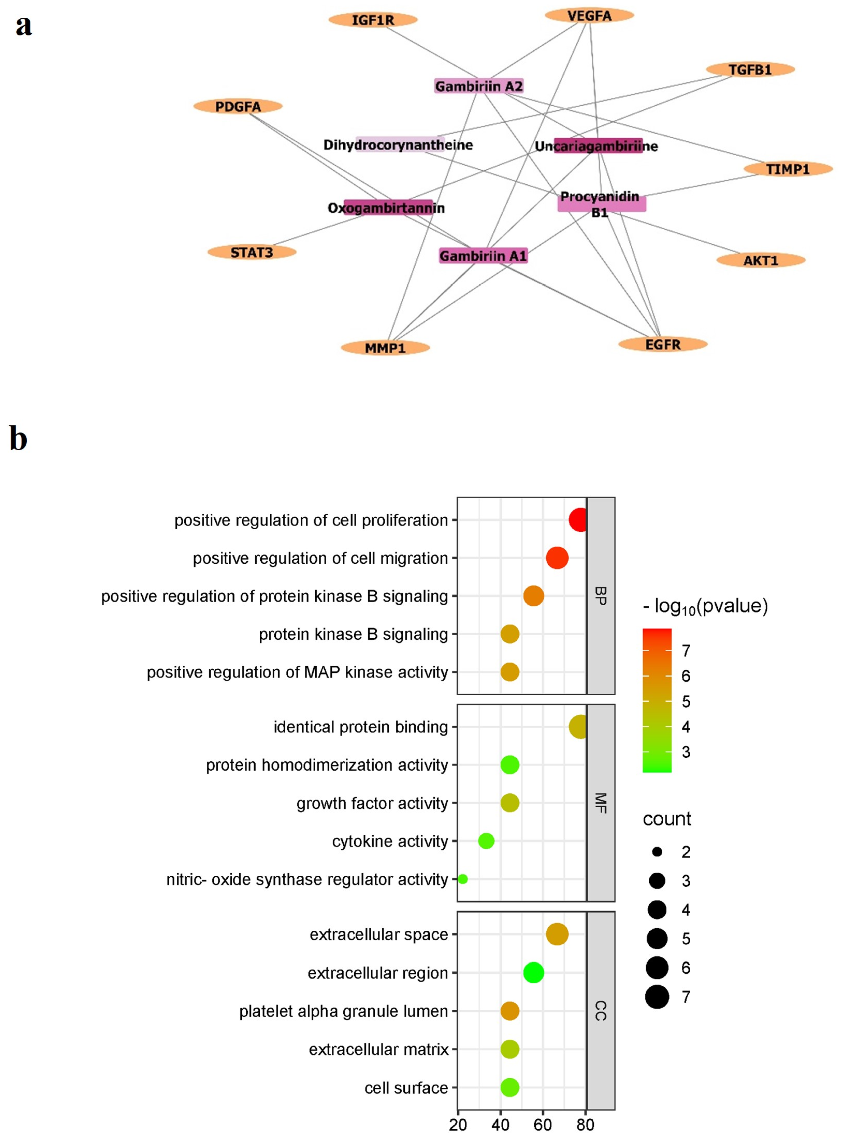
Figure 1. (a) Drug–protein interaction network and (b) Gene ontology enrichment analysis. Pink: Gambir bioactive compounds and yellow: Keloid target proteins. Biological process (BP), molecular function (MF), and cellular components (CC) related to the core target proteins.
Biologics 05 00018 g001
Figure 2. Molecular docking visualization of uncariagambiriine-TGFβ1 and uncariagambiriine-Akt complex. The dashed lines represent the specific interaction type; green: conventional hydrogen bonds, light green: carbon-hydrogen bonds; light purple: pi-alkyl interactions; violet: pi-sigma interaction; orange: pi-anion interaction; red: unfavorable donor–donor interaction.
Biologics 05 00018 g002aBiologics 05 00018 g002bBiologics 05 00018 g002cBiologics 05 00018 g002d
Figure 3. Molecular dynamics simulation of uncariagambiriine with TGFβ1 (A) and AKT1 (B). Radius of gyration of the solute (Rg), root mean square deviation (RMSD) of protein, and root mean square fluctuation (RMSF) of amino acid residues.
Biologics 05 00018 g003
Figure 4. The inhibitory effect of gambir ethanol fraction towards keloid fibroblast migration and proliferation (ex vivo) on day-3 and day-6 after treatments (AD) and Semi-quantitative analysis (E). DAPI staining was conducted at the end of day-6. The images were captured under an inverted fluorescence microscope at 100× magnification (scale bar 100 µM). Complete media was used as the negative control (NC), and the treatments were 64.38, 128.76, and 257.52 µg/mL of Gambir ethanol fraction. Data was represented as the means ± SD, n = 4. ** p < 0.01; *** p < 0.001; **** p < 0.0001.
Biologics 05 00018 g004
Table 1. Total phenolic and flavonoid contents of gambir’s crude extract and fraction.
Gambir’s SampleYield (%)Total Phenolic (mg GAE/g)Total Flavonoid (mg QE/g)
Crude extract4433.419.59
Ethanol Fraction10.9534.117.09
Hexane Fraction25.6134.012.39
Ethyl Acetate Fraction1.1433.413.33
Table 2. LC-MS analysis of Gambir extract and fraction.
SampleRT (min)Abundance (%)m/zFormulaSuspect Compound
Crude extract3.411.91579.15C30H26O12Procyanidin B1
4.270.35581.17C30H28O12Gambiriin A1
6.952.23333.16C21H20N2O2Dihydrogambirtannine
7.2814.46621.22C36H32N2O8Uncariagambiriine
Ethanol fraction1.280.97579.15C30H26O12Procyanidin B1
4.290.36581.16C30H28O12Gambiriin A1
7.014.50333.16C21H20N2O2Dihydrogambirtannine
7.268.27621.22C36H32N2O8Uncariagambiriine
8.531.96345.12C21H16N2O3Oxogambirtannine
Ethyl acetate fraction1.280.85579.15C30H26O12Procyanidin B1
4.290.34581.16C30H28O12Gambiriin A2
7.013.61333.16C21H20N2O2Dihydrogambirtannine
7.267.10621.22C36H32N2O8Uncariagambiriine
8.512.73345.12C21H16N2O3Oxogambirtannine
Hexne fraction6.994.30333.16C21H20N2O2Dihydrogambirtannine
7.248.19621.22C36H32N2O8Uncariagambiriine
Table 3. The molecular docking analysis of Gambir’s bioactive compounds with keloid target proteins.
Gambir Bioactive CompoundsBinding Affinity (Kcal/mol)
TGFβ1Akt
Procyanidin B1−8.05−9.33
Gambiriin A1−6.03−9.17
Gambiriin A2−5.41−10.51
Dihydrogambirtannine−6.75−9.18
Uncariagambiriine−8.24−11.02
Oxogambirtannine−7.16−8.84
Native ligand−7.21−10.73
Table 4. The effect of Gambir extract and fraction on keloid fibroblast viability and the IC50 value of concentration.
SampleIC50 KFIC50 NFSI (IC50 KF/NF)
Crude extract249.56 ± 0.221096.42 ± 0.524.39
Ethanol fraction128.76 ± 0.24780.58 ± 0.196.06
Ethyl acetate fraction238.79 ± 0.231012.03 ± 0.484.24
Hexane fraction162.07 ± 0.26570.23 ± 0.493.52
SI: selectivity index, KF: keloid fibroblast, NF: normal fibroblast.
Disclaimer/Publisher’s Note: The statements, opinions and data contained in all publications are solely those of the individual author(s) and contributor(s) and not of MDPI and/or the editor(s). MDPI and/or the editor(s) disclaim responsibility for any injury to people or property resulting from any ideas, methods, instructions or products referred to in the content.