Article

Insights Gained from the Immune Response and Screening of Healthcare Workers After COVID-19 Vaccination

1
Division of Infectious Diseases and Global Public Health, University of California, San Diego, CA 92103, USA
2
Genalyte Inc., San Diego, CA 92121, USA
*
Author to whom correspondence should be addressed.
COVID 2025, 5(8), 129; https://doi.org/10.3390/covid5080129
Submission received: 15 July 2025 / Revised: 7 August 2025 / Accepted: 7 August 2025 / Published: 8 August 2025
(This article belongs to the Section COVID Clinical Manifestations and Management)

Abstract

Background: COVID-19 vaccination has been a key tool in protecting healthcare workers (HCWs), but breakthrough infections have occurred. The durability of vaccine-induced immunity and its impact on HCWs remain critical for public health strategies. Methods: In this small cohort study (n = 32), we assessed antibody levels and breakthrough infection rates in HCWs over 12 months post-vaccination, providing insights for booster strategies and infection control. A cohort of 32 HCWs was screened for SARS-CoV-2 infection using weekly self-administered swabs and blood samples collected at baseline, 6 months, and 12 months. SARS-CoV-2 antibodies (IgG, IgM) targeting spike proteins and nucleocapsids were analyzed using a multi-antigen serology panel. Pooled nucleic acid testing was employed for infection detection. Results: Nine participants showed breakthrough infections, with nucleocapsid antibodies indicating prior infection. Eight of these cases occurred after the third vaccine dose during the Omicron-dominant period. Anti-spike antibody levels declined significantly in participants without prior infection, while those with breakthrough infections exhibited increased levels. The half-life of S1 and S1 receptor-binding domain (RDB) vaccine-induced antibodies was 144 and 166 days, respectively, which aligns with CDC data. These findings provide valuable insights for determining the optimal timing of booster doses. Conclusions: Our findings highlight the waning antibody levels over time and the occurrence of breakthrough infections. Although based on a small sample, these data support the need for ongoing monitoring and timely boosters.

1. Introduction

As of March 2025, there have been over seven hundred and seventy million confirmed cases of COVID-19 and seven million deaths globally [1]. During the first year of the pandemic, the World Health Organization estimated that between 80,000 and 180,000 of these deaths occurred among healthcare workers (HCW) [2]. To mitigate the direct and indirect impact of COVID-19, personal protective equipment (PPE), early detection through screening and rapid diagnosis, and isolation are necessary for HCWs to protect themselves and limit transmission to patients.
In addition to infection control methods, vaccinations have played a critical role in ensuring HCW safety. Through eliciting protective immunity from the production of anti-spike antibodies, vaccinations have become mandatory for HCWs across many countries to reduce transmission to at-risk populations [3,4]. However, according to a Centers for Disease Control and Prevention (CDC) study, only 15.3% of HCWs in acute care hospitals and 10.5% in nursing homes received the 2023–2024 COVID-19 vaccine [5], with the lowest rates among licensed independent practitioners [6]. This decline in vaccination uptake coincides with the expiration of the COVID-19 vaccine mandate for HCWs in June 2023 and the cessation of free government-funded COVID-19 vaccinations.
Public health officials emphasize the importance of vaccinations to maintain immunity and prevent the spread of diseases. However, there are currently no specific CDC recommendations for COVID-19 vaccinations for HCWs, aside from workplace policies and promotions like free on-site vaccinations [6].
Several studies have provided valuable insights into post-vaccination SARS-CoV-2 infection rates among HCWs. One study involving 3235 vaccinated HCWs reported an overall post-vaccination infection rate of 2.63%, with 76.5% of these cases occurring among individuals vaccinated with at least two COVID-19 vaccine doses [7]. Another study found a cumulative incidence of 6.3% for new SARS-CoV-2 infections among HCWs following the implementation of a vaccination program [8]. These findings highlight that while vaccinations significantly reduce the risk of infection, breakthrough cases can still occur among HCWs who have received one or more doses of a COVID-19 vaccine. Rates of reinfection may be influenced by factors such as declining immunity, the emergence of variants of concern, and ongoing occupational exposure [9].
In this 12-month observational study, we screened 32 HCWs for SARS-CoV-2 infection and evaluated the kinetics of their antibody levels post-vaccination. The goal was to better understand the durability of the immune response and how these findings can inform future booster strategies and public health measures.

2. Materials and Methods

2.1. Screening for Healthcare Workers

Between July 2021 and November 2022, weekly self-administered nasopharyngeal swabs were voluntarily taken by healthcare personnel to determine the prevalence of SARS-CoV-2 infection in the workplace. In addition to sampling, questionnaires were given to inquire about possible symptoms, COVID-19 infection in the past 14 days, and whether anyone in their household or close environment had tested positive. Detection of SARS-CoV-2 was conducted through pooled nucleic acid testing where individual swabs were placed in viral transport media (VTM). Equal volumes of VTM were then taken from these samples and combined into one pool that would undergo testing using the Fluxergy platform (RT-PCR) [10]. The remaining transport media was stored for subsequent testing and deconvolution. In the case that a pool did have a positive result, each test was run individually [10].

2.2. Detection of Antibody Levels: Multi-Antigen Serology Panel [Genalyte]

Blood was drawn at the beginning of the study post-primary vaccination—precluding direct measurement of peak antibody levels—as well as at 6 months and 12 months afterwards. Plasma was collected from these samples and run through a MaverickTM SARS-CoV-2 Multi-Antigen Panel (Genalyte, Inc., San Diego, CA, USA) which detects IgG and IgM responses to 13 antigens including 5 SARS-CoV-2 antigens: Nucleocapsid, spike S1 RBD, spike S1 subunit, spike S2 subunit, and spike S1S2 protein, 4 common Coronavirus antigens (SARS-CoV-229E S, SARS-CoV-NL63 NC, SARS-CoV-OC43 S, and SARS-CoV-HKU1 S), 2 influenza A hemagglutinin antigens (H1 and H3), and the NC and S1 antigens. Briefly, 10uL of sample was added to the assay stripwell plate, and the plate and chip were loaded into the instrument. The serum sample was then charged over the chip to bind specific antibodies to specific antigens on the chip. After washing any unbound or low affinity binders, specific antibodies were detected with anti-IgG and anti-IgM secondary antibodies. The response measurements are reported in Genalyte Response Units (GRU) which are converted to arbitrary units (AU) through a calibration process per manufacturer instructions. A proprietary machine learning algorithm based on a well-established random forest ensemble method using 3000 decision trees was applied to generate positive, negative, or indeterminate calls for SARS-CoV-2 antibodies. Although detailed information regarding the algorithm’s training data and internal workings cannot be disclosed due to proprietary restrictions, the assay and algorithm have been independently validated in prior studies demonstrating strong concordance with standard serological and neutralization assays [11,12]. Additionally, internal quality control measures and replicate testing within our laboratory confirmed the reliability and reproducibility of the antibody response calls. A linear regression analysis was used to assess the change of IgG antibody levels over the 12-month period. Analyses were performed in R (version 4.1.0) [13] using base functions and the lme4 package for mixed models where applicable.

2.3. Antibody Half-Life Estimation

To estimate the elimination half-life of SARS-CoV-2 antibodies, we applied a log-linear regression model under the assumption of first-order kinetics, where the rate of antibody decline is proportional to its concentration over time. For each participant, we modeled the natural logarithm of antibody concentration as a linear function of time since last vaccination using ordinary least squares (OLS) regression:
log C t = log C o k · t
where:
  • Ct is the antibody concentration at time t,
  • Co is the estimated concentration at time zero (intercept),
  • k is the elimination rate constant (slope of the regression),
  • t is time since injection.
The antibody half-life ( t 1 / 2 ) was derived using the standard first-order decay equation:
t 1 / 2 = l n 2 k
Regression was performed independently for each participant. To summarize group-level kinetics, we report the average half-life across all participants without breakthrough infections. Model fit was evaluated for each regression using the coefficient of determination (R2) and the p-value for the slope estimate.
Because repeated measures were taken from the same individuals over time, we also assessed potential within-subject correlation using a linear mixed-effects model as a sensitivity analysis, treating subject ID as a random intercept. These models yielded comparable estimates of k, supporting the robustness of the simple regression approach for estimating individual decay rates. Missing data, including occasional skipped weekly swabs or missed antibody timepoints, were addressed by using all available observations per participant. Regression models were fit only when at least two antibody measurements were available, and no imputation was performed. All analyses were performed in R (version 4.1.0) [13] using base functions and the lme4 package for mixed models where applicable.

3. Results

3.1. Screening HCWs for SARS-CoV-2 Infection

Thirty-two (32) HCWs were enrolled, with most of the cohort being female (66%, n = 21), and the mean age was 43 years [IQR]. All participants were vaccinated with mRNA vaccines (66% Moderna, 34% Pfizer) prior to enrollment, and some received a second (32/32), third (29/32), fourth (5/32), and fifth (1/32) dose during the study period (Table 1). Within the first six months, an average of 16 nasal swabs per person were examined (n = 523 swabs), all of which tested negative for viral RNA (Figure 1).
Blood samples were collected at baseline, 6 months, and 12 months to assess SARS-CoV-2 antibody (IgG and IgM) responses against viral spike proteins and nucleocapsids. At baseline, no participants exhibited an antibody response against nucleocapsids, suggesting no prior infection. However, by the 12-month mark, we observed an increase in nucleocapsid antibody levels in nine participants, indicating breakthrough infections. Seven of these participants reported either testing positive for COVID-19 or being in direct contact with the virus, while the remaining two likely experienced asymptomatic, undiagnosed breakthrough infections. Notably, one of these participants had a 14-day gap between consecutive nasal sampling, during which they may have been infected but not tested. Eight of the nine cases occurred after the third vaccine dose, and all but one case was reported between January and July 2022, when Omicron variants predominated.

3.2. Antibody Kinetics

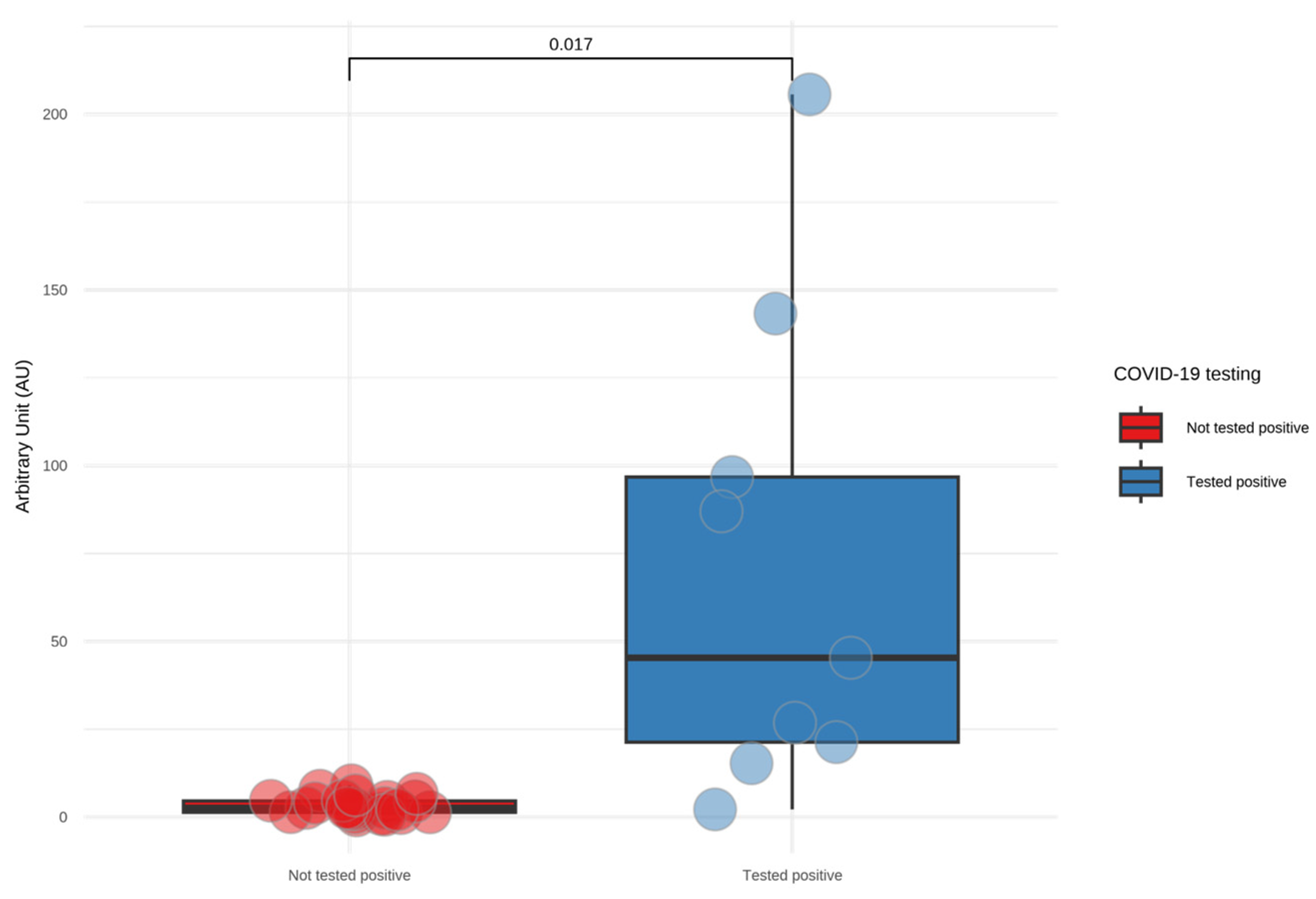
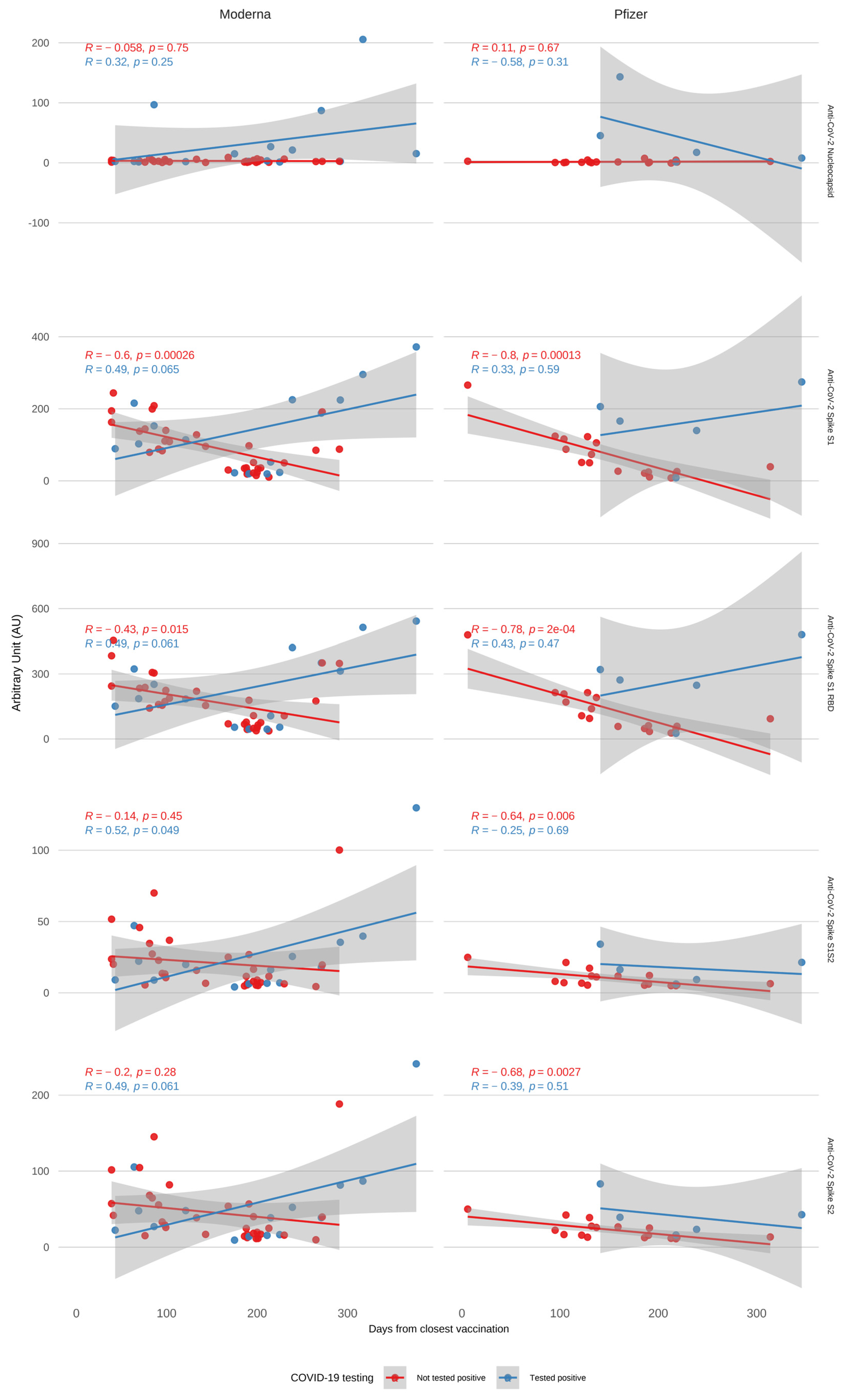
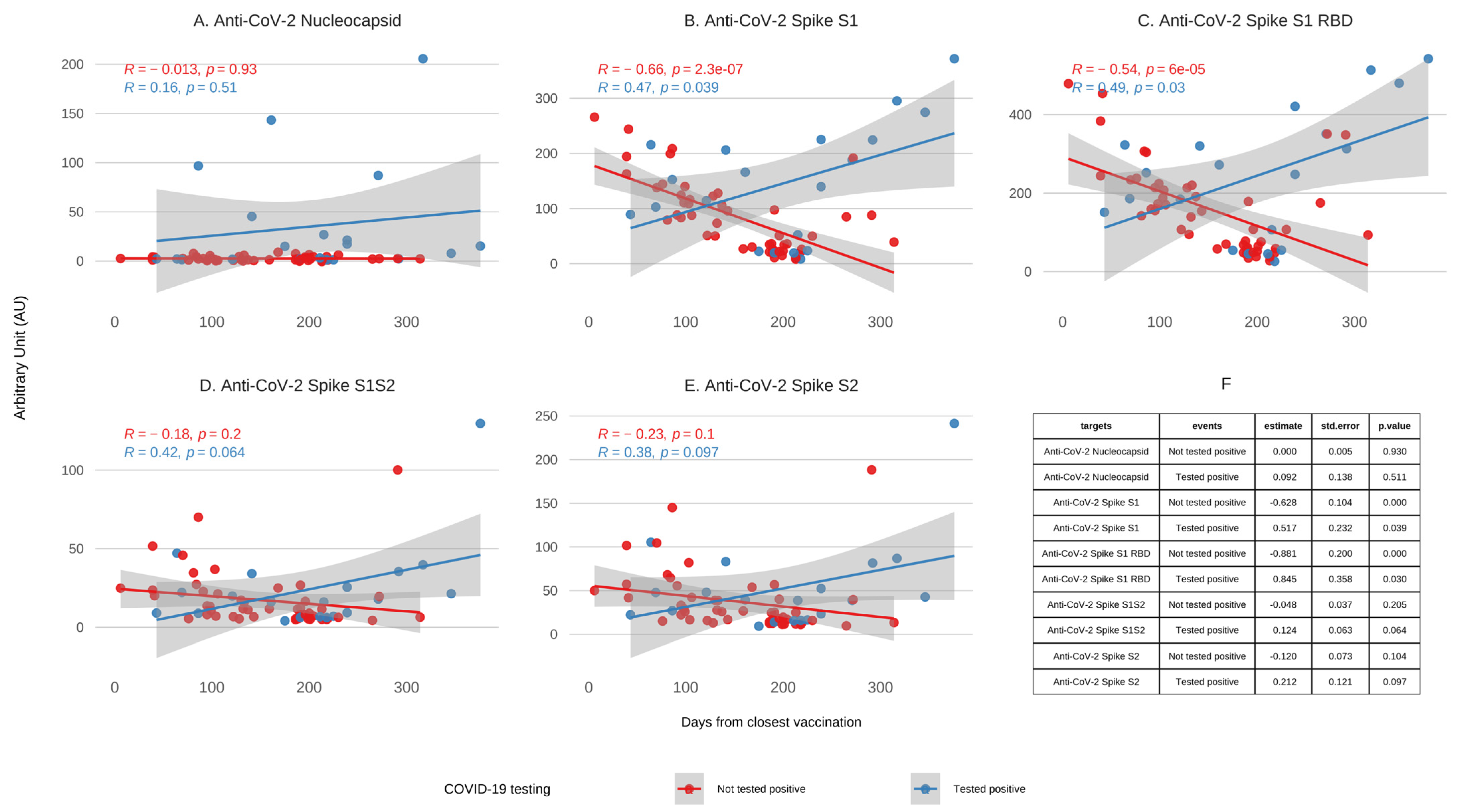
In participants with no prior evidence of COVID-19 infection (n = 23), we observed a significant decrease in IgG levels against spike proteins (S1: p < 0.001, S1 RBD: p < 0.001) over the follow-up period (Figure 2). In contrast, participants who reported a breakthrough COVID-19 infection (n = 9) showed an overall increase in anti-spike (S) protein levels (Figure 2). Participants who self-reported a positive COVID-19 test during follow-up had significantly higher peak anti-nucleocapsid IgG levels than those who did not report infection (p = 0.018); see Appendix A Figure A1. The average number of days between the detected infection and the last vaccination was 147 days, with a median of 114 days (range: 72–252 days) for participants with breakthrough infections. For those not exposed to the virus, the half-life of S1 and S1 RBD vaccine-induced antibodies was, on average, 144 and 166 days, respectively. Next, we explored whether the type of initial mRNA vaccine influenced antibody kinetics or breakthrough infection rates. We observed comparable rates of breakthrough infections among participants initially vaccinated with Moderna (5/21; 23.8%) and Pfizer (2/11; 18.2%). Antibody decay trends stratified by vaccine type are shown in Appendix A Figure A2. For participants with breakthrough infections, anti-nucleocapsid IgG levels showed a modest increase over time in Moderna recipients and a slight decrease among Pfizer recipients, although neither trend reached statistical significance. Patterns for spike protein antibodies (S1, S1 RBD, S1S2, and S2) were comparable across vaccine types, with similar rates of decline in infection-naïve individuals and elevated levels following breakthrough infection. These findings suggest that vaccine type may not have strongly influenced overall antibody kinetics in our cohort, although larger studies are needed to confirm these observations. Note: Three participants who received Moderna as their first and second doses later received a Pfizer vaccine as their third and/or fourth dose. One participant received a third dose of Moderna after having received Pfizer for their first two doses. All participants were grouped based on their initial vaccination type. Due to the limited sample size, these findings should be interpreted cautiously and were not formally tested for statistical significance.

4. Discussion

Our findings highlight that, while routine PCR screening remains an essential tool for detecting SARS-CoV-2 infections, including asymptomatic cases, it is not sufficient on its own to fully protect HCWs. In our study, two breakthrough infections were not identified during routine surveillance but were retrospectively detected through seroconversion to nucleocapsid antibodies. These findings underscore the importance of combining screening with up-to-date vaccination and robust infection control measures to reduce the risk of SARS-CoV-2 transmission in healthcare settings.
The divergent antibody kinetics observed between HCWs with and without breakthrough infection are likely attributable to the reactivation of SARS-CoV-2-specific memory B cells following re-exposure to viral antigens. While circulating anti-spike IgG antibodies gradually declined over time in individuals without infection, those with breakthrough infections exhibited an increase in antibody levels, likely reflecting memory B cell-mediated boosting. This is consistent with previous studies showing that SARS-CoV-2-specific memory B cells can persist for over a year and can rapidly differentiate into antibody-secreting cells upon re-exposure to antigen [14].
The consistency between half-life estimates from our study and CDC data provides valuable insights for determining the optimal timing of booster doses [15,16]. These data also align with current regulatory frameworks from the European Medicines Agency (EMA), the CDC, and the U.S. Food and Drug Administration, which increasingly emphasize immunobridging and the use of antibody kinetics as supportive evidence in evaluating booster vaccine strategies and updating vaccine compositions [17,18,19]. Neutralizing antibody levels have been shown to be strong correlates of protection against symptomatic SARS-CoV-2 infection, including against variants of concern with immune escape potential, underscoring the importance of maintaining sufficient antibody titers through booster vaccination to mitigate breakthrough infections [20,21]. Our findings thus contribute relevant real-world evidence that can inform regulatory and public health decisions regarding vaccine scheduling and the need for variant-adapted boosters. Waning anti-spike antibody levels, coupled with the emergence of variants of interest, underscore the urgent need for booster shots and the development of next-generation vaccines [22,23,24,25]. This is highlighted by a study by Padoan et al., which showed that the effectiveness of the BNT162b2 (Pfizer-BioNTech) vaccine against Omicron infection drops below 30% nine months after the booster dose, further highlighting the necessity for updated vaccines and safety protocols [26]. Real-world vaccine effectiveness studies from multiple settings have similarly demonstrated a marked reduction in protection against Omicron-related infection and symptomatic disease over time, reinforcing the importance of timely booster updates and variant-specific formulations [27,28].
These findings highlight the critical need for ongoing monitoring of vaccine effectiveness, particularly in high-risk populations such as HCWs, and the strategic deployment of updated, variant-specific vaccines and boosters tailored to individual risk factors to sustain population-level immunity and prevent future outbreaks [26,29,30].
Although our study cohort received only ancestral-strain vaccines, the observed waning of anti-spike antibody levels mirrors patterns reported in more recent cohorts that received variant-adapted booster vaccines. Emerging data suggest that bivalent and other updated boosters can elicit broader neutralizing responses and improved durability against newer SARS-CoV-2 variants [31,32,33]. Continued longitudinal studies are needed to directly compare the kinetics and protective effects of variant-specific boosters versus ancestral formulations, particularly in high-risk populations like HCWs.
While our cohort provides valuable longitudinal data on vaccine-induced antibody durability and breakthrough infections among HCWs in an academic medical center in California, extrapolation to other settings should be conducted cautiously. In regions with different levels of SARS-CoV-2 circulation, healthcare system burden, or vaccine access, patterns of antibody decay and breakthrough infections may vary. Similarly, HCWs in different roles (e.g., long-term care, outpatient clinics, or frontline emergency responders) may face distinct exposure risks not captured in this study. Furthermore, demographic differences including age, race/ethnicity, and comorbidities can influence immune responses and were not fully represented in our small, relatively homogeneous cohort. Future studies in more diverse and geographically varied populations are essential to validate and extend our findings.
This study has also several limitations. First, the voluntary nature of participation and testing may introduce selection bias. Individuals more engaged in their health or occupational safety may have been more likely to participate consistently and to adopt more protective behaviors, potentially influencing exposure risk and limiting the generalizability of our findings. Second, we did not perform genomic sequencing of positive cases, limiting our ability to definitively attribute breakthrough infections to specific SARS-CoV-2 variants. However, the timing of these infections aligns with the period during which Omicron-lineage viruses were predominant in the region, based on contemporaneous local surveillance data [34,35]. Third, the relatively small sample size (n = 32) restricts the generalizability of our findings and reduces the statistical power to detect meaningful differences across demographic subgroups such as sex, race/ethnicity, or vaccine type. This limitation should be recalled when interpreting both the descriptive antibody kinetics and observed infection patterns. However, this sample size is sufficient to estimate within-subject antibody kinetics with reasonable confidence, particularly given the longitudinal design and repeated measures for each participant. While our results offer valuable exploratory insights, they require confirmation in larger, more diverse cohorts to support broader public health conclusions. While we present descriptive trends in antibody kinetics and infection patterns within these subgroups, these findings should be considered exploratory and interpreted with caution. Larger, more diverse cohorts with variant-level viral characterization will be essential to confirm these patterns and support stratified public health recommendations for booster timing and vaccine strategy. Fourth, in participants with breakthrough infections, the precise timing of infection relative to their most recent vaccination remains uncertain. This limitation constrains our ability to accurately characterize vaccine-induced antibody durability and to precisely estimate antibody half-lives post-infection and post-vaccination. Consequently, interpretations regarding vaccine sustainability and immune kinetics should be made with caution. Finally, binding antibody titers, while informative, may overestimate protective immunity—especially against Omicron-lineage variants with immune escape. The absence of functional neutralization assays, such as surrogate virus neutralization tests [36,37], limits our ability to directly assess protective immune responses. However, it is well established that antibodies targeting the RBD of the spike protein, such as those measured in our study, are strongly associated with neutralizing activity, providing valuable indirect insights into immunity. While neutralization assays offer critical functional assessment, they can be resource-intensive and less feasible for longitudinal monitoring. In contrast, binding antibody measurements, as used here, enable scalable and repeated sampling to track humoral kinetics over time. Future studies incorporating functional assays will be important to confirm the neutralizing potential of these responses in our cohort and to further validate the utility of binding antibody data as a surrogate marker of protection. Finally, while our study focuses on humoral immunity, it is important to note that cellular immune responses, particularly T cell immunity, also contribute substantially to protection against severe disease and may retain cross-reactivity against SARS-CoV-2 variants including Omicron [38,39,40]. This complementary arm of the immune system may explain sustained protection despite waning antibody levels.

5. Conclusions

The results of our study demonstrate a significant decline in antibody levels among HCWs following vaccination, emphasizing the need for timely booster doses, particularly in the face of emerging variants of concern. Our findings underscore the critical importance of continuous monitoring of long-term antibody kinetics and the use of half-life data to optimize booster timing. Implementing these strategies is essential for maintaining vaccine efficacy and guiding the development of next-generation vaccines tailored to evolving SARS-CoV-2 variants.
Moreover, our study highlights the limitations of relying solely on vaccination and screening, as evidenced by undiagnosed breakthrough infections. While vaccines provide substantial protection, they do not guarantee absolute immunity. A comprehensive approach—combining up-to-date vaccination, routine screening, and stringent infection control measures—is, therefore, essential to ensure robust protection against COVID-19, particularly in high-risk populations such as HCWs.
Although our findings reflect immune dynamics following ancestral-strain vaccination, they underscore principles that remain highly relevant in the era of variant-adapted boosters—namely, the importance of timely immunization strategies informed by antibody waning kinetics and emerging variant threats. These insights can guide public health policies, optimize vaccine deployment, and support preparedness efforts for future pandemics by promoting a multifaceted and adaptive approach to disease prevention.

Author Contributions

Conceptualization, A.C.; Data Curation, A.C., M.P., and S.M.; Formal Analysis, A.C. and J.H.; Funding acquisition, A.C. and S.G.; Investigation, C.N.K.; Methodology, A.C.; Project administration, C.N.K.; Writing—original draft, J.H. and A.C.; Writing—review and editing, S.G., D.M.S., B.P., C.I., and J.H. All authors have read and agreed to the published version of the manuscript.

Funding

This work was performed with the support of the Translational Virology Core at the San Diego Center for AIDS Research. This work was supported by grants from the National Institutes of Health (San Diego Center for AIDS Research, CFAR, AI306214 and AI100665), the Department of Veterans Affairs, the John and Mary Tu Foundation, and the James B. Pendleton Charitable Trust. AC was also supported by Merck [MISP#60300] and The National Institutes of Health Grants [DA049644, AI145555, MH128153 (R01), AI106039 and DP2 CA051915]. SGW is supported by and The National Institutes of Health Grants [AI164570, MH062512, AI147821, DA051915, AI164559, AI158293].

Institutional Review Board Statement

The study was conducted in accordance with the Declaration of Helsinki and approved by the Institutional Review Board of University of California, San Diego (project #210676 approved on 14 June 2021).

Informed Consent Statement

Informed consent was obtained from all subjects involved in the study. Written informed consent has been obtained from the patients to publish this paper.

Data Availability Statement

The raw data supporting the conclusions of this article will be made available by the authors on request.

Acknowledgments

The authors would like to express their gratitude to all the participants who made this study possible and contributed to the advancement of COVID-19 research.

Conflicts of Interest

D.M.S. has consulted for the following companies Fluxergy, Gilead, Hyundai Biosciences, Model Medicines, Bayer Pharmaceuticals, and Pharma Holdings. The funders had no role in the design of the study; in the collection, analyses, or interpretation of data; in the writing of the manuscript; or in the decision to publish the results.

Abbreviations

The following abbreviations are used in this manuscript:
HCWHealthcare Worker
RDBReceptor-Binding Domain
NCNucleocapsid
PPEPersonal Protective Equipment
CDCCenters for Disease Control and Prevention
VTMVirus Transport Media
RT-PCRReverse Transcription Polymerase Chain Reaction
AUArbitrary Units
GRUGenalyte Response Units
OLSOrdinary Least Squares
IQRInterquartile Range
BMIBody Mass Index
VOIVariants of Interest

Appendix A

Figure A1. Anti-nucleocapsid IgG levels in participants with and without self-reported breakthrough infection. Boxplot comparing peak anti-nucleocapsid IgG levels between participants who self-reported a positive COVID-19 test and those who did not. Anti- nucleocapsid levels were measured using a SARS-CoV-2 Multi-Antigen Serology Panel at baseline, 6 months, and 12 months; for each participant, the highest recorded value was used. Participants with reported breakthrough infections (Tested positive) had significantly higher anti-nucleocapsid IgG levels compared to those who did not report prior infection (Not tested positive) (p = 0.017, Wilcoxon rank-sum test).
Covid 05 00129 g0a1
Figure A2. Kinetics of SARS-CoV-2 antibodies following vaccination with and without breakthrough infection, grouped by initial vaccine type. IgG antibodies directed against SARS-CoV-2 spike proteins (S1, S1 RBD, S1S2, S2) and nucleocapsid were quantified using a SARS-CoV-2 Multi-Antigen Serology Panel. Antibody levels are plotted against days since the last vaccination, stratified by initial mRNA vaccine type (Moderna vs. Pfizer) and infection status (blue = breakthrough infection; red = no infection). Ribbons around each regression line represent the standard error (SE) of the fit. While some divergence in nucleocapsid antibody kinetics was observed—particularly among breakthrough cases—there were no statistically significant differences in antibody trajectories between vaccine groups across all antibody types. Note: Three participants who received Moderna for their first and second doses later received Pfizer for their third and/or fourth dose. One participant received a third Moderna dose after two initial Pfizer doses. All participants were grouped according to their initial vaccine type.
Covid 05 00129 g0a2

References

  1. World Health Organization. WHO Coronavirus (COVID-19) Dashboard. Available online: https://covid19.who.int/ (accessed on 12 March 2025).
  2. World Health Organization. Health and Care Worker Deaths During COVID-19. Available online: https://www.who.int/news/item/20-10-2021-health-and-care-worker-deaths-during-covid-19 (accessed on 12 March 2025).
  3. Prévost, J.; Finzi, A. The great escape? SARS-CoV-2 variants evading neutralizing responses. Cell Host. Microbe 2021, 29, 322–324. [Google Scholar] [CrossRef]
  4. Stokel-Walker, C. COVID-19: The countries that have mandatory vaccination for health workers. BMJ 2021, 373, n1645. [Google Scholar] [CrossRef] [PubMed]
  5. Bell, J.; Meng, L.; Barbre, K.; Wong, E.; Lape-Newman, B.; Koech, W.; Soe, M.M.; Woods, A.; Kuhar, D.T.; Stuckey, M.J.; et al. Influenza and COVID-19 Vaccination Coverage Among Health Care Personnel—National Healthcare Safety Network, United States, 2023–2024 Respiratory Virus Season. MMWR Morb. Mortal. Wkly. Rep. 2024, 73, 966–972. [Google Scholar] [CrossRef]
  6. Axios Less than 1 in 6 Health Workers Got COVID Booster. Available online: https://www.axios.com/2024/11/04/health-workers-covid-booster-rates?utm_source=chatgpt.com (accessed on 12 March 2025).
  7. Vaishya, R.; Sibal, A.; Malani, A.; Prasad, K.H. SARS-CoV-2 infection after COVID-19 immunization in healthcare workers: A retrospective, pilot study. Indian J. Med. Res. 2021, 153, 550–554. [Google Scholar] [CrossRef]
  8. Kasztelewicz, B.; Skrok, K.; Burzyńska, J.; Migdał, M.; Dzierżanowska-Fangrat, K. Incidence of SARS-CoV-2 infection among healthcare workers before and after COVID-19 vaccination in a tertiary paediatric hospital in Warsaw: A retrospective cohort study. PLoS ONE 2024, 19, e0301612. [Google Scholar] [CrossRef]
  9. Xiang, T.; Liang, B.; Fang, Y.; Lu, S.; Li, S.; Wang, H.; Li, H.; Yang, X.; Shen, S.; Zhu, B.; et al. Declining Levels of Neutralizing Antibodies Against SARS-CoV-2 in Convalescent COVID-19 Patients One Year Post Symptom Onset. Front. Immunol. 2021, 12, 708523. [Google Scholar] [CrossRef] [PubMed]
  10. Rawlings, S.A.; Scott, B.; Layman, L.; Naranatt, P.; Heltsley, R.; Ignacio, C.; Porrachia, M.; Gianella, S.; Smith, D.; Chaillon, A. Can’t Work From Home: Pooled Nucleic Acid Testing of Laboratory Workers During the COVID-19 Pandemic. Open Forum Infect. Dis. 2021, 8, ofab129. [Google Scholar] [CrossRef]
  11. Donato, L.J.; Theel, E.S.; Baumann, N.A.; Bridgeman, A.R.; Blommel, J.H.; Wu, Y.; Karon, B.S. Evaluation of the genalyte maverick SARS-CoV-2 multi-antigen serology panel. J. Clin. Virol. Plus 2021, 1, 100030. [Google Scholar] [CrossRef] [PubMed]
  12. US Food and Drug Administraiton (FDA) Maverick™ SARS-CoV-2 Multi-Antigen Serology Panel v2; 2021. Available online: https://www.fda.gov/media/142915/download (accessed on 12 June 2025).
  13. RStudio Team. RStudio: Integrated Development for R; Version 2024.12.1.563; RStudio: Boston, MA, USA, 2020; Available online: http://www.rstudio.com/ (accessed on 12 June 2025).
  14. Marcotte, H.; Piralla, A.; Zuo, F.; Du, L.; Cassaniti, I.; Wan, H.; Kumagai-Braesh, M.; Andréll, J.; Percivalle, E.; Sammartino, J.C.; et al. Immunity to SARS-CoV-2 up to 15 months after infection. iScience 2022, 25, 103743. [Google Scholar] [CrossRef] [PubMed]
  15. Centers for Disease Control and Prevention Science Brief: SARS-CoV-2 Infection-Induced and Vaccine-Induced Immunity. Available online: https://www.cdc.gov/coronavirus/2019-ncov/science/science-briefs/vaccine-induced-immunity.html (accessed on 12 March 2025).
  16. Dai, Y.C.; Lin, Y.C.; Ching, L.L.; Tsai, J.J.; Ishikawa, K.; Tsai, W.Y.; Chen, J.J.; Nerurkar, V.R.; Wang, W.K. Determining the Time of Booster Dose Based on the Half-Life and Neutralization Titers against SARS-CoV-2 Variants of Concern in Fully Vaccinated Individuals. Microbiol. Spectr. 2023, 11, e0408122. [Google Scholar] [CrossRef]
  17. European Medicines Agency Regulatory Requirements for Vaccines Intended to Provide Protection Against Variant Strain(s) of SARS-CoV-2-Scientific Guideline 2021. Available online: https://www.ema.europa.eu/en/regulatory-requirements-vaccines-intended-provide-protection-against-variant-strains-sars-cov-2-scientific-guideline (accessed on 12 June 2025).
  18. Centers for Disease Control and Prevention Interim Clinical Considerations for Use of COVID-19 Vaccines in the United States. 2025. Available online: https://www.cdc.gov/covid/hcp/vaccine-considerations/index.html (accessed on 12 June 2025).
  19. U.S. Food and Drug Administration Emergency Use Authorization for Vaccines to Prevent COVID-19. 2022. Available online: https://www.fda.gov/emergency-preparedness-and-response/mcm-legal-regulatory-and-policy-framework/emergency-use-authorization (accessed on 12 June 2025).
  20. Khoury, D.S.; Cromer, D.; Reynaldi, A.; Schlub, T.E.; Wheatley, A.K.; Juno, J.A.; Subbarao, K.; Kent, S.J.; Triccas, J.A.; Davenport, M.P. Neutralizing antibody levels are highly predictive of immune protection from symptomatic SARS-CoV-2 infection. Nat. Med. 2021, 27, 1205–1211. [Google Scholar] [CrossRef]
  21. Gilbert, P.B.; Montefiori, D.C.; McDermott, A.B.; Fong, Y.; Benkeser, D.; Deng, W.; Zhou, H.; Houchens, C.R.; Martins, K.; Jayashankar, L.; et al. Immune correlates analysis of the mRNA-1273 COVID-19 vaccine efficacy clinical trial. Science 2022, 375, 43–50. [Google Scholar] [CrossRef]
  22. Burckhardt, R.M.; Dennehy, J.J.; Poon, L.L.M.; Saif, L.J.; Enquist, L.W. Are COVID-19 Vaccine Boosters Needed? The Science behind Boosters. J. Virol. 2022, 96, e0197321. [Google Scholar] [CrossRef] [PubMed]
  23. Wang, Q.; Wang, S.; Liu, Y.; Wang, S.; Peng, H.; Hao, Y.; Hong, K.; Li, D.; Shao, Y. Sequential Administration of SARS-CoV-2 Strains-Based Vaccines Effectively Induces Potent Immune Responses against Previously Unexposed Omicron Strain. Pathogens 2023, 12, 655. [Google Scholar] [CrossRef] [PubMed]
  24. Shen, X.; Hao, Y.; Wang, S.; Li, D.; Ren, L.; Zhu, M.; Wang, S.; Li, J.; Tang, W.; Fu, Y.; et al. Sequential heterologous immunization with COVID-19 vaccines induces broader neutralizing responses against SARS-CoV-2 variants in comparison with homologous boosters. Vaccine 2023, 41, 6645–6653. [Google Scholar] [CrossRef]
  25. World Health Organization Tracking SARS-COV-2 Variants. Available online: https://www.who.int/activities/tracking-SARS-CoV-2-variants (accessed on 12 March 2025).
  26. Padoan, A.; Cosma, C.; Della Rocca, F.; Barbaro, F.; Santarossa, C.; Dall’Olmo, L.; Galla, L.; Cattelan, A.; Cianci, V.; Basso, D.; et al. A cohort analysis of SARS-CoV-2 anti-spike protein receptor binding domain (RBD) IgG levels and neutralizing antibodies in fully vaccinated healthcare workers. Clin. Chem. Lab. Med. 2022, 60, 1110–1115. [Google Scholar] [CrossRef]
  27. Andrews, N.; Stowe, J.; Kirsebom, F.; Toffa, S.; Rickeard, T.; Gallagher, E.; Gower, C.; Kall, M.; Groves, N.; O’Connell, A.M.; et al. COVID-19 Vaccine Effectiveness against the Omicron (B.1.1.529) Variant. N. Engl. J. Med. 2022, 386, 1532–1546. [Google Scholar] [CrossRef]
  28. Tseng, H.F.; Ackerson, B.K.; Luo, Y.; Sy, L.S.; Talarico, C.A.; Tian, Y.; Bruxvoort, K.J.; Tubert, J.E.; Florea, A.; Ku, J.H.; et al. Effectiveness of mRNA-1273 against SARS-CoV-2 Omicron and Delta variants. Nat. Med. 2022, 28, 1063–1071. [Google Scholar] [CrossRef]
  29. Skrzat-Klapaczyńska, A.; Bieńkowski, C.; Kowalska, J.; Paciorek, M.; Puła, J.; Krogulec, D.; Stengiel, J.; Pawełczyk, A.; Perlejewski, K.; Osuch, S.; et al. The Beneficial Effect of the COVID-19 Vaccine Booster Dose among Healthcare Workers in an Infectious Diseases Center. Vaccines 2022, 10, 552. [Google Scholar] [CrossRef] [PubMed]
  30. Choi, J.H.; Kim, Y.R.; Heo, S.T.; Oh, H.; Kim, M.; Lee, H.R.; Yoo, J.R. Healthcare Workers in South Korea Maintain a SARS-CoV-2 Antibody Response Six Months After Receiving a Second Dose of the BNT162b2 mRNA Vaccine. Front. Immunol. 2022, 13, 827306. [Google Scholar] [CrossRef] [PubMed]
  31. Davis-Gardner, M.E.; Lai, L.; Wali, B.; Samaha, H.; Solis, D.; Lee, M.; Porter-Morrison, A.; Hentenaar, I.T.; Yamamoto, F.; Godbole, S.; et al. Neutralization against BA.2.75.2, BQ.1.1, and XBB from mRNA Bivalent Booster. N. Engl. J. Med. 2023, 388, 183–185. [Google Scholar] [CrossRef]
  32. Chalkias, S.; Harper, C.; Vrbicky, K.; Walsh, S.R.; Essink, B.; Brosz, A.; McGhee, N.; Tomassini, J.E.; Chen, X.; Ying, C.; et al. Three-month antibody persistence of a bivalent Omicron-containing booster vaccine against COVID-19. Nat. Commun. 2023, 14, 5125. [Google Scholar] [CrossRef]
  33. Collier, A.Y.; Miller, J.; Hachmann, N.P.; McMahan, K.; Liu, J.; Bondzie, E.A.; Gallup, L.; Rowe, M.; Schonberg, E.; Thai, S.; et al. Immunogenicity of BA.5 Bivalent mRNA Vaccine Boosters. N. Engl. J. Med. 2023, 388, 565–567. [Google Scholar] [CrossRef]
  34. CDC COVID-19 Response Team. SARS-CoV-2 B.1.1.529 (Omicron) Variant—United States, December 1–8, 2021. MMWR Morb. Mortal. Wkly. Rep. 2021, 70, 1731–1734. [Google Scholar] [CrossRef]
  35. Lambrou, A.S.; Shirk, P.; Steele, M.K.; Paul, P.; Paden, C.R.; Cadwell, B.; Reese, H.E.; Aoki, Y.; Hassell, N.; Zheng, X.Y.; et al. Genomic Surveillance for SARS-CoV-2 Variants: Predominance of the Delta (B.1.617.2) and Omicron (B.1.1.529) Variants—United States, June 2021–January 2022. MMWR Morb. Mortal. Wkly. Rep. 2022, 71, 206–211. [Google Scholar] [CrossRef]
  36. Tan, C.W.; Chia, W.N.; Qin, X.; Liu, P.; Chen, M.I.C.; Tiu, C.; Hu, Z.; Chen, V.C.-W.; Young, B.E.; Sia, W.R.; et al. A SARS-CoV-2 surrogate virus neutralization test based on antibody-mediated blockage of ACE2–spike protein–protein interaction. Nat. Biotechnol. 2020, 38, 1073–1078. [Google Scholar] [CrossRef] [PubMed]
  37. Wang, L.F.; Tan, C.W.; Chia, W.N.; Zhu, F.; Young, B.; Chantasrisawad, N.; Hwa, S.H.; Yeoh, A.Y.; Lim, B.L.; Yap, W.C.; et al. Differential escape of neutralizing antibodies by SARS-CoV-2 Omicron and pre-emergent sarbecoviruses. Res. Sq. 2022, rs.3.rs-1362541. [Google Scholar] [CrossRef]
  38. Sahin, U.; Muik, A.; Derhovanessian, E.; Vogler, I.; Kranz, L.M.; Vormehr, M.; Baum, A.; Pascal, K.; Quandt, J.; Maurus, D.; et al. COVID-19 vaccine BNT162b1 elicits human antibody and T(H)1 T cell responses. Nature 2020, 586, 594–599. [Google Scholar] [CrossRef] [PubMed]
  39. Grifoni, A.; Weiskopf, D.; Ramirez, S.I.; Mateus, J.; Dan, J.M.; Moderbacher, C.R.; Rawlings, S.A.; Sutherland, A.; Premkumar, L.; Jadi, R.S.; et al. Targets of T Cell Responses to SARS-CoV-2 Coronavirus in Humans with COVID-19 Disease and Unexposed Individuals. Cell 2020, 181, 1489–1501.e15. [Google Scholar] [CrossRef] [PubMed]
  40. Tarke, A.; Coelho, C.H.; Zhang, Z.; Dan, J.M.; Yu, E.D.; Methot, N.; Bloom, N.I.; Goodwin, B.; Phillips, E.; Mallal, S.; et al. SARS-CoV-2 vaccination induces immunological T cell memory able to cross-recognize variants from Alpha to Omicron. Cell 2022, 185, 847–859.e11. [Google Scholar] [CrossRef]
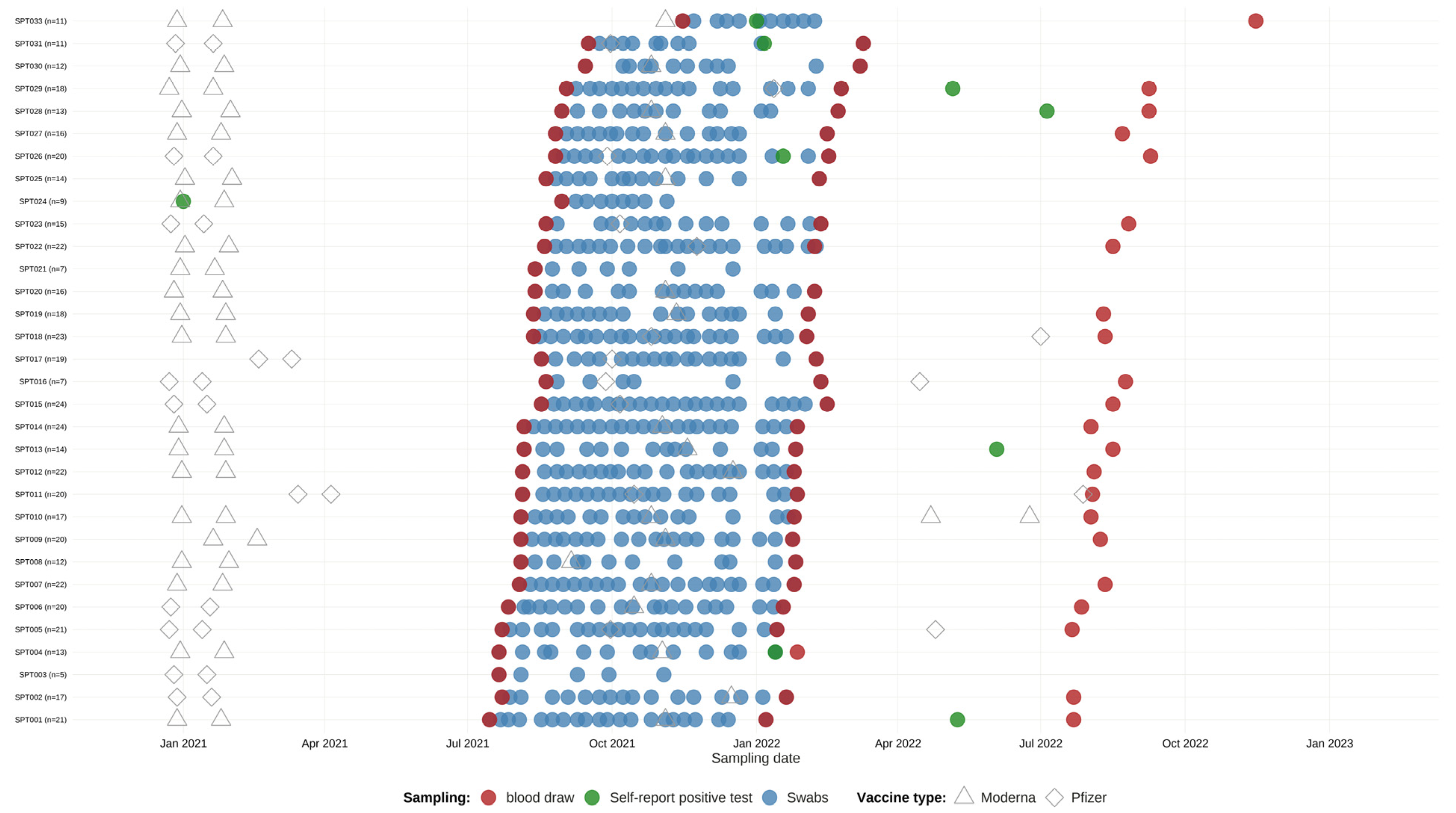
Figure 1. Participant sampling, testing and vaccination over the course of the study. Each row represents a participant and shows the total number (n) of nasopharyngeal swabs (blue), self-report of positive test for COVID-19 (green), blood draws (red), and vaccination (triangle and diamond) completed over a course of the study. Weekly swabs were proposed on a voluntary basis and blood was drawn at the baseline, month 6, and month 12 from study entry. Vaccinations and positive test results were also documented.
Covid 05 00129 g001
Figure 2. Kinetics of SARS-CoV-2 antibodies following vaccination with and without breakthrough infection. A SARS-CoV-2 Multi-Antigen Serology Panel was used to quantify the level of IgG antibodies directed against the nucleocapsid (Panel (A)) and COVID spike proteins [Panel (BE): S1, S1 RBD, S1S2, S2]. Plotted against days since last vaccination, these models show a change in antibody levels over time for participants who self-reported a positive SARS-CoV-2 test or were identified as positive through our pooled PCR screening protocol (blue) and those who had not (red). Ribbons around each regression line reflect the standard error (SE) of the fit. Panel (F): The table insert at the bottom right summarizes model estimates and SEs.
Covid 05 00129 g002
Table 1. Demographic Characteristics of HCWs.
Variables (N)Values
Age (32): 44 [40.9;47.1]
Gender (32):
Female21 (65.6%)
Male10 (31.2%)
Non-Binary1 (3.12%)
Ethnicity (32):
Hispanic or Latino/a9 (28.1%)
Not Hispanic or Latino/a23 (71.9%)
Race (32):
Asian 7 (21.9%)
Black or African American1 (3.12%)
Native American 1 (3.12%)
White Caucasian 23 (71.9%)
BMI (30):24.4 [19.5;34.3]
Prior COVID Diagnosis (32)2 (6.25%)
Vaccination (32):
Yes32 (100%)
Initial Vaccine Type (32):
Moderna 21 (65.6%)
Pfizer11 (34.3%)
Vaccine Dosage (32):
First Dose 32 (100%)
Second Dose32 (100%)
Third Dose29 (90.6%)
Fourth Dose 5 (15.6%)
Fifth Dose1 (3.1%)
Abbreviation: HCWs, Healthcare Workers; BMI: Body Mass Index. N represents the total number of participants; the bracket represents a 95% confidence interval for age, and parentheses are used to show % of individuals within each respective category.
Disclaimer/Publisher’s Note: The statements, opinions and data contained in all publications are solely those of the individual author(s) and contributor(s) and not of MDPI and/or the editor(s). MDPI and/or the editor(s) disclaim responsibility for any injury to people or property resulting from any ideas, methods, instructions or products referred to in the content.