You are currently viewing a new version of our website. To view the old version click .
Crops
  • Review
  • Open Access

17 November 2025

Peach Postharvest Fungal Diseases: Sustainable Management and an Integrative Review of Emerging Strategies

,
,
,
,
,
,
and
1
Phytohormones and Plant Development Laboratory, Department of Plant Biology (Plant Physiology), Faculty of Biology, University of Murcia, 30100 Murcia, Spain
2
Plant-Microbial-Marine-Biotechnologies and Precision Agriculture Group, Polydisciplinary Faculty of Larache, Abdelmalek Essaâdi University, Larache 92000, Morocco
3
Training and Research Center Louata, Providence Verte Company, Sefrou 31000, Morocco
*
Author to whom correspondence should be addressed.
This article belongs to the Special Issue Molecular Mechanisms and Integrated Control of Pathogen Crops

Abstract

Postharvest fungal diseases represent a major constraint to the storage, transport, and marketability of peach (Prunus persica) fruits. Pathogens such as Monilinia spp. (Brown rot), Penicillium expansum (Blue rot), Rhizopus stolonifera (Soft rot), Botrytis cinerea (Gray rot), and Geotrichum candidum (Acid rot) cause significant economic losses globally. Traditional control methods primarily rely on chemical fungicides, which are increasingly challenged by issues of resistance development, consumer health concerns, and regulatory restrictions. This review critically synthesizes the biology, infection mechanisms, and optimal environmental conditions of key fungal pathogens affecting postharvest peaches. It further evaluates the current landscape of chemical, physical, and biological control methods, emphasizing novel approaches including essential oils, microbial antagonists, induced resistance, and eco-friendly sanitizers. Comparative efficacy, sustainability, and practical implementation of these strategies are discussed. Integrated management approaches that combine multiple interventions under low-residue or residue-free systems are highlighted as the most promising direction. This review concludes that the future of peach postharvest protection lies in tailor-made, multi-faceted integrated programs that are both effective and environmentally sound.

1. Introduction

Peach (Prunus persica), a member of the Rosaceae family, is renowned for its nutritional profile and high levels of bioactive compounds such as flavonoids, vitamin C, chlorogenic acid, neochlorogenic acid, and protocatechuic acid [,]. The global production of peaches and nectarines has markedly increased to meet rising consumer demand, with China accounting for over 17.5 Mt in 2023, followed by notable contributions from Spain, Türkiye, Italy, the USA, Iran, Greece, Chile, Mexico, and Morocco [].
However, peaches are highly perishable and prone to postharvest decay, primarily due to fungal pathogens. Favorable environmental conditions, such as high humidity and warm temperatures during harvest and distribution, enhance the growth of fungi like Monilinia spp., Botrytis cinerea, Penicillium expansum, Rhizopus stolonifer, and Geotrichum candidum. These pathogens often enter through wounds or lenticels and compromise fruit integrity via enzymatic degradation and mycotoxin production [].
Conventional control relies on chemical fungicides such as fludioxonil, cyprodinil, pyrimethanil, and imazalil. However, growing concerns about fungal resistance, food safety, and environmental sustainability have encouraged the exploration of alternative approaches. These include physical treatments (UV irradiation, heat), biological agents (antagonistic yeasts and bacteria), and natural compounds (essential oils and plant extracts) [].
In the context of growing global concern over pesticide residues, environmental contamination, and the emergence of fungicide-resistant strains, the development of sustainable postharvest practices has become a key priority in modern horticulture. Regulatory frameworks such as the European Union’s “Farm to Fork” strategy and the Green Deal aim to reduce chemical pesticide use by 50% by 2030, pushing both researchers and producers to adopt eco-friendly alternatives []. Consumers are also increasingly demanding safer, residue-free fruits, thereby encouraging the transition toward integrated postharvest management systems that combine effectiveness with ecological responsibility. These shifts emphasize the urgent need for innovative, scalable, and sustainable disease control strategies that minimize chemical inputs while maintaining fruit quality and safety.
The objective of this review is to provide a critical synthesis of the main fungal pathogens responsible for postharvest decay in peaches and to assess the comparative effectiveness of chemical, physical, and biological control methods. This work aims to inform future research and support the development of sustainable strategies for managing postharvest diseases in peaches.

2. Postharvest Rot of Peaches

2.1. Brown Rot

Among the 35 species of the genus Monilinia, three are primarily responsible for brown rot in peaches: Monilinia laxa, Monilinia fructicola, and Monilinia fructigena []. These pathogens affect stone and pome fruits by colonizing flowers, shoots, and fruits. Due to high genetic similarity.
Morphologically, M. laxa produces small gray-green tufts of conidia (<0.5 mm), M. fructicola forms brownish tufts (~1 mm) with dark patches, and M. fructigena exhibits concentric yellowish conidial pads (~1.5 mm) (Figure 1) [].
Figure 1. Macroscopic images of Monilinia spp.: (a) Monilinia laxa on a peach fruit (own photo, taken at the Training and Research Center Louata, Sefrou, Morocco, 21 June 2019); (b) Monilinia fructicola on a peach fruit (own photo, taken at the Training and Research Center Louata, Sefrou, Morocco, 28 July 2019); (c) Monilinia fructigena on a peach fruit, adapted from []; (d) M. fructicola on a PDA medium (own photo, taken Department of Plant Biology (Plant Physiology), Murcia, Spain, 23 December 2002); (e) M. fructigena on a PDA medium (own photo, taken Department of Plant Biology (Plant Physiology), Murcia, Spain, 8 September 2002).
Brown rot causes severe economic losses, with over 50% of global postharvest peach losses attributed to Monilinia spp. infections in countries such as Turkey, Brazil, Spain, and Italy result in the loss of over 50% of postharvest peaches globally [,].
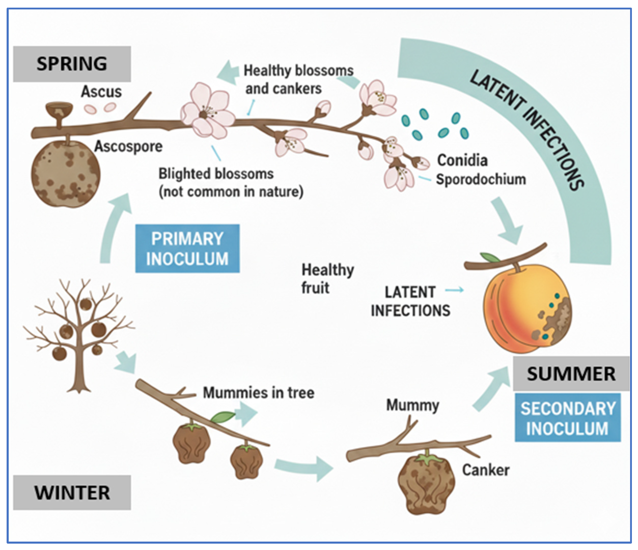
The life cycle involves overwintering on mummified fruits and twig cankers, which produce apothecia in spring (Figure 2). Spores are released and infect blossoms, initiating secondary cycles of infection []. Weather conditions—especially warm, humid climates—favor rapid disease development []. For instance, M. fructicola and M. fructigena conidia germinate in less than 2 h at 25 °C and 0.99 water activity, while M. laxa takes about 4 h under the same conditions.
Figure 2. Life cycle of Monilinia spp., adapted from [].
Temperature and humidity are critical: M. laxa grows better at low temperatures (0 °C), while M. fructicola performs better at 30–33 °C []. Notably, M. fructicola can sporulate at 33 °C, unlike M. laxa.
Despite the identification of mummified fruits as inoculum sources, the direct correlation between their presence and infection intensity on fruit surfaces remains unclear. However, orchards with high levels of mummified fruit or unpruned branches tend to have higher airborne conidial loads and more severe outbreaks [].

2.2. Blue Rot

Penicillium expansum, a member of the Talaromyces family, is the primary causal agent of blue mold in peaches and several other fruit crops. It is also a major concern for the fruit packing and processing industries, particularly for its ability to grow at low temperatures and produce toxic secondary metabolites [].
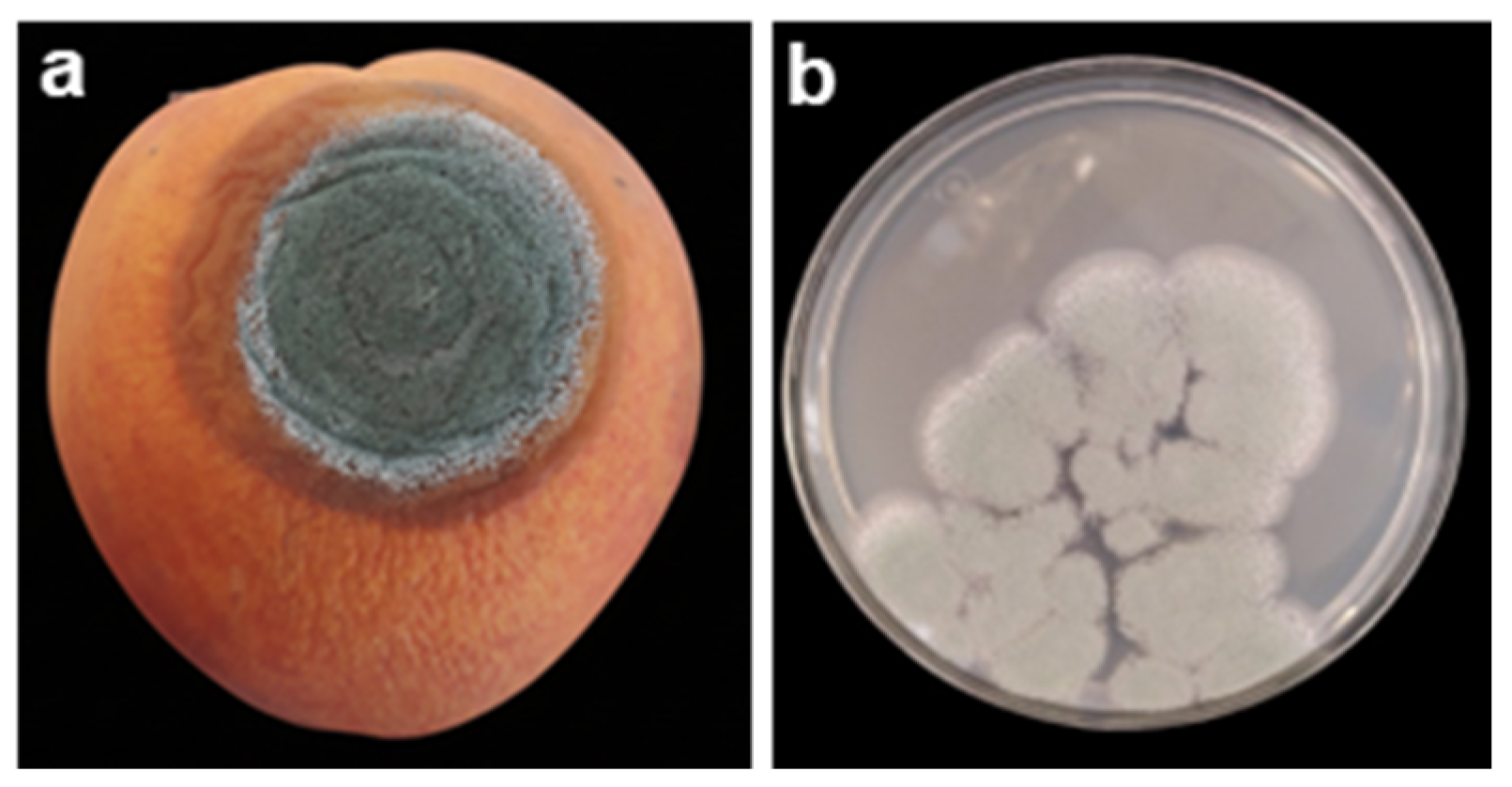
Infection generally occurs through wounds or natural openings, such as lenticels and the calyx []. Lesions initially appear soft and watery, often accompanied by white mycelial tufts, which later develop into blue-green sporulation masses (Figure 3). The infected tissue rapidly decays and becomes discolored and mushy. As the infection progresses, P. expansum acidifies the host tissue via gluconic and citric acid production, which facilitates colonization [].
Figure 3. Macroscopic images of Penicillium expansum: (a) P. expansum on a peach fruit (Own photo, taken at the Training and Research Center Louata, Sefrou, Morocco, 28 July 2019); (b) macroscopic image of P. expansum on a PDA medium (Own photo, taken at the Phytohormones and Plant Development Laboratory, Murcia, Spain, 14 October 2025).
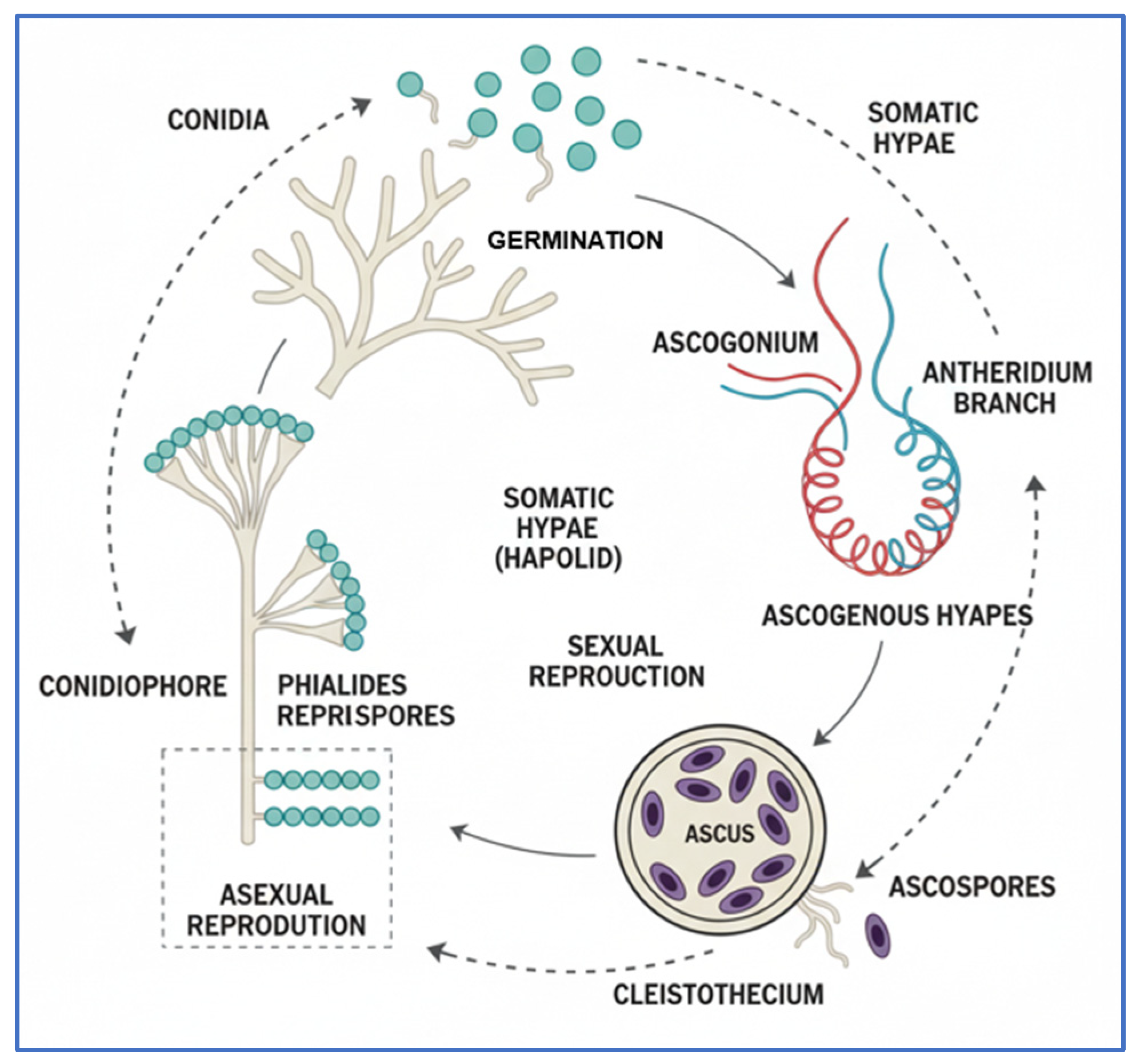
Penicillium reproduces in three main ways: vegetatively, asexually, and sexually. Vegetative reproduction occurs when hyphae divide into fragments to form independent mycelia []. In asexual reproduction, conidial spores develop from conidiophores, detach by wind, and germinate on the substrate to form a new mycelium. Sexual reproduction involves male and female parts, where the ascogonium is the male part and the antheridium is the female part. Both parts act as uninucleate hyphae, and the ascogonium divides to form nucleated hyphae that undergo meiosis and mitosis to produce ascospores. The ascospores develop on the substrate to give rise to the mother mycelium, which then continues to develop as an independent Penicillium (Figure 4).
Figure 4. Life cycle of Penicillium spp., adapted from [].
There has been limited research conducted to characterize the growth and conditions that promote the ability of P. expansum to develop, despite its frequent involvement in food contamination. Therefore, it is urgent to conduct further studies on the factors that influence the growth and toxigenesis of this fungus in order to better understand the risks associated with its presence in food and develop effective prevention strategies. Studies have been conducted to assess the impact of storage temperature, relative humidity (RH), and water activity on the growth rate of P. expansum. By understanding how these environmental factors affect microbial growth, it is possible to develop more effective control and prevention methods to avoid fruit contamination. Generally, maximum growth of P. expansum occurs at 25 °C, with minimal growth at 5 °C. However, moderate growth occurs at temperatures between 15 °C and 35 °C []. RH has influenced fungal growth significantly. Tannous et al. (2016) found that when they controlled the growth of P. expansum across a range of water activity from 85% to 99%, optimal growth was observed at 99%. At this humidity level, the fungus had the shortest latency phase and the largest colony growth (8.3 cm). This highlights the importance of environmental factors, such as RH, in controlling the development of P. expansum and understanding how to prevent its growth in food industries [,]. The results of these studies can be used to develop mathematical models that predict microbial growth based on environmental conditions, which can be useful for the food industry.
Moreover, Penicillium species can pose risks to human health through the synthesis of toxic secondary metabolites, including patulin (C7H6O4), citrinin (C13H14O5), commonins, chaetoglobosins A and C, cytochalasins, geosmin, expansolides A and B, and andrastins A, B, and C. Among these, patulin and citrinin are the most well-known and widely studied []. Patulin has been detected in both raw and processed products such as juices. Its production is favored by the presence of glucose, nitrogen, and acidic conditions. Studies have shown that it has hepatotoxic and genotoxic effects, and it increases the mortality rate, but it is not carcinogenic. The United States Food and Drug Administration (FDA) has set a limit for the allowable amount of patulin in processed products at 0.05 µg/mL []. Citrinin is also known for its toxicity. Toxicity tests conducted on mice have shown kidney hypertrophy and disruption of cellular organelles such as mitochondria, the endoplasmic reticulum, and lysosomes [].
Given its pathogenicity and toxigenic potential, P. expansum poses both economic and public health risks. Effective postharvest management requires a combination of proper handling to avoid wounds and appropriate cold chain storage, alongside biological or chemical control strategies.

2.3. Soft Rot

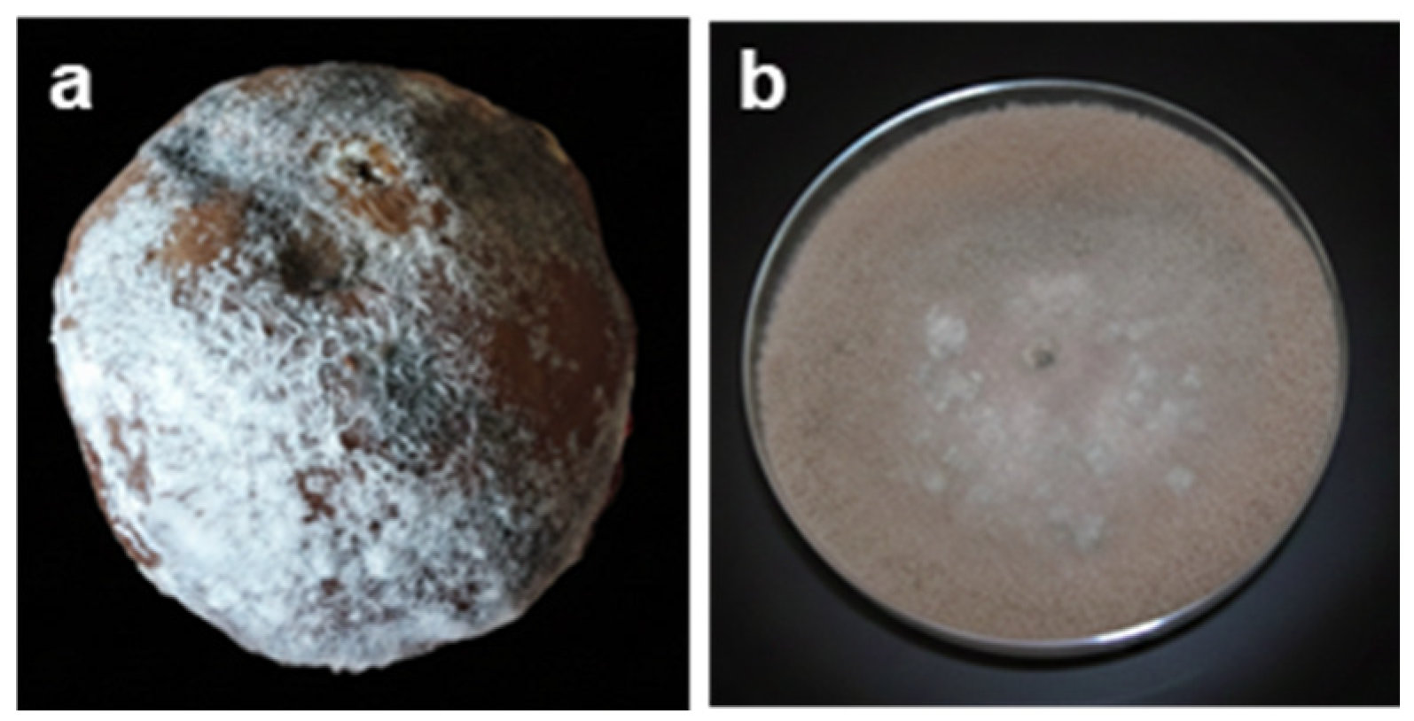
Rhizopus stolonifer, a filamentous fungus belonging to the Zygomycetes class, the Mucorales order, the Mucoraceae family, and the Rhizopus genus, is the primary causal agent of soft rot (also known as Rhizopus rot or black rot) in peaches and other perishable fruits (Figure 5). This pathogen is responsible for rapid postharvest decay, leading to significant economic losses during storage and distribution [].
Figure 5. Macroscopic images of Rhizopus stolonifer: (a) R. stolonifer on a peach fruit (own photo, taken at the Training and Research Center Louata, Sefrou, Morocco, 12 August 2019); (b) R. stolonifer on PDA medium (own photo, taken at the Training and Research Center Louata, Sefrou, Morocco, 19 May 2023).
The disease is also known as black rot or Rhizopus rot. According to the morphological classification by Schipper (1984), the common features of the R. stolonifer group include well-developed, complex rhizoids, sporangiophores, sporangia, and sporangiospores []. The main column (mycelium) is conical-cylindrical, gray or brownish in color, and can reach up to 140 μm in height, with branching forming a tangled mycelium. This mycelium sends upright aerial hyphae from various points, forming a whitish growth in cultures that eventually turns into black spots, called sporangiophores, which measure about 1 to 3 mm long and up to 20 to 25 μm in diameter. Each sporangiophore carries a single spherical sporangium that can reach about 250 μm in diameter and contains many sporangiospores, which can reach 13 μm in length and diameter, ranging from 100 to 275 μm. The spores of Rhizopus come in different shapes: angular, subglobular, and ellipsoidal, with distinct ridges on the surface depending on their maturity [].
It is frequently reported that R. stolonifer infection gains better access through wounds, injuries, and abrasions on the entire plant. A study conducted by Holmes and Stange (2002) indicated that the incidence of the disease in sweet potatoes is closely related to the type of injury. During a two-year evaluation period, bruising injuries followed by puncture wounds resulted in the highest infection rates []. Spores already present on the fruits serve as a source of inoculation for subsequent infections. As a result, water systems used in packing plants to wash produce postharvest can become a source of contamination. This contamination can spread to other products requiring washing, increasing the risk of future infections.
The growth and biomass production of R. stolonifer are influenced by various environmental factors. It grows rapidly at optimal temperatures between 23 °C and 28 °C but is sensitive to temperatures below 10 °C. The ideal pH for the germination of sporangiospores ranges from 3.0 to 10.0. A carbon and nitrogen source, such as sucrose and glutamine, is necessary to stimulate mycelial growth []. Generally, Rhizopus rot is characterized by high water absorption and exudation of clear leachate. According to Mena-Névarez et al. (2012), the production of leachate on infected mangoes is four times higher than on non-infected fruit after 35 days of storage, while on oranges, it is ten times higher. At the onset of infection, a thin layer of cotton-like fungal structures may cover the surface. Once R. stolonifer has penetrated the wounded tissues, its mycelium spreads around the infection site [].
Infection by this pathogen is closely linked to enzymatic activities that enable its mycelium to penetrate directly into uninjured fruits, inducing maceration within 2 to 3 days. Al-Hindi et al. (2011) found that this fungus produces approximately 3.37 ± 0.16 units/mL, 2.81 ± 0.22 units/mL, 0.58 ± 0.02 units/mL, and 0.67 ± 0.03 units/mL of xylanase, phosphoglucomutase, cellulase, and amylase, respectively, to degrade the fruit cell wall []. From the first day of infection, the surface may be covered with fine, fluffy, white fungal structures resembling cotton. Shortly after, the affected areas will be covered with gray, hairy mycelium, which will eventually form a mass of black sporangia.
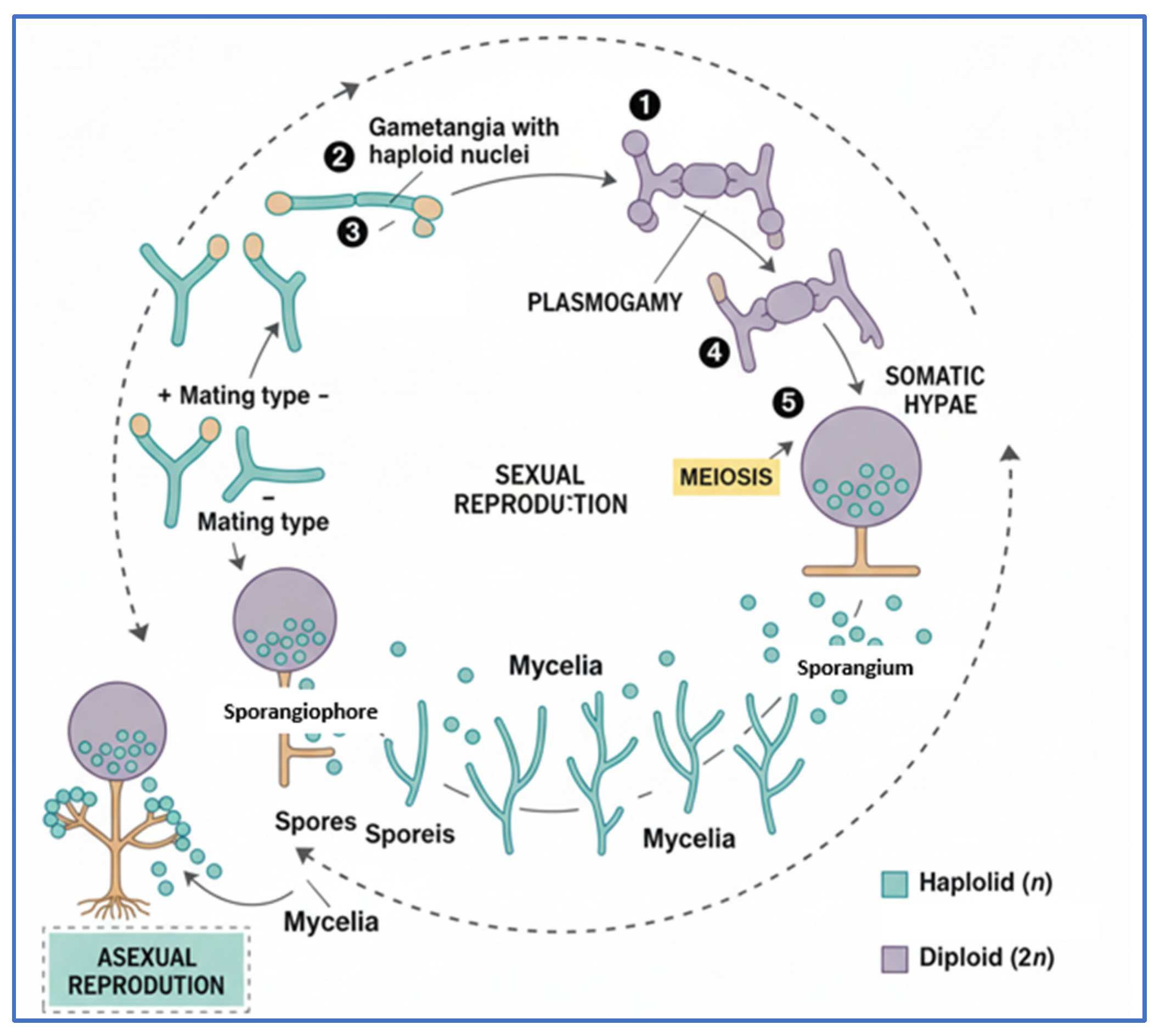
R. stolonifer reproduces both sexually and asexually during its life cycle, which enables the fungus to adapt to a range of environmental circumstances (Figure 6). When two mating types—referred to as “+” and “−”—come into contact, their gametangia, which have haploid nuclei, fuse, starting the sexual reproduction process. A young zygosporangium is created as a result of this fusion, which is referred to as plasmogamy. Karyogamy is the process by which the haploid nuclei inside this structure unite to produce a diploid nucleus. After going through meiosis, this diploid stage produces haploid spores that are released into the environment. Rhizopus produces sporangia, which are structures that generate spores without requiring sexual union, during asexual reproduction. Following their dispersal and germination, these spores produce new mycelium that can carry on the growth and reproduction cycle. Through the rapid expansion made possible by asexual reproduction or the genetic variation created by sexual reproduction, this dual reproductive strategy guarantees R. stolonifer’s survival and proliferation under a variety of situations [].
Figure 6. Life cycle of Rhizopus stolonifer, adapted from [].

2.4. Gray Rot

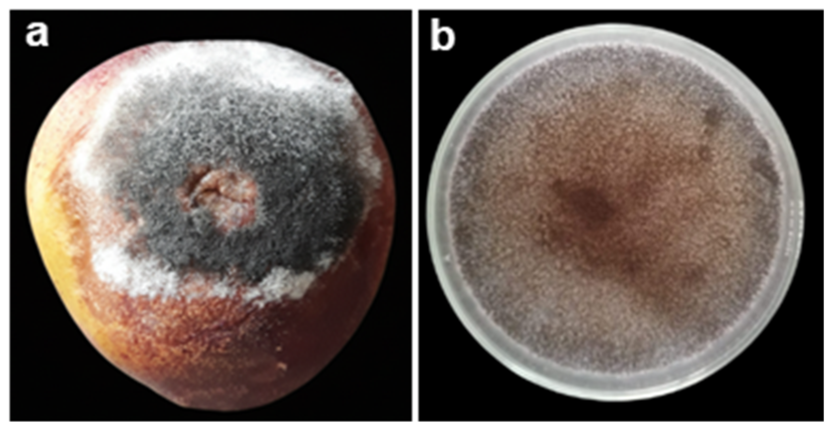
Botrytis cinerea, a fungus belonging to the Ascomycetes group, can infect a wide range of hosts, making it a common pathogen in many crops, both before and after harvest. Recently, a study ranked this fungus as the second most significant global pathogen based on its scientific and economic importance. It is a highly impactful phytopathogen capable of causing substantial economic losses, ranging from USD 10 to 100 billion worldwide []. Unlike other species in the Botrytis genus, B. cinerea is particularly notable for its ability to infect a wide range of hosts, including more than 1400 plant species. It also affects numerous fruits of the Rosaceae family, both ripe and those in storage, such as peaches, nectarines, apples, and strawberries. This fungus causes maceration accompanied by gray sporulation, a characteristic symptom of its infection [,]. However, it is the agent responsible for noble rot in grapes, inducing the production of geosmin []. Gray mold is caused by conidiophores, and the release of water accompanies the digestion of cell walls, facilitated by the array of enzymes and toxins secreted by this necrotrophic fungus []. The exogenous availability of nutrients, such as glucose, favors the germination of spores and the growth of hyphae. This is a critical factor for the development and spread of B. cinerea on its host plants [].
The fungus B. cinerea has a mycelium that is branched, septate, and can vary in color from hyaline to brown (Figure 7). The conidiophores, which can emerge directly from the mycelium or from sclerotia, are tall, slender, and irregularly branched at their tips. The apical cells of the conidiophores are enlarged or rounded and carry clusters of conidia simultaneously on short denticles. The conidia themselves are smooth and can vary in color from hyaline to gray. They are oval in shape, with an average length of 10 μm and a width of 5 μm [].
Figure 7. Macroscopic images of Botrytis cinerea: (a) B. cinerea on a peach fruit (Own photo, taken at the Training and Research Center Louata, Sefrou, Morocco, 19 April 2024); (b) B. cinerea on a PDA medium (own photo, taken in the Faculty of Science, Tetouan, Morocco, 10 June 2011).
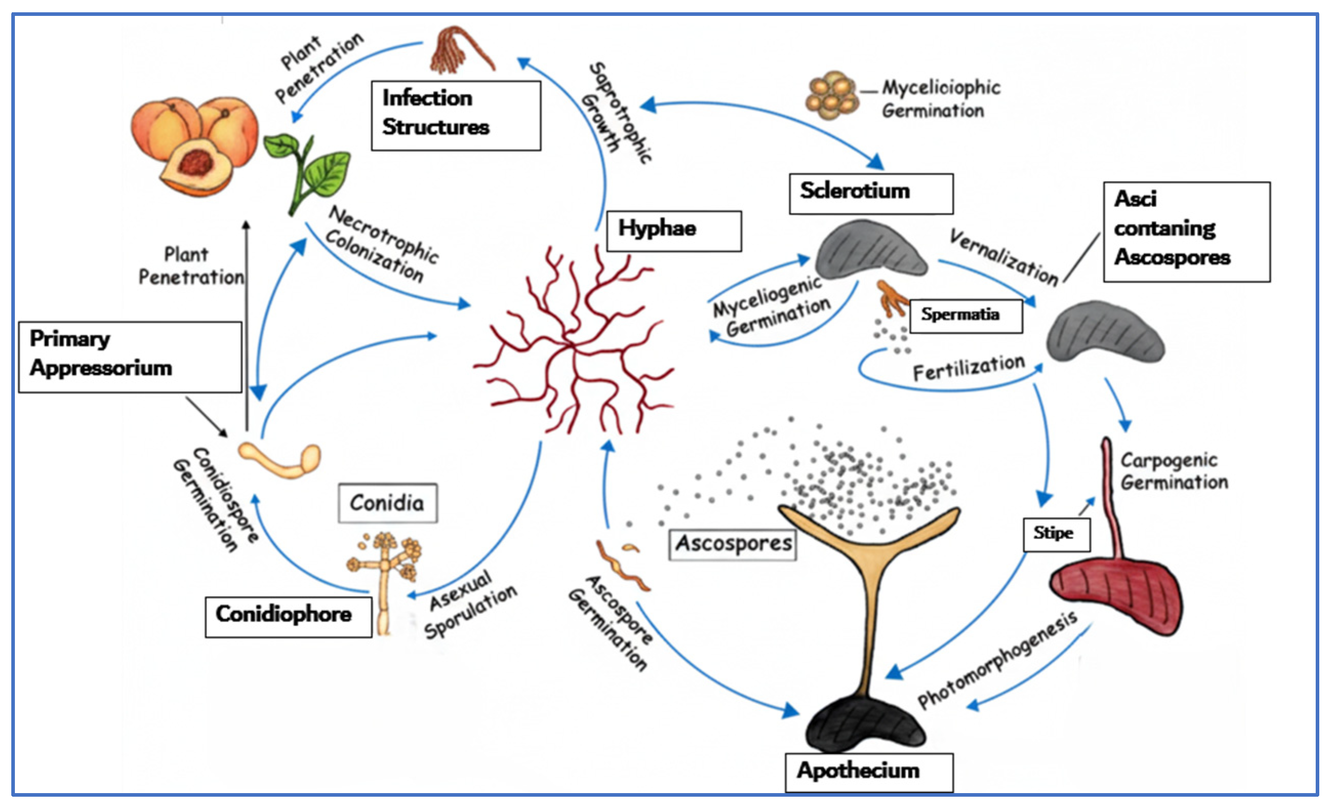
Sclerotia are globular masses formed by the fusion of mycelial branches, which protect the fungus from desiccation, UV rays, and microbial attacks over extended periods. The melanized layer and the β-glucans surrounding the internal mycelium are responsible for this protection []. When environmental conditions are favorable, the sclerotia can germinate by emitting conidiophores or by producing an apothecium after a sexual process []. Chlamydospores are short-term survival structures that help the fungus overcome unfavorable periods on plant surfaces. Microconidia, which develop from germ tubes produced by macroconidia or endogenously, can also function as spermatia during the sexual reproduction of certain fungi, such as B. cinerea. Asci contain eight ascospores and arise in a bundle from the base of the apothecial caps [].
Several factors influence conidia germination, but the most important are temperature, high humidity, and nutrient availability. The fungus can grow within a temperature range of 5 °C to 30 °C, with an optimal range between 15 °C and 20 °C. Additionally, conidia production is affected by RH, which promotes their increase when it ranges between 65.5% and 92% []. Sporulation occurs in both light and darkness []. Moreover, it has been demonstrated that germination can be induced by certain gases, notably ethylene. Once the conidia have germinated, B. cinerea is capable of invading tissues in two ways: either through active or passive invasion. In other words, the fungus can penetrate tissues via pre-existing wounds or stomata, or it can directly attack healthy tissues without requiring a pre-established entry point []. When the fungus invades tissues, it triggers a classic defense response against pathogens, namely programmed cell death []. The production of diffusible factors with phytotoxic activity, such as toxins, oxalic acid, and reactive oxygen species (ROS), is responsible for this reaction.
The life cycle of the plant pathogen B. cinerea, which can reproduce both sexually and asexually, is depicted in this graphic (Figure 8). The fungus alternates between growing saprotrophically on decaying organic matter and colonizing living plants in a necrotrophic manner. Conidiospores germinate to produce hyphae that develop into conidiophores, marking the start of their life cycle. Conidia produced by these structures enable asexual reproduction. B. cinerea shifts to saprotrophic growth, breaking down plant remnants, after its host dies. When sclerotia germinate to form spermatia or develop apothecia, sexual reproduction begins under ideal circumstances. By releasing ascospores, these apothecia complete the life cycle and allow the fungus to infect new hosts. B. cinerea’s ability to reproduce and survive in both living and dead substances demonstrates its versatility and tenacity [].
Figure 8. Life cycle of Botrytis cinerea, adapted from [].

2.5. Acid Rot

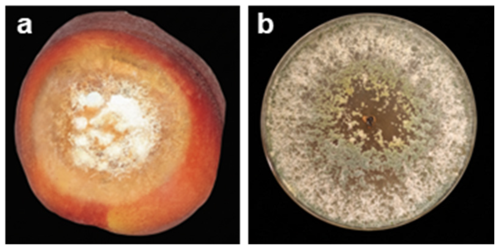
Geotrichum candidum is responsible for the sour rot observed in several fruits, such as strawberries, kiwis, and other fruits [,]. It generally affects only ripe fruits but can also impact heavily bruised green fruits. It is characterized by dark, depressed areas with purple borders. Later, these areas are covered with masses of white spores (Figure 9). Inside the affected fruits, the rotting flesh is moist, dark, and sometimes has cavities filled with white spores, accompanied by a sour smell. The rot can extend to the core.
Figure 9. Peach Acid Rote and Geotrichum candidum images: (a) G. candidum on a peach fruit (own photo, taken at the Training and Research Center Louata, Sefrou, Morocco, 7 October 2023). (b) G. candidum on a PDA medium (own photo, taken at the Phytohormones and Plant Development Laboratory, Murcia, Spain, 14 October 2025).
After cultivating the fungus on agar and incubating it at a temperature of 30 °C for seven days in dark conditions, it was observed that the fungal colonies were flat and presented a fluffy white mycelium. The mycelium consisted of septate and transparent cells. The conidia, also transparent, were cylindrical to subglobose in shape [].
In June 2020, the first case of this pathogen was detected on peaches in Yangshan (China), causing damage estimated at more than 10% of the total harvest. The colonies of this pathogen appear white, flat, and milky, resembling yeasts, with radial mycelium. The hyphae of this pathogen, observed under the microscope, are septate and branched, breaking apart into arthroconidia measuring between 3.39 and 9.27 × 2.05–7.71 μm [].
The optimal temperature for the development and growth of G. candidum mycelium was 25 °C. However, this pathogen was also very active at temperatures ranging between 15 and 30 °C. The conidia germinated after an incubation period of 8 h []. After storing the fruits at 0 ± 1 °C for 28 days, approximately 70% of the fruits exhibited soft, water-soaked lesions with white mycelial growth on their external surface. These fruits had a sour odor and lesions measuring between 10 and 23 mm in diameter [].

2.6. Comparative Overview of Major Postharvest Fungal Pathogens in Peach

After reviewing the main postharvest fungal diseases affecting peach, Table 1 provides a comparative summary of the key pathogens, infection conditions, characteristic symptoms, and major control strategies, and major control strategies. Table 1 aims to synthesize the essential information from the preceding sections, allowing readers to quickly understand the primary threats to peaches during postharvest storage and the current approaches to managing them effectively.
Table 1. Major postharvest fungal pathogens of peach: symptoms and infection conditions.
Knowledge of the infection mechanisms and environmental conditions favoring each fungal pathogen underpins the development of an effective control strategy. The information is directly used in the choice and optimization of postharvest management approaches discussed in the next section to ensure that methods of control are matched with the pathogen biology and dynamics of disease.

3. Methods for Controlling Postharvest Rots of Peach

Today, the control of fruit fungi during storage has become a concern due to the quantitative and qualitative losses caused by infection, particularly in the context of the global food crisis. However, numerous studies have been conducted to extend the fresh storage time while maintaining fruit quality. Chemical, physical, and biological control methods have been developed.

3.1. Chemical Controls

Numerous well-established processes contribute to the development of fungal resistance to antifungal drugs. Target-site modification is a significant mechanism in which mutations in important genes, including ERG11 (Ergosterol biosynthesis gene 11), lower the binding affinity of azoles, hence decreasing susceptibility. Efflux pump overexpression, which is mediated by transcriptional regulators such as TAC1 (Tachykinin precursor 1), is another popular gene tactic. It lowers the intracellular concentrations of antifungal medications by actively expelling them from the cell []. Furthermore, fungus may have changes in metabolic pathways, such as mutations in URA3 (Orotidine 5′-phosphate decarboxylase) or ERG3, which reduce the effectiveness of medications. The development of biofilms improves resistance and pathogenicity by creating a physical and biochemical barrier []. Mutations in transcription factors, like TAC1b, can potentially cause resistance by triggering efflux-independent processes. Finally, genetic plasticity and chromosomal alterations, including gene copy number variations, allow rapid adaptation to antifungal exposure [].
Knowledge of the mechanisms of resistance is essential in designing effective antifungal strategies and the management of resistant fungal infections. These form the basis for different control methods that are discussed in the subsections that follow.

3.1.1. Fungicides Synthetic/Chemical Fungicides

Reduced-risk fungicides such as fenhexamid and fludioxonil are highly effective against postharvest brown rot and gray mold on stone fruits like peaches, nectarines, and plums. Tebucozole, on the other hand, is more effective against brown rot than gray mold but significantly reduces the incidence of Rhizopus rot. The effective concentrations required to inhibit 50% of mycelial growth in vitro were low (≤6.3 × 10⁻⁵ mg/mL) for fenhexamid and fludioxonil, confirming their high efficacy against brown rot and gray mold. It has been established that fludioxonil acts by excessively activating the high-osmolarity glycerol (HOG) signaling pathway through hybrid histidine kinases of group III (HK). This pathway is responsible for cellular adaptation to environmental changes such as osmotic pressure variations. It is widely accepted that the fungicidal activity of fludioxonil is due to a disorderly transduction of the osmotic stress signal, which leads to a disruption of the HOG pathway. This disruption prevents fungal cells from effectively adapting to changes in their environment, making them more vulnerable to the action of the fungicide. In other words, fludioxonil disrupts the HOG pathway by inducing artificial osmotic stress, leading to a hyper-accumulation of glycerol, which ultimately results in fungal cell death [,].
Fenhexamid is a chemical used to kill unwanted fungi, and it was developed by Bayer CropScience Co. in 2000. This fungicide acts in a specific way, as it blocks an important step in the production of ergosterol, an essential component of fungal cell membranes. More specifically, fenhexamid inhibits the enzyme responsible for C4 demethylation in ergosterol biosynthesis. As a result, fungal cells are unable to produce ergosterol, making them vulnerable and ultimately leading to their death [].
Fenhexamid and the combination of cyprodinil and fludioxonil have proven effective in protecting peach fruits against infection by M. fructicola, while iprodione, difenoconazole, tebuconazole, and pyrimethanil did not show the same protective effect.
However, the study of cross-resistance relationships in Alternaria solani demonstrated that resistance mutations to fludioxonil reduce sensitivity to the fungicides quintozene, iprodione, and vinclozolin. However, no cross-resistance was observed with fungicides having different modes of action, such as imazalil (IMZ), flusilazole, and boscalid. Moreover, the study of fitness parameters revealed that resistance mutations did not negatively affect growth and germination but had a pleiotropic effect on virulence and conidia production in resistant mutants. These results suggest that fludioxonil is an effective fungicide against A. solani and that it carries a medium risk of resistance development, making it an ideal alternative to high-risk fungicides such as boscalid and pyraclostrobin [].
Pyrimethanil is a fungicide from the anilinopyrimidine class. It works by inhibiting methionine biosynthesis, as observed by Daniels and Lucas (1995) and deeply studied by Fritz et al. (1997), demonstrating the secretion of cell wall-degrading enzymes [,]. This dual action makes the development of cross-resistance unlikely with commonly used fungicides such as thiabendazole, IMZ, or sodium o-phenylphenate in the many resistant isolates of P. digitatum []. It was introduced in 1990 and is classified by the United States Environmental Protection Agency (USEPA) as a “low-risk” fungicide. It is primarily used to control gray mold on horticultural and ornamental crops. An old study examined the effects of IMZ on the microbiological, sensory, and physical characteristics of individually wrapped fruits and vegetables between 0 and 21 °C, including peaches.

3.1.2. Plant Defense Activators

  • β-aminobutyric Acid
Plant defense activators differ from fungicides in that they do not directly kill fungal pathogens but enhance the fruit’s innate resistance mechanisms. Treatment with β-aminobutyric acid (BABA) improved defense-related enzymatic activities, such as chitinase (PpCHI) and β-1,3-glucanase (GLU), increased lignin accumulation, and maintained a higher energy status, while boosting the transcription of defense-related genes, thereby creating fruit resistance to Rhizopus rot. BABA induced a priming effect, with early activation of redox-regulated genes and PpTGA1 modification, followed by a hydrogen peroxide (H2O2) burst through respiratory burst oxidase homolog (PpRBOH) upregulation and mitogen-activated protein kinase (MAPK) cascade activation upon pathogen invasion, resulting in robust defense [].
  • 2,4-epibrassinolide and Methyl Jasmonate
2,4-epibrassinolide (EBR) at 5 μM increased the expression of defense enzymes and PR genes, including PpCHI, PpGns1, phenylalanine ammonia-lyase (PpPAL), nonexpressor of pathogenesis-related genes 1 (PpNPR1), and pathogenesis-related proteins 1 and 4 (PpPR1 and PpPR4), enhancing phenolic compounds and lignin biosynthesis. EBR-treated peaches accumulated antifungal compounds upon inoculation, indicating a primed defense response []. Similarly, 10 μM methyl jasmonate (MeJA) reduced lesion development by R. stolonifer, stimulated the activity of PpCHI and GLU, promoting a primed defense response specific to Rhizopus rot [].
  • Benzothiadiazole
Postharvest treatment of peaches with 0.2 mg/mL of S-methyl ester of benzo-(1,2,3)-thiodiazole-7-carbothioic acid (BTH) significantly reduced lesion areas caused by P. expansum. Additionally, significant increases in PpCHI and GLU activities, total phenolic and chlorogenic acid levels, and lignin content, without negatively affecting solid soluble content, soluble pectin, titratable acidity, and ascorbic acid (AA) levels [].
  • 6-benzylaminopurine
6-benzylaminopurine (BAP) at 0.1 mg/mL markedly decreased disease incidence from 77.3% in control fruits to 2.6% in treated peaches and maintained fruit firmness at 13.8 MPa. BAP treatment also enhanced superoxide dismutase (SOD) activity and preserved cell membrane integrity, without affecting total phenolics or flavonoid content [].
  • Nitric Oxide and Sodium Metasilicate
Other treatments such as 15 μM nitric oxide (NO) enhance the resistance of harvested peaches against M. fructicola by inducing signals such as endogenous NO, ROS, salicylic acid (SA), and jasmonic acid (JA), while inhibiting aconitase activity through sodium nitroprusside (SNP) and H2O2 []. However, the use of sodium metasilicate helped maintain high fruit firmness and increased total polyphenol synthesis in response to fungal attack. However, it did not affect titratable acidity or soluble solids. An application of 0.006 g/mL can potentially prevent brown rot in peaches both before and after harvest, in addition to boosting polyphenol synthesis and maintaining a high firmness of the flesh [].
  • Essential oils treatments
Recently, the use of essential oils (EOs) has sparked great interest as a natural alternative to conventional synthetic pesticides. This is mainly due to concerns surrounding ecosystem pollution and pesticide resistance in pests and fungal pathogens. Researchers conducted a study to test the effectiveness of two EOs, verbena EO (Verbena officinalis) at 1000 ppm and thyme EO (Thymus vulgaris) at 500 ppm, in combating rot caused by Monilinia spp. on peaches. The results showed that thyme EO, which mainly contained o-cymene (56.2%), had smaller lesion diameters than verbena EO, which is rich in citral (44.5%) and isobornyl formate (45.4%). Specifically, the lesions caused by M. laxa, M. fructigena, and M. fructicola were 6, 9, and 23 mm for thyme EO, while for verbena EO, the lesion diameters were 17, 25, and 27 mm. However, no phytotoxic effects were observed on the fruit tissues, even at high concentrations of the studied EO []. Powerful inhibitors of B. cinerea and M. fructicola were identified in a study on the antifungal activity of compounds from Thymus capitatus (L.). Carvacrol and thymol proved to be highly effective inhibitors of spore germination and mycelial growth in these fungi. In volatility tests, even at a concentration of 10 μg/mL of medium (0.25 mg/Petri dish), carvacrol and thymol showed significant inhibition of 85% and 82% at 48 h against B. cinerea, and 38% and 57% against M. fructicola, respectively [].
The use of thyme EO in treatment mixtures also proved effective. By combining thyme and mint EO with Arabic gum and polyoxyethylene sorbitan monolaurate (Tween 20), a solution was created that strongly inhibited the growth of three different pathogens: B. cinerea, P. expansum, and R. stolonifer. The tests revealed a significant reduction in pathogen growth six days after application, with reductions of 83%, 77%, and 88%, respectively. This solution was effective in protecting peaches from postharvest rot, even at temperatures ranging from 5 to 30 °C. When used to store peaches at 4 °C, it successfully protected the peaches from the three pathogens for 10 days and significantly slowed the progression of the disease for 30 days compared to the controls (Arabic gum and Tween 20). The use of this solution also reduced the incidence of the disease to 25–30% and its severity to 26–46% during the entire storage period of the peaches []. When compared to cinnamon EO vapor, thyme EO demonstrated greater effectiveness in reducing the occurrence of brown rot in two peach varieties. This improvement was attributed to an increase in defense-related enzymatic activities, as well as a rise in the total phenolic compound content [].
On the other hand, tea tree EO (TTO) proved to be the most effective against M. fructicola compared to thyme EO, rosemary EO, and lemon EO, both in vitro and in inoculated peaches. Scanning electron microscope (SEM) and transmission electron microscope (TEM) observations showed that TTO significantly altered the mycelial morphology and ultrastructure in M. fructicola. The untreated M. fructicola mycelium appeared smooth and regular with normal, even surfaces. In contrast, the mycelium of M. fructicola treated with TTO at a concentration of 2 MIC (2.8 mg/mL) was indented, flattened, and showed irregular structures. TEM observations revealed a normal fungal ultrastructure in the control, with cells having normal wall thickness, a regular plasmalemma, uniform cytoplasm, clearly visible internal organelles, and typical fungal mycelial septa. However, treatment with TTO significantly altered the general ultrastructure of the cell, with thickened and weakened cell walls, altered cytoplasm, increased vacuolization, and plasmalemma rupture. Furthermore, TTO treatment also led to the accumulation of ROS, which may be due to membrane integrity disruption, damaging the mitochondria []. The application of TTO on B. cinerea also caused a significant increase in ROS levels, which can disrupt the normal functioning of the mitochondria and damage the cells through oxidative stress [].
The results obtained regarding the antifungal effect of TTO are consistent with those reported by Jiang et al. (2021). They showed that TTO releases TTO/HP-β-CD IC in a prolonged manner for a period of one month, whether at a temperature of 4 °C or 20 °C. Moreover, the use of TTO/HP-β-CD IC, which had been stored for 12 months, inhibited the growth of M. fructicola [].
A study was conducted to examine the antifungal activity of EOs from three Monarda species (Monarda didyma L., M. fistulosa L., and M. didyma var 80-1A L.) on the fungus B. cinerea. The researchers evaluated the inhibitory effects of the EOs in their volatile phase and by contact on the hyphae and spores of the fungus. The results showed that all three tested EOs inhibited mycelial growth and spore germination. M. didyma was found to be the most effective species, followed by M. didyma var 80-1A, and then M. fistulosa. The EOs exhibited greater toxicity to the fungal growth in the volatile phase than by direct contact. The antifungal activity was mainly attributed to the composition of the EOs: linalool, thymol, and geraniol being the main components of the EOs from M. didyma var. 80-1A, M. didyma, and M. fistulosa, respectively [].
Camélé et al. (2012) conducted an in vitro study to assess the effectiveness of various extracts from Mediterranean aromatic plants (Verbena officinalis, T. vulgaris, and Origanum vulgare), such as β-phellandrene, β-pinene, camphene, carvacrol, citral, o-cymene, γ-terpinene, and thymol, in combating five pathogens responsible for postharvest fruit rot. According to their results, phenolic compounds, particularly carvacrol and thymol, could potentially interfere with cell wall enzymes such as chitin synthase/CHI, as well as with the α- and β-GLU of target species [].
The EO of lavender exhibited strong inhibitory activity against M. fructicola in flat peaches, with nearly complete growth inhibition at a concentration of 0.8 mg/mL. This inhibition was associated with changes in the structure of the hyphae and rupture of the spores, as well as leakage of the cytoplasmic content. Furthermore, the results also showed an increase in the expression of the apoptosis genes RTG1 and RLM1 in fungi treated with lavender EO. These findings suggest that lavender EO can inhibit M. fructicola by inducing damage to the cytoplasmic membrane and stimulating fungal cell apoptosis. The main active ingredients in lavender EO are monoterpenes and sesquiterpenes, which are believed to contribute to its inhibitory activity [].
Four EOs from Mentha spicata, Mentha piperita, T. vulgaris, carvacrol, and T. vulgaris CT thymol (Tt) showed strong and continuous inhibition against R. stolonifer after 72 h of incubation, with inhibition rates of up to 98.8%. Other plant extracts at the same concentration (Lavandula latifolia, Lavandula angustifolia, Cymbopogon martinii, Cymbopogon khasans, Cymbopogon citratus, Syzygium aromaticum, Thymus satureoides CT bornéol, T. vulgaris CT linalool, Thymus zygis CT linalool, Artemisia indica, Ocimum sanctum, Ocimum basilicum, O. vulgare, and Origanum heracleoticum) showed mild inhibitory effectiveness against R. stolonifer, with inhibition ranging from 30.1% to 75.1% after 72 h of incubation []. In summary, the use of EOs can be beneficial in combating postharvest peach rot, although their effectiveness may vary depending on the concentration and the targeted fungus. Therefore, it is important to continue research to better understand the mechanisms of action and optimize the use of these EOs to protect fruits from this disease.

3.1.3. Sanitizers

  • Neutral electrolyzed water
Consumers are currently exposed to synthetic active ingredients used to combat postharvest diseases. Neutral electrolyzed water (NEW), containing between 12 ppm and 53 ppm of available chlorine, prevented 100% of the spore germination of various fungal species such as Alternaria alternata, B. cinerea, Cladosporium australiense, Colletotrichum gloeosporioides and Colletotrichum siamense, Fusarium solani and Fusarium oxysporum, as well as Lasiodiplodia theobromae. This inhibition was observed after a contact time of 3.5 or 10 min []. A daily soaking and spraying of peaches inoculated with M. fructicola with electrolyzed oxidizing water prevented infection for 12 days and resulted in an infection incidence of 10% and a disease severity of 6% after 14 days of storage at 25 °C [].
  • Hydrogen peroxide
Hydrogen peroxide (H2O2) damaged P. digitatum conidia, affecting viability, germination, and mycelium development in a concentration-dependent manner. A direct correlation was observed between the concentration of H2O2 and the mortality and germination of conidia. When a lethal treatment was applied, the conidia suffered severe damage, and their cytoplasm became disorganized, although their fungal cell wall remained intact. Furthermore, the treatment caused oxidative damage at various cellular levels, leading to membrane disruptions in the conidia and hyphae of P. digitatum []. The exposure time to H2O2 vapor led to a logarithmic decrease in the germination rate of B. cinerea spores. Treatments with 2.7 × 10⁻⁴ mg/mL of H2O2 vapor at 20 °C and 5.5 × 10⁻⁴ mg/mL of H2O2 vapor at 30 °C required 10.5 and 5.7 min, respectively, to eliminate 99% of the spores. However, prolonged exposure to this vapor resulted in increased water loss and a decrease in the firmness of grapes. Additionally, injuries were observed in the form of yellow-brown discoloration of the fruits and stems, which increased in intensity depending on the duration of exposure and the concentration of H2O2 vapor [].
  • Ozone
The average natural incidence caused by M. fructicola on stone fruits was reduced after spraying 1.5 ppm ozone (O3) for 400 min. No visible ozone toxicity was observed []. An inhibition of the normal aerial mycelial growth and prevention of sporulation of M. fructicola, B. cinerea, and P. expansum. While the normal symptoms of the disease were observed at 5 °C on the control fruits, M. fructicola did not develop aerial mycelium, whereas B. cinerea developed short, compact mycelium that did not produce conidia. While P. expansum developed normal external mycelium on the treated fruits, the area where the aerial mycelium was present was noticeably smaller than on the control fruits, and sporulation was prevented []. In addition to concentration, the number of applications of ozonated water affects the quality of the treated fruits. Intensive use of grapevines has a negative effect on chromatic parameters, glycosylated aroma precursors, and phenolic compounds. On the other hand, a single application of ozonated water had a beneficial effect on technological maturity, chromatic parameters, seed maturity, and phenolic compound content [].
  • Peracetic Acid
Using a treatment of 0.2 mg/mL of peracetic acid (PAA) at 40 °C for 40 s, brown rot was effectively controlled when peaches and nectarines were infected prior to treatment. However, when the infection time was extended to 24, 48, and 72 h, the treatment was ineffective. This treatment was also effective against different inoculum concentrations (103, 104, 105, and 106 conidia/mL) in peaches and nectarines but did not offer protection against future infections. For the peaches, the incidence of brown rot was significantly reduced by 61% and 36%, respectively, but no effect was observed on nectarines when applied to naturally infected fruits [].
  • Acetic Acid
The growth of B. cinerea was affected by acetic acid in a dose- and time-dependent manner. Specifically, exposure to a fumigation containing 4 × 10⁻³ or 6 × 10⁻³ mg/mL of acetic acid for 6 min, as well as exposure to a fumigation containing 8 × 10⁻³ mg/mL of acetic acid for 3 or 6 min, all resulted in a complete inhibition of B. cinerea growth []. The effectiveness of acetic acid also extends to the control of M. fructicola, P. expansum, and R. stolonifer. Specifically, the use of acetic acid reduced core rot caused by P. expansum from 98% to 16% without harming the fruit. Similarly, citrus decay caused by P. digitatum was significantly reduced from 86% to only 11% with the use of acetic acid []. Damage was observed on Harbrite peaches that were fumigated with 2.7 × 10⁻³ mg/mL of acetic acid. These damages were characterized by light brown streaks, indicating slight phytotoxicity. However, despite these minor damages, this dose effectively prevented decay caused by M. fructicola and R. stolonifer on peaches. In contrast, the use of higher concentrations of acetic acid led to an increase in injuries, characterized by darker and more pronounced streaks []. However, a study was conducted on the effect of coating white prickly pear with chitosan containing 1.0% acetic acid, on fruit preservation and sensory quality, as well as their content in phenolic compounds and antioxidant activity. The results showed that this coating helped delay weight loss, maintain fruit firmness, and preserve their color throughout the storage period. Moreover, the sensory values of the chitosan-coated white prickly pears with 1.0% and 2.5% acetic acid were higher than those obtained for uncoated fruits. The phenolic compound content and antioxidant activity of the white prickly pears were not affected by the chitosan coating with 1.0% and 2.5% acetic acid. Finally, the microbial populations of the chitosan-coated white prickly pears remained unchanged throughout the storage period, with a quantity lower than 10 CFU/g of fruit [].
  • Calcium Chloride and Sodium Hypochlorite
Fruits can be successfully stored for a period of 20 days under cold storage conditions by soaking them in a solution composed of 1% calcium chloride (CaCl2) and 1.5% AA []. Experiments have shown that immersing fruits in a CaCl2 solution, followed by packaging and storing them at a temperature of 10 °C with a RH of 75 ± 5%, resulted in a significant decrease in firmness and AA content, as well as a significant increase in the decay index and percentage of weight loss during storage []. However, it was found that a hot water treatment (at 45 °C for 1 min) was more effective than using a 1% calcium hypochlorite solution or tap water in preserving the quality of tomatoes and reducing the incidence of decay during storage in an evaporative cooler []. Thus, it is possible to use nonionic surfactants such as polyoxyethylene sorbitan monooleate (Tween 80), Tween 20, and sorbitan monolaurate (Span 20) in combination with sodium hypochlorite (NaOCl), which could be an effective solution to limit the proliferation of P. expansum in flotation tanks, with a potential reduction of up to 5 log CFU [].

3.2. Physical Controls

Physical treatments have gained significant interest in recent years to combat various postharvest diseases in fruits and vegetables due to the complete absence of residues in the treated product and minimal environmental impact.

3.2.1. Heat Treatments

Heat treatment of food is a commonly used process for preserving fruits. Among the different types of heat treatments, hot water (HW) treatment and hot air (HA) are included. These techniques are particularly suitable for commercial use to reduce fruit rot, delay ripening, and preserve quality.
  • Hot water Treatments
Jemric et al. (2011) showed that dipping peaches in HW at 48 °C for 12 min significantly reduced the germination of M. laxa conidia, with a germination rate of 9% in treated fruits and 93% in control fruits. No significant differences were found between heat-treated fruits and control fruits in terms of firmness, titratable acidity, and soluble solids concentration. No visible heat damage symptoms were observed on the fruits after any of the treatments tested []. Experiments were conducted to evaluate the use of HW as a method to control the decay of peaches and nectarines infected with M. fructicola. The fruits were immersed in HW at temperatures of 46 or 50 °C for 2.5 min, and the results showed that this method significantly reduced the incidence of rotten fruits from 82.8% to 59.3% and 38.8%, respectively. Next, the effect of 10% ethanol in combination with HW was evaluated. The fruits were dipped in either HW alone or with 10% ethanol at temperatures of 46 or 50 °C for 2.5 min. The results showed that the use of 10% ethanol significantly improved the control of brown rot compared to HW alone. In fact, immersing the fruits in 10% ethanol at 46 or 50 °C for 2.5 min reduced rot to 33.8% and 24.5%, respectively. These results suggest that using 10% ethanol in combination with HW could be an effective method to control rot in fruits infected by M. fructicola []. This treatment was also used to control gray mold. Immersions in HW at 46, 48, and 50 °C for 4, 8, or 15 min were tested to control Botrytis rot during kiwi storage. The fruits were stored at 0 °C under air conditions for up to 13 weeks after the treatments. Dipping the fruits at 46 °C for 15 min and at 48 °C for 8 min provided almost complete control of the disease without heat damage. The firmness of the fruits was not affected by these treatments.
  • Hot Air Treatments
Wang et al. (2015) studied the impact of HA at a temperature of 44 °C for 114 min on the reduction in blue mold in sweet cherries. The results of the study showed that the fruits treated with HA had a significantly lower incidence of disease and smaller lesion diameter compared to the control fruits. The HA treatment also increased the activities of CHI and GLU but inhibited the activities of polygalacturonase (PG) and pectin methylesterase (PME). Furthermore, the fruits treated with HA showed significantly higher activities of superoxide dismutase (SOD), catalase (CAT), peroxidase (POD), and polyphenol oxidase (PPO), but a lower activity of ascorbate peroxidase (APX) compared to the control fruits. Defense-related genes such as PaGLU, PaCAT, and PaNPR1 were strongly induced in the HA-treated fruits during storage, while the expression of expansins (EXP) was downregulated by the HA treatment []. A previous study also observed that HA (40 °C, 2 days) had a significant impact on the degradation of organic acids, particularly citric acid, as well as the accumulation of soluble sugars, such as fructose and glucose, in mandarins. The researchers identified genes involved in citric acid degradation (CitAco1, CitAco2, CitAco3, CitIDH1, CitIDH2, CitIDH3, CitGAD4, CitGAD5, and CitGS2) and in sucrose metabolism (CitAI1, CitAI3, CitNI1, and CitNI3) and examined their expression. The HA treatment significantly increased the expression of CitAco3, CitIDH2/3, and CitGAD4, while having little effect on CitGS2. Genes related to sucrose metabolism also responded differently to HA treatment, with induction of CitAI genes and a relatively absent effect on CitNI genes [].
HA at 38 °C for 3 h or HW at 48 °C for 10 min maintained peach quality while reducing ROS and increasing the antioxidant activity of the fruit. However, the results showed that HW treatment was more effective than HA treatment in alleviating internal browning symptoms in the fruit. This increased effectiveness of the HW treatment can be explained by better heat transmission in water, which can more easily affect the antioxidant system of peaches. The HA treatment, on the other hand, had no effect on the metabolism of AA or glutathione (GSH). In contrast, the HW treatment improved AA metabolism early in storage and GSH metabolism at a later stage of storage [].
By directly inhibiting pathogens and inducing physiological resistance in the fruits, heat treatments significantly reduce fungal infections (Monilinia spp., B. cinerea, and Penicillium spp.) in peaches. They should be incorporated into a postharvest management plan since they aid in preserving fruit quality, nutritional content, and shelf life but do not totally prevent rot. For maximum effectiveness while maintaining fruit quality, the time × temperature combination must be optimized [].

3.2.2. UV Treatments

UV irradiation treatments (UV-B, UV-C, UV-B + UV-C) have recently gained significant interest due to their positive effects on the preservation of ripe fruits. The UV-C treatment (0.72 kJ·m−2) improved the physicochemical and sensory properties of peaches stored at 4 °C for 25 days compared to the UV-B treatment (0.36 kJ·m−2). Furthermore, the UV-C + UV-B treatment (28.8 kJ·m−2) showed the best results compared to the control, resulting in a 22.1% reduction in weight loss, 29.0% reduction in rot, and a 10% reduction in total soluble solids (TSS) of peaches, as well as a 34.7% increase in vitamin C, 22.8% increase in firmness, 4.7% increase in total phenolic content (TPC), 92.8% increase in titratable acidity, and 17.7% increase in pH [].
Irradiation of peaches with UV-C at 11.74 mW·cm−2 was effective for the curative control of soft rot, with the best results obtained after an exposure time of 10 min. The application of UV-C for 10–15 min reduced the mycelial growth of R. stolonifer, and exposure for 30 min completely inhibited the mycelial growth of this pathogen. No effect was observed in the protective or curative control of M. fructicola []. However, the effectiveness of UV-C inactivation of P. expansum was dependent on the intensity and the condition of the fruit tissues. Indeed, a higher UV-C intensity was required to achieve a similar reduction in the P. expansum population on wounded pear disks (3.1 kJ·m−2 for a 2.7 log reduction) compared to intact pear disks (1.7 kJ·m−2 for a 2.8 log reduction). However, no significant effects on weight loss, TSS content, and texture were observed between UV-C-treated pears and untreated pears. On the other hand, alterations in color, flavor, and texture were observed on UV-C-treated pears after 4 and 8 weeks of storage []. The UV-C treatment at a dose of 3 kJ·m−2 decreased the lesion diameter as well as the disease index caused by R. stolonifer on inoculated nectarines, compared to untreated fruits. Furthermore, the UV-C treatment increased the activities of antioxidant enzymes such as SOD, CAT, and APX, as well as the content of AA and GSH. The UV-C treatment also stimulated the phenylpropanoid metabolic pathway and anthocyanin biosynthesis, leading to an increase in phenolic compounds, total flavonoids, anthocyanins, and lignin content. Additionally, the UV-C treatment promoted the activities of CHI and GLU. These results demonstrated that UV-C treatment could strengthen the antioxidant system, stimulate the phenylpropanoid metabolic pathway, and enhance anthocyanin biosynthesis, thus reducing the severity of the disease caused by R. stolonifer on nectarines during storage. In conclusion, this study showed that UV-C is an environmentally friendly control method to limit the occurrence of diseases on nectarine fruits postharvest [].

3.2.3. Other Physical Techniques

The possibility of other recently developed physical methods for the prevention and management of peach postharvest illnesses has also been investigated. Recent research showed that plasma-processed air (PPA) treatments (30–240 s) significantly decreased the incidence of M. fructicola on peaches during storage, with the best effect occurring at 240 s exposure. This is primarily due to the action of reactive species generated in the plasma that damage fungal cell walls and membranes. Plasma-based technologies are among the most promising []. Similarly to other fruits, plasma-activated water (PAW) has been extensively researched for its capacity to produce ROS and reactive nitrogen species (RNS) that deactivate fungal spores, break up biofilms, and postpone decay; while there are currently few studies on peaches, it is very possible to incorporate PAW into postharvest washing lines []. As shown in sweet cherries, high-voltage electrostatic fields (HVEF) modify the fruit-associated microbial ecology and change membrane permeability and cellular ion balance to produce antifungal action []. As demonstrated in blueberries, pulsed light (PL) reduces contamination and maintains fruit quality by delivering high-energy bursts of broad-spectrum light that cause oxidative stress and DNA damage in fungal cells []. These results suggest that these technologies could be modified for postharvest peach control, providing sustainable and residue-free substitutes for traditional fungicides.

3.3. Biological Controls

Antagonists Treatment

Biological control is an ecological and effective method of disease management that involves introducing a third organism to control pathogens, rather than using chemical pesticides. This method relies on the action of antagonistic microorganisms, which can control diseases through mechanisms such as spatial or nutritional competition, inducible resistance, and the production of metabolites.
  • Bacterial treatment
Many research studies have been conducted to assess the effectiveness of various Bacillus spp. strains in the biocontrol of fungi that can develop on fruits postharvest. Peaches treated with Bacillus licheniformis HG03 showed increased resistance to R. stolonifer when stored at 20 °C and high RH. Compared to the control group, treatment with HG03 increased the activity of related enzymes and rebalanced the metabolism of ROS. This treatment reduced the accumulation of lipid peroxidation products and improved the ability to scavenge free radicals in peaches []. However, a 71.3% reduction in disease incidence and a 73.5% reduction in the lesion diameter of B. cinerea were obtained when peaches were treated with a concentration of 1 × 10⁷ CFU mL−1 of Bacillus subtilis JK-14. This treatment also increased the activity of antioxidant enzymes in peaches inoculated with B. cinerea. Specifically, the average activities of SOD, POD, and CAT were 36.5%, 17.6%, and 20.3% higher, respectively, compared to the control treatment. This highlights the potential of defense-related enzymatic activities in enhancing peach resistance against pathogens. Furthermore, the induced activity of defense-related enzymes seems to play a crucial role in the mechanism of B. subtilis JK-14 for controlling postharvest diseases in peaches []. Other studies have focused on the effectiveness of different Bacillus species in combating postharvest fungi. Bacillus amyloliquefaciens PPCB004 has been shown to be an effective antagonist in inhibiting the mycelial growth of B. cinerea, P. expansum, and R. stolonifer, with inhibition zones of 31 ± 0.28 mm, 24 ± 0.27 mm, and 11 ± 0.1 mm, respectively [], Bacillus vallismortis and Bacillus altitudinis produce volatile substances that exhibit significant antagonistic activity. Mycelial growth of M. fructicola was inhibited by 80.3% and 68.4%, along with cellular damage in the presence of B. vallismortis and B. altitudinis strains, respectively. Among the volatile compounds produced are 6-methyl-2-heptanone and 2-pentylfuran [].
In addition to being present in the rhizosphere and on the surface of plants, Gram-negative bacteria such as Pseudomonas spp. can also have beneficial effects by combating phytopathogenic fungi []. Several studies have focused on different strains belonging to the Pseudomonas genus to assess their potential as antagonistic microorganisms against a wide range of fungal pathogens. These studies also investigated the ability of these strains to produce antifungal compounds []. Aiello et al. (2019) studied the effect of Pseudomonas synxantha (strain DLS65) on brown rot. The results showed that this strain significantly reduced the incidence and severity of the disease caused by M. fructicola and M. fructigena after 5 days of storage at 25 °C and 10 °C. After storage at 25 °C, the reduction in incidence and severity was 50% and 76%, respectively. At 10 °C, the incidence and severity caused by M. fructicola were reduced by 68% and 92%, while those caused by M. fructigena were reduced by 55% and 90% []. Two strains of Pseudomonas (P. fluorescens CHA0 and P. aeruginosa 7NSK2) can induce resistance in grapevines against B. cinerea. Both strains triggered an oxidative burst and accumulation of phytoalexins in grapevine cells, preparing the leaves for an accelerated phytoalexin production when challenged with B. cinerea. Crude bacterial cell extracts also enhanced the oxidative burst and induced comparable amounts of phytoalexins and resistance to B. cinerea as those induced by live bacteria. Different Pseudomonas strains showed varying levels of resistance, suggesting the importance of pyochelin, pyoverdine, and/or SA in initiating phytoalexin responses and bacterial-induced resistance in grapevines against B. cinerea [].
A treatment with the EPS125 strain of Pantoea agglomerans significantly reduced the diameter of rot lesions on wounded fruits inoculated with 1 × 10³ spores/mL of M. laxa and stored at 20 °C. The lesion sizes ranged from 67.7 to 22.3 mm for untreated fruits and from 7.3 to 4 mm for those treated preventively with EPS125. No significant difference was observed between the preventive treatment with EPS125 and a fungicide treatment (tebuconazole at 0.125 mg/mL) [].
  • Yeast Treatment
Biological control of postharvest fruit diseases by antagonistic yeasts has been considered an effective and promising strategy to reduce fruit loss after harvest. The application of antagonistic yeasts to control brown rot is a promising alternative to chemical fungicides. C. laurentii has been studied for its effectiveness in limiting gray mold, blue mold, and Rhizopus mold on peach fruit after harvest. Concentrations of C. laurentii at 1 × 10⁹ CFU/mL were sufficient to completely prevent gray mold after 4 days of incubation at 25 °C. The incidence of blue mold or Rhizopus mold was reduced by 78.6% and 80%, respectively. C. laurentii did not affect fruit quality characteristics after storage at 2 °C for 30 days and at 20 °C for 7 days [].
The use of C. laurentii has shown a significant ability to reduce brown rot at both 25 °C and 1 °C. Although yeast populations increased more rapidly in the presence of the pathogen, they then declined rapidly. In fruits inoculated with M. fructicola alone or combined with C. laurentii, an increase in lipid peroxidation and the activities of antioxidant enzymes such as SOD, CAT, and POD were observed. Moreover, the isoenzyme pattern of PPO changed significantly after the appearance of symptoms, inducing new isoforms of PPO. In contrast, the induction of lipid peroxidation and the activities of SOD, CAT, and POD were low in fruits inoculated with the antagonist C. laurentii alone, although no significant changes were observed in the PPO isoenzymes []. Studies have examined different mechanisms of action in C. laurentii, including competition for nutrients, particularly sucrose. Scanning electron microscopy results showed that the yeast biofilm adheres to the fruit and the hyphae of C. gloeosporioides, indicating competition for space. Unlike the statistical analysis, which showed that only N-acetylglucosaminidase (NAGase) and CHI were significantly stimulated on injured fruits, the activity of the three hydrolytic enzymes was detected in vitro, but only NAGase was induced by the addition of autoclaved pathogen mycelium. The wounds treated with yeast biocontrol stimulated GLU activity and suppressed CHI activity on the fruit wounds, with or without the presence of autoclaved pathogen mycelium, but did not significantly affect NGase activity [].
The yeast Pichia membranefaciens has proven to be very effective in combating Rhizopus rot in peaches. Additionally, it appears that this yeast could potentially enhance the activities of defense-related enzymes, including PPO, POD, PAL, and CAT []. The addition of 0.2 mg/mL of BTH improved the biological control activity of P. membranefaciens against blue mold in peaches. This combined treatment had a synergistic effect on the induction of SOD, CAT, APX, CHI, and GLU activities, which increased the fruit’s disease resistance compared to BTH or yeast alone. It also led to smaller lesion diameters and reduced incidence of blue mold in peach fruits. Furthermore, the combined treatment did not adversely affect the quality parameters of the peaches, including firmness, TSS content, titratable acidity, and vitamin C content after 6 days of storage at 20 °C. These results suggest that the use of BTH can enhance the biological activity of P. membranefaciens, but further research is needed to determine whether it reduces BTH residue production [].
Thus, Pichia guilliermondii has been shown to stimulate the gene expression of PAL and promote the accumulation of total phenolic compounds, total flavonoids, and major individual phenolic compounds []. A significant increase in the endogenous SA content was stimulated in peaches infected by R. stolonifer and P. expansum, and treated with P. guilliermondii []. In addition to P. membranefaciens, Kloeckera apiculata appears to be capable of parasitizing M. fructicola. In vivo tests showed that their presence led to a significant reduction in the incidence of rot caused by M. fructicola, with a reduction of 76.0% for P. membranefaciens and 65.8% for K. apiculata. Furthermore, K. apiculata was shown to be capable of attaching to the hyphae of M. fructicola [].

3.4. Comparative Evaluation of Postharvest Rot Control Methods

Numerous strategies have been developed to control postharvest fungal rots in peaches, ranging from conventional fungicides to innovative biological and physical approaches. Each method presents distinct advantages and limitations, particularly in terms of effectiveness, safety, environmental sustainability, cost, and practical feasibility. A comparative assessment of these approaches is essential to support the development of integrated and efficient disease management systems.
Table 2 provides a side-by-side evaluation of the major control methods for peach postharvest rots, summarizing their relative efficacy, residue risk, environmental impact, industrial applicability, and other key considerations. This section is intended to synthesize the preceding content and offer stakeholders a practical overview to select strategies that best match production scale, pathogen pressure, and regulatory requirements.
The interest in integrated techniques that combine several treatment types, like heat therapy with biological control, is highlighted by several recent studies. To improve postharvest disease management in peaches and litchis, Bacillus subtilis CF-3 VOCs (Volatile Organic Compounds) combined with heat treatment effectively control M. fructicola by preventing fungal growth and rot development, preserve fruit quality by preserving firmness, soluble solids, and minimizing weight loss, and significantly increasing the activity of antioxidant and disease-resistance enzymes []. Likewise, it has been demonstrated that combining chemical fungicides with biological control agents improves disease control while lowering the dosages of chemicals needed. Cryptococcus laurentii and thiabendazole, for instance, enhanced control of B. cinerea in postharvest conditions.
While lowering chemical inputs, the combination of strobilurins (fungicides that prevent fungi from blocking mitochondrial respiration) and SA enhanced the management of leaf spot and downy mildew diseases [,]. Furthermore, CHI mixed with low-dose fungicides improved protection against B. cinerea on strawberries and apple scab (Venturia inaequalis) [], while sodium bicarbonate with IMZ improved control of P. digitatum on citrus, including resistant isolates []. The promise of integrated management strategies in sustainable agriculture is highlighted by these examples, which collectively show how combining biological, chemical, and physical treatments can maximize disease control, minimize chemical usage, and maintain crop quality.
Table 2. Comparative evaluation of postharvest rot control methods.
Control MethodEfficacyResidue RiskResistance RiskEnvironmental ImpactCostIndustrial ApplicabilityCommentsRefs.
Chemical FungicidesHighHighHighNegativeModerateHighFast action, widely used, but declining acceptance[,]
Biological ControlModerateNoneLowPositiveModerate–HighMediumEco-friendly, needs formulation and storage stability[]
Physical TreatmentsModerate–HighNoneNoneNeutral to positiveHighMediumEffective if optimized, may stress fruit tissues[,]
Integrated MethodsHigh
(synergistic)
LowLowPositiveVariableMedium–HighCombines benefits, requires protocol optimization[]

4. Conclusions and Research Gaps

Postharvest fungal diseases remain one of the most significant challenges in peach production and supply chains, often leading to substantial economic losses, reduced fruit quality, and shortened shelf life. As this review highlights, major fungal pathogens such as Monilinia spp. (brown rot), Penicillium expansum (blue mold), Rhizopus stolonifer (soft rot), Botrytis cinerea (gray mold), and Geotrichum candidum (sour rot) are responsible for the most common and destructive postharvest rots in peaches. These fungi differ in terms of infection mechanisms, environmental preferences, and susceptibility to control measures, making their management complex and multifactorial.
For decades, chemical fungicides have served as the primary line of defense against postharvest decay. However, the emergence of fungicide-resistant strains, growing regulatory restrictions, and increasing consumer demand for residue-free products have pushed researchers and industry stakeholders to explore alternative and more sustainable control methods. Recent developments in biological control (using antagonistic yeasts and bacteria), physical approaches (such as UV-C and controlled atmospheres), and natural products (essential oils, plant extracts, and biopolymers) have demonstrated promising results both in laboratory and semi-commercial settings. Nonetheless, few of these solutions have achieved large-scale industrial adoption due to challenges related to cost, stability, reproducibility, or regulatory approval.
Despite the progress made, several important knowledge gaps and unresolved issues still limit the effectiveness of postharvest rot management in peaches. There is still a lack of predictive modeling tools simulating pathogen dynamics under real-world cold chain logistics and fluctuating environmental conditions, as well as an incomplete understanding of the fruit microbiome and its role in enhancing or inhibiting fungal colonization during storage. Furthermore, standardization of biocontrol agents remains limited, particularly regarding formulation stability, compatibility with other treatments, and efficacy across different peach cultivars and environments. The exploration of synergistic combinations, such as biocontrol agents with UV or EOs, is still insufficient, and data on consumer perception, sensory impact, and the nutritional implications of alternative treatments over extended storage periods remain scarce. Addressing these challenges requires integrated efforts across plant pathology, microbiology, postharvest technology, food safety, and supply chain logistics.
Looking ahead, several avenues appear particularly promising. These include the development of multi-strain or microbiome-based biocontrol products that are more resilient and effective under variable conditions, the application of nanotechnology for controlled release and targeted delivery of antifungal agents, and the use of artificial intelligence and machine learning for early detection, monitoring, and prediction of rot outbreaks. The integration of “omics” tools such as genomics, metabolomics, and transcriptomics could help identify molecular targets of resistance and provide a deeper understanding of host–pathogen interactions. Furthermore, emerging approaches such as metagenomics to study the fruit microbiome for the development of next-generation probiotic preparations, the use of CRISPR technology to edit fruit disease resistance genes, and the development of real-time disease early warning systems based on the Internet of Things (IoT), all point to clear technological breakthroughs. The design of sustainable packaging systems incorporating antimicrobial materials or sensors to prolong shelf life and monitor fruit status also represents a significant opportunity.
Ultimately, the goal is not only to reduce rot incidence but also to ensure fruit safety, maintain high quality, reduce postharvest losses, and comply with international standards for sustainable and responsible agriculture. To achieve this, bridging the gap between experimental research and commercial application will be essential, supported by policy, innovation, and interdisciplinary collaboration.

Author Contributions

S.E.M. conceived the review, performed the literature search, and drafted the manuscript; A.A. contributed to structuring and revising the manuscript; A.H. revised the manuscript and collected the photographs; A.A.H., A.C. and J.H.-R. critically revised the manuscript; M.E. and M.B.A. co-supervised the work and provided guidance. All authors have read and agreed to the published version of the manuscript.

Funding

This research did not receive any specific grant from funding agencies in the public, commercial, or not-for-profit sectors.

Data Availability Statement

No new data were created or analyzed in this study. Data sharing is not applicable to this article.

Acknowledgments

The authors would like to thank the Faculty of Biology, University of Murcia (Spain), the Polydisciplinary Faculty of Larache, Abdelmalek Essaâdi University, and the Training and Research Center Louata (Morocco) for their institutional and technical support.

Conflicts of Interest

The authors declare no conflicts of interest.

References

  1. Liu, H.; Cao, J.; Jiang, W. Evaluation and Comparison of Vitamin C, Phenolic Compounds, Antioxidant Properties and Metal Chelating Activity of Pulp and Peel from Selected Peach Cultivars. LWT-Food Sci. Technol. 2015, 63, 1042–1048. [Google Scholar] [CrossRef]
  2. Zhao, X.; Zhang, W.; Yin, X.; Su, M.; Sun, C.; Li, X.; Chen, K. Phenolic Composition and Antioxidant Properties of Different Peach [Prunus Persica (L.) Batsch] Cultivars in China. Int. J. Mol. Sci. 2015, 16, 5762–5778. [Google Scholar] [CrossRef]
  3. FAOSTAT. Available online: https://www.fao.org/faostat/fr/#rankings/countries_by_commodity (accessed on 14 September 2025).
  4. Zhang, S.; Zheng, Q.; Xu, B.; Liu, J. Identification of the Fungal Pathogens of Postharvest Disease on Peach Fruits and the Control Mechanisms of Bacillus subtilis JK-14. Toxins 2019, 11, 322. [Google Scholar] [CrossRef]
  5. Li, Z.; Nie, J.; Yan, Z.; Cheng, Y.; Lan, F.; Huang, Y.; Chen, Q.; Zhao, X.; Li, A. A Monitoring Survey and Dietary Risk Assessment for Pesticide Residues on Peaches in China. Regul. Toxicol. Pharmacol. 2018, 97, 152–162. [Google Scholar] [CrossRef]
  6. Schebesta, H.; Bernaz, N.; Macchi, C. The European Union Farm to Fork Strategy. Eur. Food Feed Law Rev. 2020, 15, 420–427. [Google Scholar]
  7. Lesik, K. Monilinia Species Causing Fruit Brown Rot, Blossom and Twig Blight in Apple Orchards in Belarus. Proc. Latv. Acad. Sci. Sect. B Nat. Exact Appl. Sci. 2013, 67, 192–194. [Google Scholar] [CrossRef]
  8. Monilinia fructigena-Brown Rot of Fruit—Discover Life. Available online: https://www.discoverlife.org/mp/20q?search=Monilinia+fructigena (accessed on 24 September 2025).
  9. Oliveira Lino, L.; Pacheco, I.; Mercier, V.; Faoro, F.; Bassi, D.; Bornard, I.; Quilot-Turion, B. Brown Rot Strikes Prunus Fruit: An Ancient Fight Almost Always Lost. J. Agric. Food Chem. 2016, 64, 4029–4047. [Google Scholar] [CrossRef] [PubMed]
  10. De Cal, A.; Gell, I.; Usall, J.; Viñas, I.; Melgarejo, P. First Report of Brown Rot Caused by Monilinia fructicola in Peach Orchards in Ebro Valley, Spain. Plant Dis. 2009, 93, 763. [Google Scholar] [CrossRef]
  11. Obi, V.I.; Barriuso, J.J.; Gogorcena, Y. Peach Brown Rot: Still in Search of an Ideal Management Option. Agriculture 2018, 8, 125. [Google Scholar] [CrossRef]
  12. Villarino, M.; Melgarejo, P.; Usall, J.; Segarra, J.; De Cal, A. Primary Inoculum Sources of Monilinia Spp. in Spanish Peach Orchards and Their Relative Importance in Brown Rot. Plant Dis. 2010, 94, 1048–1054. [Google Scholar] [CrossRef] [PubMed]
  13. Casals, C.; Viñas, I.; Torres, R.; Griera, C.; Usall, J. Effect of Temperature and Water Activity on in Vitro Germination of Monilinia spp. J. Appl. Microbiol. 2010, 108, 47–54. [Google Scholar] [CrossRef]
  14. Bernat, M.; Segarra, J.; Xu, X.-M.; Casals, C.; Usall, J. Influence of Temperature on Decay, Mycelium Development and Sporodochia Production Caused by Monilinia fructicola and M. laxa on Stone Fruits. Food Microbiol. 2017, 64, 112–118. [Google Scholar] [CrossRef]
  15. Bellamy, S.; Shaw, M.; Xu, X. Using Microbes to Suppress Overwintering Inoculum of Monilinia Laxa on Mummified Fruits and to Protect Cherry Blossoms and Fruit from Infection. Front. Hortic. 2024, 3, 1504505. [Google Scholar] [CrossRef]
  16. Luciano-Rosario, D.; Keller, N.P.; Jurick, W.M. Penicillium expansum: Biology, Omics, and Management Tools for a Global Postharvest Pathogen Causing Blue Mould of Pome Fruit. Mol. Plant Pathol. 2020, 21, 1391–1404. [Google Scholar] [CrossRef]
  17. Prusky, D.; Lichter, A. Activation of Quiescent Infections by Postharvest Pathogens during Transition from the Biotrophic to the Necrotrophic Stage. FEMS Microbiol. Lett. 2007, 268, 1–8. [Google Scholar] [CrossRef] [PubMed]
  18. Wang, C.W.; Ai, J.; Lv, H.Y.; Qin, H.Y.; Yang, Y.M.; Liu, Y.X.; Fan, S.T. First Report of Penicillium expansum Causing Postharvest Decay on Stored Kiwifruit (Actinidia arguta) in China. Plant Dis. 2015, 99, 1037. [Google Scholar] [CrossRef]
  19. Errampalli, D. Penicillium expansum (Blue Mold). In Postharvest Decay; Bautista-Baños, S., Ed.; Academic Press: San Diego, CA, USA, 2014; pp. 189–231. ISBN 978-0-12-411552-1. [Google Scholar]
  20. Razzaq, A. Penicillium|Life Cycle|Importance|Occurrence|. Study Solut. 2016. Available online: https://istudy.pk/penicillium/ (accessed on 5 November 2025).
  21. Sattar, A.; Riaz, A.; Mehmood, N.; Altaf, R.; Afzal, A.; Jabeen, Z. Effect of Incubation Temperature on Lesion Diameter of Penicillium expansum on Apple Fruit Varieties. Int. J. Biosci. 2018, 12, 1–5. [Google Scholar]
  22. Lahlali, R.; Friel, D.; Serrhini, M.N.; Jijakli, M. Effect of Incubation Temperature and Relative Humidity on Lesion Diameter of Botrytis cinerea Pers. and Penicillium expansum Link. on Apple Fruits. Commun. Agric. Appl. Biol. Sci. 2006, 71, 1161. [Google Scholar]
  23. Tannous, J.; Atoui, A.; El Khoury, A.; Francis, Z.; Oswald, I.P.; Puel, O.; Lteif, R. A Study on the Physicochemical Parameters for Penicillium expansum Growth and Patulin Production: Effect of Temperature, pH, and Water Activity. Food Sci. Nutr. 2016, 4, 611–622. [Google Scholar] [CrossRef]
  24. Tannous, J.; Keller, N.P.; Atoui, A.; El Khoury, A.; Lteif, R.; Oswald, I.P.; Puel, O. Secondary Metabolism in Penicillium expansum: Emphasis on Recent Advances in Patulin Research. Crit. Rev. Food Sci. Nutr. 2018, 58, 2082–2098. [Google Scholar] [CrossRef]
  25. Saleh, I.; Goktepe, I. The Characteristics, Occurrence, and Toxicological Effects of Patulin. Food Chem. Toxicol. 2019, 129, 301–311. [Google Scholar] [CrossRef]
  26. Sun, M.-H.; Li, X.-H.; Xu, Y.; Xu, Y.; Pan, Z.-N.; Sun, S.-C. Citrinin Exposure Disrupts Organelle Distribution and Functions in Mouse Oocytes. Environ. Res. 2020, 185, 109476. [Google Scholar] [CrossRef] [PubMed]
  27. Zhou, D.; Wang, Z.; Li, M.; Xing, M.; Xian, T.; Tu, K. Carvacrol and Eugenol Effectively Inhibit Rhizopus stolonifer and Control Postharvest Soft Rot Decay in Peaches. J. Appl. Microbiol. 2018, 124, 166–178. [Google Scholar] [CrossRef] [PubMed]
  28. Schipper, M.A.A. A Revision of the Genus Rhizopus. I. The Rhizopus stolonifer-Group and Rhizopus Oryzae. Stud. Mycol. 1984, 25, 19. [Google Scholar]
  29. Swingle, D.B. Formation of the Spores in the Sporangia of Rhizopus Nigricans and of Phycomyces Nitens; US Government Printing Office: Washington, DC, USA, 1903.
  30. Holmes, G.J.; Stange, R.R. Influence of Wound Type and Storage Duration on Susceptibility of Sweetpotatoes to Rhizopus Soft Rot. Plant Dis. 2002, 86, 345–348. [Google Scholar] [CrossRef]
  31. Amiri, A.; Chai, W.; Schnabel, G. Effect of Nutrient Status, pH, Temperature and Water Potential on Germination and Growth of Rhizopus stolonifer and Gilbertella Persicaria. J. Plant Pathol. 2011, 93, 603–612. [Google Scholar]
  32. Mena-Nevarez, G.; Valencia-Del Toro, G.; Piña-Guzmán, A.B.; Villanueva-Arce, R.; Durán-Páramo, E.; Robles-Martínez, F. Degradation Capacity of Fungi (Colletotrichum sp., Penicillium sp. and Rhizopus sp.) on Mangoes and Oranges. Afr. J. Agric. Res. 2012, 7, 4564–4574. [Google Scholar] [CrossRef]
  33. Al-Hindi, R.R.; Al-Najada, A.R.; Mohamed, S.A. Isolation and Identification of Some Fruit Spoilage Fungi: Screening of Plant Cell Wall Degrading Enzymes. Afr. J. Microbiol. Res. 2011, 5, 443–448. [Google Scholar]
  34. Vazhacharikal, P.J.; Mathew, J.J.; Sajeshkumar, N.K.; Prathap, P. Effect of Concentration and pH on the Preservative Action of Calcium Propionate against Black Bread Mold (Rhizopus stolonifer) in Kerala. CIBTech J. Biotechnol. 2015, 4, 1–12. [Google Scholar]
  35. Hua, L.; Yong, C.; Zhanquan, Z.; Boqiang, L.; Guozheng, Q.; Shiping, T. Pathogenic Mechanisms and Control Strategies of Botrytis cinerea Causing Post-Harvest Decay in Fruits and Vegetables. Food Qual. Saf. 2018, 2, 111–119. [Google Scholar] [CrossRef]
  36. Elad, Y.; Pertot, I.; Cotes Prado, A.M.; Stewart, A. Plant Hosts of Botrytis Spp. In Botrytis—The Fungus, the Pathogen and Its Management in Agricultural Systems; Fillinger, S., Elad, Y., Eds.; Springer International Publishing: Cham, Switzerland, 2016; pp. 413–486. ISBN 978-3-319-23370-3. [Google Scholar]
  37. Morales-Valle, H.; Silva, L.C.; Paterson, R.R.M.; Venâncio, A.; Lima, N. Effects of the Origins of Botrytis cinerea on Earthy Aromas from Grape Broth Media Further Inoculated with Penicillium expansum. Food Microbiol. 2011, 28, 1048–1053. [Google Scholar] [CrossRef]
  38. Williamson, B.; Tudzynski, B.; Tudzynski, P.; Van Kan, J.A.L. Botrytis cinerea: The Cause of Grey Mould Disease. Mol. Plant Pathol. 2007, 8, 561–580. [Google Scholar] [CrossRef]
  39. Yoder, O.C.; Whalen, M.L. Factors Affecting Postharvest Infection of Stored Cabbage Tissue by Botrytis cinerea. Can. J. Bot. 1975, 53, 691–699. [Google Scholar] [CrossRef]
  40. Barnett, H.L.; Hunter, B.B. Illustrated Genera of Imperfect Fungi; APS Press: St. Paul, MN, USA, 1998. [Google Scholar]
  41. Jarvis, W.R. Botryotinia and Botrytis Species: Taxonomy, Physiology and Pathogenicity—A Guide to the Literature; Agriculture and Agri Food Canada: Ottawa, ON, Canada, 1977. [Google Scholar]
  42. Fukumori, Y.; Nakajima, M.; Akutsu, K. Microconidia Act the Role as Spermatia in the Sexual Reproduction of Botrytis cinerea. J. Gen. Plant Pathol. 2004, 70, 256–260. [Google Scholar] [CrossRef]
  43. Ciliberti, N.; Fermaud, M.; Roudet, J.; Languasco, L.; Rossi, V. Environmental Effects on the Production of Botrytis cinerea Conidia on Different Media, Grape Bunch Trash, and Mature Berries: Sporulation Patterns in Botrytis cinerea. Aust. J. Grape Wine Res. 2016, 22, 262–270. [Google Scholar] [CrossRef]
  44. Tan, K.K.; Epton, H.A.S. Effect of Light on the Growth and Sporulation of Botrytis cinerea. Trans. Br. Mycol. Soc. 1973, 61, 145–157. [Google Scholar] [CrossRef]
  45. van Kan, J.A. Infection Strategies of Botrytis cinerea. In Proceedings of the VIII International Symposium on Postharvest Physiology of Ornamental Plants, Doorwerth, The Netherlands, 10–14 August 2003; Volume 669, pp. 77–90. [Google Scholar]
  46. Wojtaszek, P. Oxidative Burst: An Early Plant Response to Pathogen Infection. Biochem. J. 1997, 322, 681–692. [Google Scholar] [CrossRef] [PubMed]
  47. Amselem, J.; Cuomo, C.A.; van Kan, J.A.; Viaud, M.; Benito, E.P.; Couloux, A.; Coutinho, P.M.; de Vries, R.P.; Dyer, P.S.; Fillinger, S. Genomic Analysis of the Necrotrophic Fungal Pathogens Sclerotinia Sclerotiorum and Botrytis cinerea. PLoS Genet. 2011, 7, e1002230. [Google Scholar] [CrossRef]
  48. Halfeld-Vieira, B.A.; Terao, D.; Nechet, K.L. First Report of Geotrichum Candidum Causing Sour-Rot of Melon in Brazil. Plant Dis. 2020, 104, 1537. [Google Scholar] [CrossRef]
  49. Cheng, H.; Tang, W.; Wang, H.; Liu, Q.; Li, H.; Liu, Y. First Report of Geotrichum candidum Causing Postharvest Sour Rot on Kiwifruits in China. Plant Dis. 2021, 105, 1566. [Google Scholar] [CrossRef]
  50. Chantarasiri, A.; Boontanom, P.; Siriprom, W.; Kongsriprapan, S. First Report of Geotrichum candidum Causing Sour Rot of Longkong Fruits (Lansium domesticum) in Southern Thailand. New Dis. Rep. 2021, 43, e12016. [Google Scholar] [CrossRef]
  51. Alonzo, G.; Lopes, U.P.; Wang, N.-Y.; Peres, N.A. First Report of Sour Rot of Strawberry Caused by Geotrichum Candidum in the United States. Plant Dis. 2021, 105, 225. [Google Scholar] [CrossRef]
  52. Thomidis, T.; Prodromou, I.; Farmakis, A.; Zambounis, A. Effect of Temperature on the Growth of Geotrichum candidum and Chemical Control of Sour Rot on Tomatoes. Trop. Plant Pathol. 2021, 46, 545–552. [Google Scholar] [CrossRef]
  53. Alam, M.W.; Rehman, A.; Malik, A.U.; Iqbal, Z.; Amin, M.; Ali, S.; Hameed, A.; Sarfraz, S. First Report of Geotrichum Candidum Causing Postharvest Sour Rot of Peach in Punjab, Pakistan. Plant Dis. 2017, 101, 1543. [Google Scholar] [CrossRef]
  54. Li, Y.; Hind, C.; Furner-Pardoe, J.; Sutton, J.M.; Rahman, K.M. Understanding the Mechanisms of Resistance to Azole Antifungals in Candida Species. JAC-Antimicrob. Resist. 2025, 7, dlaf106. [Google Scholar] [CrossRef]
  55. Kaur, J.; Nobile, C.J. Antifungal Drug-Resistance Mechanisms in Candida Biofilms. Curr. Opin. Microbiol. 2023, 71, 102237. [Google Scholar] [CrossRef] [PubMed]
  56. Jay, A.; Jordan, D.F.; Gerstein, A.; Landry, C.R. The Role of Gene Copy Number Variation in Antimicrobial Resistance in Human Fungal Pathogens. npj Antimicrob. Resist. 2025, 3, 1. [Google Scholar] [CrossRef]
  57. Yoshimi, A.; Kojima, K.; Takano, Y.; Tanaka, C. Group III Histidine Kinase Is a Positive Regulator of Hog1-Type Mitogen-Activated Protein Kinase in Filamentous Fungi. Eukaryot Cell 2005, 4, 1820–1828. [Google Scholar] [CrossRef] [PubMed]
  58. Jacob, J.J.; Bevan, J.S. Should All Patients with Acromegaly Receive Somatostatin Analogue Therapy before Surgery and, If so, for How Long? Clin. Endocrinol. 2014, 81, 812–817. [Google Scholar] [CrossRef]
  59. Billard, A. Fenhexamid: Mode d’Action et Résistance Chez le Complex d’Espèces Botrytis spp., Responsable de La Pourriture Grise de La Vigne; Université Paris Sud-Paris 11: Paris, France, 2011. [Google Scholar]
  60. Malandrakis, A.A.; Lafka, E.; Flouri, F. Impact of Fludioxonil Resistance on Fitness and Cross-Resistance Profiles of Altrenaria Solani Laboratory Mutants. Eur. J. Plant Pathol. 2021, 161, 665–676. [Google Scholar] [CrossRef]
  61. Daniels, A.; Lucas, J.A. Mode of Action of the Anilino-pyrimidine Fungicide Pyrimethanil. 1. In-vivo Activity against Botrytis Fabae on Broad Bean (Vicia faba) Leaves. Pestic. Sci. 1995, 45, 33–41. [Google Scholar] [CrossRef]
  62. Fritz, R.; Lanen, C.; Colas, V.; Leroux, P. Inhibition of Methionine Biosynthesis in Botrytis cinerea by the Anilinopyrimidine Fungicide Pyrimethanil. Pestic. Sci. 1997, 49, 40–46. [Google Scholar] [CrossRef]
  63. Jacob, S.; Brandhorst, T.T. The Mechanism of Phenylpyrrole Fungicides Cannot Be Explained by a Stearic Effect upon Triososephosphate Isomerase Alone. J. Med. Microbiol. 2020, 96, 523–538. [Google Scholar]
  64. Li, C.; Wang, K.; Huang, Y.; Lei, C.; Cao, S.; Qiu, L.; Xu, F.; Jiang, Y.; Zou, Y.; Zheng, Y. Activation of the BABA-induced Priming Defence through Redox Homeostasis and the Modules of TGA1 and MAPKK5 in Postharvest Peach Fruit. Mol. Plant Pathol. 2021, 22, 1624–1640. [Google Scholar] [CrossRef]
  65. Li, C.H.; Du, M.Y.; Wang, K.T. 2, 4-Epibrassionolide Activates Priming Resistance against Rhizopus stolonifer Infection in Peach Fruit. Acta Aliment. 2020, 49, 135–143. [Google Scholar] [CrossRef]
  66. Ji, N.; Wang, J.; Zuo, X.; Li, Y.; Li, M.; Wang, K.; Jin, P.; Zheng, Y. PpWRKY45 Is Involved in Methyl Jasmonate Primed Disease Resistance by Enhancing the Expression of Jasmonate Acid Biosynthetic and Pathogenesis-Related Genes of Peach Fruit. Postharvest Biol. Technol. 2021, 172, 111390. [Google Scholar] [CrossRef]
  67. Liu, H.X.; Wang, B.G.; Bi, Y.; Luo, Y.B.; Jiang, W.B. Improving Disease Resistance in Peach Fruit during Storage Using Benzo-(1,2,3)-Thiodiazole-7-Carbothioic Acid S-Methyl Ester (BTH). J. Hortic. Sci. Biotechnol. 2005, 80, 736–740. [Google Scholar] [CrossRef]
  68. Zhang, Y.; Zeng, L.; Yang, J.; Zheng, X.; Yu, T. 6-Benzylaminopurine Inhibits Growth of Monilinia fructicola and Induces Defense-Related Mechanism in Peach Fruit. Food Chem. 2015, 187, 210–217. [Google Scholar] [CrossRef] [PubMed]
  69. Shi, J.Y.; Liu, N.; Gu, R.X.; Zhu, L.Q.; Zhang, C.; Wang, Q.G.; Lei, Z.H.; Liu, Y.Y.; Ren, J.Y. Signals Induced by Exogenous Nitric Oxide and Their Role in Controlling Brown Rot Disease Caused by Monilinia fructicola in Postharvest Peach Fruit. J. Gen. Plant Pathol. 2015, 81, 68–76. [Google Scholar] [CrossRef]
  70. Pavanello, E.P.; Brackmann, A.; da Costa, I.F.D.; Both, V.; Ludwig, V. Use of Sodium Metasilicate for Management of Peach Brown Rot. Pesqui. Agropecuária Trop. 2016, 46, 245–253. [Google Scholar] [CrossRef]
  71. Elshafie, H.S.; Mancini, E.; Camele, I.; De Martino, L.; De Feo, V. In Vivo Antifungal Activity of Two Essential Oils from Mediterranean Plants against Postharvest Brown Rot Disease of Peach Fruit. Ind. Crops Prod. 2015, 66, 11–15. [Google Scholar] [CrossRef]
  72. Tsao, R.; Zhou, T. Antifungal Activity of Monoterpenoids against Postharvest Pathogens Botrytis cinerea and Monilinia fructicola. J. Essent. Oil Res. 2000, 12, 113–121. [Google Scholar] [CrossRef]
  73. Alamri, S.A.M.; Hashem, M.; Alqahtani, M.S.A.; Alshehri, A.M.A.; Mohamed, Z.A.; Ziedan, E.S.H. Formulation of Mint and Thyme Essential Oils with Arabic Gum and Tween to Enhance Their Efficiency in the Control of Postharvest Rots of Peach Fruit. Can. J. Plant Pathol. 2020, 42, 330–343. [Google Scholar] [CrossRef]
  74. Cindi, M.D.; Soundy, P.; Romanazzi, G.; Sivakumar, D. Different Defense Responses and Brown Rot Control in Two Prunus Persica Cultivars to Essential Oil Vapours after Storage. Postharvest Biol. Technol. 2016, 119, 9–17. [Google Scholar] [CrossRef]
  75. Xu, Y.; Wei, J.; Wei, Y.; Han, P.; Dai, K.; Zou, X.; Jiang, S.; Xu, F.; Wang, H.; Sun, J. Tea Tree Oil Controls Brown Rot in Peaches by Damaging the Cell Membrane of Monilinia fructicola. Postharvest Biol. Technol. 2021, 175, 111474. [Google Scholar] [CrossRef]
  76. Li, Y.; Shao, X.; Xu, J.; Wei, Y.; Xu, F.; Wang, H. Tea Tree Oil Exhibits Antifungal Activity against Botrytis cinerea by Affecting Mitochondria. Food Chem. 2017, 234, 62–67. [Google Scholar] [CrossRef]
  77. Jiang, S.; Zhao, T.; Wei, Y.; Cao, Z.; Xu, Y.; Wei, J.; Xu, F.; Wang, H.; Shao, X. Preparation and Characterization of Tea Tree Oil/Hydroxypropyl-β-Cyclodextrin Inclusion Complex and Its Application to Control Brown Rot in Peach Fruit. Food Hydrocoll. 2021, 121, 107037. [Google Scholar] [CrossRef]
  78. Adebayo, O.; Bélanger, A.; Khanizadeh, S. Variable Inhibitory Activities of Essential Oils of Three Monarda Species on the Growth of Botrytis cinerea. Can. J. Plant Sci. 2013, 93, 987–995. [Google Scholar] [CrossRef]
  79. Camele, I.; Altieri, L.; De Martino, L.; De Feo, V.; Mancini, E.; Rana, G.L. In Vitro Control of Post-Harvest Fruit Rot Fungi by Some Plant Essential Oil Components. Int. J. Mol. Sci. 2012, 13, 2290–2300. [Google Scholar] [CrossRef] [PubMed]
  80. Xiong, X.; Zhang, L.; Li, X.; Zeng, Q.; Deng, R.; Ren, X.; Kong, Q. Antifungal Mechanisms of Lavender Essential Oil in the Inhibition of Rot Disease Caused by Monilinia fructicola in Postharvest Flat Peaches. Can. J. Microbiol. 2021, 67, 724–736. [Google Scholar] [CrossRef] [PubMed]
  81. Yan, J.; Wu, H.; Shi, F.; Wang, H.; Chen, K.; Feng, J.; Jia, W. Antifungal Activity Screening for Mint and Thyme Essential Oils against Rhizopus stolonifer and Their Application in Postharvest Preservation of Strawberry and Peach Fruits. J. Appl. Microbiol. 2021, 130, 1993–2007. [Google Scholar] [CrossRef]
  82. Vásquez-López, A.; Gómez-Jaimes, R.; Villarreal-Barajas, T. Effectiveness of Neutral Electrolyzed Water and Copper Oxychloride on Fungi Spores Isolated from Tropical Fruits. Heliyon 2021, 7, e07935. [Google Scholar] [CrossRef] [PubMed]
  83. Guentzel, J.L.; Lam, K.L.; Callan, M.A.; Emmons, S.A.; Dunham, V.L. Postharvest Management of Gray Mold and Brown Rot on Surfaces of Peaches and Grapes Using Electrolyzed Oxidizing Water. Int. J. Food Microbiol. 2010, 143, 54–60. [Google Scholar] [CrossRef] [PubMed]
  84. Yang, M.; Zhang, Y.; Tan, X.; Li, L.; OuYang, Q.; Tao, N. Sodium Cuminate Inhibits the Mycelial Growth of Penicillium Digitatum by Inducing Oxidative Stress and Damaging the Cell Membrane. J. Fungi 2025, 11, 612. [Google Scholar] [CrossRef]
  85. Rij, R.E.; Forney, C.F. Phytotoxocity of Vapour Phase Hydrogen Peroxide to Thompson Seedless Grapes and Botrytis cinerea Spores. Crop Prot. 1995, 14, 131–135. [Google Scholar] [CrossRef]
  86. Drogoudi, P.; Pantelidis, G.; Thomidis, T. Impact of Ozonated Water on Brown Rot Development and Storage Potential of Nectarine and Plum. Ozone Sci. Eng. 2023, 45, 410–418. [Google Scholar] [CrossRef]
  87. Palou, L.; Crisosto, C.H.; Smilanick, J.L.; Adaskaveg, J.E.; Zoffoli, J.P. Effects of Continuous 0.3 Ppm Ozone Exposure on Decay Development and Physiological Responses of Peaches and Table Grapes in Cold Storage. Postharvest Biol. Technol. 2002, 24, 39–48. [Google Scholar] [CrossRef]
  88. Campayo, A.; De La Hoz, K.S.; García-Martínez, M.M.; Sánchez-Martínez, J.F.; Salinas, M.R.; Alonso, G.L. Spraying Ozonated Water on Bobal Grapevines: Effect on Grape Quality. Food Res. Int. 2019, 125, 108540. [Google Scholar] [CrossRef]
  89. Sisquella, M.; Casals, C.; Viñas, I.; Teixidó, N.; Usall, J. Combination of Peracetic Acid and Hot Water Treatment to Control Postharvest Brown Rot on Peaches and Nectarines. Postharvest Biol. Technol. 2013, 83, 1–8. [Google Scholar] [CrossRef]
  90. Lagopodi, A.L.; Cetiz, K.; Koukounaras, A.; Sfakiotakis, E.M. Acetic Acid, Ethanol and Steam Effects on the Growth of Botrytis cinerea in Vitro and Combination of Steam and Modified Atmosphere Packaging to Control Decay in Kiwifruit. J. Phytopathol. 2009, 157, 79–84. [Google Scholar] [CrossRef]
  91. Sholberg, P.L. Fumigation of Fruit with Short-Chain Organic Acids to Reduce the Potential of Postharvest Decay. Plant Dis. 1998, 82, 689–693. [Google Scholar] [CrossRef]
  92. Sholberg, P.L.; Gaunce, A.P. Fumigation of Stonefruit with Acetic Acid to Control Postharvest Decay. Crop Prot. 1996, 15, 681–686. [Google Scholar] [CrossRef]
  93. Ochoa-Velasco, C.E.; Guerrero-Beltrán, J.Á. Postharvest Quality of Peeled Prickly Pear Fruit Treated with Acetic Acid and Chitosan. Postharvest Biol. Technol. 2014, 92, 139–145. [Google Scholar] [CrossRef]
  94. Shalan, A.M. Post-Harvest Applications by Calcium Chloride and Ascorbic Acid Enhanced Storage Ability of Peach Fruits Cv. Floridaprince. J. Plant Prod. 2020, 11, 179–188. [Google Scholar] [CrossRef]
  95. Sohail, M.; Ayub, M.; Khalil, S.A.; Zeb, A.; Ullah, F.; Afridi, S.R.; Ullah, R. Effect of Calcium Chloride Treatment on Post Harvest Quality of Peach Fruit during Cold Storage. Int. Food Res. J. 2015, 22, 2225. [Google Scholar]
  96. Benitez1i, M.M.; Saludes, B.; Rivera, F.R.; Adonis, M.; Sudaria, E.H.; Sudaria, E.; Salabao, A. 1Post-Harvest Characteristics of Tomatoes (Solanum lycopersicum L.) as Affected by Treatment with Hot Water and Sodium Hypochlorite under Three Simple Evaporative Coolers. Asian J. Resour. Manag. Gov. 2021, 1, 24. [Google Scholar]
  97. Okull, D.O.; Demirci, A.; Rosenberger, D.; LaBorde, L.F. Susceptibility of Penicillium expansum Spores to Sodium Hypochlorite, Electrolyzed Oxidizing Water, and Chlorine Dioxide Solutions Modified with Nonionic Surfactants. J. Food Prot. 2006, 69, 1944–1948. [Google Scholar] [CrossRef]
  98. Jemric, T.; Ivic, D.; Fruk, G.; Matijas, H.S.; Cvjetkovic, B.; Bupic, M.; Pavkovic, B. Reduction of Postharvest Decay of Peach and Nectarine Caused by Monilinia laxa Using Hot Water Dipping. Food Bioprocess Technol. 2011, 4, 149–154. [Google Scholar] [CrossRef]
  99. Margosan, D.A.; Smilanick, J.L.; Simmons, G.F.; Henson, D.J. Combination of Hot Water and Ethanol to Control Postharvest Decay of Peaches and Nectarines. Plant Dis. 1997, 81, 1405–1409. [Google Scholar] [CrossRef] [PubMed]
  100. Wang, L.; Jin, P.; Wang, J.; Gong, H.; Zhang, S.; Zheng, Y. Hot Air Treatment Induces Resistance against Blue Mold Decay Caused by Penicillium expansum in Sweet Cherry (Prunus cerasus L.) Fruit. Sci. Hortic. 2015, 189, 74–80. [Google Scholar] [CrossRef]
  101. Chen, M.; Jiang, Q.; Yin, X.-R.; Lin, Q.; Chen, J.-Y.; Allan, A.C.; Xu, C.-J.; Chen, K.-S. Effect of Hot Air Treatment on Organic Acid-and Sugar-Metabolism in Ponkan (Citrus reticulata) Fruit. Sci. Hortic. 2012, 147, 118–125. [Google Scholar] [CrossRef]
  102. Huan, C.; Han, S.; Jiang, L.; An, X.; Yu, M.; Xu, Y.; Ma, R.; Yu, Z. Postharvest Hot Air and Hot Water Treatments Affect the Antioxidant System in Peach Fruit during Refrigerated Storage. Postharvest Biol. Technol. 2017, 126, 1–14. [Google Scholar] [CrossRef]
  103. Di Francesco, A.; Ippolito, A.; Romanazzi, G. Heat Treatments for the Control of Postharvest Decay of Fresh Fruit: Case Studies of Peach Brown Rot, Kiwifruit Gray Mold and Citrus Green and Blue Molds. Postharvest Biol. Technol. 2026, 231, 113868. [Google Scholar] [CrossRef]
  104. Abdipour, M.; Hosseinifarahi, M.; Naseri, N. Combination Method of UV-B and UV-C Prevents Post-Harvest Decay and Improves Organoleptic Quality of Peach Fruit. Sci. Hortic. 2019, 256, 108564. [Google Scholar] [CrossRef]
  105. Bassetto, E.; Amorim, L.; Benato, E.A.; Gonçalves, F.P.; Lourenço, S.A. Effect of UV-C Irradiation on Postharvest Control of Brown Rot (Monilinia fructicola) and Soft Rot (Rhizopus stolonifer) of Peaches. Fitopatol. Bras. 2007, 32, 393–399. [Google Scholar] [CrossRef]
  106. Syamaladevi, R.M.; Lupien, S.L.; Bhunia, K.; Sablani, S.S.; Dugan, F.; Rasco, B.; Killinger, K.; Dhingra, A.; Ross, C. UV-C Light Inactivation Kinetics of Penicillium expansum on Pear Surfaces: Influence on Physicochemical and Sensory Quality during Storage. Postharvest Biol. Technol. 2014, 87, 27–32. [Google Scholar] [CrossRef]
  107. Zhang, W.; Jiang, H.; Cao, J.; Jiang, W. UV-C Treatment Controls Brown Rot in Postharvest Nectarine by Regulating ROS Metabolism and Anthocyanin Synthesis. Postharvest Biol. Technol. 2021, 180, 111613. [Google Scholar] [CrossRef]
  108. Wu, Q.; Shen, C.; Li, J.; Wu, D.; Chen, K. Application of Indirect Plasma-Processed Air on Microbial Inactivation and Quality of Yellow Peaches during Storage. Innov. Food Sci. Emerg. Technol. 2022, 79, 103044. [Google Scholar] [CrossRef]
  109. Cheng, J.-H.; He, L.; Sun, D.-W.; Pan, Y.; Ma, J. Inhibition of Cell Wall Pectin Metabolism by Plasma Activated Water (PAW) to Maintain Firmness and Quality of Postharvest Blueberry. Plant Physiol. Biochem. 2023, 201, 107803. [Google Scholar] [CrossRef]
  110. Liu, Y.; Zhang, L.; Hu, T.; Liu, Q.; Zhou, S.; Zhao, Y.; Jatt, A.-N.; Zhang, C.; Gong, H. A New Strategy for Enhancing Postharvest Quality of Sweet Cherry: High-Voltage Electrostatic Field Improves the Physicochemical Properties and Fungal Community. Foods 2024, 13, 3670. [Google Scholar] [CrossRef] [PubMed]
  111. Pratap-Singh, A.; Shojaei, M.; Singh, A.; Ye, Y.; Mandal, R.; Yan, Y.; Pico, J.; Gerbrandt, E.M.; Castellarin, S.D. Effects of Pulsed Light on the Postharvest Quality and Shelf-Life of Highbush Blueberries (Cv. Draper). Appl. Food Res. 2023, 3, 100273. [Google Scholar] [CrossRef]
  112. Wang, X.; Zhu, J.; Wei, H.; Ding, Z.; Li, X.; Liu, Z.; Wang, H.; Wang, Y. Biological Control Efficacy of Bacillus Licheniformis HG03 against Soft Rot Disease of Postharvest Peach. Food Control. 2023, 145, 109402. [Google Scholar] [CrossRef]
  113. Arrebola, E.; Sivakumar, D.; Bacigalupo, R.; Korsten, L. Combined Application of Antagonist Bacillus Amyloliquefaciens and Essential Oils for the Control of Peach Postharvest Diseases. Crop Prot. 2010, 29, 369–377. [Google Scholar] [CrossRef]
  114. Liu, C.; Yin, X.; Wang, Q.; Peng, Y.; Ma, Y.; Liu, P.; Shi, J. Antagonistic Activities of Volatiles Produced by Two Bacillus Strains against Monilinia fructicola in Peach Fruit. J. Sci. Food Agric. 2018, 98, 5756–5763. [Google Scholar] [CrossRef]
  115. Zhang, H.; Qiu, Y.; Li, M.; Song, F.; Jiang, M. Functions of Pipecolic Acid on Induced Resistance against Botrytis cinerea and Pseudomonas syringae Pv. Tomato DC3000 in Tomato Plants. J. Phytopathol. 2020, 168, 591–600. [Google Scholar] [CrossRef]
  116. Lugtenberg, B.; Kamilova, F. Plant-Growth-Promoting Rhizobacteria. Annu. Rev. Microbiol. 2009, 63, 541–556. [Google Scholar] [CrossRef]
  117. Aiello, D.; Restuccia, C.; Stefani, E.; Vitale, A.; Cirvilleri, G. Postharvest Biocontrol Ability of Pseudomonas Synxantha against Monilinia fructicola and Monilinia fructigena on Stone Fruit. Postharvest Biol. Technol. 2019, 149, 83–89. [Google Scholar] [CrossRef]
  118. Verhagen, B.W.; Trotel-Aziz, P.; Couderchet, M.; Höfte, M.; Aziz, A. Pseudomonas Spp.-Induced Systemic Resistance to Botrytis cinerea Is Associated with Induction and Priming of Defence Responses in Grapevine. J. Exp. Bot. 2010, 61, 249–260. [Google Scholar] [CrossRef] [PubMed]
  119. Bonaterra, A.; Mari, M.; Casalini, L.; Montesinos, E. Biological Control of Monilinia laxa and Rhizopus stolonifer in Postharvest of Stone Fruit by Pantoea Agglomerans EPS125 and Putative Mechanisms of Antagonism. Int. J. Food Microbiol. 2003, 84, 93–104. [Google Scholar] [CrossRef] [PubMed]
  120. Zhang, H.; Zheng, X.; Yu, T. Biological Control of Postharvest Diseases of Peach with Cryptococcus laurentii. Food Control 2007, 18, 287–291. [Google Scholar] [CrossRef]
  121. Wang, Y.; Tian, S. Interaction between Cryptococcus laurentii, Monilinia fructicola, and Sweet Cherry Fruit at Different Temperatures. Agric. Sci. China 2008, 7, 48–57. [Google Scholar] [CrossRef]
  122. Bautista-Baños, S.; Bosquez-Molina, E.; Barrera-Necha, L.L. Rhizopus stolonifer (Soft Rot). In Postharvest decay; Elsevier: Amsterdam, The Netherlands, 2014; pp. 1–44. [Google Scholar]
  123. Zhang, X.; Wu, F.; Gu, N.; Yan, X.; Wang, K.; Dhanasekaran, S.; Gu, X.; Zhao, L.; Zhang, H. Postharvest Biological Control of Rhizopus Rot and the Mechanisms Involved in Induced Disease Resistance of Peaches by Pichia Membranefaciens. Postharvest Biol. Technol. 2020, 163, 111146. [Google Scholar] [CrossRef]
  124. Cao, S.; Yang, Z.; Hu, Z.; Zheng, Y. The Effects of the Combination of Pichia Membranefaciens and BTH on Controlling of Blue Mould Decay Caused by Penicillium expansum in Peach Fruit. Food Chem. 2011, 124, 991–996. [Google Scholar] [CrossRef]
  125. Li, Y.; Ji, N.; Zuo, X.; Hou, Y.; Zhang, J.; Zou, Y.; Jin, P.; Zheng, Y. PpMYB308 Is Involved in Pichia guilliermondii-Induced Disease Resistance against Rhizopus Rot by Activating the Phenylpropanoid Pathway in Peach Fruit. Postharvest Biol. Technol. 2023, 195, 112115. [Google Scholar] [CrossRef]
  126. Zhao, Y.; Li, Y.; Zhang, B. Induced Resistance in Peach Fruit as Treated by Pichia guilliermondii and Their Possible Mechanism. Int. J. Food Prop. 2020, 23, 34–51. [Google Scholar] [CrossRef]
  127. Zhang, J.; Xie, J.; Zhou, Y.; Deng, L.; Yao, S.; Zeng, K. Inhibitory Effect of Pichia Membranaefaciens and Kloeckera Apiculata against Monilinia fructicola and Their Biocontrol Ability of Brown Rot in Postharvest Plum. Biol. Control 2017, 114, 51–58. [Google Scholar] [CrossRef]
  128. Wu, S.; Zhen, C.; Wang, K.; Gao, H. Effects of Bacillus subtilis CF-3 VOCs Combined with Heat Treatment on the Control of Monilinia fructicola in Peaches and Colletotrichum gloeosporioides in Litchi Fruit. J. Food Sci. 2019, 84, 3418–3428. [Google Scholar] [CrossRef]
  129. Bartlett, D.W.; Clough, J.M.; Godwin, J.R.; Hall, A.A.; Hamer, M.; Parr-Dobrzanski, B. The Strobilurin Fungicides. Pest Manag. Sci. 2002, 58, 649–662. [Google Scholar] [CrossRef]
  130. Wilingham, S.L.; Pegg, K.G.; Langdon, P.W.B.; Cooke, A.W.; Peasley, D.; Mclennan, R. Combinations of Strobilurin Fungicides and Acibenzolar (Bion) to Reduce Scab on Passionfruit Caused by Cladosporium Oxysporum. Australas. Plant Pathol. 2002, 31, 333–336. [Google Scholar] [CrossRef]
  131. Rahman, H.; Shovan, L.R.; Hjeljord, L.G.; Aam, B.B.; Eijsink, V.G.H.; Sørlie, M.; Tronsmo, A. Inhibition of Fungal Plant Pathogens by Synergistic Action of Chito-Oligosaccharides and Commercially Available Fungicides. PLoS ONE 2014, 9, e93192. [Google Scholar] [CrossRef]
  132. Smilanick, J.L.; Mansour, M.F.; Margosan, D.A.; Gabler, F.M.; Goodwine, W.R. Influence of pH and NaHCO3 on Effectiveness of Imazalil to Inhibit Germination of Penicillium Digitatum and to Control Postharvest Green Mold on Citrus Fruit. Plant Dis. 2005, 89, 640–648. [Google Scholar] [CrossRef]
  133. Mombert, P.; Guijarro Díaz-Otero, B.; Alonso-Prados, J.L. Study of the Different Evaluation Areas in the Pesticide Risk Assessment Process. EFSA J. 2022, 20, e200412. [Google Scholar] [CrossRef]
  134. Lazarević-Pašti, T.; Milanković, V.; Tasić, T.; Petrović, S.; Leskovac, A. With or Without You?—A Critical Review on Pesticides in Food. Foods 2025, 14, 1128. [Google Scholar] [CrossRef] [PubMed]
  135. Cucu, M.A.; Choudhary, R.; Trkulja, V.; Garg, S.; Matić, S. Utilizing Environmentally Friendly Techniques for the Sustainable Control of Plant Pathogens: A Review. Agronomy 2025, 15, 1551. [Google Scholar] [CrossRef]
  136. Strano, M.C.; Altieri, G.; Admane, N.; Genovese, F.; Renzo, G.C.D.; Strano, M.C.; Altieri, G.; Admane, N.; Genovese, F.; Renzo, G.C.D. Advance in Citrus Postharvest Management: Diseases, Cold Storage and Quality Evaluation. In Citrus Pathology; IntechOpen: London, UK, 2017; ISBN 978-953-51-3072-7. [Google Scholar]
  137. Mahajan, P.V.; Caleb, O.J.; Singh, Z.; Watkins, C.B.; Geyer, M. Postharvest Treatments of Fresh Produce. Philos. Trans. A Math. Phys. Eng. Sci. 2014, 372, 20130309. [Google Scholar] [CrossRef] [PubMed]
  138. Ons, L.; Bylemans, D.; Thevissen, K.; Cammue, B.P.A. Combining Biocontrol Agents with Chemical Fungicides for Integrated Plant Fungal Disease Control. Microorganisms 2020, 8, 1930. [Google Scholar] [CrossRef]
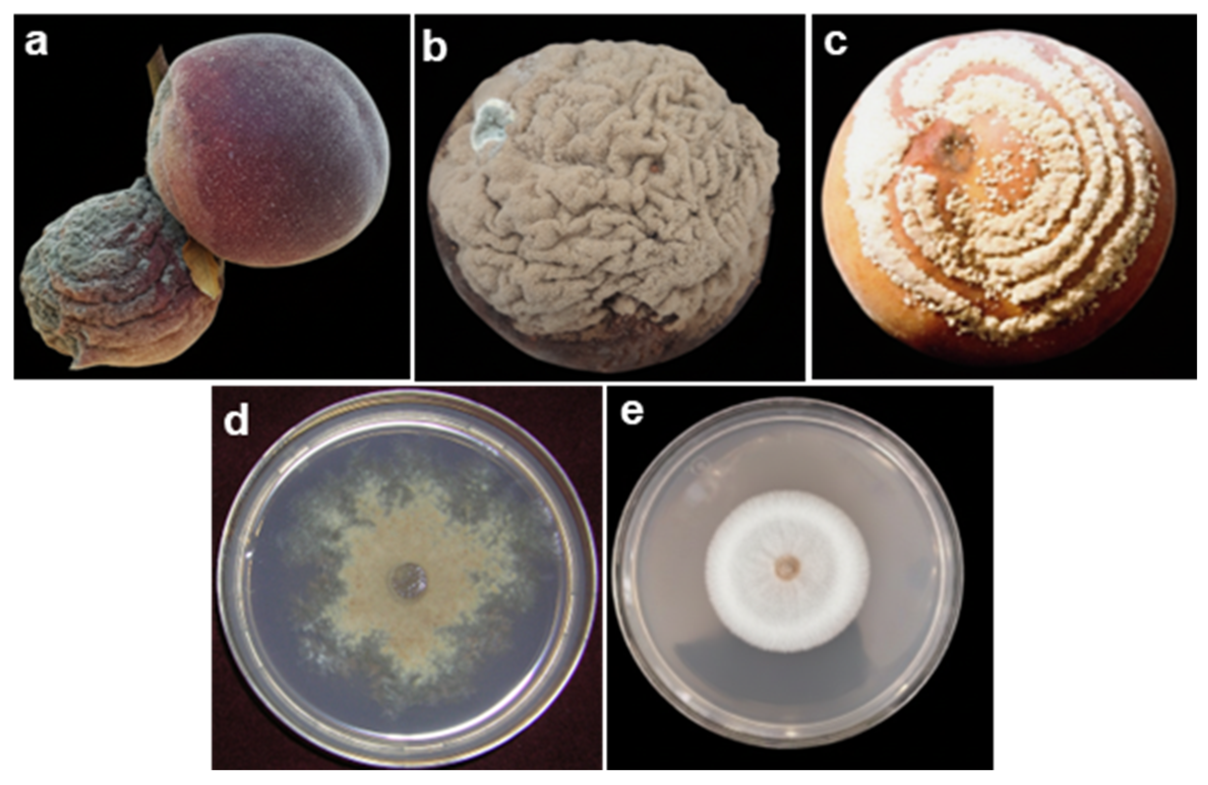
Disclaimer/Publisher’s Note: The statements, opinions and data contained in all publications are solely those of the individual author(s) and contributor(s) and not of MDPI and/or the editor(s). MDPI and/or the editor(s) disclaim responsibility for any injury to people or property resulting from any ideas, methods, instructions or products referred to in the content.

Article Metrics

Citations

Article Access Statistics

Multiple requests from the same IP address are counted as one view.