Article

Efficient Room-Temperature Luminescence of Indole-5-Carboxamide in Poly(vinyl alcohol) Films

1
Department of Physics and Astronomy, Texas Christian University, Fort Worth, TX 76129, USA
2
Department of Chemistry and Biochemistry, Texas Christian University, Fort Worth, TX 76129, USA
*
Authors to whom correspondence should be addressed.
Photochem 2025, 5(2), 14; https://doi.org/10.3390/photochem5020014
Submission received: 30 April 2025 / Revised: 29 May 2025 / Accepted: 2 June 2025 / Published: 4 June 2025

Abstract

:
N-phenyl-1H-Indole-5-carboxamide (Ind-CA) exhibits previously unknown room-temperature phosphorescence (RTP) when immobilized in poly (vinyl alcohol) film (PVA film). High-fluorescence anisotropy of Ind-CA in PVA suggests that the fluorophores are strongly immobilized in a polymer matrix, while a relatively low (ca. 0.1) quantum yield indicates a strong non-radiative singlet excited state deactivation. With an increased triplet-state population, Ind-CA can be used for various phosphorescence studies. The room-temperature phosphorescence (RTP) capability of Ind-CA indicates that there is an intricate balance between RTP and the structure of the indole-containing luminophore, as an isomeric N-1H-indole-5-ylbenzamide (Ind-BA) does not show any appreciable levels of RTP. Moreover, the phosphorescence lifetime of Ind-CA is about two orders of magnitude longer than many other 5-substituted indoles. These results further highlight the prospects for the potential rational designs of small molecules with desired triplet-state configuration and RTP characteristics.

1. Introduction

Room-temperature phosphorescence (RTP) has been commonly observed from inorganic materials [1]. RTP has been employed for safety uses, information encryption and decryption, and anti-counterfeiting [2,3,4]. In order to improve and extend its applications, the use of organic molecules has been of interest, particularly for physical/chemical sensing [5] and biological imaging [6]. Recently, RTP obtained from direct triplet excitation has paved new avenues to study large protein conformational studies [7,8]. Moreover, long-wavelength excitation has the potential to enable the study of optically dense samples and limit unwanted background such as autofluorescence and scattering [9]. Due to the relative ease of synthesis and access to various structural motifs, small organic molecules play an integral part in the quest for new materials with desired photophysical properties. Arguably, the prospect of introducing RTP capabilities using efficient, facile, and potentially modular approaches to make structurally and functionally diverse small organic molecules should ultimately establish structure–property relationships that will aid in the guided design of all-organic RTP materials [10,11,12,13,14].
Indole is one of the most prevalent organic motifs, found in numerous applications in the chemical, biological, and materials areas of modern sciences. The RTP of indoles, including tryptophan, has already been extensively studied in deoxygenated solutions [15,16,17,18] and in PVA by our group and others [19,20,21,22,23,24,25]. In view of the demonstrated RTP by indole and several substituted indoles, such as 5-bromoindole, 5-chloroindole, and 2-phenylindole, we decided to investigate the effect of an amide functionality on RTP. Amide groups could be efficiently installed under mild conditions, as many amine and acid derivatives are readily available, thus providing opportunities for establishing structure–property relationships. Here, as an initial entry to this venue, N-phenyl-1H-Indole-5-carboxamide (Ind-CA) and the isomeric N-1H-indole-5-ylbenzamide (Ind-BA) were synthesized using conventional amide coupling methodology (Scheme 1).
To achieve efficient RTP of an organic dye, the fluorophore of interest should be isolated from oxygen (efficient quencher of the triplet state) and placed in an environment that increases spin–orbit interaction. This is necessary to facilitate spin-forbidden transitions (intersystem crossing and triplet–singlet as well as singlet–triplet). The immobilization of fluorophores in a polymer matrix, like PVA, effectively fulfills these requirements. Non-covalent bonds formed between the polymer’s functional groups and those from fluorophores effectively restrict molecular motion in a solid matrix [26,27,28] and reduce oxygen diffusion, promoting the forbidden transitions [29,30,31,32,33,34,35]. Transparent and flexible polymer films have often been used to enhance RTP in practical applications [2,3,36,37,38]. The cost and ease of making polymer films have made it a viable way to detect RTP from novel and existing fluorophores.

2. Materials and Methods
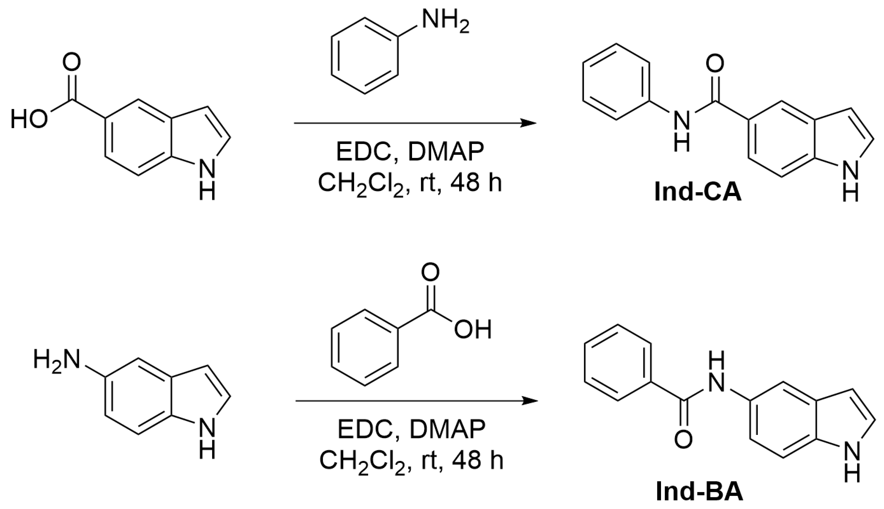
2.1. Synthesis

All reagents and solvents were purchased from commercial sources (Sigma-Aldrich, Acros, St. Louis, MI, USA, and TCI, Portland, OR, USA) and were used as received. NMR spectra were acquired on a Bruker (Billerica, MA, USA) Ascend 400 (400 MHz) spectrophotometer, and the chemical shifts are reported in ppm (δ) from the residual solvent peak (acetone: 2.05 ppm). Multiplicities are reported as follows: s—singlet, d—doublet, dd—doublet of doublets, t—triplet, tt—triplet of triplets, and m—multiplet.
The synthesis of Ind-CA [39]: A 50 mL round bottom flask was sequentially charged with a stirring bar, 1H-indole-5-carboxylic acid (0.30 g, 1.86 mmol), dimethylaminopyridine/DMAP (0.24 g, 1.88 mmol), 1-(3-dimethylaminopropy)-3-ethylcarbodiimide hydrochloride/EDC (0.36 g, 1.88 mmol), CH2Cl2 (10 mL), and aniline (0.17 mL, 1.86 mmol) and stirred under room temperature for 48 h. Next, the reaction mixture was diluted with CH2Cl2 (40 mL) and washed with water (40 mL), 1 M HCl (40 mL × 2), 1 M NaOH (40 mL × 2), and brine (40 mL × 2). The organic fraction was dried over Na2SO4, and volatiles were removed in vacuum. The residue was dissolved in ethyl acetate and filtered, and volatiles were removed in vacuum to give Ind-CA (0.25 g, 57% yield) as a beige solid.
1H NMR (400 MHz, acetone-d6): δ 10.54 (s, 1H), 9.42 (s, 1H), 8.30 (m, 1H), 7.90 (dd, J = 8.4 Hz, 1.0 Hz, 2H), 7.81 (dd, J = 8.8, 1.8 Hz, 1H), 7.51 (d, J = 8.4 Hz, 1H), 7.45 (m, 1H), 7.34 (m, 2H), 7.08 (tt, J = 7.4, 1.0 Hz, 1H), 6.60 (m, 1H).
13C NMR (100 MHz, acetone-d6): δ 166.48, 140.06, 138.08, 128.53, 127.66, 126.61, 126.38, 123.13, 121.05, 120.34, 119.96, 111.03, 102.63.
Synthesis of Ind-BA [40]: A 50 mL round bottom flask was sequentially charged with a stirring bar, benzoic acid (0.23 g, 1.86 mmol), DMAP (0.24 g, 1.88 mmol), EDC (0.36 g, 1.88 mmol), CH2Cl2 (10 mL), and 5-aminoindole (0.24 g, 1.86 mmol) and stirred under room temperature for 48 h. Next, the reaction mixture was diluted with CH2Cl2 (40 mL) and washed with water (40 mL), 1 M HCl (40 mL × 2), 1 M NaOH (40 mL × 2), and brine (40 mL × 2). The organic fraction was dried over Na2SO4, and volatiles were removed in vacuum. The residue was dissolved in ethyl acetate and filtered, and volatiles were removed in vacuum to give Ind-BA (0.32 g, 73% yield) as an off-gray solid.
1H NMR (400 MHz, acetone-d6): δ 10.20 (s, 1H), 9.37 (s, 1H), 8.14 (d, J = 1.6 Hz, 1H), 8.02 (m, 2H), 7.51 (m, 4H), 7.39 (d, J = 8.4 Hz, 1H), 7.33 (t, J = 2.8 Hz, 1H), 6.46 (m, 1H).
13C NMR (100 MHz, acetone-d6): δ 165.02, 136.04, 133.49, 131.60, 131.02, 128.29, 128.11, 127.33, 125.47, 115.95, 112.09, 110.94, 101.64.

2.2. PVA Film Preparation

Polyvinyl alcohol (PVA) [MW 130,000, 98% hydrolyzed] obtained from Sigma-Aldrich was used for film preparation. The PVA films with and without Ind-CA and Ind-BA were made from PVA-containing solutions following the literature procedures [41].

2.3. Absorption Measurements

Room-temperature absorption spectra were measured using the Varian Cary 60 UV–Vis Spectrophotometer (Agilent Technologies, Inc., Santa Clara, CA, USA). The baseline signal correction was performed with PVA film as the blank.

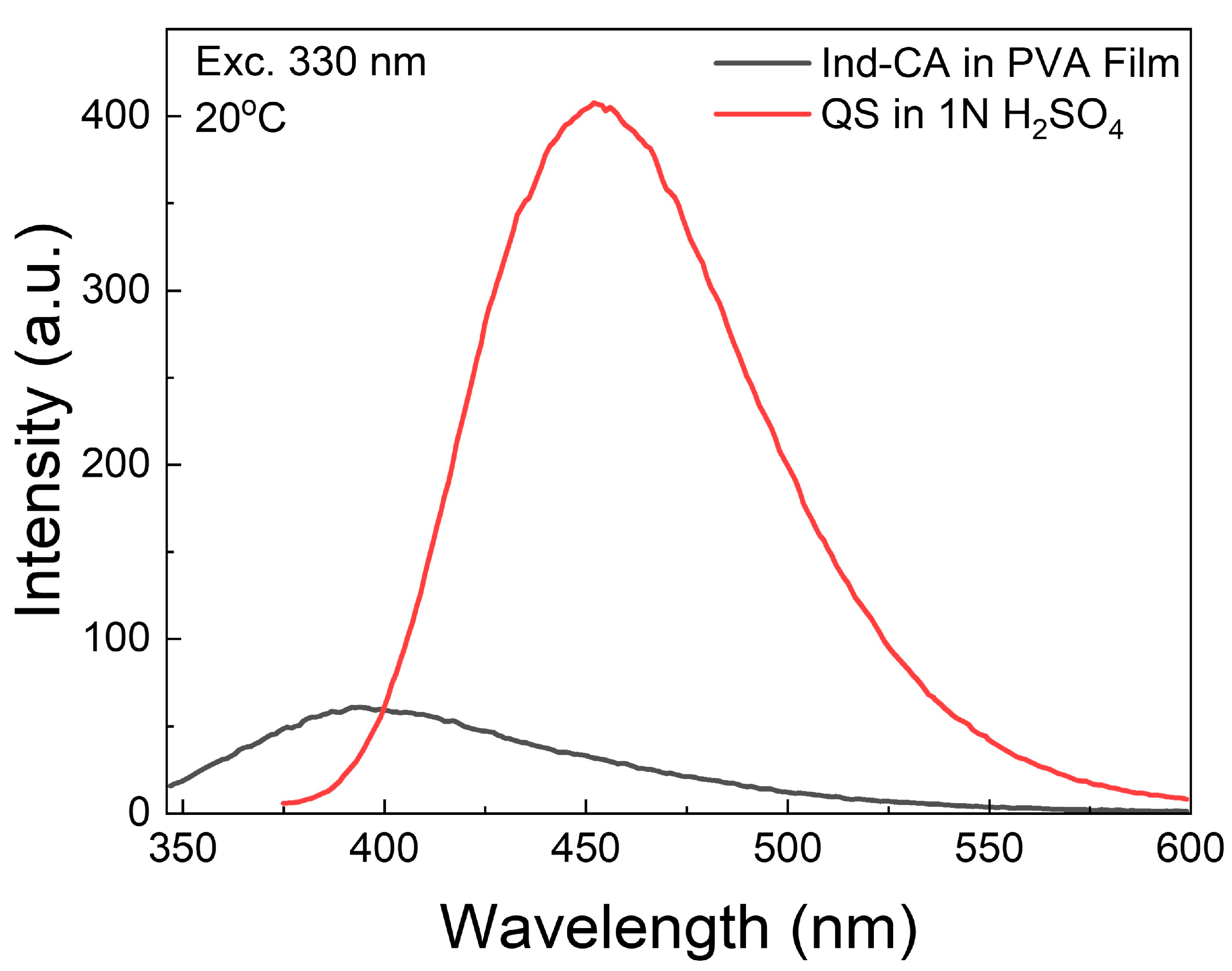
2.4. Steady-State Fluorescence Measurements

A Varian Cary Eclipse Spectrofluorometer (Agilent Technologies, Inc., Santa Clara, CA, USA) was used to conduct steady-state fluorescence measurements in square geometry.

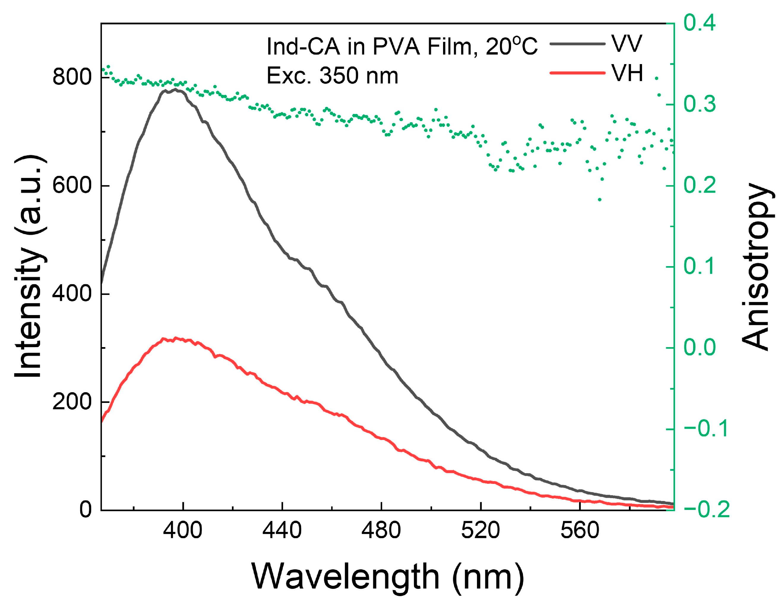
2.5. Fluorescence Anisotropy Measurements

For fluorescence anisotropy measurements, the Varian Cary Eclipse spectrofluorometer was fitted with a UV grid polarizer on the excitation and a plastic sheet polarizer on the observation. The anisotropies were calculated using the following expression:
r = I VV GI VH   I VV + 2 GI VH
where the I VV and I VH components are the fluorescence intensities when excited with vertical polarization (V) and observed with horizontal and vertical polarization, respectively. The G-Factor, represented as G , was used to compensate for the uneven transmission of different polarizations through the detection path.

2.6. Fluorescence Quantum Yield Measurements

To calculate the fluorescence quantum yield (QY) of Ind-CA-doped PVA film, a solution of quinine sulfate (QS) in 1 N H2SO4 (with a quantum yield of 0.54 [42]) was used as a standard for relative QY measurements. The absorbance spectra of both samples were measured with the absorbances of the reference and sample being equal at 335 nm. In this case, the quantum yield of Ind-CA in PVA film was obtained using the following equation:
QY Ind CA = QY QS · I Ind CA I QS · n 1 N   H 2 SO 4 2 n PVA 2
where QY QS = 0.54 is the quantum yield of QS in 1 N H2SO4 [42], I Ind CA   and I QS are the integrated intensities of Ind-CA in PVA film and QS in 1 N H2SO4 fluorescence emissions, and n 1 N   H 2 SO 4 2  and n PVA 2 are the refractive indexes of 1 N H2SO4 (1.35) and PVA film (1.48).

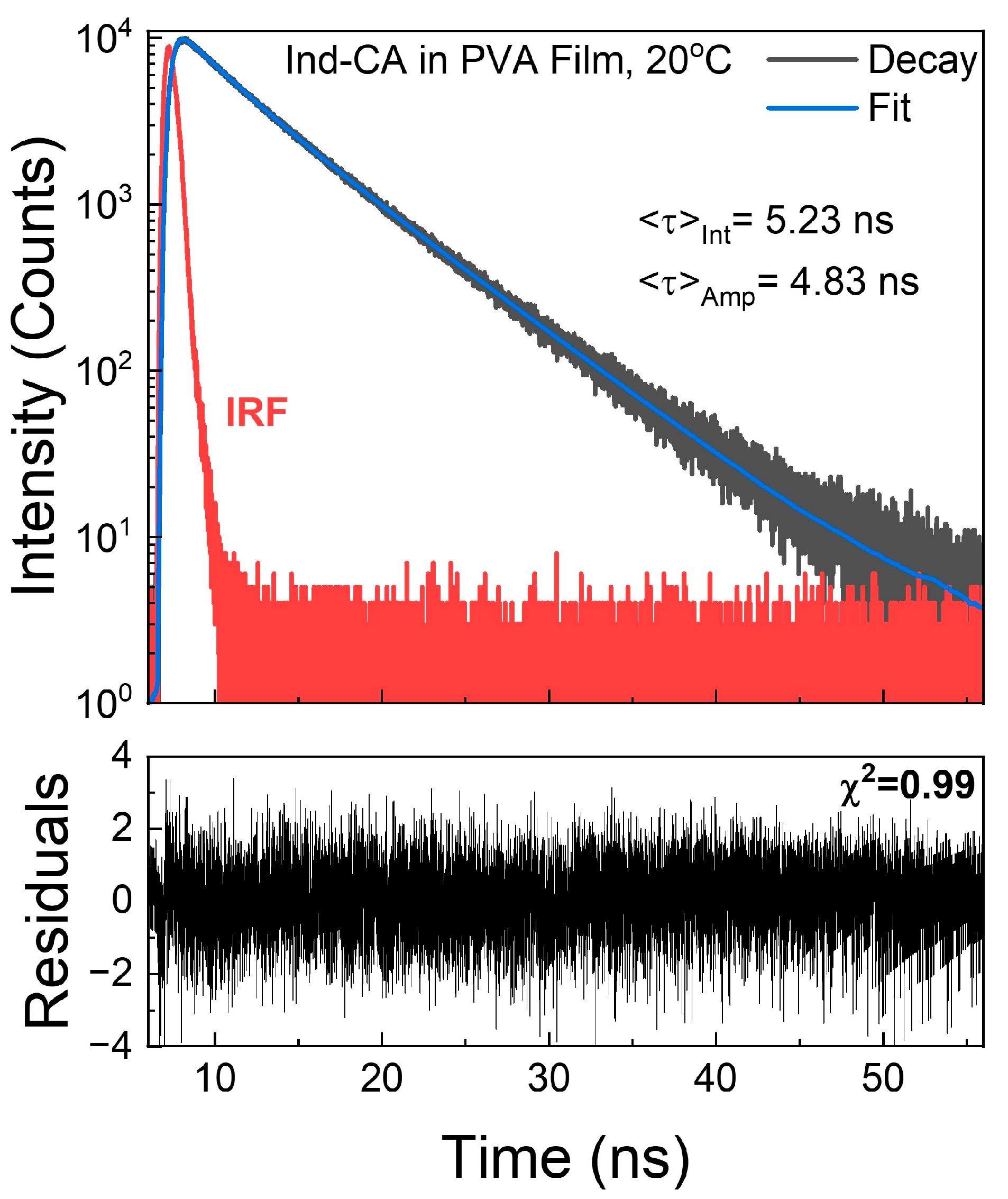
2.7. Fluorescence Lifetime Measurements

Fluorescence lifetime measurements were conducted using an FT200 fluorometer (PicoQuant GmBH, Berlin, Germany) equipped with a time-correlated single-photon counting module capable of a 4 ps time resolution (PicoHarp 300, PicoQuant GmBH, Berlin, Germany). For the excitation, a UV pulsed LED (PicoQuant GmBH, Berlin, Germany) with a repetition rate of 5 MHz was employed. The time-dependent data were subsequently analyzed by FluoFit4 (PicoQuant GmBH, Berlin, Germany) to fit the appropriate decay models. The collected time-dependent intensities were fitted to a multi-exponential model:
I t = I 0 i α i e t τ i
where the α i term represents the amplitude for the ith intensity decay component at a time t0, and τ i represents the lifetime of that component.
For calculating the intensity ( < τ > int ) and amplitude ( < τ > amp ) average decays, the following formulas were used:
< τ > int = f i τ i
< τ > amp = α i τ i
f i = α i τ i α i τ i
The applicability of the appropriate average values has been described in [43].

2.8. Phosphorescence Spectra Measurements

Phosphorescence excitation and emission spectra were measured using the time-gated phosphorescence detection mode of the Varian Cary Eclipse. The following parameters were used unless otherwise specified: Total Decay Time—1.0 s, Number of Flashes—10, Delay Time—0.2 ms, and Gate Time—5 ms. These phosphorescence mode parameters were set to remove short-lived emission components such as fluorescence background signals and scattering, which include but are not limited to Raleigh and Raman scattering.
Phosphorescence anisotropies were measured in a similar manner to the fluorescence anisotropy.

2.9. Phosphorescence Lifetime Measurements

For phosphorescence lifetime estimation, an Apple iPhone 14 Pro Max in slow-motion video mode (240 fps, 1080 p) was used to record the emission decay of the Ind-CA-doped PVA film following UV excitation. The film was kept in focus and framed consistently. The resulting video, consisting of 1321 frames (with the first frame at t = 0), spanned approximately 5.5 s and was exported as individual images.
A fixed region of interest (ROI) covering the sample was defined and consistently applied across all frames. The mean grayscale intensity of each ROI was measured, yielding a dataset of relative intensity versus frame number.
Frame indices were converted to elapsed time by dividing the frame number by the acquisition rate (240 fps). A constant background offset derived from the intensity plateau at later times when emission had ceased was subtracted from all intensity measurements. The background-corrected decay data were then fitted using a single-exponential decay model:
I t = I 0 e t τ
where I 0 represents the initial amplitude of the decay, and τ corresponds to the phosphorescence lifetime.

3. Results and Discussion

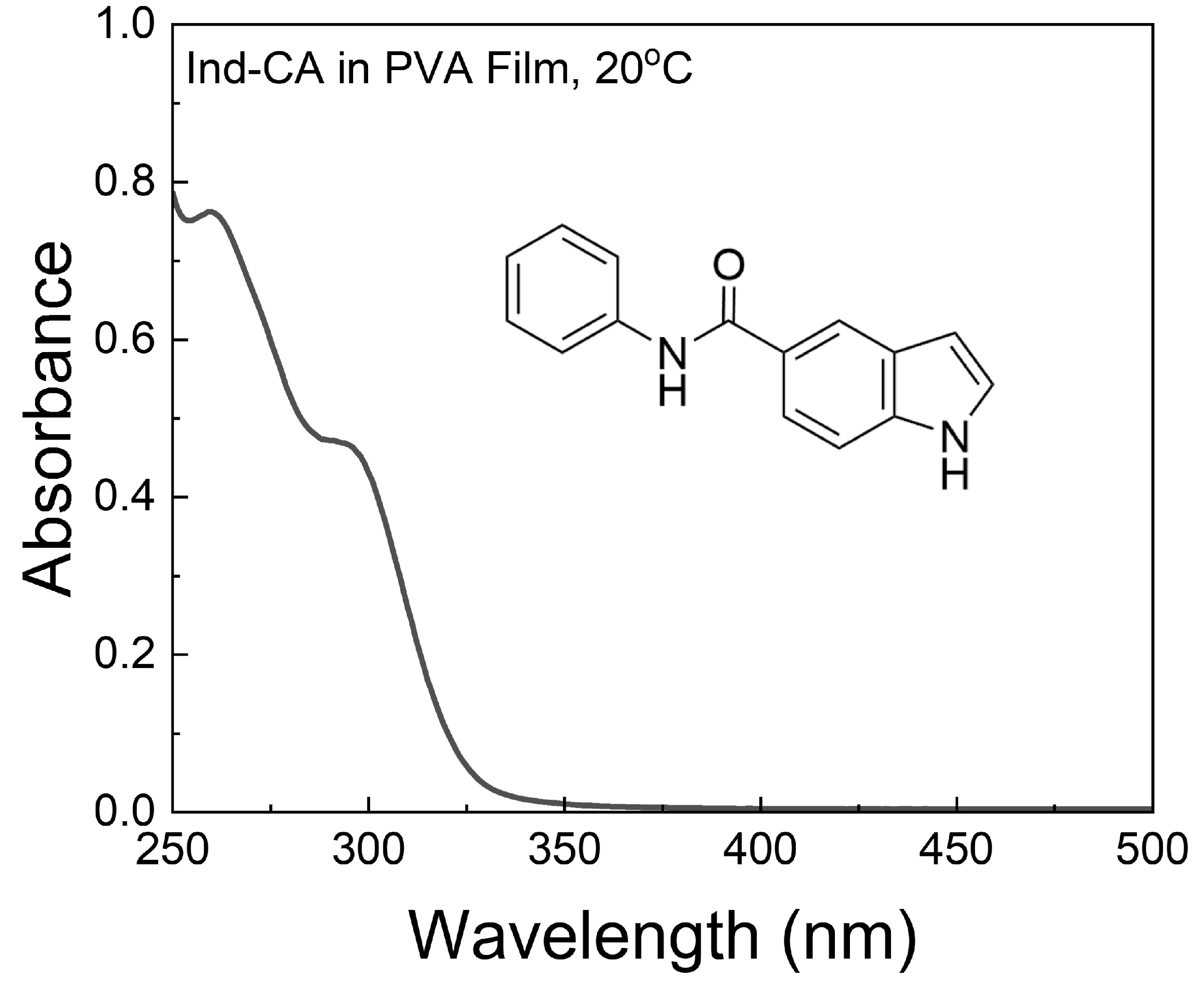
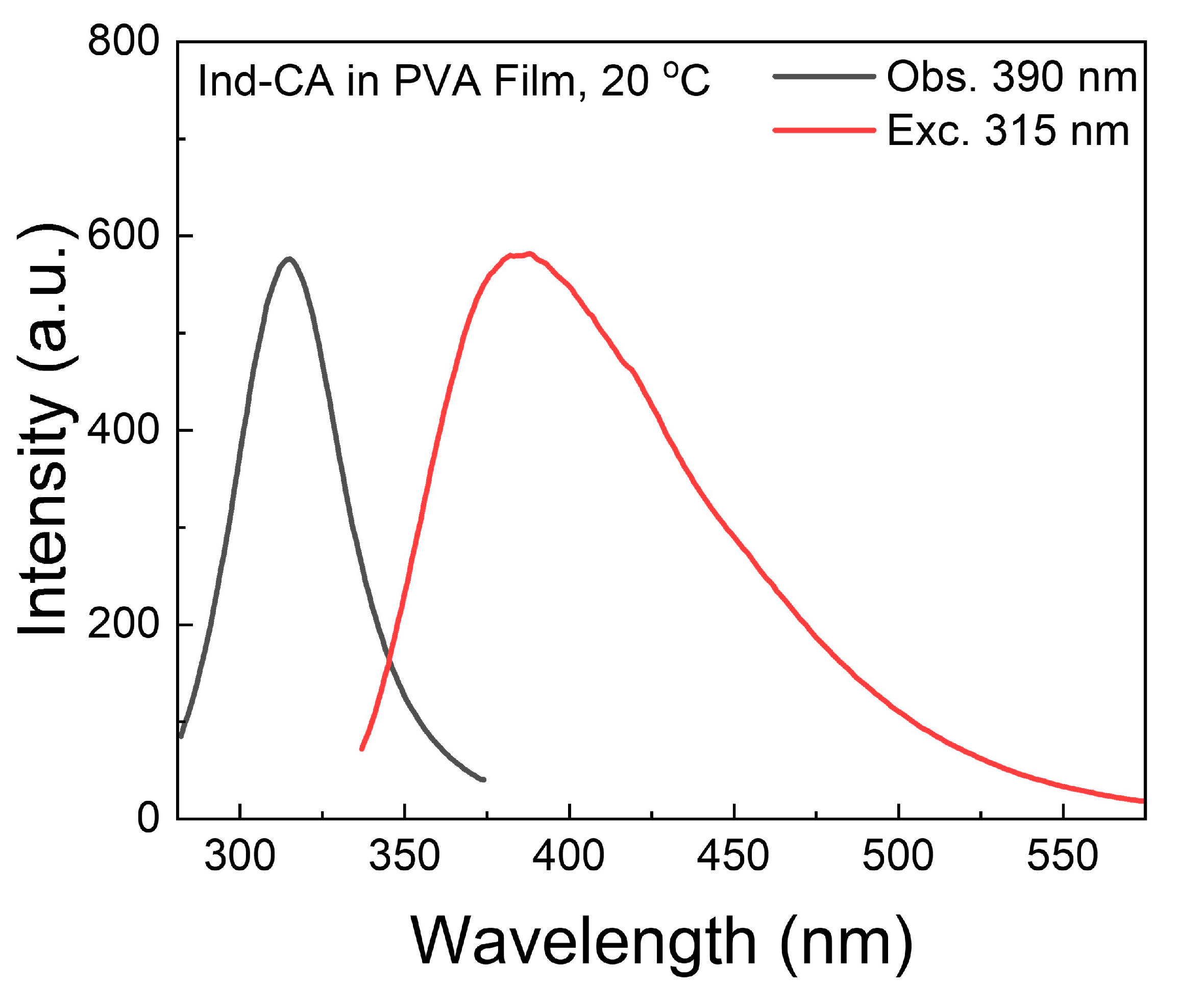
3.1. Absorption and Fluorescence Spectra of Ind-CA

The absorption of Ind-CA in PVA film occurs in the UV region, below 350 nm (Figure 1). The shape of the absorption spectrum indicates the presence of closely overlapping excited states. Similar absorption profiles were observed in water and ethanol (Figure S1).
The fluorescence spectrum of Ind-CA-doped PVA film is broad, and spans from 330 nm to 580 nm (Figure 2), exhibiting a strong blue phosphorescence at room temperature, and is substantially stronger than several other 5-substituted indole derivatives. Excited at equal absorbances (Figure S2), Ind-BA shows its fluorescence to be red-shifted but comparable in intensity to Ind-CA (Figure S3).
The fluorescence excitation spectrum (Figure 2) does not exactly mimic the absorption spectrum (Figure 1), as only the band corresponding to absorption to the first excited state is observed. This suggests that higher excited states are effectively quenched by a non-radiative pathway.

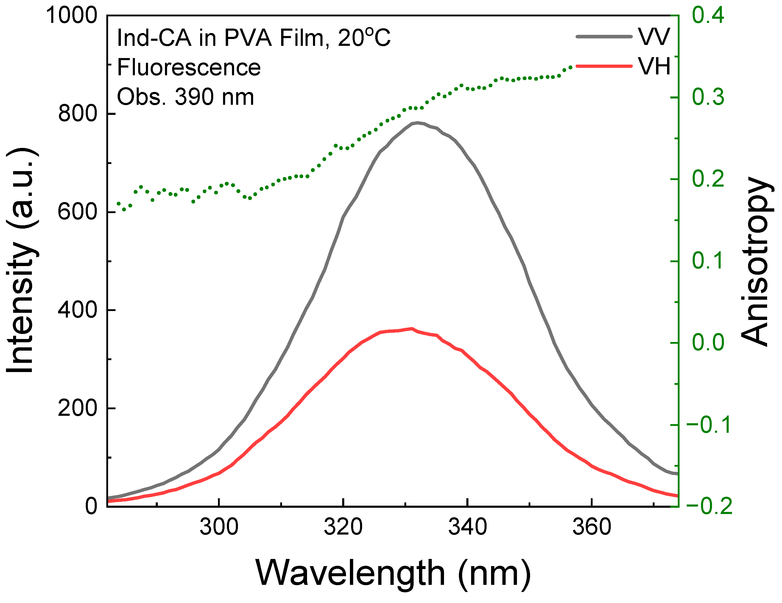
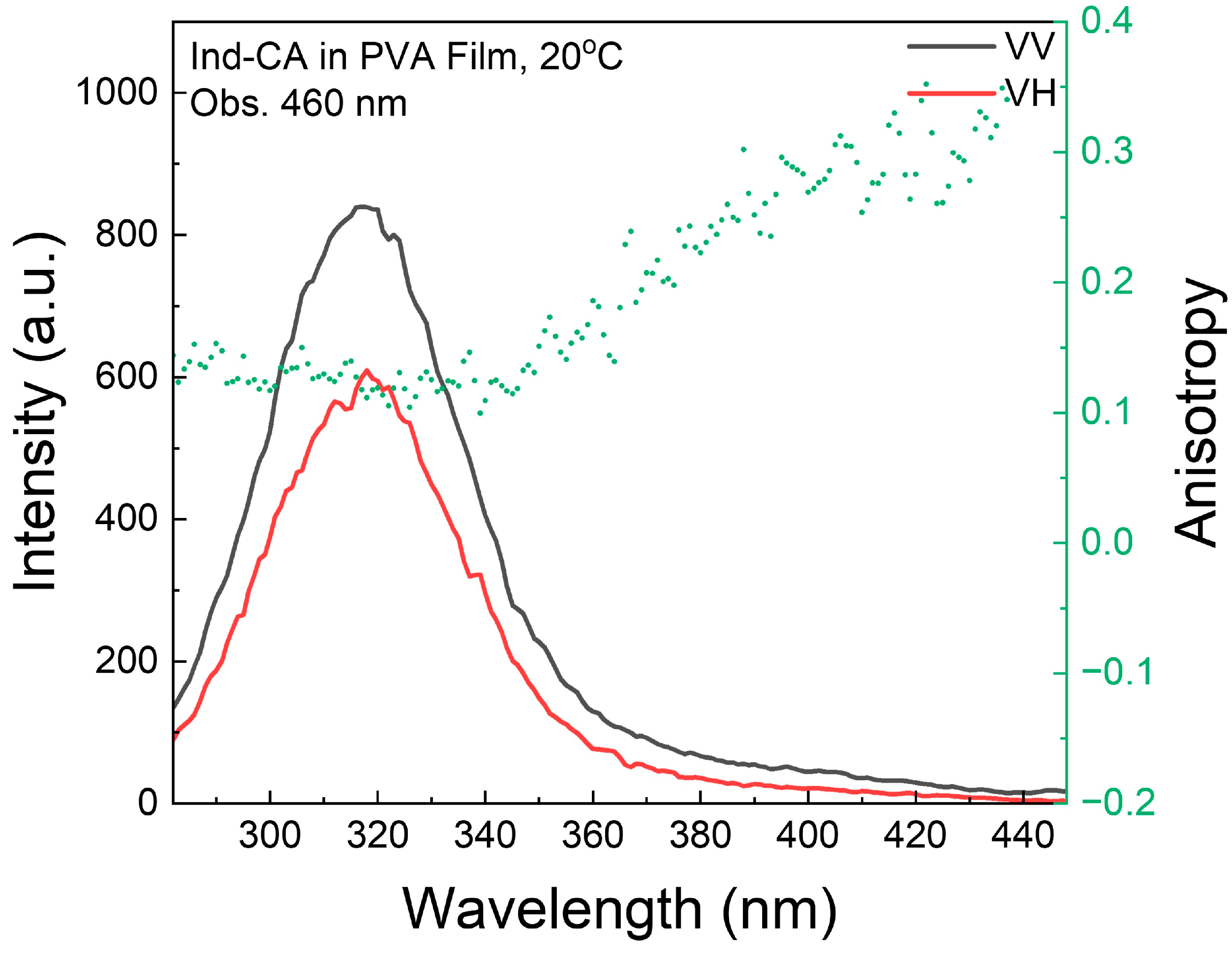
3.2. Fluorescence Anisotropy

The involvement of a higher excited state in the fluorescence emission should be evident in the fluorescence excitation anisotropy, since its transition moment is typically orthogonal. Indeed, at shorter wavelengths, the fluorescence excitation anisotropy spectrum of Ind-CA indicates the participation of a higher excited state (Figure 3). This is not surprising, as indoles are known to possess closely overlapping electronic excited states [30,44].
The fluorescence emission anisotropy slightly decreases at longer wavelengths (Figure 4). A high value of fluorescence anisotropy suggests that fluorophores are effectively immobilized in the polymer matrix.

3.3. Fluorescence Quantum Yield

The fluorescence efficiency of Ind-CA-doped PVA was estimated by comparison with the fluorescence of quinine sulfate (QS) in 1 N H2SO4 (Figure 5). At an excitation wavelength of 330 nm, the absorbances of Ind-CA and QS are equal (Figure S4). The estimated quantum yield of Ind-CA in PVA is 0.11, which is more than twice as low as that of indole in PVA film [22].

3.4. Fluorescence Lifetime

The fluorescence intensity decay of Ind-CA in PVA film is shown in Figure 6. The decay is slightly heterogeneous and reveals a mean lifetime of about 5 ns, comparable to that of indole [22].

3.5. Room-Temperature Phosphorescence

The phosphorescence of Ind-CA in PVA film can be observed with the naked eye on the UV illuminator, as the blue glow emitted by the Ind-CA-doped PVA persists for several seconds after the UV illuminator is turned off (Figure 7).

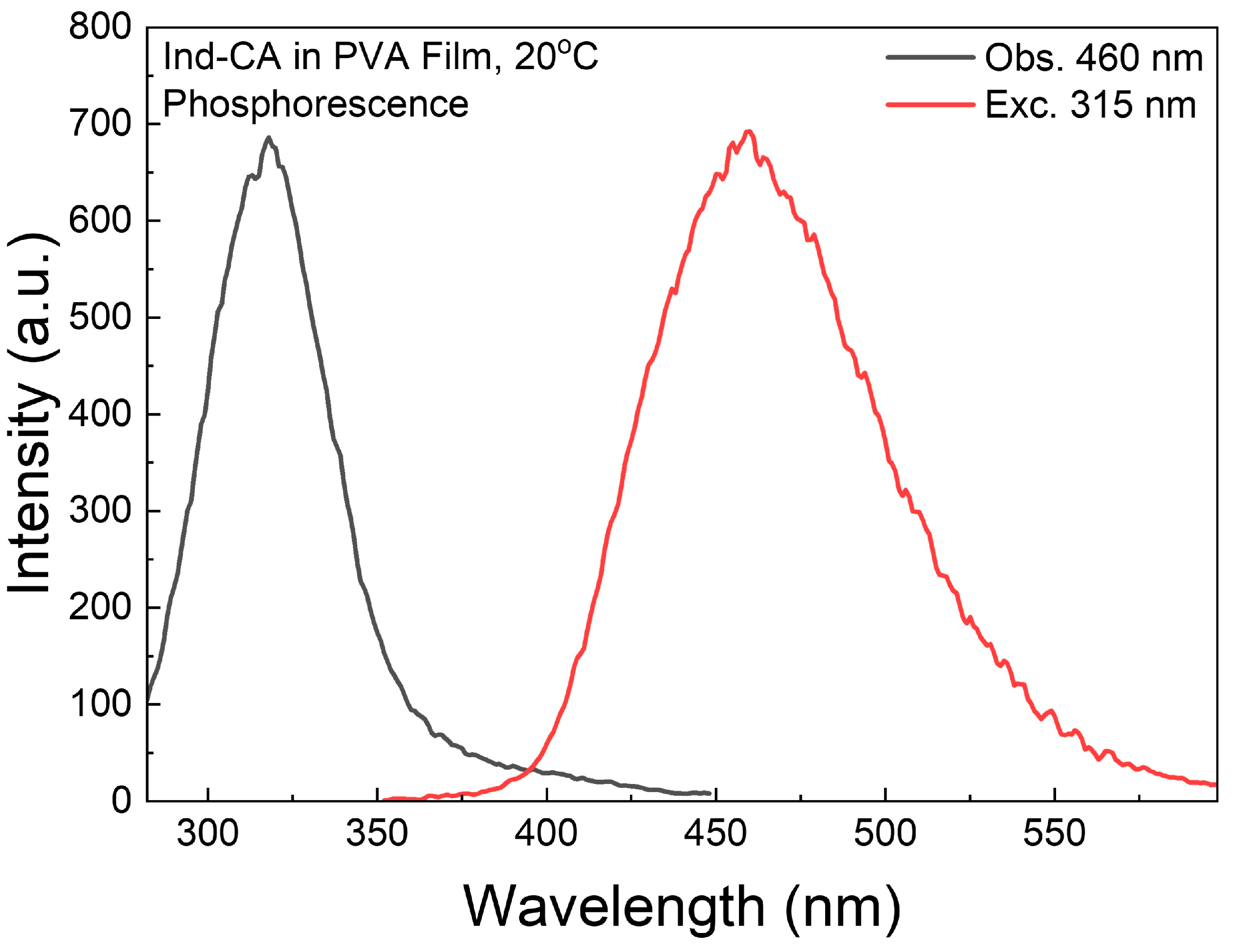
3.6. Phosphorescence Spectra

The phosphorescence excitation and emission spectra of Ind-CA in PVA film reveal a bell-shaped phosphorescence emission profile, spanning from 350 nm to 600 nm, with a peak centered at about 450 nm (Figure 8). The excitation phosphorescence spectrum mimics the fluorescence excitation spectrum, except for the long-wavelength range above 350 nm. The direct singlet–triplet absorption measurements at room temperature are beyond our instrument’s capabilities, as the absorbance for such transitions is extremely low and falls within the noise range of any detector. However, the phosphorescence is easily observable.
The phosphorescence of Ind-CA can be observed with excitation wavelengths well above those corresponding to the absorption peaks. However, the phosphorescence of Ind-BA is orders of magnitude weaker than that of Ind-CA (Figures S5 and S6). It is possible that Ind-CA interacts more strongly with the polymer, increasing the spin–orbit perturbation and altering the spin-forbidden transitions (S-T and T-S). Also, it might be that, in PVA, these two compounds adopt slightly different conformations. Such a drastic difference could potentially be explained by the ability of Ind-CA to adopt a sufficient degree of distortion in the PVA matrix between the indolyl and phenyl moieties, which amounts to efficient RTP. On the other hand, Ind-BA appears to adopt a more planar conformation in PVA, which might favor non-radiative processes, which suppress spin–orbit coupling. This is consistent with the fact that fluorescence emission of Ind-CA is at ca. 415 nm, whereas fluorescence emission of Ind-BA is at 460 nm (Figure S3).

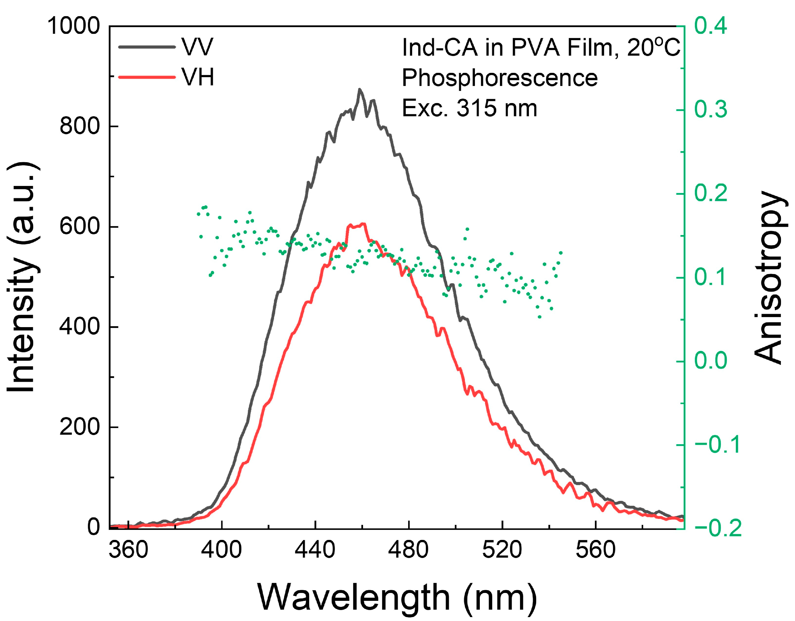
3.7. Phosphorescence Anisotropy

The phosphorescence excitation anisotropy spectrum indicates that, at shorter excitation wavelengths corresponding to the singlet excited state (absorption), the anisotropy is low, approximately 0.1 (Figure 9). This behavior is anticipated, as the triplet state is populated through an intersystem crossing process, resulting in differing directions of absorption and emission (phosphorescence) moments. At longer wavelengths, above 340 nm, the phosphorescence anisotropy increases to about 0.3, resembling the anisotropy observed in fluorescence (Figure 3). This also alludes to the possibility of direct excitation to the triplet state of the Ind-CA fluorophore.
The phosphorescence emission anisotropy (Figure 10) remains low and steady within the emission spectrum, suggesting that the triplet–singlet transition is orthogonal to the singlet–singlet transition.

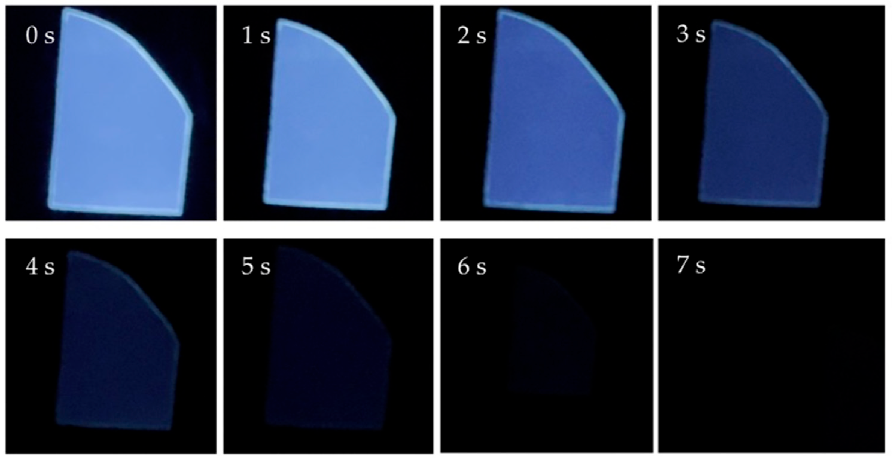
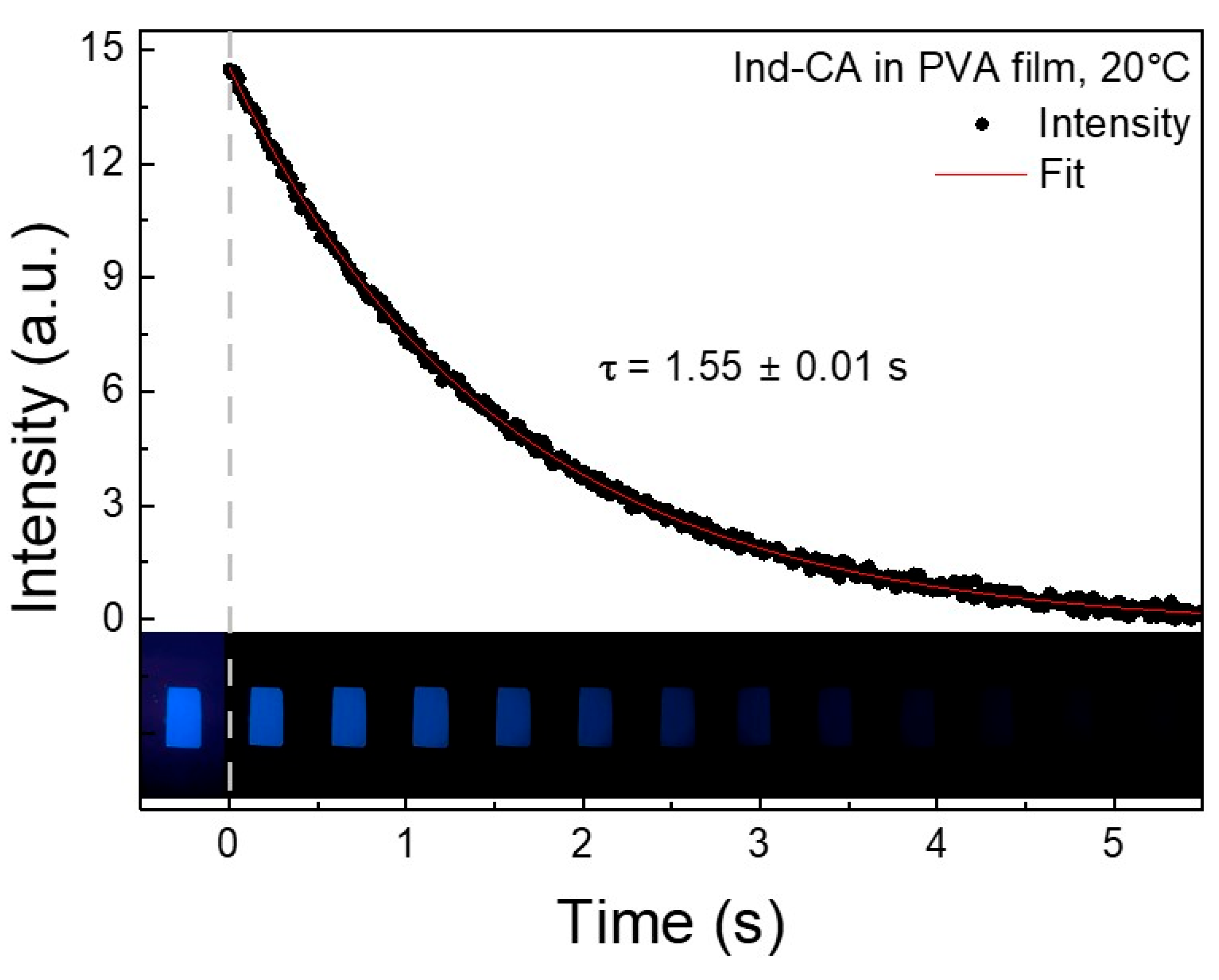
3.8. Phosphorescence Lifetime

The phosphorescence lifetime of Ind-CA in PVA film is relatively long and has been estimated by continuous monitoring with a cell phone after UV excitation (Figure 11).
The images within the graph are of the PVA film at 0.5 s intervals. The initial frame corresponds to the moment when the excitation is still on. Subsequent frames depict emission decay after excitation is switched off. The single-exponent fit (---) reveals a mean lifetime of about 1.5 s (Figure 11), as the triplet state of Ind-CA can be populated directly with long-wavelength light excitation. The spectral region above 400 nm offers stronger laser excitation, which is not easily accessible in UV. Moreover, the phosphorescence anisotropy with long-wavelength excitation is high (Figure S7), enabling the eventual steady-state and time-resolved study of slower molecular processes.
Another advantage of direct triplet excitation is the possibility of using samples in a conventional square geometry configuration. For this reason, it is convenient to use multiple strips of dye-doped polymer immersed in an index-matching fluid, which reduces reflections and makes strips invisible (see Figures S8 and S9). The UV excitation results in a saturated phosphorescence signal because of increased sample absorbance (inner filter effect type I). However, the long-wavelength excitation is not being significantly absorbed (see Figure S10).
The possibility of exciting virtually within a non-absorbing region and observing at a longer wavelength emission (not reabsorbed) enables the use of high optical density samples. This significantly extends the possible applications of RTP. Also, this enables easy anisotropy measurements (Figure S7). The long-wavelength excitation and extremely long lifetime of Ind-CA make this fluorophore an ideal donor in triplet–singlet FRET measurements. Such triplet–singlet FRET measurements do not involve intersystem crossing and can be used for the estimation of triplet-state parameters [45].

4. Conclusions

The results of this study unambiguously highlight the importance of the structure of the small-molecule scaffolds in modulating room-temperature phosphorescence (RTP) in a polymer matrix. We investigated the properties of two amide-containing indole scaffolds, i.e., Ind-CA and Ind-BA, which differ only in the type of amide linker connecting the indolyl and phenyl moieties (see Scheme 1). Both Ind-CA and Ind-BA embedded in PVA film fluoresce in the visible region with Ind-BA being slightly red-shifted.
Interestingly, Ind-CA exhibits a long-lived, strong blue phosphorescence at room temperature (~1.5 s) and is substantially greater than that of Ind-BA. This may be attributed to the stronger interaction that Ind-CA has with the polymer, enhancing the spin–orbit coupling and altering the spin-forbidden transitions (S-T and T-S). This result also implies the slightly different conformations that these two compounds have with the polymer matrix, where Ind-BA adopts a planar structure, promoting non-radiative pathways and inhibiting spin–orbit perturbation. Ind-CA embedded in PVA film also exhibits higher phosphorescence anisotropy with direct triplet excitation when compared to that of UV excitation. We believe that the possibility of direct triplet-state excitation will expand the repertoire of spectroscopic and imaging techniques to elucidate macromolecular dynamics. Further studies on indole derivatives, including the regioisomers of Ind-CA, as a viable scaffold for the structure–property relationship responsible for efficient RTP properties, are underway in our laboratories.

Supplementary Materials

The following supporting information can be downloaded at: https://www.mdpi.com/article/10.3390/photochem5020014/s1, Figure S1. Absorption of Ind-CA in ethanol. Figure S2. Left: Absorption spectrum of N-1H-indole-5-ylbenzamide (Ind-BA) in PVA film. Figure S3. Comparison of fluorescence emission spectra of Ind-CA and Ind-BA in PVA films. Figure S4. Absorption spectra of Ind-CA in PVA and QS in 1NH2SO4. Figure S5. Comparison of phosphorescence emission spectra of Ind-CA and Ind-BA in PVA films. Figure S6. Comparison of phosphorescence excitation spectra of Ind-CA and Ind-BA in PVA films. Figure S7. Phosphorescence emission anisotropy of Ind-CA in PVA film, measured upon direct triplet state excitation at 420 nm. Figure S8. Photographs of microcuvette containing an Ind-CA in PVA strip with and without an index-matching fluid (Benzene). Figure S9. Photographs of microcuvette containing an Ind-CA in PVA strip illuminated with a 405 nm laser pointer. Figure S10. Comparison of directly excited phosphorescence emissions of Ind-CA in PVA film strips. 1H and 13C NMR spectra of Ind-CA and Ind-BA.

Author Contributions

Conceptualization, B.L., S.V.D., Z.G., and I.G.; methodology, B.L. and I.G.; validation, B.L., A.J., and I.G.; formal analysis, B.L., A.J., and I.G.; investigation, B.L., A.J., R.S., D.P., T.T.P., S.V.D., Z.G., and I.G.; resources, S.V.D., Z.G., and I.G.; data curation, S.V.D., Z.G., and I.G.; writing—original draft preparation, I.G.; writing—review and editing, B.L., A.J., R.S., D.P., T.T.P., S.V.D., Z.G., and I.G.; supervision, Z.G. and I.G.; project administration, S.V.D., Z.G., and I.G.; funding acquisition, S.V.D., Z.G., and I.G. All authors have read and agreed to the published version of the manuscript.

Funding

This work was supported in part by the Dean’s Research Opportunity Fund from Texas Christian University (S.V.D.) and The Welch Foundation Grant No. P-2238-20250403 (Z.G.).

Data Availability Statement

The original contributions presented in this study are included in the article/Supplementary Material. Further inquiries can be directed to the corresponding authors.

Acknowledgments

Z.G. acknowledges the support from the “Tex” Moncrief Jr. Endowment Fund. We dedicate this paper to Sabato D’Auria on the occasion of his 65th birthday.

Conflicts of Interest

The authors declare no conflicts of interest.

Abbreviations

The following abbreviations are used in this manuscript:
Ind-CAN-phenyl-1H-Indole-5-carboxamide
Ind-BAN-1H-indole-5-ylbenzamide
PVAPolyvinyl Alcohol
RTPRoom-Temperature Phosphorescence

References

  1. Qiu, J.; Li, Y.; Jia, Y. Persistent Phosphors: From Fundamentals to Applications; Woodhead Publishing: Cambridge, UK, 2020. [Google Scholar]
  2. Gao, R.; Yan, D.; Evans, D.G.; Duan, X. Layer-by-layer assembly of long-afterglow self-supporting thin films with dual-stimuli-responsive phosphorescence and antiforgery applications. Nano Res. 2017, 10, 3606–3617. [Google Scholar] [CrossRef]
  3. Su, Y.; Phua, S.Z.F.; Li, Y.; Zhou, X.; Jana, D.; Liu, G.; Zhao, Y. Ultralong room temperature phosphorescence from amorphous organic materials toward confidential information encryption and decryption. Sci. Adv. 2018, 4, eaas9732. [Google Scholar] [CrossRef] [PubMed]
  4. Sun, H.; Liu, S.; Lin, W.; Zhang, K.Y.; Lv, W.; Huang, X.; Huo, F.; Yang, H.; Jenkins, G.; Zhao, Q.; et al. Smart responsive phosphorescent materials for data recording and security protection. Nat. Commun. 2014, 5, 3601. [Google Scholar] [CrossRef] [PubMed]
  5. Yang, J.; Fang, M.; Li, Z. Stimulus-responsive room temperature phosphorescence materials: Internal mechanism, design strategy, and potential application. Acc. Mater. Res. 2021, 2, 644–654. [Google Scholar] [CrossRef]
  6. Fateminia, S.M.A.; Mao, Z.; Xu, S.; Yang, Z.; Chi, Z.; Liu, B. Organic nanocrystals with bright red persistent room-temperature phosphorescence for biological applications. Angew. Chem. Int. Ed. 2017, 56, 12160–12164. [Google Scholar] [CrossRef]
  7. Vanderkooi, J.; Calhoun, D.; Englander, S. On the prevalence of room-temperature protein phosphorescence. Science 1987, 236, 568–569. [Google Scholar] [CrossRef]
  8. Gacintov, N.; Brenner, H. The triplet state as a probe of dynamics and structure in biological macromolecules. Photochem. Photobiol. 1989, 50, 841–858. [Google Scholar] [CrossRef]
  9. Ceresa, L.; Chavez, J.; Kitchner, E.; Kimball, J.; Gryczynski, I.; Gryczynski, Z. Imaging and detection of long-lived fluorescence probes in presence of highly emissive and scattering background. Exp. Biol. Med. 2022, 247, 1840–1851. [Google Scholar] [CrossRef]
  10. Zhao, W.; He, Z.; Tang, B.Z. Room-temperature phosphorescence from organic aggregates. Nat. Rev. Mater. 2020, 5, 869–885. [Google Scholar] [CrossRef]
  11. Wu, Z.; Nitsch, J.; Marder, T.B. Persistent room-temperature phosphorescence from purely organic molecules and multi-component systems. Adv. Opt. Mater. 2021, 9, 2100411. [Google Scholar] [CrossRef]
  12. Nidhankar, A.D.; Goudappagouda; Wakchaure, W.C.; Babu, S.S. Efficient metal-free organic phoshpors. Chem. Sci. 2021, 12, 4216–4236. [Google Scholar] [CrossRef] [PubMed]
  13. Wang, X.; Dong, M.; Li, Z.; Wang, P.; Liang, F.-S. Recent advances of room-temperature phosphorescence and long persistent luminescence by doping system of purely organic molecules. Dyes Pigm. 2022, 204, 110400. [Google Scholar] [CrossRef]
  14. Shen, X.; Wu, W.; Yang, C. Recent progress in solid-state room temperature afterglow based on pure organic small molecules. Molecules 2024, 29, 3236. [Google Scholar] [CrossRef]
  15. Papp, S.; Vanderkooi, J. Tryptophan phosphorescence at room temperature as a tool to study protein structure and dynamics. Photochem. Photobiol. 1989, 49, 775–784. [Google Scholar] [CrossRef]
  16. Fischer, C.; Gafni, A.; Steel, D.; Schauerte, J. The triplet-state lifetime of indole in aqueous and viscous environments: Significance to the interpretation of room temperature phosphorescence in proteins. J. Am. Chem. Soc. 2002, 124, 10359–10366. [Google Scholar] [CrossRef] [PubMed]
  17. Schlyer, B.; Schauerte, J.; Steel, D.; Gafni, A. Time-resolved room temperature protein phosphorescence: Nonexponential decay from single emitting tryptophans. Biophys. J. 1994, 67, 1192–1202. [Google Scholar] [CrossRef] [PubMed]
  18. Strambini, G.; Gonelli, M.; Galley, W. Room temperature phosphorescence of Trp 314 as monitor of subunit communication in alcohol dehydrogenase from horse liver. Biochemistry 1990, 29, 203–208. [Google Scholar] [CrossRef]
  19. Kawski, A.; Gryczynski, I.; Gryczynski, Z. Fluorescence and phosphorescence anisotropy spectra of indole in poly (vinyl alcohol) film at room temperature. Z. Naturforsch. A 1994, 49, 1091–1092. [Google Scholar] [CrossRef]
  20. Kowalska-Baron, A.; Chan, M.; Gałęcki, K.; Wysocki, S. Photophysics of indole, tryptophan and N-acetyl-L-tryptophanamide (NATA): Heavy atom effect. Spectrochim. Acta A Mol. Biomol. Spectrosc. 2012, 98, 282–289. [Google Scholar] [CrossRef]
  21. Kowalska-Baron, A.; Gałęcki, K.; Wysocki, S. Room temperature phosphorescence study on the structural flexibility of single tryptophan containing proteins. Spectrochim. Acta A Mol. Biomol. Spectrosc. 2015, 134, 380–387. [Google Scholar] [CrossRef]
  22. Chavez, J.; Ceresa, L.; Kitchner, E.; Kimball, J.; Shtoyko, T.; Fudala, R.; Borejdo, J.; Gryczynski, Z.; Gryczynski, I. On the possibility of direct triplet state excitation of indole. J. Photochem. Photobiol. B Biol. 2020, 208, 111897. [Google Scholar] [CrossRef] [PubMed]
  23. Chavez, J.; Ceresa, L.; Kimball, J.; Kitchner, E.; Gryczynski, Z.; Gryczynski, I. Room temperature luminescence of 5-chloroindole. J. Mol. Liq. 2022, 360, 119482. [Google Scholar] [CrossRef]
  24. Kowalska-Baron, A.; Gałęcki, K.; Wysocki, K. Photophysics of indole-2-carboxylic acid (I2C) and indole 5-carboxylic acid (I5C): Heavy atom effect. Spectrochim. Acta A Mol. Biomol. Spectrosc. 2013, 116, 183–195. [Google Scholar] [CrossRef] [PubMed]
  25. Zhou, W.; Wen, G.; Li, K.; Xiong, H.; Zhang, J.; Lu, S.; Chen, X. Room temperature phosphorescence of heavy-atom-free indole carboxylic acid/polyacrylamide: Low cost, long lifetime and good luminescence efficiency. Dyes Pigm. 2022, 205, 110481. [Google Scholar] [CrossRef]
  26. Ma, H.; Peng, Q.; An, Z.; Huang, W.; Shuai, Z. Efficient and long-lived room-temperature organic phosphorescence: Theoretical descriptors for molecular designs. J. Am. Chem. Soc. 2018, 141, 1010–1015. [Google Scholar] [CrossRef]
  27. Gu, L.; Shi, H.; Miao, C.; Wu, Q.; Cheng, Z.; Cai, S.; Gu, M.; Ma, C.; Yao, W.; Gao, Y. Prolonging the lifetime of ultralong organic phosphorescence through dihydrogen bonding. J. Mater. Chem. C. 2018, 6, 226–233. [Google Scholar] [CrossRef]
  28. Yao, X.; Ma, H.; Wang, X.; Wang, H.; Wang, Q.; Zou, X.; Song, Z.; Jia, W.; Li, Y.; Mao, Y. Ultralong organic phosphorescence from isolated molecules with repulsive interactions for multifunctional applications. Nat. Commun. 2022, 13, 4890. [Google Scholar] [CrossRef]
  29. Valeur, B.; Berberan-Santos, M.N. Molecular Fluorescence: Principles and Applications, 2nd ed.; Wiley-VCH: Weinheim, Germany, 2013. [Google Scholar]
  30. Lakowicz, J.R. Principles of Fluorescence Spectroscopy, 3rd ed.; Springer: New York, NY, USA, 2006. [Google Scholar]
  31. McGlynn, P.; Azumi, T.; Kinoshita, M. Molecular Spectroscopy of the Triplet State; Prentice-Hall: Englewood Cliffs, NJ, USA, 1969. [Google Scholar]
  32. El-Sayed, M.A.; Brewer, R.G. Polarization of the π * π and π *-n Phosphorescence Spectra of N-Heterocyclics. J. Phys. Chem. 1963, 39, 1623–1628. [Google Scholar] [CrossRef]
  33. Lim, E.C.; Yu, J.M.H. Vibronic Spin-Orbit Interactions in Heteroaromatic Molecules. I. Polycyclic Monazines. J. Chem. Phys. 1967, 47, 3270–3275. [Google Scholar] [CrossRef]
  34. Lee, D.; Bolton, O.; Kim, B.C.; Youk, J.H.; Takayama, S.; Kim, J. Room Temperature Phosphorescence of Metal-Free Organic Materials in Amorphous Polymer Matrices. J. Am. Chem. Soc. 2013, 135, 6325–6329. [Google Scholar] [CrossRef]
  35. Liu, Y.; Zhan, G.; Liu, Z.-W.; Bian, Z.-Q.; Huang, C.-H. Room-temperature phosphorescence from purely organic materials. Chin. Chem. Lett. 2016, 27, 1231–1240. [Google Scholar] [CrossRef]
  36. Cai, S.; Ma, H.; Shi, H.; Wang, H.; Wang, X.; Xiao, L.; Ye, W.; Huang, K.; Cao, X.; Gan, N.; et al. Enabling long-lived organic room temperature phosphorescence in polymers by subunit interlocking. Nat. Commun. 2019, 10, 4247. [Google Scholar] [CrossRef] [PubMed]
  37. He, Z.; Gao, H.; Zhang, S.; Zheng, S.; Wang, Y.; Zhao, Z.; Ding, D.; Yang, B.; Zhang, Y.; Yuan, W.Z. Achieving persistent, efficient, and robust room-temperature phosphorescence from pure organics for versatile applications. Adv. Mater. 2019, 31, 1807222. [Google Scholar] [CrossRef] [PubMed]
  38. Dai, X.Y.; Huo, M.; Liu, Y. Phosphorescence resonance energy transfer from purely organic supramolecular assembly. Nat. Rev. Chem. 2023, 7, 854–874. [Google Scholar] [CrossRef] [PubMed]
  39. Rousseau, J.-F.; Dodd, R.H. Regioselective ortho-directed metalation and electrophilic substitution of indole–And indoline-5-(N-phenyl) carboxamides. Heterocycles 2001, 55, 2289–2304. [Google Scholar] [CrossRef]
  40. Macor, J.E.; Blank, D.H.; Fox, C.B.; Lebel, L.A.; Newman, M.E.; Post, R.J.; Ryan, K.; Schmidt, A.W.; Schulz, D.W.; Koe, B.K. 5-[(3-Nitropyrid-2-yl) amino] indoles: Novel serotonin agonists with selectivity for the 5-HT1D receptor. Variation of the C3 substituent on the indole template leads to increased 5-HT1D receptor selectivity. J. Med. Chem. 1994, 37, 2509–2512. [Google Scholar] [CrossRef]
  41. Gryczynski, Z.; Gryczynski, I. Practical Fluorescence Spectroscopy; CRC Press: Boca Raton, FL, USA, 2019. [Google Scholar]
  42. Eaton, D.F. Reference materials for fluorescence measurement. Pure Appl. Chem. 1988, 60, 1107–1114. [Google Scholar] [CrossRef]
  43. Sillen, A.; Engelborghs, Y. The correct use of ‘average’ fluorescence parameters. Photochem. Photobiol. 1998, 67, 475–486. [Google Scholar] [CrossRef]
  44. Jameson, D.M. Introduction to Fluorescence; CRC Press: Boca Raton, FL, USA, 2014. [Google Scholar]
  45. Lee, B.; Gryczynski, I.; Gryczynski, Z. Demonstration of intermolecular triplet−singlet FRET in dye doped PVA films at room temperature. J. Phys. Chem. A 2025, 129, 2734–2737. [Google Scholar] [CrossRef]
Scheme 1. Synthesis of Ind-CA (top) and Ind-BA (bottom) used in this study.
Photochem 05 00014 sch001
Figure 1. Absorption of Ind-CA in PVA film.
Photochem 05 00014 g001
Figure 2. Fluorescence emission and excitation spectra of Ind-CA in PVA film.
Photochem 05 00014 g002
Figure 3. Fluorescence excitation anisotropy of Ind-CA in PVA film.
Photochem 05 00014 g003
Figure 4. Fluorescence emission anisotropy of Ind-CA in PVA film.
Photochem 05 00014 g004
Figure 5. Comparison of fluorescence emissions of Ind-CA in PVA film and quinine sulfate in 1 N H2SO4.
Photochem 05 00014 g005
Figure 6. Fluorescence lifetime decay of Ind-CA-doped PVA film analyzed with a double exponential model with recovered parameters, τ 1 = 5.85 ns and τ 2 = 2.94 ns, and amplitudes α 1 = 0.65 and α 2 = 0.35 . Instrument response function (IRF) graphed in red.
Photochem 05 00014 g006
Figure 7. Phosphorescence of Ind-CA on the UV illuminator. The first photograph (upper left) was taken with the UV illuminator ON, followed by snapshots captured at approximately 1 s intervals with the UV illuminator OFF.
Photochem 05 00014 g007
Figure 8. Phosphorescence excitation and emission spectra of Ind-CA in PVA film.
Photochem 05 00014 g008
Figure 9. Phosphorescence excitation anisotropy of Ind-CA in PVA film.
Photochem 05 00014 g009
Figure 10. Phosphorescence emission anisotropy of Ind-CA in PVA film.
Photochem 05 00014 g010
Figure 11. Phosphorescence intensity decay of Ind-CA in PVA film.
Photochem 05 00014 g011
Disclaimer/Publisher’s Note: The statements, opinions and data contained in all publications are solely those of the individual author(s) and contributor(s) and not of MDPI and/or the editor(s). MDPI and/or the editor(s) disclaim responsibility for any injury to people or property resulting from any ideas, methods, instructions or products referred to in the content.

