Abstract
This study investigates the enhanced production of key therapeutic metabolites (ellagic acid, serotonin, and chlorogenic acid) in response to abiotic stress in in vitro cultures of Quercus suber somatic embryos. Findings indicate significant increases in metabolite levels under various stress conditions, highlighting the potential for commercial-scale production of these compounds, known for their antioxidant, anticancer, and anti-COVID-19 properties. Under osmotic/saline stress, ellagic acid production significantly increased, representing an 80% increase compared to control conditions. In embryos exposed to different stressors, serotonin accumulation showed a six-fold increase under osmotic/saline stress. Although the elicitors used did not increase chlorogenic acid levels, exploring alternative stress types may enhance its production. This research paves the way for sustainable, large-scale production of health-beneficial metabolites, addressing global health challenges and promoting resource sustainability.
1. Introduction
Plants produce a vast array of organic compounds, known as metabolites, essential for processes such as growth, cell division, respiration, photosynthesis, reproduction, and storage. These are classified as primary metabolites. In addition to these, plants synthesize secondary metabolites, compounds that, while not directly involved in primary metabolic functions, play crucial roles in plant defense, adaptation, and interaction with the environment [1].
Secondary metabolites, also referred to as natural products, are structurally diverse and often specific to particular plant families, genera, or species. This specificity allows them to serve as diagnostic markers in chemotaxonomic studies [2]. They have also been pivotal in medicine, as exemplified by the discovery of aspirin, and in plant defense mechanisms, such as phytoalexins. Additionally, these compounds contribute to allelopathy, by inhibiting the growth of neighboring plants, and provide protection against ultraviolet (UV) radiation [3]. As a result, secondary metabolites are critical in how plants respond to abiotic stresses, such as salinity and extreme temperatures [4].
Abiotic stressors significantly impact plant health and productivity by challenging homeostasis. Research has demonstrated that abiotic stress can serve as a potent elicitor of secondary metabolite synthesis in plants [5,6]. For instance, high-temperature stress can induce the biosynthesis of phenolic compounds, flavonoids, alkaloids, and terpenoids [7], while salinity stress often promotes the production of osmoprotectants and antimicrobial agents that enhance plant resilience [8]. Additionally, cerium nanoparticles (CeNPs) and ultraviolet (UV) radiation are significant abiotic stressors, with CeNPs inducing oxidative stress and modulating antioxidant defenses [9], and UV radiation causing oxidative damage and cellular disruption [10].
To maximize the production of these valuable secondary metabolites under stress conditions, it is crucial to establish plant production systems that allow for precise control over the stress environment. In vitro culture systems offer such control, providing an environment where stress factors can be meticulously adjusted [11]. This approach not only facilitates the study of stress-induced metabolite production, but also ensures consistent and efficient production of bioactive compounds.
In this context, somatic embryogenesis in Quercus suber L. [12] emerges as a promising technique for the in vitro production of bioactive metabolites. This method allows for a consistent supply of Q. suber embryos, which can serve as biofactories for the sustainable production of secondary metabolites. Cork oak acorns are already known for their bioactive compounds and significant antioxidant capacity, making them valuable as functional food ingredients and natural antioxidant sources [13]. This species is rich in diverse phenolic compounds, including those with notable antioxidant activity [14,15], and its acorns and leaves exhibit inhibitory effects on key enzymes, potentially alleviating symptoms of various diseases [16].
Among the therapeutic metabolites produced by plants, ellagic acid, chlorogenic acid, and serotonin stand out for their health benefits. Ellagic acid, abundant in cork oak tissues [14,16,17], is known for its strong antioxidant and anti-inflammatory properties, making it a potential candidate for combating oxidative stress-related diseases [18]. Chlorogenic acid, valued for its antioxidant, anti-inflammatory, and antimicrobial properties [19,20,21,22,23,24], has been less documented in Q. suber acorns. Serotonin, known for its role in neurotransmission and mood regulation, also exhibits potential antiviral properties, particularly in relation to the cytokine storm in COVID-19 infections [25,26], although its presence in cork oak has not been previously documented [27].
The growing demand for natural compounds with therapeutic properties underscores the importance of exploring new sources and production methods. Utilizing Q. suber somatic embryos for the production of ellagic acid, chlorogenic acid, and serotonin offers a sustainable and scalable approach to meet this demand. This study aims to investigate the potential of in vitro cultures of cork oak embryos as biofactories for these secondary metabolites, employing abiotic elicitation techniques such as thermal stress, osmotic stress, ultraviolet radiation, and cerium nanoparticles to enhance production.
2. Results
2.1. Establishment of Cork Oak Somatic Embryo Cultures
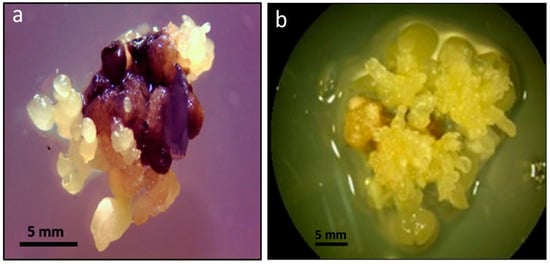
The induction of somatic embryogenesis in cork oak (Figure 1) was successful, resulting in numerous globular structures on the zygotic embryos, along with proembryos and secondary immature dicotyledonary embryos. Fused embryos were also observed, with an overall induction percentage of 71.42%.

Figure 1.
(a) Direct somatic embryogenesis on an immature zygotic embryo after 30 days in SM medium. (b) Embryos and embryonic callus on SM medium supplemented with 500 mg/L Glutamine, showing recurrent embryogenesis and numerous dicotyledonary embryos.
The cultivation system demonstrated efficient embryo formation and multiplication, with secondary embryogenesis resulting in a 25-fold increase in the number of secondary embryos within one month, highlighting the system’s potential for scalable production. The morphological development remained similar to the control in high temperature, UV radiation, and cerium oxide nanoparticle stress treatments. However, embryos under osmotic/saline stress (200 mM NaCl) exhibited darkening and a slower rate of recurrent embryogenesis, which prevented the analysis of their ethanolic extracts due to severe osmotic stress and interference with colorimetric reactions.
2.2. Antioxidant Capacity, Total Phenols, and Flavonoids
The total antioxidant capacity, determined by DPPH (2,2-diphenyl-1-picrylhydrazyl) reduction, was significantly increased in embryos subjected to high temperature (35 °C) and moderate saline stress (50 mM NaCl) compared to the control. Other treatments did not show significant differences (Table 1).

Table 1.
Total antioxidant capacity, total phenol content, and total flavonoid content in cork oak somatic embryos under abiotic stress treatments. DW: dry weight. Data are mean ± SD, n = 3. Significant differences (Duncan’s test, * p < 0.05); and ** p < 0.01), NQ: not quantified.
No significant differences in total phenol content were observed between treatments and the control. Total flavonoid content increased in embryos under high temperature and UV radiation, with UV having a higher increment. Other treatments showed no significant differences compared to the control, with a slight reduction in moderate saline stress (50 mM NaCl).
2.3. Presence and Quantification of Ellagic Acid, Chlorogenic Acid, and Serotonin
Among all the abiotic stress treatments, including high temperature (35 °C), UV radiation, moderate osmotic/saline stress (50 mM NaCl), severe osmotic/saline stress (200 mM NaCl), and exposure to nanoceria at concentrations of 100 mg/L and 200 mg/L, positive results for chlorogenic acid presence were observed. Furthermore, the presence of serotonin, another important compound, was detected in all the abiotic stress treatments, as well as in the control group (Table 2).

Table 2.
Determination of the presence of chlorogenic acid and serotonin in Quercus suber embryos subjected to different abiotic stress treatments. The positive chlorogenic acid determination corresponds to the appearance of the Prussian blue-colored complex by the reduction of ferricyanide (Wang et al., 2019) [28].
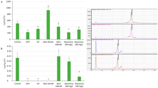
Ellagic acid content (Figure 2a) significantly increased under moderate salt stress (50 mM NaCl) compared to control, while other treatments showed reductions, especially under thermal stress and 100 mg/L cerium nanoparticles.

Figure 2.
Plant polyphenols: Ellagic acid (a) and Chlorogenic acid (b) content (mg/kg dry weight, DW) measured by LC-QQQ-MS in Quercus suber somatic embryos subjected to different abiotic stress treatments: high temperature (35 °C), UV radiation, moderate osmotic/saline stress (50 mM NaCl), severe osmotic/saline stress (200 mM NaCl), and cerium oxide nanoparticles (nanoceria) at concentrations of 100 mg/L and 200 mg/L in the culture medium. Treatments sharing the same letter were not statistically significant at the 0.05 level, as determined by Duncan’s test. The inset on the right shows the chromatograms of Ellagic acid detection in control sample under two different collision energies (CE: −41.0, black, and −33.0, pink), and Chlorogenic acid (CE: −17.0, blue, and −47.0, red).
Chlorogenic acid was detected in all samples through the reduction of ferricyanide, resulting in a positive Prussian blue colored complex reaction. However, the quantification of chlorogenic acid using LC-QQQ-MS (Liquid Chromatography-Triple Quadrupole Mass Spectrometry; Figure 2b) revealed a significant reduction in its content in embryos subjected to high temperature stress, UV radiation, and moderate osmotic/saline stress treatments. Embryos subjected to UV radiation did not show detectable levels of chlorogenic acid. Furthermore, the treatment with 200 mg/L of nanoceria resulted in a significant decrease in chlorogenic acid content (0.04 ± 0.01 mg/Kg DW) compared to the control embryos (0.23 ± 0.02 mg/Kg DW). No significant differences were observed in embryos subjected to severe osmotic/saline stress (0.25 ± 0.04 mg/Kg DW) or 100 mg/L nanoceria (0.20 ± 0.02 mg/Kg DW).
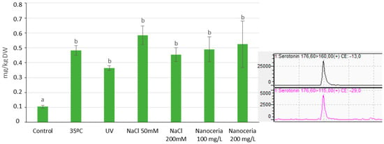
Serotonin levels (Figure 3) increased in cork oak somatic embryos subjected to all stress treatments compared to the control group (0.11 ± 0.009 mg/Kg DW). LC-QQQ-MS analysis confirmed the increase in serotonin content. Although no statistically significant differences were observed among the treatments, embryos subjected to moderate saline stress (0.58 ± 0.06 mg/Kg DW) exhibited the highest level of serotonin. The lowest value of serotonin was observed in embryos subjected to UV radiation stress (0.36 ± 0.01 mg/Kg DW).

Figure 3.
Serotonin content (mg/kg dry weight, DW) measured by LC-QQ-MS in Quercus suber somatic embryos subjected to different abiotic stress treatments: high temperature (35 °C), UV radiation, moderate osmotic/saline stress (50 mM NaCl), severe osmotic/saline stress (200 mM NaCl), and cerium oxide nanoparticles (nanoceria) at concentrations of 100 mg/L and 200 mg/L in the culture medium. Treatments sharing the same letter were not statistically significant at the 0.05 level, as determined by Duncan’s test. The inset on the right shows the chromatograms of serotonin detection in control sample under two different collision energies (CE: −13.0 and −29.0), highlighting the transition from m/z 176.60 to 160.00 (black) and m/z 176.60 to 115.00 (pink).
Overall, different abiotic stress treatments variably affected the antioxidant capacity and phytochemical composition of cork oak somatic embryos. High temperature and moderate saline stress increased antioxidant capacity and flavonoid content. Moderate saline stress also increased ellagic acid. Conversely, thermal stress and cerium nanoparticles reduced ellagic and chlorogenic acid, while UV radiation increased flavonoid content but negatively impacted chlorogenic acid. Serotonin levels generally increased with stress treatments, peaking under moderate saline stress.
3. Discussion
The importance of secondary metabolites and plant biotechnologies is well recognized for their potential benefits in human health and economic applications. Acorns, traditionally used as animal forage, are rich in fiber, proteins, vitamins (especially A and E), minerals, and unsaturated fatty acids. They also contain secondary metabolites, such as phenolic compounds, which offer various health benefits due to their high antioxidant activity [29]. However, the high tannin content imparts an astringent taste, limiting their consumption.
The pursuit of novel strategies for producing secondary metabolites with health benefits is a crucial objective in the bioeconomy. Plants possess defense mechanisms against biotic and abiotic stress, which involve the production of valuable secondary metabolites. Abiotic stress elicitation presents a simple and potentially economically viable system for enhancing these compounds. Recurrent somatic embryogenesis in cork oak can yield significant quantities of metabolites and can be scaled up in bioreactors [30]. However, the use of cork oak somatic embryos as biofactories has not been explored until now.
Our results show that cork oak somatic embryogenesis produces plant material with substantial antioxidant capacity (68.09 ± 3.99% Percent Scavenging Activity), a significant amount of phenolic compounds (173.80 ± 9.77 mg GA/g DW), and moderate levels of flavonoids (2.42 ± 0.21 mg quercetin/g DW) [31,32].
The application of abiotic stress elicitors led to changes in the phytochemical composition of cork oak somatic embryos. High temperatures and moderate osmotic/saline stress increased antioxidant capacity (Table 1). The impact of high temperature on antioxidant activity varies among species and plant parts. For instance, strawberries show increased antioxidant activity with rising temperatures [33], while lettuce experiences reduced antioxidant activity [34]. In Brassica oleracea, temperature increases have minimal effects [35]. Similarly, osmotic/saline stress has been shown to increase antioxidant activity in plants like Suaeda maritima [36] and Trianthema triquetra [37].
The total phenol content did not show significant changes with the elicitors tested. Although stress conditions like UV radiation, high temperature, and toxicity are expected to increase phenolic compounds [38], the production of ethylene and phenolic compounds are closely interconnected. In vitro, ethylene inhibitors promote recurrent embryogenesis [39]). The low ethylene concentration in our cultures, except those under severe osmotic/saline stress, might explain the steady levels of phenolic compounds. Increased phenolic compounds can trigger ethylene synthesis, which negatively affects recurrent embryogenesis [38].
Flavonoid content increased in cultures subjected to high temperature and UV radiation (Table 1). While low temperatures typically increase flavonoid content [40], high temperatures also play a role. UV radiation, a known elicitor of flavonoids, acts as a light filter protecting plant tissues [41,42]. UV light exposure increases flavonoid production in vivo [43] and in vitro callus cultures [44], indicating a stress tolerance mechanism [45].
Previous studies identified ellagic acid in Q. suber acorns [46] and cork tissue [14]. Our quantification using LC-QQ-MS revealed levels exceeding 500 mg/Kg DW, comparable to high levels found in red fruits and nuts [47]. Moderate osmotic/saline stress significantly increased ellagic acid production, exceeding 900 mg/Kg DW, an 80% increase compared to control conditions. This aligns with findings in strawberries [48]. The reduction in ellagic acid under other stress treatments might be due to oxidation, as it reacts with free radicals [49]. Ellagic acid’s high cost makes its production from cork oak somatic embryo cultures commercially appealing [50].
The presence of chlorogenic acid in Q. suber was previously unreported. The detected quantity (0.23 ± 0.02 mg/Kg DW) was significantly lower than in coffee, which has the highest chlorogenic acid levels [51]. Elicitors reduced chlorogenic acid levels, necessitating further investigations with different stress types to enhance its content. This study is the first to report chlorogenic acid in cork oak somatic embryos.
Serotonin presence in cork oak somatic embryos under control conditions (0.1 ± 0.01 mg/kg DW) is comparable to levels found in the edible tissues of other Fagaceae species [52]. Notably, all tested elicitors significantly increased serotonin levels, with a six-fold increase observed under moderate osmotic/saline stress. Serotonin, known for its role in regulating abiotic stress tolerance and protecting against reactive oxygen species, contributes to the maintenance of recurrent embryogenesis in cork oak somatic embryo cultures [5].
One of the major successes in our elicitation process was the substantial increase in serotonin levels. Serotonin is naturally present in numerous plant species, including those in the Fagaceae family, to which Q. suber belongs [27]. Under control conditions, the amount of serotonin detected in cork oak somatic embryos (0.1 ± 0.01 mg/kg DW) was comparable to that in the edible tissues of other Fagaceae species [52]. Interestingly, all elicitors tested significantly increased serotonin levels, with a particularly notable six-fold increase under moderate osmotic/saline stress. While vegetables generally contain moderate levels of serotonin [53], seeds, which lack vacuoles for the storage or excretion of hydrophilic secondary metabolites, may store serotonin in protein bodies developed in the cotyledons during ripening [54].
Serotonin plays a crucial role in regulating tolerance to abiotic stress in plants [55]. Additionally, serotonin is believed to have a protective effect against reactive oxygen species, leading to a delay in the process of senescence [5]. This attribute contributes to the maintenance of the secondary or recurrent embryogenesis process in cork oak somatic embryo cultures subjected to abiotic elicitors. Therefore, the tissue culture of cork oak somatic embryos serves as a valuable source of serotonin, a compound of high interest in human health with significant economic potential. In conclusion, our study demonstrates that cork oak somatic embryogenesis yields plant material with significant antioxidant capacity, high levels of phenolic compounds, and moderate levels of flavonoids. The application of abiotic stress elicitation techniques, such as high temperature and moderate osmotic/saline stress, enhances the phytochemical composition of cork oak somatic embryos, including their antioxidant activity. Notably, moderate osmotic/saline stress triggers a substantial increase in ellagic acid production, suggesting the potential for commercial-scale production from biofactories. Furthermore, our findings reveal the presence of serotonin and its significant accumulation in cork oak somatic embryos, highlighting its relevance in human health and the potential therapeutic implications, particularly in the context of the COVID-19 pandemic.
Our study demonstrates that cork oak somatic embryogenesis produces plant material with significant antioxidant capacity, high levels of phenolic compounds, and moderate flavonoid levels. Abiotic stress elicitation, especially moderate osmotic/saline stress, enhances the phytochemical composition, including ellagic acid and serotonin. This work opens new opportunities for sustainable phytochemical production with applications in pharmaceuticals, nutraceuticals, and functional foods. Future research should focus on optimizing elicitation processes, exploring alternative stressors, and investigating the health benefits of phytochemicals from cork oak somatic embryos. While our study primarily focuses on the experimental aspects of somatic embryogenesis, it is important to consider the commercial viability of producing metabolites such as ellagic acid. Although we have not conducted a detailed economic analysis, we recognize that the current production levels may not yet justify commercial costs. However, the experimental nature of our work suggests that there is potential for future optimizations that could improve both yield and cost-efficiency, which are critical for commercial application. This study highlights the potential of biotechnology and bioeconomy in addressing global challenges related to human health and sustainable resource utilization.
4. Materials and Methods
4.1. Cork Oak Somatic Embryo Culture
The induction of somatic embryogenesis in Quercus suber and the subsequent culture of somatic embryos were carried out following the protocol described by Bueno and Manzanera [56]. Immature acorns were collected in El Pardo forest (Madrid, Spain) between September and October. The zygotic embryos, ranging from 5 to 10 mm in diameter, were cultured for one month in Petri dishes containing an initiation medium. The culture chamber provided a photoperiod of 16 h of light and 8 h of darkness, with a temperature maintained at 20 ± 2 °C.
To induce secondary or recurrent embryogenesis (Gomez-Garay et al., 2014a) [57], the embryos were transferred to basal SM medium supplemented with 500 mg/L glutamine (SM + Gln medium). This medium consisted of Sommer et al.’s (1975) [58] macronutrients and Murashige and Skoog’s (1962) [59] micronutrients. The initial three months of culture in SM + Gln medium were conducted under photoperiod conditions of 16 h of light and 8 h of darkness, at a temperature of 20 ± 1 °C. Subsequently, the embryos were maintained in darkness throughout the experiment and subcultured on fresh SM + Gln medium every 30 days during the proliferation phase.
4.2. Abiotic Elicitation Strategies
The somatic embryos in the proliferation phase were subjected to different abiotic stress treatments as elicitors. The stress treatments included high temperature, UV radiation, saline/osmotic stress, and cerium oxide nanoparticles (nanoceria):
- 1.
- High-Temperature Stress Treatment: The somatic embryos were cultured on SM + Gln medium for 7 days at 35 ± 1 °C in darkness. This temperature was chosen based on previous studies indicating that it is a critical threshold for inducing stress responses in Quercus suber somatic embryos without causing irreversible tissue damage.
- 2.
- UV Stress Treatment: Somatic embryos were cultured on SM + Gln medium for 7 days and subjected to UV-C light for 15 min each day. The UV exposure was administered at specific intervals: 0 h, 24 h, 96 h, 120 h, 144 h, and 168 h from the start of the culture. This exposure schedule was designed to deliver a controlled dosage sufficient to induce stress responses while minimizing the risk of excessive necrosis or cellular damage. The UV-C exposure was conducted in a laminar flow hood to maintain a controlled environment and prevent contamination. For the remainder of the time, the embryos were kept in darkness.
- 3.
- Saline/Osmotic Stress Treatment: Two saline/osmotic stress treatments were applied. The somatic embryos were cultured on SM + Gln medium supplemented with NaCl at two different concentrations: 50 mM or 200 mM. The cultures were maintained for seven days in darkness.
- 4.
- Cerium Oxide Nanoparticle Stress (Nanoceria) Treatment: Two cerium oxide nanoparticle stress treatments were applied. The somatic embryos were cultured on SM + Gln medium supplemented with cerium oxide nanoparticles at two different concentrations: 100 mg/L or 200 mg/L. Commercially available nano-CeO2 particles (<25 nm particle size) purchased from Sigma-Aldrich Chemical Co. (Saint Louis, MO 63103, USA) were used as received. The dispersion of nano-CeO2 particles was achieved following the method described by Gomez-Garay et al. (2014) [9].
After seven days of treatment, the somatic cork oak embryos from the different treatments were collected in their entirety. The embryos were frozen at −20 °C for 24 h and then freeze-dried. The freeze-dried embryos were ground into a fine powder, and the obtained material was stored in the dark for further analysis. All experiments were conducted in triplicate.
4.3. Analysis of Secondary Metabolites
4.3.1. Ethanolic Extracts
The ethanolic extracts of the somatic Q. suber embryos from the different treatments and the control were obtained as follows: 200 mg of each of the lyophilized samples obtained in the previous section were homogenized in 10 mL of 80 % ethanol at 60 °C with agitation for 30 min. Afterward, the extracts were centrifuged at 5000 rpm for 10 min, and the supernatant was collected and refrigerated. Three extractions were performed for each treatment.
For the analysis of flavonoids, the extracts were obtained using a similar procedure, but 200 mg of ground lyophilized embryos were homogenized in 9 mL of 80% ethanol, to which 1 mL of 35% HCl was added. Three extractions were carried out for each treatment.
The antioxidant capacity of each extract was evaluated using the DPPH method described by Brand-Williams et al. (1995) [60].
The total phenol content was determined using the method proposed by Singleton and Rossi (1965) [61].The results were expressed as gallic acid equivalents (mg gallic acid/g dry weight of somatic embryos) by interpolating the absorbance values of the test on a gallic acid standard line. The analysis was conducted in triplicate for each extract of the different treatments.
The total flavonoid content was determined using colorimetry based on the method described by Lock et al. (2006) [62]. The results were expressed as quercetin equivalents (mg quercetin/g dry weight of freeze-dried somatic embryos) by interpolating the absorbance values of the test on a quercetin standard line. The analysis was conducted in triplicate for each extract of the different treatments.
4.3.2. Methanolic Extracts
The methanolic extracts of the cork oak somatic embryos from the different treatments and the control were obtained by homogenizing 5 g of each of the lyophilized samples in 100 mL of methanol. The homogenates were kept in the dark for 24 h before being filtered with a 0.45 µm filter. Three extractions were carried out for each treatment.
The presence of chlorogenic acid (CGA) in the methanolic extracts of each sample was determined using the protocol described by Wang et al. (2019) [28]. This protocol involves the reduction of ferricyanide to its reduced form by CGA, resulting in the formation of a colored complex (Prussian blue).
The presence of serotonin in the methanolic extracts of each sample was determined according to the protocol described by Jin et al. (2008) [63]. The determination is based on a color reaction between serotonin derivatives and p-dimethylaminobenzaldehyde (Ehrlich’s reagent), which follows the electrophilic substitution reaction mechanism at the indole ring.
The quantification of ellagic acid, chlorogenic acid, and serotonin in the methanolic extracts was performed using a Shimadzu model LCMS8030 UHPLC coupled triple quadrupole mass spectrometer. The conditions for multiple reaction monitoring during liquid chromatography coupled to a triple quadrupole mass spectrometer, MRM (LC-QQQ-MS) (Shimadzu, Kyoto, Japan), analysis were as follows: Column Phenomenex Gemini 5u C18 110 A 150 × 2mm; injection volume: 40 μL. Gradient mode: From 5% phase B to 15% phase B in 3 min to 95% phase B in 7 min; return to initial conditions from 7 to 10 min. Phase A: H2O + 0.1% formic acid; Phase B: MeOH + 0.1% formic acid. The flow rate was set at 0.4 mL/min, and the total run time was 10 min.
The quantification of ellagic acid, chlorogenic acid, and serotonin was performed using multiple reaction monitoring (MRM) transitions:
- 1.
- Chlorogenic acid:
- (1)
- Quantifier (m/z): 353.30 > 191.1 (EC: 17)
- (2)
- Qualifier (m/z): 353.30 > 93.1 (EC: 47)
- 2.
- Ellagic acid:
- (1)
- Quantifier (m/z): 300.80 > 144.95 (EC: 41)
- (2)
- Qualifier (m/z): 300.80 > 284.00 (EC: 33)
- 3.
- Serotonin:
- (1)
- Quantifier (m/z): 176.60 > 160.00 (EC: −13)
- (2)
- Qualifier (m/z): 176.60 > 115.00 (EC: −29)
The detection limits for the analysis were as follows: Ellagic acid (0.032 mg/L), chlorogenic acid (0.001 mg/L), and serotonin (0.004 mg/L). The quantification limits were: Ellagic acid (0.105 mg/L), chlorogenic acid (0.002 mg/L), and serotonin (0.012 mg/L).
This analytical method allowed for the identification and quantification of ellagic acid, chlorogenic acid, and serotonin in the methanolic extracts of the somatic Quercus suber embryos from different elicitation treatments and the control.
4.4. Statistical Analysis
All experiments were performed in triplicate to ensure reliable results. Statistical analysis was conducted to evaluate the effect of the different treatments on the total antioxidant capacity, total phenols, total flavonoids, ellagic acid, chlorogenic acid, and serotonin.
The data obtained from the experiments were analyzed using analysis of variance (ANOVA) to determine the statistical significance of the treatment effects. To compare the means and identify significant differences among treatments, the Duncan multiple range test was performed. Treatments with significantly different means were identified at a p-value of less than 0.05.
Author Contributions
Conceptualization, B.P.L. and A.G.-G.; methodology, B.P.L. and E.P.-U.; validation, A.G.-G. and J.A.M.; formal analysis, A.G.-G.; investigation, C.J. and A.M.; resources, A.G.-G.; data curation, J.A.M. and E.P.-U.; writing—original draft preparation, A.M. and C.J.; writing—review and editing, A.G.-G. and J.A.M.; visualization, B.P.L.; project administration, A.G.-G. All authors have read and agreed to the published version of the manuscript.
Funding
This research received no external funding.
Data Availability Statement
Data is contained within the article.
Acknowledgments
We thank Raquel Alonso Valenzuela for her technical assistance and to Estefanía García Calvo and Cristina Gutiérrez López (Unidad de Espectrometría de Masas-CAI Técnicas Físicas y Químicas UCM) for the assistance with MRM (LC-QQQ-MS) analysis.
Conflicts of Interest
The authors declare no conflicts of interest.
References
- Ávalos, A.; Pérez-Urria, C.E. Plant secondary metabolism. Reduca (Biol.) Ser. Veg. Physiol. 2009, 2, 119–145. [Google Scholar]
- Wink, M.; Botschen, F.; Gosmann, C.; Schäfer, H.; Waterman, P.G. Chemotaxonomy seen from a phylogenetic perspective and evolution of secondary metabolism. In Annual Plant Reviews Volume 40: Biochemistry of Plant Secondary Metabolism; John Wiley & Sons Ltd.: Hoboken, NJ, USA, 2010; pp. 364–433. [Google Scholar]
- Moore, B.D.; Andrew, R.L.; Külheim, C.; Foley, W.J. Explaining intraspecific diversity in plant secondary metabolites in an ecological context. New Phytol. 2014, 201, 733–750. [Google Scholar] [CrossRef] [PubMed]
- Ashraf, M.A.; Iqbal, M.; Rasheed, R.; Hussain, I.; Riaz, M.; Arif, M.S. Environmental stress and secondary metabolites in plants: An overview. In Plant Metabolites and Regulation under Environmental Stress; Academic Press: Cambridge, MA, USA, 2018; pp. 153–167. [Google Scholar]
- Akula, R.; Ravishankar, G.A. Influence of abiotic stress signals on secondary metabolites in plants. Plant Signal. Behav. 2011, 6, 1720–1731. [Google Scholar] [CrossRef] [PubMed]
- Qaderi, M.M.; Martel, A.B.; Strugnell, C.A. Environmental factors regulate plant secondary metabolites. Plants 2023, 12, 447. [Google Scholar] [CrossRef]
- Humbal, A.; Pathak, B. Influence of exogenous elicitors on the production of secondary metabolite in plants: A review (“VSI: Secondary metabolites”). Plant Stress 2023, 8, 100166. [Google Scholar] [CrossRef]
- Acosta-Motos, J.R.; Penella, C.; Hernández, J.A.; Díaz-Vivancos, P.; Sánchez-Blanco, M.J.; Navarro, J.M.; Barba-Espín, G. Towards a sustainable agriculture: Strategies involving phytoprotectants against salt stress. Agronomy 2020, 10, 194. [Google Scholar] [CrossRef]
- Gomez-Garay, A.; Pintos, B.; Manzanera, J.A.; Lobo, C.; Villalobos, N.; Martín, L. Uptake of CeO2 nanoparticles and its effect on growth of Medicago arborea in vitro plantlets. Biol. Trac. Elem. Res. 2014, 161, 143–150. [Google Scholar] [CrossRef]
- Xie, X.; He, Z.; Chen, N.; Tang, Z.; Wang, Q.; Cai, Y. The roles of environmental factors in regulation of oxidative stress in plant. BioMed Res. Int. 2019, 2019, 9732325. [Google Scholar] [CrossRef]
- Gupta, P.K.; Durzan, D.J. In vitro establishment and multiplication of juvenile and mature Douglas-fir and sugar pine. Acta Hortic. 1987, 212, 483–488. [Google Scholar] [CrossRef]
- Bueno, M.A.; Astorga, R.; Manzanera, J.A. Plant regeneration through somatic embryogenesis in Quercus suber L. Phys. Plant. 1992, 85, 30–34. [Google Scholar] [CrossRef]
- Makhlouf, F.Z.; Barkat, M.; Caponio, F. Comparative study of total phenolic content and antioxidant properties of Quercus fruit: Flour and oil. North Afr. J. Food Nutr. Res. 2019, 3, 148–155. [Google Scholar] [CrossRef]
- Fernandes, A.; Fernandes, I.; Cruz, L.; Mateus, N.; Cabral, M.; de Freitas, V. Antioxidant and biological properties of bioactive phenolic compounds from Quercus suber L. J. Agri. Food Chem. 2009, 57, 11154–11160. [Google Scholar] [CrossRef] [PubMed]
- Bejarano, I.; Godoy-Cancho, B.; Franco, L.; Martínez-Cañas, M.A.; Tormo, M.A. Quercus suber L. Cork extracts induce apoptosis in human myeloid leukaemia HL-60 cells. Phytother. Res. 2015, 29, 1180–1187. [Google Scholar] [CrossRef] [PubMed]
- Custódio, L.; Patarra, J.; Alberício, F.; da Rosa Neng, N.; Nogueira JM, F.; Romano, A. Phenolic composition, antioxidant potential and in vitro inhibitory activity of leaves and acorns of Quercus suber on key enzymes relevant for hyperglycemia and Alzheimer’s disease. Ind. Crops Prod. 2015, 64, 45–51. [Google Scholar] [CrossRef]
- Santos, S.A.; Pinto, P.C.; Silvestre, A.J.; Neto, C.P. Chemical composition and antioxidant activity of phenolic extracts of cork from Quercus suber L. Ind. Crops Prod. 2010, 31, 521–526. [Google Scholar] [CrossRef]
- Vattem, D.A.; Shetty, K. Biological functionality of ellagic acid: A review. J. Food Biochem. 2005, 29, 234–266. [Google Scholar] [CrossRef]
- Meng, S.; Cao, J.; Feng, Q.; Peng, J.; Hu, Y. Roles of chlorogenic acid on regulating glucose and lipids metabolism: A review. Evid.-Based Complement. Altern. Med. 2013, 1, 801457. [Google Scholar] [CrossRef]
- Kabir, F.; Katayama, S.; Tanji, N.; Nakamura, S. Antimicrobial effects of chlorogenic acid and related compounds. J. Korean Soc. Appl. Biol. Chem. 2014, 57, 359–365. [Google Scholar] [CrossRef]
- Liang, N.; Kitts, D.D. Role of chlorogenic acids in controlling oxidative and inflammatory stress conditions. Nutrients 2016, 8, 16. [Google Scholar] [CrossRef]
- Barahuie, F.; Saifullah, B.; Dorniani, D.; Fakurazi, S.; Karthivashan, G.; Hussein, M.Z.; Elfghi, F.M. Graphene oxide as a nanocarrier for controlled release and targeted delivery of an anticancer active agent, chlorogenic acid. Mater. Sci. Eng. C 2017, 74, 177–185. [Google Scholar] [CrossRef]
- Murai, T.; Matsuda, S. The chemopreventive effects of chlorogenic acids, phenolic compounds in coffee, against inflammation, cancer, and neurological diseases. Molecules 2023, 28, 2381. [Google Scholar] [CrossRef] [PubMed]
- Mohapatra, P.K.; Chopdar, K.S.; Dash, G.C.; Mohanty, A.K.; Raval, M.K. In silico screening and covalent binding of phytochemicals of Ocimum sanctum against SARS-CoV-2 (COVID 19) main protease. J. Biomol. Struct. Dyn. 2023, 41, 435–444. [Google Scholar] [CrossRef] [PubMed]
- Anderson, G.; Reiter, R.J. Melatonin: Roles in influenza, COVID-19, and other viral infections. Rev. Med. Virol. 2020, 30, e2109. [Google Scholar] [CrossRef] [PubMed]
- Deng, J.; Rayner, D.; Ramaraju, H.B.; Abbas, U.; Garcia, C.; Heybati, K.; Moskalyk, M. Efficacy and safety of selective serotonin reuptake inhibitors in COVID-19 management: A systematic review and meta-analysis. Clin. Microbiol. Infect. 2023, 29, 578–586. [Google Scholar] [CrossRef]
- Erland, L.A.; Turi, C.E.; Saxena, P.K. Serotonin: An ancient molecule and an important regulator of plant processes. Biotechnol. Adv. 2016, 34, 1347–1361. [Google Scholar] [CrossRef]
- Wang, X.; Zeng, Z.; Tian, Z.; Sun, J.; Li, Y.; Fan, X. Validation of spectrophotometric determination of chlorogenic acid in fermentation broth and fruits. Food Chem. 2019, 278, 170–177. [Google Scholar] [CrossRef]
- Vinha, A.F.; Barreira, J.C.; Costa, A.S.; Oliveira MB, P. A new age for Quercus spp. fruits: Review on nutritional and phytochemical composition and related biological activities of acorns. Compr. Rev. Food Sci. Food Saf. 2016, 15, 947–981. [Google Scholar] [CrossRef]
- Valdiani, A.; Hansen, O.K.; Nielsen, U.B.; Johannsen, V.K.; Shariat, M.; Georgiev, M.I.; Omidvar, V.; Ebrahimi, M.; Tavakoli Dinanai, E.; Abiri, R. Bioreactor-based advances in plant tissue and cell culture: Challenges and prospects. Crit. Rev. Biotechnol. 2019, 39, 20–34. [Google Scholar] [CrossRef]
- Pérez-Jiménez, J.; Neveu, V.; Vos, F.; Scalbert, A. Identification of the 100 richest dietary sources of polyphenols: An application of the Phenol-Explorer database. Eur. J. Clin. Nutr. 2010, 64, S112–S120. [Google Scholar] [CrossRef]
- Haytowitz, D.B.; Wu, X.; Bhagwat, S. USDA Database for the Flavonoid Content of Selected Foods; Release 3.3; US Department of Agriculture: Beltsville, MD, USA, 2018; 173p. [Google Scholar]
- Wang, S.Y.; Zheng, W. Effect of plant growth temperature on antioxidant capacity in strawberry. J. Agric. Food Chem. 2001, 49, 4977–4982. [Google Scholar] [CrossRef]
- Boo, H.O.; Heo, B.G.; Gorinstein, S.; Chon, S.U. Positive effects of temperature and growth conditions on enzymatic and antioxidant status in lettuce plants. Plant Sci. 2011, 181, 479–484. [Google Scholar] [CrossRef] [PubMed]
- Soengas, P.; Rodríguez, V.M.; Velasco, P.; Cartea, M.E. Effect of temperature stress on antioxidant defenses in Brassica oleracea. ACS Omega 2018, 3, 5237–5243. [Google Scholar] [CrossRef]
- Alhdad, G.M.; Seal, C.E.; Al-Azzawi, M.J.; Flowers, T.J. The effect of combined salinity and waterlogging on the halophyte Suaeda maritima: The role of antioxidants. Environ. Exp. Bot. 2013, 87, 120–125. [Google Scholar] [CrossRef]
- Sharma, V.; Ramawat, K.G. Salt stress enhanced antioxidant response in callus of three halophytes (Salsola baryosma, Trianthema triquetra, Zygophyllum simplex) of Thar Desert. Biologia 2014, 69, 178–185. [Google Scholar] [CrossRef]
- Chalker-Scott, L.; Fuchigami, L.H. The role of phenolic compounds in plant stress responses. In Low Temperature Stress Physiology in Crops; CRC Press: Boca Raton, FL, USA, 2018; pp. 67–80. [Google Scholar]
- Kumar, V.; Ramakrishna, A.; Ravishankar, G.A. Influence of different ethylene inhibitors on somatic embryogenesis and secondary embryogenesis from Coffea canephora P ex Fr. In Vitro Cell. Dev. Biol.-Plant 2007, 43, 602–607. [Google Scholar] [CrossRef]
- Coberly, L.C.; Rausher, M.D. Analysis of a chalcone synthase mutant in Ipomoea purpurea reveals a novel function for flavonoids: Amelioration of heat stress. Mol. Ecol. 2003, 12, 1113–1124. [Google Scholar] [CrossRef]
- Rausher, M.D. The evolution of flavonoids and their genes. In The Science of Flavonoids; Springer: New York, NY, USA, 2006; pp. 175–211. [Google Scholar]
- Petrussa, E.; Braidot, E.; Zancani, M.; Peresson, C.; Bertolini, A.; Patui, S.; Vianello, A. Plant flavonoids—Biosynthesis, transport and involvement in stress responses. Int. J. Mol. Sci. 2013, 14, 14950–14973. [Google Scholar] [CrossRef]
- Çakırlar, H.; Çiçek, N.; Fedina, I.; Georgieva, K.; Doğru, A.; Velitchkova, M. NaCl induced cross-acclimation to UV-B radiation in four Barley (Hordeum vulgare L.) cultivars. Acta Physiol. Plant. 2008, 30, 561–567. [Google Scholar] [CrossRef]
- Antognoni, F.; Zheng, S.; Pagnucco, C.; Baraldi, R.; Poli, F.; Biondi, S. Induction of flavonoid production by UV-B radiation in Passiflora quadrangularis callus cultures. Fitoterapia 2007, 78, 345–352. [Google Scholar] [CrossRef]
- Ryan, K.G.; Swinny, E.E.; Markham, K.R.; Winefield, C. Flavonoid gene expression and UV photoprotection in transgenic and mutant Petunia leaves. Phytochemistry 2002, 59, 23–32. [Google Scholar] [CrossRef]
- Cantos, E.; Espín, J.C.; López-Bote, C.; de la Hoz, L.; Ordóñez, J.A.; Tomás-Barberán, F.A. Phenolic compounds and fatty acids from acorns (Quercus spp.), the main dietary constituent of free-ranged Iberian pigs. J. Agric. Food Chem. 2003, 51, 6248–6255. [Google Scholar] [CrossRef]
- Daniel, E.M.; Krupnick, A.S.; Heur, Y.H.; Blinzler, J.A.; Nims, R.W.; Stoner, G.D. Extraction, stability, and quantitation of ellagic acid in various fruits and nuts. J. Food Compos. Anal. 1989, 2, 338–349. [Google Scholar] [CrossRef]
- Sarker, U.; Oba, S. Augmentation of leaf color parameters, pigments, vitamins, phenolic acids, flavonoids and antioxidant activity in selected Amaranthus tricolor under salinity stress. Sci. Rep. 2018, 8, 12349. [Google Scholar] [CrossRef] [PubMed]
- Häkkinen, S.H.; Kärenlampi, S.O.; Mykkänen, H.M.; Heinonen, I.M.; Törrönen, A.R. Ellagic acid content in berries: Influence of domestic processing and storage. Eur. Food Res. Technol. 2000, 212, 75–80. [Google Scholar] [CrossRef]
- Gumienna, M.; Szwengiel, A.; Górna, B. Bioactive components of pomegranate fruit and their transformation by fermentation processes. Eur. Food Res. Technol. 2016, 242, 631–640. [Google Scholar] [CrossRef]
- Fujioka, K.; Shibamoto, T. Chlorogenic acid and caffeine contents in various commercial brewed coffees. Food Chem. 2008, 106, 217–221. [Google Scholar] [CrossRef]
- Feldman, J.M.; Lee, E.M. Serotonin content of foods: Effect on urinary excretion of 5-hydroxyindoleacetic acid 1 3. Am. J. Clin. Nutr. 1985, 42, 639–643. [Google Scholar] [CrossRef]
- Ravishankar, G.A.; Ramakrishna, A. (Eds.) Serotonin and Melatonin: Their Functional Role in Plants, Food, Phytomedicine, and Human Health; CRC Press: Boca Raton, FL, USA, 2016. [Google Scholar]
- Grobe, W. Function of serotonin in seeds of walnuts. Phytochemistry 1982, 21, 819–822. [Google Scholar] [CrossRef]
- Kaur, H.; Mukherjee, S.; Baluska, F.; Bhatla, S.C. Regulatory roles of serotonin and melatonin in abiotic stress tolerance in plants. Plant Signal. Behav. 2015, 10, e1049788. [Google Scholar] [CrossRef]
- Bueno, M.A.; Manzanera, J.A. Primeros ensayos de inducción de embriones somáticos de Quercus suber L. Sci. Gerund. 1992, 18, 29–37. [Google Scholar]
- Gomez-Garay, A.; Manzanera, J.A.; Pintos-Lopez, B. Embryogenesis in Oak species. A review. For. Syst. 2014, 23, 191–198. [Google Scholar] [CrossRef]
- Sommer, H.E.; Brown, C.L.; Kormanik, P.P. Differentiation of plantlets in longleaf pine (Pinus palustris Mill.) tissue cultured in vitro. Bot. Gaz. 1975, 136, 196–200. [Google Scholar] [CrossRef]
- Murashige, T.; Skoog, F. A revised medium for rapid growth and bio assays with tobacco tissue cultures. Physiol. Plant. 1962, 15, 473–497. [Google Scholar] [CrossRef]
- Brand-Williams, W.; Cuvelier, M.E.; Berset CL, W.T. Use of a free radical method to evaluate antioxidant activity. LWT-Food Sci. Technol. 1995, 28, 25–30. [Google Scholar] [CrossRef]
- Singleton, V.L.; Rossi, J.A. Colorimetry of total phenolics with phosphomolybdic-phosphotungstic acid reagents. Am. J. Enol. Vitic. 1965, 16, 144–158. [Google Scholar] [CrossRef]
- Lock, O.; Cabello, I.; Doroteo, V.H. Analysis of Flavonoids in Plants; Sección Química, Departamento de Ciencias, Pontificia Universidad Católica del Perú: Lima, Peru, 2006. [Google Scholar]
- Jin, Q.; Shan, L.; Yue, J.; Wang, X. Spectrophotometric determination of total serotonin derivatives in the safflower seeds with Ehrlich’s reagent and the underlying color reaction mechanism. Food Chem. 2008, 108, 779–783. [Google Scholar] [CrossRef]
Disclaimer/Publisher’s Note: The statements, opinions and data contained in all publications are solely those of the individual author(s) and contributor(s) and not of MDPI and/or the editor(s). MDPI and/or the editor(s) disclaim responsibility for any injury to people or property resulting from any ideas, methods, instructions or products referred to in the content. |
© 2024 by the authors. Licensee MDPI, Basel, Switzerland. This article is an open access article distributed under the terms and conditions of the Creative Commons Attribution (CC BY) license (https://creativecommons.org/licenses/by/4.0/).