1. Introduction
In many cases, human remains found at sea are subjected to medico-legal and anthropological investigations [
1,
2]. The sea is an often-unfamiliar forensic context due to the infinite variables that compromise identification investigations [
3]. Globally, drowning is the third leading cause of death from unintentional injury and accounts for 7% of all injury deaths worldwide [
4]. To this percentage, one must then add deaths, also in a marine context, caused by natural disasters, transport accidents, intentional deaths resulting from suicide and homicide [
5,
6,
7] and, although rare, deaths caused by the depredation of sharks or other marine animals [
8].
Taphonomy, in a marine context, is a topic discussed by many researchers, not only because the differences between marine and terrestrial taphonomy are very obvious, but also because the variables of the marine context cause a decomposition that, to date, has no clear phases useful to create a chronology [
9]. The degradation of bone tissue in a marine context is caused by variables of a predominantly environmental nature, primarily the time and mode of residence in the water, followed by the deposition of debris on the surface of the bone, the flow of the marine current, salinity, pH, temperature, depth, surface chemistry in contact with the specimen, flora and fauna [
10].
These variables during the decomposition phases cause different types of degradation that result, in most court cases, in limitations in identification and in the substantial time in solving the case. In this work, which fully embraces the marine taphonomic context, two forensic cases are described, one found on the coast of Reggio Calabria and the other only 9 km away, on the coast of Vibo Valentia.
Case A, partial, disarticulated and unidentified skeletal remains, and Case B, an isolated intact foot with soft tissues. Three missing persons options were considered among the identification hypotheses, as investigations were opened simultaneously for a homicide case, a missing fisherman case and a case of shipwreck due to increased migration flows.
2. Case Report A—Skeletal Remains
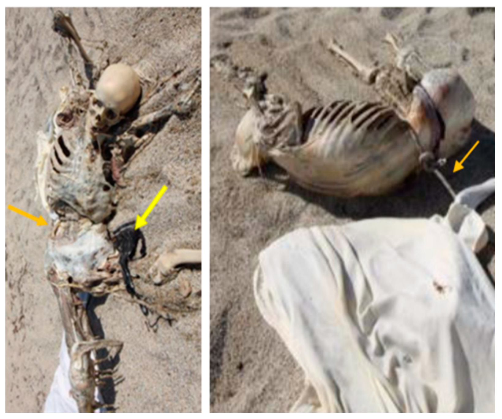
On 20 June 2020, a partially skeletonised and disarticulated body was found in the sea, a few metres from the coastline in Southern Italy (Reggio Calabria). The recovery of the body required the intervention of divers and the intervention of law enforcement agencies via a helicopter.
The body was found in an obvious state of decomposition, in skeletal condition with some remains of internal organs inside the chest, and was missing both feet. The body, as documented in the attached photos (
Figure 1, orange arrow), had a rope tied to its pelvis, which was used by rescuers to retrieve it from the water and transport it to the beach. Subsequently, the body was transferred to the Department of Forensic Medicine in Bari (Italy) to undergo anthropological and medico-legal investigations and to reconstruct the biological profile of the unidentified subject.
Before carrying out the external examination, the corpse was gently washed under low pressure, in order to remove the excess of sandy substrate covering the bone segments, as well as the residues of muscles and epidermis, that were in an advanced state of decomposition and in a pre-adipoceration phase.
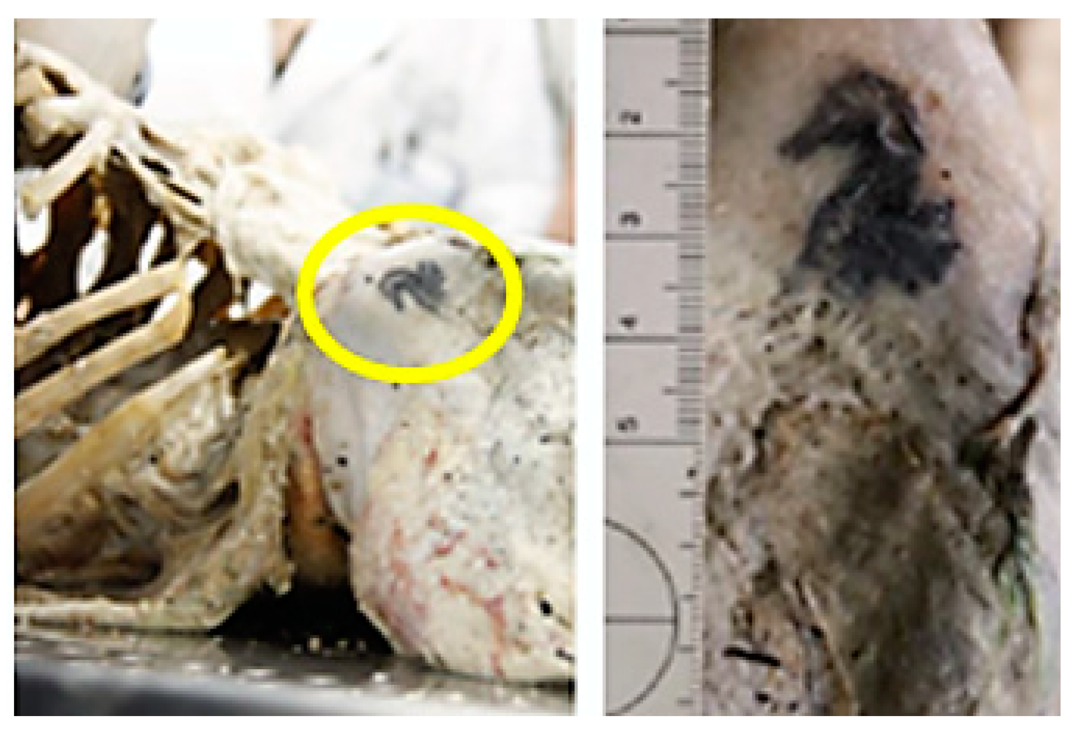
The external examination determined the subject to be male, given the residues of the genitals and the presence of residues of black briefs, Calvin Klein brand (
Figure 1, yellow arrow). In correspondence with the skin of the right iliac wing, a tattoo was identified, referring to a seahorse (
Figure 2).
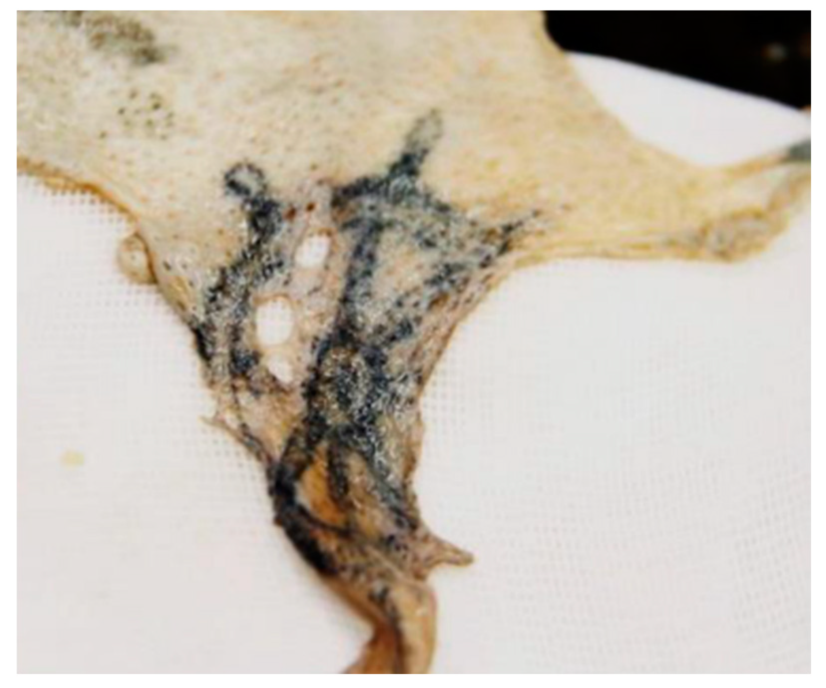
In correspondence with shreds of skin, probably attributable to the left deltoid region, a second tattoo identifiable as a boat rudder was identified (
Figure 3). In the region of the trunk, other intact shreds of skin were highlighted, extending from the occipital region to the shoulders and from the dorsal region continuing to the buttocks, where they ended, leaving the legs skeletonised.
After the first phase of cleaning and macroscopic observation, the corpse was subjected to radiological investigations and then an autopsy was performed, taking a series of samples for histological examination and samples of muscle tissue where possible, given the advanced state of decomposition, to be subsequently subjected to genetic investigation. Finally, an anthropological analysis was carried out to reconstruct the biological profile and proceed with further forensic investigations.
3. Case Report B—Isolated Foot
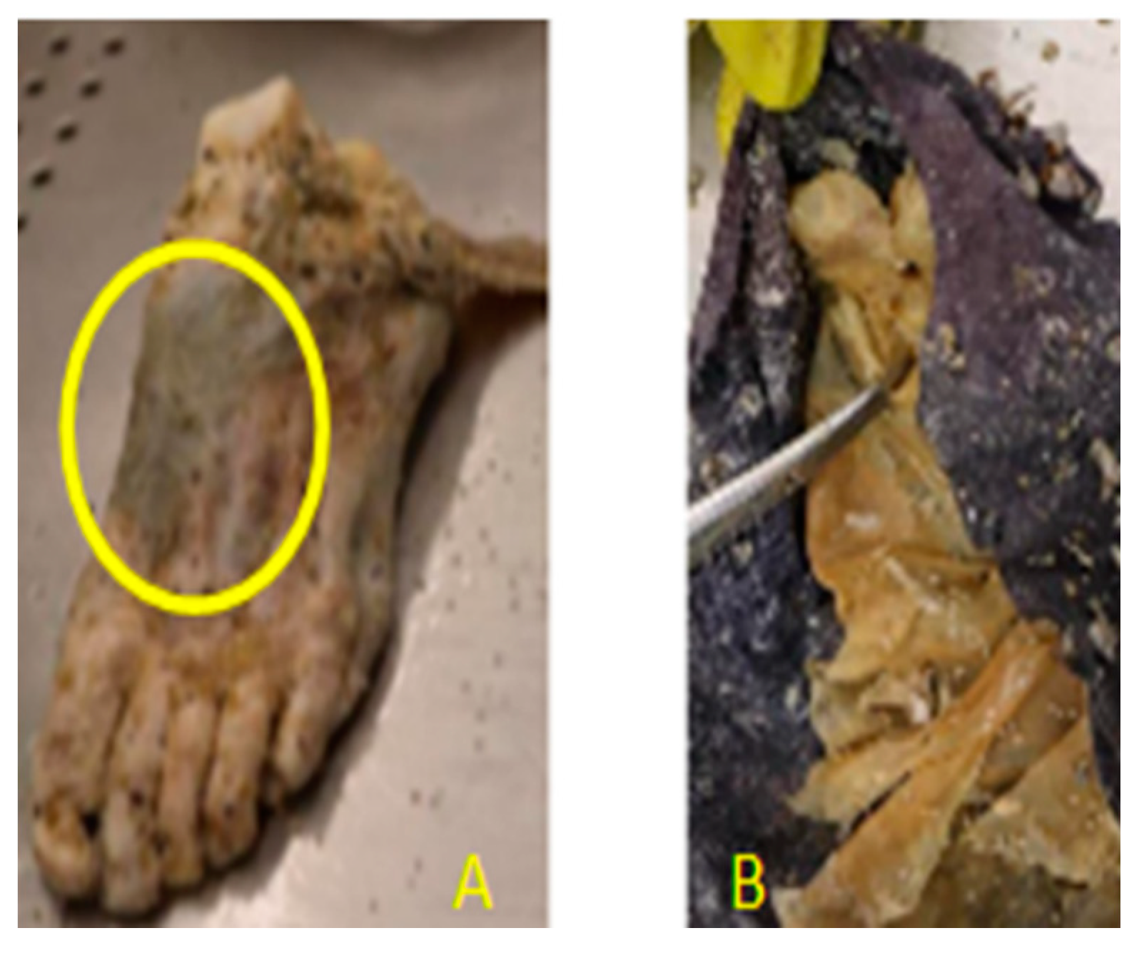
On 20 June 2020, an isolated foot was found stranded on a coastline in Southern Italy (Vibo Valentia).
The informed police forces detected the presence of an isolated foot with a strip of soft tissue still attached. A dark blue sock with residues of epidermis inside was also found. The area was marked off to document the presence of the find (
Figure 4).
Given the uncertainty of the origin and the discovery of the skeletal remains only 9 km away, on the same day, the find was transported to the Department of Forensic Medicine in Bari (Italy) for anthropometric and genetic investigations. In the department’s laboratories, before external examination, the foot was washed at low pressure to remove the excess sandy substrate covering the soft tissues and the pigmentation of the epidermis.
The medico-legal investigation determined a left foot, disarticulated at the level of the tibio-tarsus, complete, with persistence of all the soft tissues, in the first phase of adipocerization; a violaceous ecchymosis was identified on the back of the foot with the dimensions of 7 × 5 cm and a sample was taken for histological analysis. The maximum length of the foot was 27 cm; the maximum width was 8.7 cm; a dark-coloured sock diffusely colonised by baleen was evident with the presence of epidermal residues inside (
Figure 5).
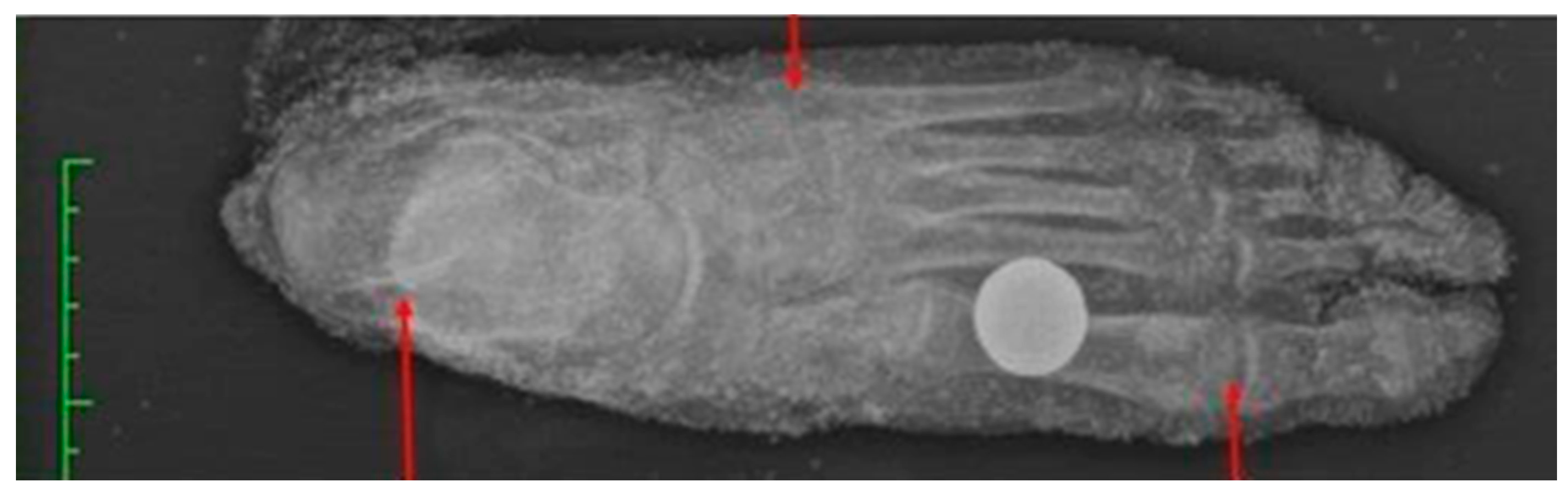
After the macroscopic observation, the foot was subjected to radiological investigations which allowed, in the following phase, an anthropometric analysis to determine the biological profile of the subject to whom the foot belonged. Finally, samples of soft tissue were taken for genetic investigation in order to compare them with the results of the genetic analysis carried out on the samples of muscular soft tissue taken from the skeletal remains found 9 km away on the same day.
4. Materials and Methods
4.1. Case A
For the reconstruction of the biological profile of the skeletal remains, Case A, traditional anthropological methods were applied; the skeleton was 90% intact with total absence of both feet [
11]. Morphological parameters were used for sex determination through the analysis of the skull, complete with mandible and hip [
12].
The age at death was determined by applying the method of the degree of epiphyseal fusion of the entire skeletal system, the degree of fusion of the cranial sutures and the method of macroscopic observation of the IV rib [
13,
14,
15]. The method of the degree of dental wear through a dental analysis and the study of the morphology of the pubic symphysis were also used [
16,
17]. For the determination of stature, the methods by Trotter and Gleser were used [
18]. To determine the time of death, the method of the degree of adipocerization and disarticulation of the corpse was applied [
19,
20].
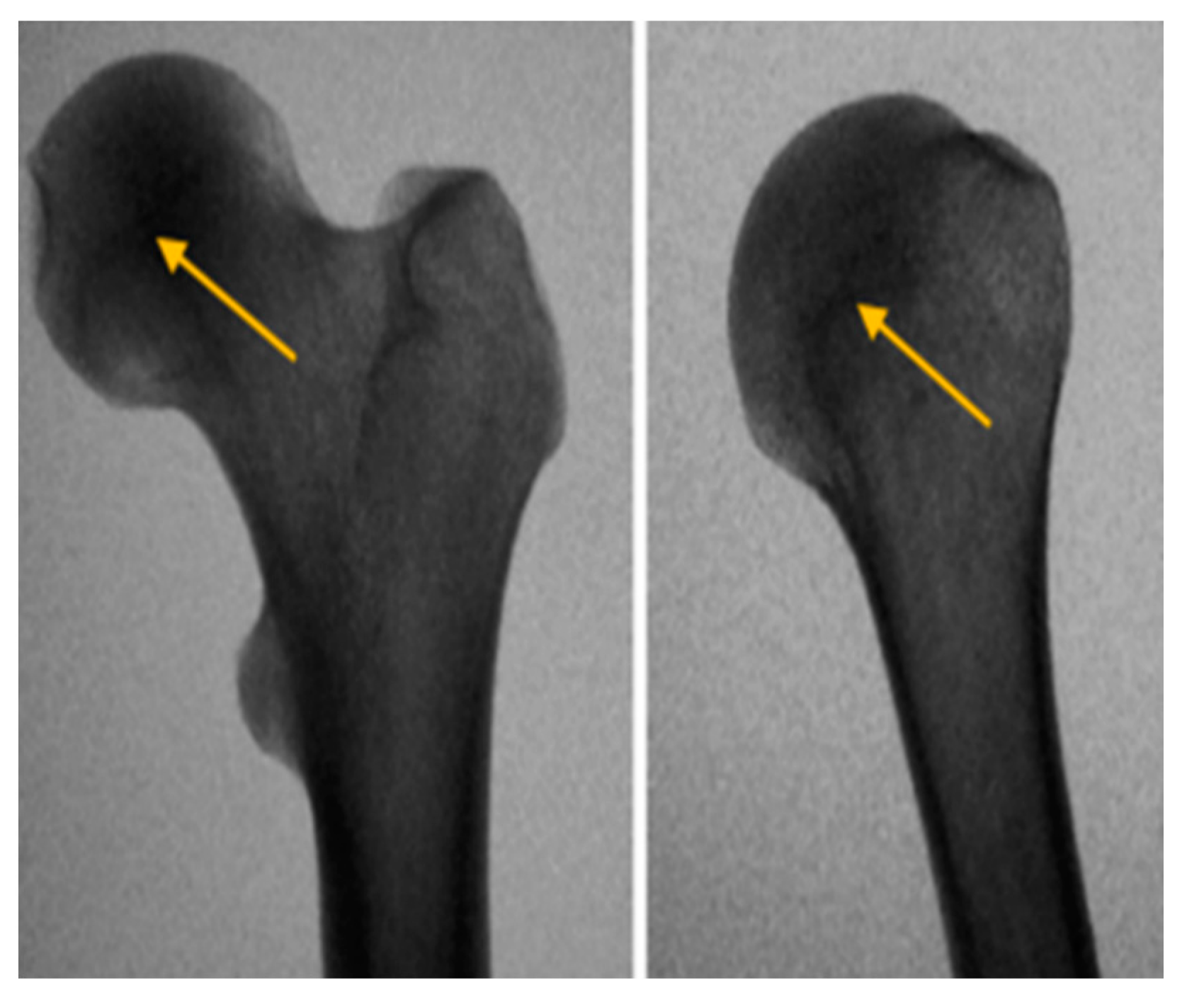
Radiological investigations were carried out to exclude the presence of gunshot ammunition material, to exclude further microfractures and to determine the degree of fusion of the ossification nuclei (
Figure 6) and, finally, several soft tissue samples were taken, where possible, for histological and genetic investigations. The samples taken were subjected to DNA extraction using the commercial NucleoSpin
®DNA Tissue kit from MACHEREY-NAGEL, following the specific protocol indicated by the manufacturer.
4.2. Case B
With regard to the anthropometric investigation of the isolated foot, Case B, only metric and morphological methods were applied, based on radiographic-digital investigation through the use of the Horos software (version 3.3.6, Horos Project, Annapolis, MD, USA) that allowed the digital extrapolation of metric measurements to be conducted (
Figure 7) [
21].
For the determination of sex, the method of discriminant analysis applied to the relative measurements of the calcaneus was applied [
22]. For the determination of age, the ossification centres of the nuclei of the calcaneus, talus, metatarsals and phalanges were analysed [
13]. For the determination of stature, a formula applied only to the maximum plantar length was used [
23].
Again, the method of observing the degree of adipocerization and disarticulation was applied to determine the time of death of the subject to whom the specimen belonged; in addition, a sample of foot tissue was taken for genetic investigation. In this case as well, soft tissue samples were taken for both histological and genetic investigation by DNA extraction using the commercial NucleoSpin®DNA Tissue kit by MACHEREY-NAGEL, following the specific protocol indicated by the manufacturer.
5. Results
The anthropological–anthropometric analyses affirmed that the skeletal remains of Case A, found at sea, a few metres from the coast in southern Italy (Reggio Calabria), belonged to a Caucasoid male subject, with height at waist of approximately 1.78 ± 3.3 and age of 25–35 years at the time of death.
Radiological investigations excluded the presence of fractures and traces of gunshot ammunition. Considering the adipocerisation phases in a marine context, it was established that the corpse had been in the water between 1 and 2 months before recovery. Histological examinations did not yield any noteworthy results, except for the lungs, where the presence of traces of blood from congestion was detected under the light microscope.
With regard to the isolated foot, Case B, the anthropometric investigations affirmed that the foot under examination belonged to an adult male subject (with a minimum classification error of 15%), approximately 1.80, with age at the time of death between 20 and 30 years. Radiological investigations excluded the presence of peri-mortem and ante-mortem fractures, abnormalities and dysmorphisms. In addition, in this case, considering the phases of adipocerization and the integrity conditions of the foot, a time of death between 1 and 2 months prior was confirmed. The histological examinations carried out on a single fragment taken from the dorsal muscle of the foot, in the direction of the purplish ecchymosis, revealed a reddish–purple blood effusion at the level of the adipose layer and the muscle fasciae, most probably caused by antemortem trauma.
The genetic investigation revealed that the genotypes of the muscle fragments of the corpse under examination and those of the isolated foot were identical. This confirms that the remains under examination belonged to the same person.
6. Discussion
The autopsy investigations carried out on the skeleton, Case A, were limited by the ex-ante loss of soft tissue due to the spoliative action of the marine micro and macro fauna. The residual soft tissue was affected by initial adipocerisation. Adipocerisation is a particular transformative phenomenon that occurs in cadavers due to prolonged residence in water or particularly moist soils and is characterised by the formation of free and in-soluble fatty acids by post-mortem hydrogenation and hydrolysis of adipose tissues [
24].
It gives the remains a slimy, fatty, cretaceous appearance and a grey-brown colour. It is believed to start quite rapidly when in water or a humid environment [
25]. However, for the effects to become visible, it takes a sufficiently long time, a minimum of one month. On the other hand, for complete adipocerisation of a corpse, a minimum residence of 3–6 months in a favourable environment is required.
The decomposition of human soft tissue in water occurs at a slower rate than decomposition outside a marine context and there are obvious differences in taphonomy between fresh and salt water. In a marine context, in salt water, decomposition has an accelerated process if the temperature is high, such as in summer. On the other hand, if the temperature is very low, as in winter, the decomposition process that subsequently leads to disarticulation is rather slow [
26,
27].
In the present case, taking into account the initial phase of adipocerisation of the external soft tissue remains, the advanced state of putrefaction of the exposed internal organs, the spring–summer period during which the corpse remained in the marine environment and the temperature and pH of the Tyrrhenian Sea, it was established that the corpse would have remained in the water from 1 to 2 months before recovery.
With regard to the cause of death, the absence of ante-mortem and peri-mortem injuries and the advanced state of skeletonization and decomposition of the internal organs, it was not possible to express a definitive opinion.
With regard to the analysis concerning the determination of the time of death of Case B, there were initially several doubts, given the integrity of the soft tissues of the foot and the presence of adipocere. The presence of a stocking with residual epidermis inside questioned the hypotheses of the initial investigations, which excluded a time of death similar to that of the skeleton. In this case, the stocking acted as a wrapping, protecting the foot from the attack of marine macro- and microfauna and slowing decomposition [
2,
28,
29]; in fact, this has also been analysed in other court cases, where bodies found at sea were protected by clothing and therefore showed slower decomposition than at the time of disappearance.
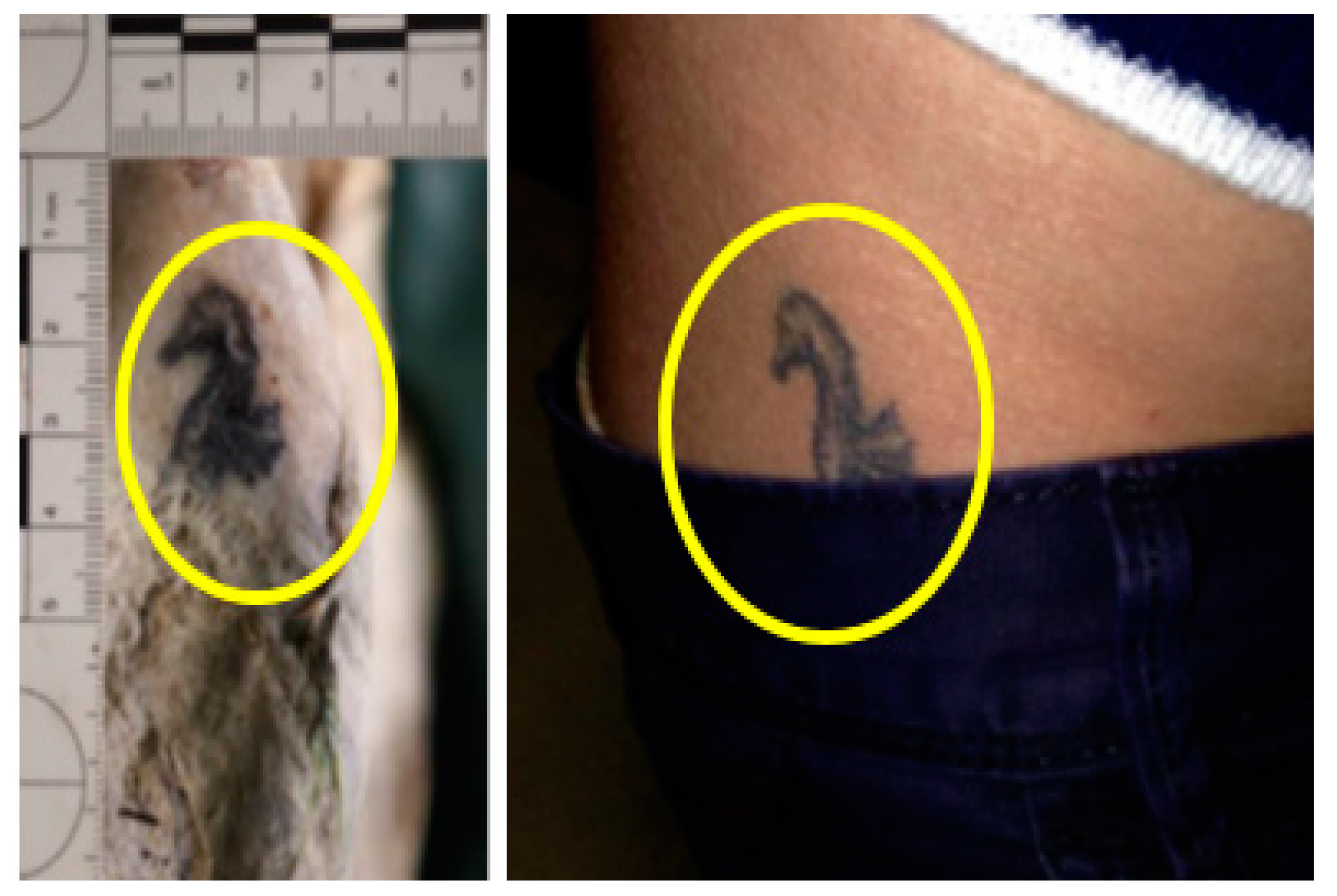
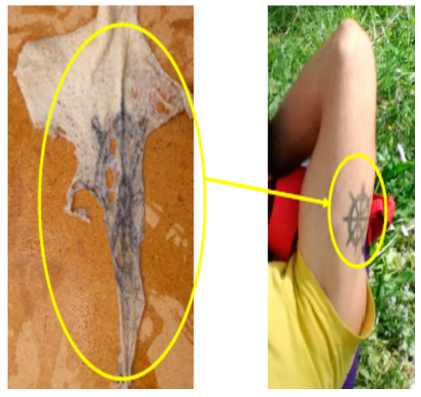
Therefore, in Case B, a time of death of between 1–2 months was confirmed. During the external examination of the corpse, two partial tattoos were identified: tattoo No. 1, which refers to a seahorse, located on the right side of the body at the level of the iliac crest; tattoo No. 2, which refers to a ship’s rudder, located on the skin of the left scapulo-humeral region. Tattoos are identification marks that determine a person’s identity and ensure their identification even after death [
30].
In the case at hand, an initial web-based survey was carried out on individuals missing at sea. The search revealed that one of these missing persons, in a photo, had a tattoo of a seahorse (
Figure 8) and, in a subsequently analysed photo, had a second tattoo indicating a sea rudder (
Figure 9), similar to what was found in the autopsy. These elements contributed strongly to the identification of the unknown skeleton.
As mentioned above, during the autopsy investigation of both cases, soft tissue samples were taken from both the skeleton of Case A and the foot of Case B for subsequent genetic investigation [
31]. In addition to these samples, a saliva sample taken from the missing person’s mother was subjected to genetic investigation in order to determine a compatibility in terms of parentage. With the three samples available, DNA typing was carried out on genetic polymorphisms of autosomal chromosomes. Tissue fragments from the muscle of the corpse and the isolated foot were subjected to DNA extraction using the commercial NucleoSpin
®DNA Tissue kit from MACHEREY-NAGEL, following the specific protocol indicated by the manufacturer.
The saliva sample was fixed on a Copan Nucleic Card™ and then DNA extraction was performed using an aliquot of the Nucleic Card™ and the FTA Purification Reagent (Whatman
®) following the manufacturer’s protocol. DNA extraction from saliva samples [
32] was performed in a multiplex amplification reaction, using the reagents of the AmpFlSTR Identifiler Plus PCR Amplification Kit and AmpFlSTR
® NGM SElect™ PCR Amplification Kit, both supplied by Life Technologies Italia, following the conditions indicated by the manufacturer. After amplification, an aliquot of each sample was subjected to capillary electrophoresis on an ABI PRISM310 Genetic Analyzer automated sequencer (Applied Biosystems). The identification of the genetic characteristics of the test samples, related to the DNA polymorphisms sought on the autosomal chromosomes, was performed with the help of an allelic ladder containing the main Caucasian variants.
The results obtained confirmed that the genotypes of the skeletal muscle fragment profile are identical to those obtained from the isolated foot fragment. In addition, the results of the typing of the autosomal markers obtained from the saliva sample allowed us to state that the genetic characteristics of the DNA are compatible in terms of relatedness.
The result of the genetic analysis confirmed the hypothesis of the anthropological investigation due to the closeness of the results of Case A (stature, 1.78 ± 3.3; age at death, 25–35 years) and Case B (stature, 1.80 with an error of 15%; age at death, 20–30 years). The minimal difference between the results, both in the determination of stature and age range, cast doubt on a probable association between the two cases at the beginning of the investigation. The hypotheses were eventually confirmed by these results; in addition, the biostatistical calculation carried out with the Familias program version 3.2.8 gave a result of probability of maternal relationship of 99.99% with a LR (likelihood ratio) value of more than 10,000.
7. Conclusions
The application of a multidisciplinary investigation allowed the identification and correlation of a partially skeletonised corpse and an intact isolated foot, found on the same day at a distance of only 9 km. The results of the forensic anthropological analyses, genetic analyses and the presence of highly identifying tattoos made it possible to reconstruct the biological profile of an unknown individual who had been missing at sea for two months. In conclusion, this result made it possible to identify the victim among the three initial options, thus determining the profile of the subject who had disappeared due to a shipwreck of fishermen two months before being found.


