Abstract
The gut microbiota plays a crucial role in the health and well-being of primates. This study applied 16S rRNA high-throughput sequencing technology, aiming to investigate the differences in gut microbiota composition and function between captive ring-tailed lemurs (Lemur catta) in different zoos across the north–south demarcation line in China. Results revealed significant differences in gut microbiota composition between northern and southern groups. Alpha diversity indices were higher in the southern group (p < 0.05), while beta diversity analysis showed distinct clustering based on geographic location (p < 0.001). Bacteroidetes were more abundant in the northern group (49.22% vs. 28.44%), while Firmicutes predominated in the southern group (59.10% vs. 32.78%). Functional prediction analysis indicated higher levels of membrane transport and lipid metabolism pathways in the southern group, suggesting differences in nutrient absorption and energy metabolism. These findings suggest that geographic location and associated environmental factors significantly influence the gut microbiota of captive ring-tailed lemurs, even under similar dietary and husbandry conditions. Our study provides insights into the impact of geographic location on gut microbiota in captive primates, highlighting the importance of considering regional factors in zoo animal management and informing future strategies for optimizing the care and conservation of captive primates across different geographic regions.
1. Introduction
The gut microbiota plays an important role in host immunity, nutrient metabolism, and health status [,]. The structure of gut microbial communities is shaped by many factors including genotype, physiology, and sociality [,]. The significant relationship between sociality and gut microbiota had been found in primates [] and other mammals []. Due to human activities and other historical reasons, wildlife faces many threats including habitat fragmentation [], illegal hunting [], and some other challenges to survival. Since wild populations are relatively limited and inaccessible, as well as difficult to track and observe directly, fecal samples by a non-invasive sampling method provide a great source for us to investigate and understand animal health under captive or wild conditions. Therefore, a comparative study on the gut microbiota of endangered species between different populations can help us to comprehensively understand the factors affecting the gut microbiota in order to promote scientific conservation []. For instance, improving gut microbiota by changing the controllable food supply and the artificial environment could improve the health status of the endangered species in captivity [,].
Lemurs (Lemuroide) are among the most ancient extant primate species []. However, habitat loss, hunting, and climate change have pushed many lemur species to the brink of extinction, with several, such as the ring-tailed lemur (Lemur catta) [], crowned lemur (Eulemur coronatus) [], and white-footed sportive lemur (Lepilemur leucopus) [], classified as endangered (EN) by the International Union for Conservation of Nature (IUCN).
Studies on the gut microbiota of ex situ lemur populations have involved various genera, including Lemur [], Eulemur [], and Varecia []. These investigations have revealed significant inter-genus [] and inter-species differences [] in gut microbiota composition. Moreover, intraspecific variations have been observed across geographic regions [], seasons [], and between sexes [], revealing the complex dynamics of gut microbiomes [,].
In lemurs, this may be particularly important for their adaptation to diverse diets and environments []. Comparative studies of lemur gut microbiota can provide comprehensive assessments of the health status, influencing factors, and adaptive conditions of different ex situ populations [], contributing to better conservation strategies for these ancient and rare primate species.
This study focuses on the ring-tailed lemur, a representative species of the family Lemuridae. Although current research on the gut microbiota of ring-tailed lemurs has included both wild [] and captive [] populations, studies on captive groups are geographically limited, primarily concentrated in Europe [] and North America []. Evidence suggests that climate is an important factor influencing the gut microbiota structure of captive primates [,]. However, there are many unknown research fields of the gut microbiomes from Asian captive populations, particularly in the context of varying climatic conditions.
Given that climate has been identified as a key driver of the gut microbiota structure in captive primates, the two following hypotheses were proposed: (1) climatic differences between northern and southern China will lead to significant variations in gut microbiota composition (e.g., relative abundance of dominant phyla/genera) and metabolic function (e.g., lipid metabolism) of captive ring-tailed lemurs. (2) Captive ring-tailed lemurs across different sites will share a core gut microbiota, reflecting the conserved microbial structure of this species.
In this study, the gut microbiota of captive ring-tailed lemur populations in three zoos located at different latitudes in China were analyzed using 16S rRNA high-throughput sequencing, providing the first comparative evidence of gut microbiota in Asian ex situ populations of this species. This study will contribute to our understanding of the factors shaping the gut microbiota in captive lemurs and provide valuable insights for the management and conservation of these endangered primates in ex situ environments.
2. Materials and Methods
2.1. Study Sites and Sample Collection
In this study, a total of 26 fresh fecal samples of L. catta were collected from three different zoos (Taiyuan Zoo, Nanjing Hongshan Forest Zoo, and Dongguan Xiangshi Zoo) in China using non-invasive sampling methods, and the sampling season was in spring. According to different geographical locations, we classify Taiyuan Zoo as the northern group and Nanjing Hongshan Forest Zoo and Dongguan Xiangshi Zoo as the southern group based on the Qinling–Huaihe River boundary (please refer to the Supplementary Materials for detailed information) (Supplementary Table S1). Among them, the southern group belongs to the subtropical zone, while the northern group belongs to the temperate zone. All samples in each group were raised in groups. All fecal samples were collected and placed into 5 mL sterile tubes immediately then kept at –80 °C in a laboratory until subsequent analysis.
2.2. DNA Extraction and Sequencing
Total DNA was extracted using the TIANamp Stool DNA Kit (Tiangen, Beijing, China). DNA quality was verified via 2% agarose gel electrophoresis, and its concentration was measured with a NanoDrop 2000 spectrophotometer (Thermo Fisher Scientific, Wilmington, DE, USA). High-throughput sequencing targeting the V3~V4 region of the 16S rDNA gene was performed on fecal samples. PCR amplification used forward primer 338F (5′-ACTCCTACGGGAGGCAGCA-3′) and reverse primer 806R (5′-GGACTACHVGGGTWTCTAAT-3′). The 20 μL reaction system included 4.0 μL 5 × FastPfu Buffer, 2.0 μL 2.5 mmol/L dNTPs, 0.8 μL each of 1 μmol/L forward/reverse primers, 0.4 μL FastPfu polymerase, 0.2 μL BSA, 10 ng template DNA, and ddH2O to 20 μL. Reaction conditions: initial denaturation at 95 °C for 3 min; 30 cycles of 95 °C (30 s), 55 °C (30 s), and 72 °C (45 s); and final extension at 72 °C for 10 min. Purified PCR products were sequenced on the Illumina MiSeq platform by Shanghai Majorbio Biopharm Technology Co., Ltd. (Shanghai, China).
2.3. Data Analysis
High-throughput sequencing determined sequences, followed by bioinformatics analysis to explore microbial community diversity and structural differences. Raw sequences were filtered, dereplicated, and denoised using DADA2 in QIIME2. The Bayesian algorithm of the RDP classifier (v2.11) was applied to analyze representative sequences of operational taxonomic units (OTUs, 97% similarity) based on the SILVA database, and QIIME (v1.9.1) generated abundance tables for each taxonomic level []. Alpha diversity indices (Ace, Chao for abundance; Shannon, Simpson for diversity) were compared via Mothur (v1.30.2), with inter-group significance evaluated by Wilcoxon rank-sum tests (* p ≤ 0.05, ** p ≤ 0.01, *** p ≤ 0.001). Principal component analysis (PCA) and unweighted UniFrac-based principal coordinate analysis (PCoA) were conducted to analyze community structure differences. PICRUSt2 predicted gut microbiota function, and Bugbase predicted its phenotypic potential pathogenicity.
3. Results
3.1. Sequencing Information
In this study, a total of 923364 optimized sequences were obtained after high-throughput sequencing (Supplementary Table S2), and they could be divided into 20 phyla, 32 classes, 87 orders,154 families, 351 genera, and 645 species (Table 1). Shannon index curves based on the OTU level tended to become flat, and thus the amount of sequencing data was sufficiently large enough and reasonable to reflect the majority of gut microorganisms (Supplementary Figure S1). Comparative analysis using Venn diagrams revealed substantial overlap and distinct differences in microbial composition between the northern and southern groups. The analysis identified 14 shared phyla and 191 shared genera across both groups. Notably, the southern group exhibited greater microbial diversity, with 5 unique phyla and 84 unique genera. In contrast, the northern group displayed no unique phyla but harbored 39 unique genera. As a result, it can be found that the microbial specificity in the southern samples was higher (Supplementary Figure S2).
Table 1.
Intestinal flora taxa for each sample group.
3.2. Composition of Intestinal Flora from L. catta
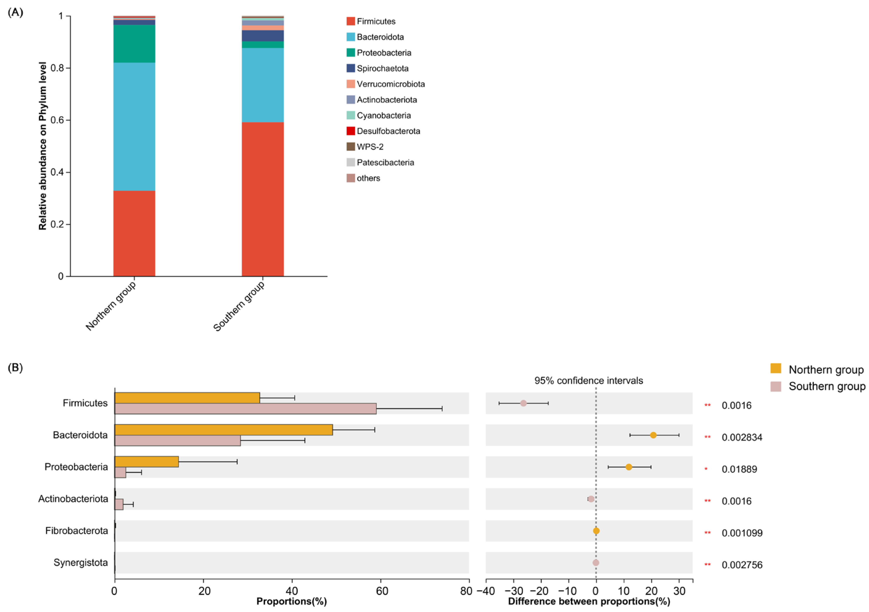
The dominant phyla of the northern group included Bacteroidota (49.22%), Firmicutes (32.78%), Proteobacteria (14.46%), and Spirechaetota (1.92%), while in the southern group, the dominant phyla were Firmicutes (59.10%), Bacteroidota (28.44%), Spirochaetota (4.31%), Proteobacteria (2.57%), Actinobacteriota (1.96%), and Verrucomierobiota (1.82%) (Table 2, Figure 1). The common dominant phyla were Firmicutes, Bacteroidota, Proteobacteria, and Spirechaetota in both of the two groups (Table 2, Figure 1).
Table 2.
Dominant phyla and genera among different sample groups.
Figure 1.
Comparison of the proportion and differences in gut microbiota at the phylum level between the northern group and southern group. (A) Microbial structure of all fecal samples at the phylum level; (B) differential analysis of dominant bacterial phyla between the northern group and southern group based on Wilcoxon rank-sum test. Taxa with relative abundance below 1% were listed as “others”. * p < 0.05, and ** p < 0.01.
Based on the Wilcoxon rank-sum test, there were significant differences in six phyla between the northern group and southern group, and the relative abundance of Firmicutes, Actinobacteriota, and Synergistota in the southern group was significantly higher than that in the northern group; the relative abundance of Fibrobacterota, Bacteroidota, and Proteobacteria in the northern group were significantly higher than those in the southern group (Figure 1).
The dominant genera of the northern group included Prevotella (19.20%), Prevotellaceae_NK3B3l_group (8.70%), Succinivibrio (8.68%), norank_f__Prevotellaceae (5.73%), and Faecalibacterium (5.17%), while the dominant genera of the southern group included unclassified_f__Lachnospiraceae (11.60%), Bacteroides (6.15%), and Christensenellaceae_R-7_group (6.17%). Compared to the northern group and southern group, we found that there was no shared dominant genus (Table 2, Figure 2).
Figure 2.
Comparison of the proportion and differences in gut microbiota at the genus level between the northern group and southern group. (A) Microbial structure of all fecal samples at the genus level; (B) differential analysis of dominant bacterial genus between the northern group and southern group based on the Wilcoxon rank-sum test. Taxa with relative abundance below 5% were listed as “others”. * p < 0.05, ** p < 0.01, and *** p < 0.001.
Based on the Wilcoxon rank-sum test, the results showed that the relative abundance of norank_f__norank_o__Clostridia_UCG-014, Prevotellaceae_UCG-001, Marvinbryantia, and Sarcina in the southern group were significantly higher than those in the northern group; the relative abundance of Prevotella, Succinivibrio, Prevotellaceae_NK3B31_group, Faecalibacterium, norank_f__Prevotellaceae, and UCG-004 in the northern group were significantly higher than those in the southern group (Figure 2).
3.3. Alpha Diversity Analysis and Beta Diversity Analysis
The alpha diversity, which refers to the diversity and richness of a microbial community, was analyzed between the northern group and southern group. The results showed that the Simpson index of the northern group was significantly higher than that of the southern group, while there were no significant difference in the Ace, Chao, and Shannon indexes (Table 3, Figure 3).
Table 3.
Mean values of alpha diversity indices between different groups.
Figure 3.
Alpha diversity analysis of gut microbiota between the northern group and southern group included Ace index (A), Chao index (B), Shannon index (C), and Simpson index (D). Significant differences were indicated by “*”.
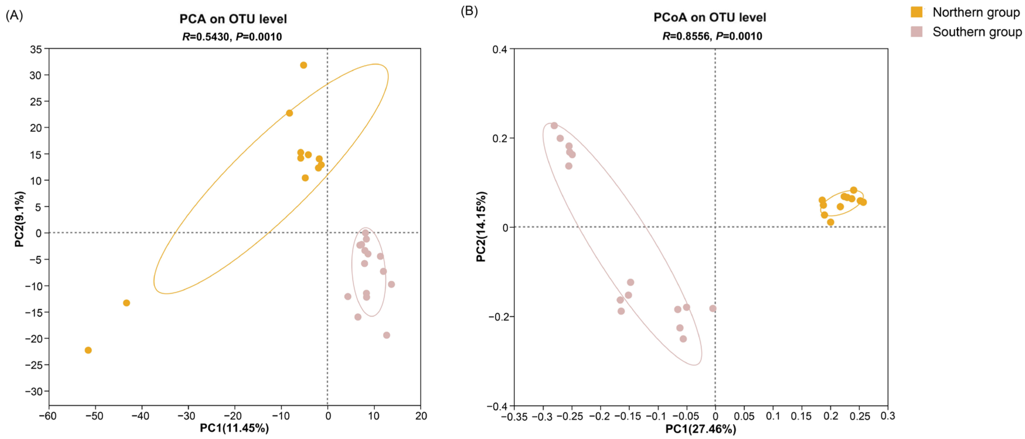
The principal coordinate analysis (PCoA) results showed that the contribution rates of PC1 and PC2 were 27.46% and 21.15%, respectively. As shown by principal component analysis (PCA), the contribution rates of the first two principal components (PC1 and PC2) stood at 11.45% and 9.1%, respectively. There were significant differences in the beta diversity of the gut microbiota from L. catta between the southern group and the northern group (Figure 4).
Figure 4.
Beta diversity analysis of the gut microbiota between the northern group and southern group including PCA (A) and PCoA based on unweighted UniFrac (B).
3.4. Functional Prediction Analysis
Analysis of metabolic pathways revealed significant differences between the northern and southern groups across multiple levels of organization. At KEGG level 1, three out of six pathways showed significant disparities, with the northern group exhibiting elevated levels in the organic systems and human diseases pathways, while the southern group demonstrated higher activity in environmental information processing pathways (Figure 5).
Figure 5.
Differential analysis of KEGG metabolic pathways between the northern group and southern group: (A) KEGG metabolic pathway level 1; (B) KEGG metabolic pathway level 2; (C) KEGG metabolic pathway level 3. * p < 0.05 and ** p < 0.01.
A total of 46 KEGG level 2 metabolic pathways were obtained, among which 16 pathways showed significant differences in gut microbiota between the southern group and northern group. Among them, membrane transport lipid metabolism, xenobiotics biodegradation and metabolism, cancer, the nervous system, cardiovascular disease, and substance dependence in the southern group were significantly higher than these from the northern group. The metabolism of the sensory system, cofactors and vitamins, the metabolism of other amino acids, infectious bacterial disease, environmental adaptation, antineoplastic drug resistance, and infectious viral disease in the northern group were significantly higher than in the southern group (Figure 5).
Further differential analysis at KEGG level 3 identified 12 pathways with significant variations. The northern group showed significantly higher activity in Pantothenate and CoA biosynthesis, the citrate cycle (TCA cycle), homologous recombination, alanine, aspartate, and glutamate metabolism, and ribosome biosynthesis. Conversely, the southern group demonstrated elevated activity in arginine biosynthesis, galactose metabolism, porphyrin and chlorophyll metabolism, the pentose phosphate pathway, pyruvate metabolism, quorum sensing, and ABC transporters. These findings suggest distinct metabolic profiles between the two groups, potentially reflecting adaptations to different environmental conditions or dietary patterns (Figure 5).
4. Discussion
Studying the gut microbiota of species in different environments is of great significance for understanding their status and maintaining their health, especially for endangered species. When the wild living environment changes, captive conditions provide a barrier for the protection of species. Therefore, improving the living conditions of endangered species in captivity is beneficial for us to better carry out wildlife conservation work and also lays the foundation for future wild release.
In this study, we focused on the composition characteristics of intestinal flora in ring-tailed lemurs under different captive environments in southern and northern China. The results showed that Firmicutes, Bacteroidota, Spirochaetota, and Proteobacteria were the dominant phyla in both the southern and northern groups, which is consistent with previous studies on the same species [,,,,] and other primate species (such as Macaca mulatta [] and Gorilla beringei beringei []). This indicates that the main components of the gut microbiota in ring-tailed lemurs are similar to those in many primates. Among them, Firmicutes can help the host absorb nutrients such as carbohydrates, polysaccharides, and fatty acids in food [,]. Bacteroidota can degrade proteins and polysaccharides in the host and can also produce butyrate, which is considered to have anti-tumor properties [,]; it is of great significance to maintain the health of the host. Some bacteria in Spirochaetota can degrade cellulose and produce short-chain fatty acids [].
At the phylum level, the proportion of Firmicutes and Actinobacteriota in the northern group was significantly higher than that in the southern group (Supplementary Table S3). The change in the ratio of Firmicutes and Bacteroidetes is considered to be an important parameter reflecting the host’s energy requirements []. It is now known that most bacteria of the Firmicutes phylum could affect nutrition and metabolism, and the ratio of Firmicutes to Bacteroidetes (F/B) is considered to be an ecological imbalance []. The increase in this ratio is often associated with obesity and metabolic disorders, which may be related to increased caloric extraction from food, fat deposition and fat production, and impaired insulin sensitivity [,]. When the ratio decreases, it is associated with inflammatory bowel disease and other related conditions, which may be related to the reduction in the production of short-chain fatty acids [,]. In many studies, a higher F/B ratio indicates a greater energy requirement for the host and a stronger ability of its gut microbiota to extract energy from food [,]. In this study, F/B in the northern group of ring-tailed lemurs was less than 1, while the F/B ratio in the southern group was greater than 1. In previous studies, the F/B ratio of the intestinal flora of the black-hat capuchin monkeys in the Daqingshan Zoo in Hohhot was 0.8 [], and Hohhot also belongs to the north. Therefore, the F/B ratio of lemurs in the north is relatively low, which suggests that in northern regions, due to the influence of northern climate conditions or other living environments, the F/B ratio in the intestinal flora of lemurs is low, and their demand for external energy is not high. At the same time, the comparison of metabolic pathways between the two groups also revealed that lipid metabolism in the southern group was significantly higher than that in the northern group.
Research has shown that there are many types of bacteria in the Bacteroidetes phylum, which have important effects on the physiological functions of the host []. These bacteria play a key role in digesting complex polysaccharides, producing SCFAs, and regulating the intestinal immune response []. Bacteroidetes play an important role in maintaining intestinal health and preventing pathogen invasion []. The abundance of Bacteroidetes is negatively correlated with the host’s energy accumulation, especially fat accumulation [,]. Although the abundance of Bacteroidetes is not conducive to energy accumulation in the host, studies have shown that Bacteroidetes are more abundant than Firmicutes in the expression of proteins related to metabolic pathways such as lipid metabolism, amino acid metabolism, and gluconeogenesis []. In our study, the relative abundance of Bacteroidetes was markedly different between the southern and northern groups, comprising 28.44% and 49.22% of the gut microbiota, respectively. Metabolic pathway analysis revealed significantly higher lipid metabolism in the southern group compared to the northern group. This finding aligns with the established negative correlation between fat accumulation and Bacteroidetes abundance [,]. Consequently, we hypothesize that fat accumulation in the northern group may be lower than in the southern group, potentially manifesting in differences in the body fat composition of ring-tailed lemurs. However, further investigation is necessary to elucidate this correlation definitively. The presence of Bacteroidetes in the intestinal tracts of captive mammals has been previously documented. For instance, Adams et al. (2021) reported a high proportion of Bacteroidetes in the gut microbiota of the island fox (Urocyon littoralis) in the Taiwan Strait []. Similarly, Chen et al. (2022) observed that Bacteroidetes constituted the most abundant bacterial phylum in the gut microbiota of captive wolves []. These findings collectively suggest that Bacteroidetes play a crucial role in the gut microbiome of various mammalian species, including ring-tailed lemurs, and may influence host metabolism and physiology.
Furthermore, studies have shown that changes in environmental temperature can affect the intestinal microbiota structure of reptiles [], mammals [], and birds []. For example, Liu et al. (2019) investigated the diversity of gut microbiota in Siberian flying squirrels during the spring and summer of 2013 and 2014; PCoA analysis showed that the changes in the diversity of gut microbiota in Siberian flying squirrels during different seasons were significantly correlated with environmental temperature (p < 0.001) []. Therefore, it is speculated that due to its adaptation to different climatic environments, the intestinal flora structure of the captive ring-tailed lemurs in the northern and southern regions shows differences.
At the genus level, there are no dominant genera shared by the gut microbiota of ring-tailed lemurs from the northern and southern groups (Supplementary Table S4). Prevotella had the highest relative abundance in the northern group, and its proportion was significantly higher than that in the southern group. According to reports, Prevotella can help degrade proteins [], carbohydrates, and monosaccharides in host food, and its abundance changes can reflect the level of carbohydrate digestion in the host. The relative abundance of the southern group unclassifeid_f__Lachnospiraceae is the highest. This genus belongs to the Lachnospiraceae family. Previous studies have shown that Lachnospiraceae and Ruminococcaceae are the two families with the highest number of Clostridiales in the mammalian intestinal environment. They are associated with the maintenance of intestinal health and can also help to decompose plant cellulose [].
Diet is often considered to be one of the most important factors affecting the gut microbiota of animals [,,,]. However, in this study, there are some differences in the food fed by different zoos, but overall, they are similar, mainly consisting of fruits and vegetables (Supplementary Table S5). Based on previous reports, the dominant phyla of the gut microbiota in wild ring-tailed lemurs in Madagascar include Firmicutes, Bacteroidetes, Proteobacteria, and Tenericutes. The dominant bacterial genera include RFN20, Prevotella, unrecognized genera of the clostridiales order, and unrecognized genera of the Ruminococcaceae family []. The dominant bacterial phyla in the research results are consistent with this study, indicating that the composition of the gut microbiota of the same species is similar in different environments. However, the differences in dominant genera are very large, indicating that different environments can indeed change the structure of animal gut microbiota. This is also confirmed in the work of McKenzie et al. (2017), who found that captivity affects the gut microbiota of mammals by altering animal diet, animal interaction, and animal contact with a changing environment and that captivity has a particularly significant impact on the gut microbiota of primates []. Meanwhile, Bennett et al. (2016) used high-throughput sequencing to describe the changes in the gut microbiota of the endangered ring-tailed lemur in the Beza Mahafaly Special Reserve in southwestern Madagascar []. The results showed that habitat disturbance may not affect the gut microbiota of lemurs as strongly as it does other primates. Bornbusch et al. (2022) [] conducted 16S rRNA sequencing of the intestinal and soil microbial communities of lemurs from the wild and different captive environments. The results showed that there were significant differences in the diversity of intestinal microorganisms between captivity and the environment. The microbial diversity of wild lemurs is not always greater than that of captive lemurs. In addition, although the commercial diets of captive lemurs on two continents are similar, the gut microbiome composition of lemurs in Madagascar is most similar, suggesting that non-dietary factors control some variability [].
This study initially hypothesized that climate factors would influence the gut microbiota composition of ring-tailed lemurs across three research sites, leading to observable differences. The results corroborated this hypothesis, revealing significant variations in both bacterial species proportions and metabolic levels between the northern and southern groups (Supplementary Figure S3). The second hypothesis posited the existence of a shared core microbiome among the lemur populations across the three research sites. Experimental analysis confirmed that at the phylum level, Firmicutes and Bacteroidetes constitute the core phyla shared by the gut microbiota of ring-tailed lemurs in all three locations. This finding is consistent with some previous studies on the gut microbiota in different locations [,] and supports our initial speculation. However, at the genus level, the gut microbiota of ring-tailed lemurs from the northern and southern populations did not share dominant genera.
In addition to the gut microbiota, this study also focused on the differences in metabolic levels (Supplementary Figures S4–S6). Our analysis revealed significantly elevated metabolic levels of transport and lipid metabolism in the southern group compared to the northern group. Membrane transport is known to influence the intestinal absorption efficiency of nutrients, while lipid metabolism plays a crucial role in regulating the host’s energy balance and obesity risk [,]. The observed higher metabolic levels in the southern group suggest enhanced absorption and conversion efficiency of nutrients. Given that the captive animals in both groups were maintained on similar diets, we hypothesize that the observed metabolic differences may be attributed to the distinct climatic conditions between the northern and southern regions. Climate has been shown to influence various physiological processes in mammals, including metabolism []. Nanjing and Dongguan, located in the lower Yangtze River basin, are characterized by a subtropical monsoon climate, while Taiyuan experiences a warm temperate semi-humid continental monsoon climate. These distinct climatic regimes result in significant environmental differences among these cities []. These findings highlight the potential impact of environmental factors on gut microbial metabolism, even in captive conditions where dietary factors are controlled. Further research is warranted to elucidate the specific mechanisms by which climate may modulate gut microbial metabolism and, consequently, host physiology in ring-tailed lemurs. Such insights could have important implications for the management and health of captive populations in diverse geographic locations. Predictive tests for pathogenic bacteria also revealed differences between the southern and northern groups (Supplementary Figure S7).
Thus, this study compared the compositional characteristics of gut microbiota in ring-tailed lemurs in different captive habitats in southern and northern China and found significant differences in gut microbiota at both the phylum and genus levels. At the phylum level, analysis via the Wilcoxon rank-sum test revealed that the relative abundance of Firmicutes was significantly higher in the southern group than in the northern group (p < 0.01), while Bacteroidota showed the opposite trend in abundance (p < 0.05). At the genus level, combined with LEFSE analysis, we also identified that the key taxa enriched in the southern group included unclassified_f__Lachnospiraceae and Christensenellaceae_R-7_group, while the northern group showed more significant enrichment of Prevotella and Succinivibrio. In future research, it is necessary to collect more samples from different research sites with different living conditions to further explore the potential impact of various factors on the gut microbiota of ring-tailed lemurs. Due to the limitations of sampling, the gender and age information of ring-tailed lemurs was not collected for correlation analysis. These factors will also be taken into account when conducting gut microbiota correlation analysis in the future. Based on the findings of this study, we propose the following evidence-based management recommendations for zoos housing ring-tailed lemurs: firstly, for environmental microbiome optimization, zoos should characterize the microbial composition of enclosure substrates (e.g., soil, bedding) and water sources, and where possible, introduce microbial taxa consistent with wild Madagascar habitats to stabilize core gut microbiota and reduce geographic variability in the microbial structure; additionally, for dietary fine-tuning based on regional metabolic needs, while diets are generally consistent across zoos, subtle adjustments should align with regional metabolic differences, and regular dietary audits should be paired with gut microbiota monitoring to ensure alignment with host needs. Finally, for inter-zoo collaboration for standardized care, given the geographic variability in gut microbiota, zoos housing ring-tailed lemurs should establish a shared database of microbial, dietary, and climatic data to facilitate the development of region-specific care guidelines, ensure consistency in ex situ conservation practices, and support data-driven reintroduction planning. In summary, this study compares the compositional characteristics of gut microbiota in ring-tailed lemurs in different captive habitats from southern and northern China and provides more reliable evidence and support for the ex situ conservation of this endangered species.
Supplementary Materials
The following supporting information can be downloaded at: https://www.mdpi.com/article/10.3390/jzbg6040057/s1, Figure S1: Rarefaction analysis of bacterial communities in gut of ring-tailed lemurs based on Shannon index; Figure S2: Phylum-level (A) and genus-level (B) species Venn diagram analysis of intestinal flora; Figure S3: Comparison of alpha and beta diversity of gut microbiota in ring-tailed lemurs at two different locations within the southern group; Figure S4: Functional prediction analysis of KEGG metabolic pathways, level 1; Figure S5: Functional prediction analysis of KEGG metabolic pathways, level 2; Figure S6: Functional prediction analysis of KEGG metabolic pathways, level 3; Figure S7: Prediction of intestinal flora pathogenicity; Table S1: Basic information of fecal samples of ring-tailed lemurs; Table S2: Raw sequencing data of each sample; Table S3: The top 5 phyla in terms of proportion; Table S4: The top 5 genera in terms of proportion; Table S5: The diet of animals in different zoos.
Author Contributions
Formal analysis, M.S. and H.W.; funding acquisition, H.W. and D.Z.; investigation, H.Y.; methodology, M.S., H.Y., N.W., H.W. and D.Z.; resources, M.S., H.Y., N.W. and D.Z.; software, M.S. and N.W.; supervision, D.Z.; writing—original draft, M.S., H.W. and D.Z.; writing—review and editing, M.S., H.W. and D.Z. All authors have read and agreed to the published version of the manuscript.
Funding
This work is funded by Tianjin Bureau of Planning and Natural Resources, and the Tianjin Municipal Education Commission Scientific Research Program (2023KJ183).
Institutional Review Board Statement
All research did not involve any animal experiments and is in compliance with the law.
Data Availability Statement
The datasets for this study can be found in the NCBI Sequence Read Archive (SRA) under accession number PRJNA1108298.
Acknowledgments
We are highly grateful to the staff of Taiyuan Zoo, Nanjing Hongshan Forest Zoo, and Dongguan Xiangshi Zoo for their support in this study. We also thank the editors and reviewers of the Journal of Zoological and Botanical Gardens for their professional suggestions for improving the manuscript.
Conflicts of Interest
The authors declare that there are no conflicts of interest.
References
- Belkaid, Y.; Hand, T.W. Role of the microbiota in immunity and inflammation. Cell 2014, 157, 121–141. [Google Scholar] [CrossRef]
- Flint, H.J.; Scott, K.P.; Louis, P.; Duncan, S.H. The role of the gut microbiota in nutrition and health. Nat. Rev. Gastroenterol. Hepatol. 2012, 9, 577–589. [Google Scholar] [CrossRef]
- Amato, K.R.; Sanders, J.G.; Song, S.J.; Nute, M.; Metcalf, J.L.; Thompson, L.R.; Morton, J.T.; Amir, A.; McKenzie, V.J.; Humphrey, G.; et al. Evolutionary trends in host physiology outweigh dietary niche in structuring primate gut microbiomes. ISME J. 2019, 13, 576–587. [Google Scholar] [CrossRef]
- Zhernakova, D.V.; Wang, D.; Liu, L.; Andreu-Sánchez, S.; Zhang, Y.; Ruiz-Moreno, A.J.; Peng, H.; Plomp, N.; Del Castillo-Izquierdo, Á.; Gacesa, R.; et al. Host genetic regulation of human gut microbial structural variation. Nature 2024, 625, 813–821. [Google Scholar] [CrossRef]
- Pinacho-Guendulain, B.; Montiel-Castro, A.J.; Ramos-Fernández, G.; Pacheco-López, G. Social complexity as a driving force of gut microbiota exchange among conspecific hosts in non-human primates. Front. Integr. Neurosci. 2022, 16, 876849. [Google Scholar] [CrossRef] [PubMed]
- Raulo, A.; Allen, B.E.; Troitsky, T.; Husby, A.; Firth, J.A.; Coulson, T.; Knowles, S.C.L. Social networks strongly predict the gut microbiota of wild mice. ISME J. 2021, 15, 2601–2613. [Google Scholar] [CrossRef] [PubMed]
- Hanski, I. Habitat fragmentation and species richness. J. Biogeogr. 2015, 42, 989–993. [Google Scholar] [CrossRef]
- Rija, A.A.; Critchlow, R.; Thomas, C.D.; Beale, C.M. Global extent and drivers of mammal population declines in protected areas under illegal hunting pressure. PLoS ONE 2020, 15, e0227163. [Google Scholar] [CrossRef] [PubMed]
- Ni, Q.; He, X.; Zeng, B.; Meng, X.; Xu, H.; Li, Y.; Yang, M.; Li, D.; Yao, Y.; Zhang, M.; et al. Variation in gut microbiota of captive Bengal slow lorises. Curr. Microbiol. 2020, 77, 2623–2632. [Google Scholar] [CrossRef]
- Bornbusch, S.L.; Greene, L.K.; Rahobilalaina, S.; Calkins, S.; Rothman, R.S.; Clarke, T.A.; LaFleur, M.; Drea, C.M. Gut microbiota of ring-tailed lemurs (Lemur catta) vary across natural and captive populations and correlate with environmental microbiota. Anim. Microbiome 2022, 4, 29. [Google Scholar] [CrossRef]
- Yang, H.; Leng, X.; Du, H.; Luo, J.; Wu, J.; Wei, Q. Adjusting the prerelease gut microbial community by diet training to improve the postrelease fitness of captive-bred Acipenser dabryanus. Front. Microbiol. 2020, 11, 488. [Google Scholar] [CrossRef] [PubMed]
- Kistler, L.; Ratan, A.; Godfrey, L.R.; Crowley, B.E.; Hughes, C.E.; Lei, R.; Cui, Y.; Wood, M.L.; Muldoon, K.M.; Andriamialison, H.; et al. Comparative and population mitogenomic analyses of Madagascar’s extinct, giant ‘subfossil’ lemurs. J. Hum. Evol. 2015, 79, 45–54. [Google Scholar] [CrossRef] [PubMed]
- LaFleur, M.; Gould, L. Lemur catta. The IUCN Red List of Threatened Species 2020: E.T11496A115565760. 2020. Available online: https://www.iucnredlist.org/species/11496/115565760 (accessed on 9 February 2025).
- Reute, K.E.; Eppley, T.M.; Hending, D.; Pacifici, M.; Semel, B.; Zaonarivelo, J. Eulemur coronatus (Amended Version of 2020 Assessment). The IUCN Red List of Threatened Species 2020: E.T8199A182239524. 2020. Available online: https://www.iucnredlist.org/species/8199/182239524 (accessed on 9 February 2025).
- Eppley, T.M.; Ferguson, B.; Louis, E.E.; Rakotondranary, S.J.; Ganzhorn, J. Lepilemur leucopus. The IUCN Red List of Threatened Species 2020: E.T11618A115566334. 2020. Available online: https://www.iucnredlist.org/species/11618/115566334 (accessed on 9 February 2025).
- McKenzie, V.J.; Song, S.J.; Delsuc, F.; Prest, T.L.; Oliverio, A.M.; Korpita, T.M.; Alexiev, A.; Amato, K.R.; Metcalf, J.L.; Kowalewski, M.; et al. The effects of captivity on the mammalian gut microbiome. Integr. Comp. Biol. 2017, 57, 690–704. [Google Scholar] [CrossRef] [PubMed]
- McKenney, E.A.; Rodrigo, A.; Yoder, A.D. Patterns of gut bacterial colonization in three primate species. PLoS ONE 2015, 10, e0124618. [Google Scholar] [CrossRef]
- Greene, L.K.; McKenney, E.A.; Gasper, W.; Wrampelmeier, C.; Hayer, S.; Ehmke, E.E.; Clayton, J.B. Gut site and gut morphology predict microbiome structure and function in ecologically diverse lemurs. Microb. Ecol. 2023, 85, 1608–1619. [Google Scholar] [CrossRef]
- Springer, A.; Fichtel, C.; Al-Ghalith, G.A.; Koc, F.; Amato, K.R.; Clayton, J.B.; Knights, D.; Kappeler, P.M. Patterns of seasonality and group membership characterize the gut microbiota in a longitudinal study of wild Verreaux’s sifakas (Propithecus verreauxi). Ecol. Evol. 2017, 7, 5732–5745. [Google Scholar] [CrossRef]
- Bennett, G.; Malone, M.; Sauther, M.L.; Cuozzo, F.P.; White, B.; Nelson, K.E.; Stumpf, R.M.; Knight, R.; Leigh, S.R.; Amato, K.R. Host age, social group, and habitat type influence the gut microbiota of wild ring-tailed lemurs (Lemur catta). Am. J. Primatol. 2016, 78, 883–892. [Google Scholar] [CrossRef]
- Clayton, J.B.; Gomez, A.; Amato, K.; Knights, D.; Travis, D.A.; Blekhman, R.; Knight, R.; Leigh, S.; Stumpf, R.; Wolf, T.; et al. The gut microbiome of nonhuman primates: Lessons in ecology and evolution. Am. J. Primatol. 2018, 80, e22867. [Google Scholar] [CrossRef]
- Clemente, J.C.; Ursell, L.K.; Parfrey, L.W.; Knight, R. The impact of the gut microbiota on human health: An integrative view. Cell 2012, 148, 1258–1270. [Google Scholar] [CrossRef]
- Lan, L.Y.; You, Y.Y.; Hong, Q.X.; Liu, Q.X.; Xu, C.Z.; Chen, W.; Zhu, Y.; Du, X.; Fan, P. The gut microbiota of gibbons across host genus and captive site in China. Am. J. Primatol. 2022, 84, e23360. [Google Scholar] [CrossRef]
- Fogel, A.T. The gut microbiome of wild lemurs: A comparison of sympatric Lemur catta and Propithecus verreauxi. Folia Primatol. 2015, 86, 85–95. [Google Scholar] [CrossRef]
- Amato, K.R.; Yeoman, C.J.; Ken, A.; Righini, N.; Carbonero, F.; Estrada, A.; Gaskins, H.R.; Stumpf, R.M.; Yildirim, S.; Torralba, M.; et al. Habitat degradation impacts black howler monkey (Alouatta pigra) gastrointestinal microbiomes. ISME J. 2013, 7, 1344–1353. [Google Scholar] [CrossRef]
- Clayto, J.B.; Vangay, P.; Huang, H.; Ward, T.; Hillmann, B.M.; Al-Ghalith, G.A.; Travis, D.A.; Long, H.T.; Van Tuan, B.; Van Minh, V.; et al. Captivity humanizes the primate microbiome. Proc. Natl. Acad. Sci. USA 2016, 113, 10376–10381. [Google Scholar] [CrossRef] [PubMed]
- Edgar, R.C. UPARSE: Highly accurate OTU sequences from microbial amplicon reads. Nat. Methods 2013, 10, 996–998. [Google Scholar] [CrossRef] [PubMed]
- Zeng, Y.; Pu, Y.; Niu, L.; Deng, J.; Zeng, D.; Amato, K.; Li, Y.; Zhou, Y.; Lin, Y.; Wang, J.; et al. Comparison of gastrointestinal microbiota in golden snub-nosed monkey (Rhinopithecus roxellanae), green monkey (Chlorocebus aethiops sabaeus), and ring-tailed lemur (Lemur catta) by high throughput sequencing. Glob. Ecol. Conserv. 2022, 33, e01946. [Google Scholar] [CrossRef]
- Yasuda, K.; Oh, K.; Ren, B.; Tickle, T.L.; Franzosa, E.A.; Wachtman, L.M.; Miller, A.D.; Westmoreland, S.V.; Mansfield, K.G.; Vallender, E.J.; et al. Biogeography of the intestinal mucosal and lumenal microbiome in the rhesus macaque. Cell Host Microbe 2015, 17, 385–391. [Google Scholar] [CrossRef]
- Fre, J.C.; Rothman, J.M.; Pell, A.N.; Nizeyi, J.B.; Cranfield, M.R.; Angert, E.R. Fecal bacterial diversity in a wild gorilla. Appl. Environ. Microbiol. 2006, 72, 3788–3792. [Google Scholar] [CrossRef]
- Flint, H.J.; Bayer, E.A.; Rincon, M.T.; Lamed, R.; White, B.A. Polysaccharide utilization by gut bacteria: Potential for new insights from genomic analysis. Nat. Rev. Microbiol. 2008, 6, 121–131. [Google Scholar] [CrossRef]
- Li, Y.; Hu, X.; Yang, S.; Zhou, J.; Qi, L.; Sun, X.; Fan, M.; Xu, S.; Cha, M.; Zhang, M.; et al. Comparison between the fecal bacterial microbiota of healthy and diarrheic captive musk deer. Front. Microbiol. 2018, 9, 300. [Google Scholar] [CrossRef]
- Schloss, P.D.; Handelsman, J. Introducing DOTUR, a computer program for defining operational taxonomic units and estimating species richness. Appl. Environ. Microbiol. 2005, 71, 1501–1506. [Google Scholar] [CrossRef]
- Thomas, F.; Hehemann, J.H.; Rebuffet, E.; Czjzek, M.; Michel, G. Environmental and gut bacteroidetes: The food connection. Front. Microbiol. 2011, 2, 93. [Google Scholar] [CrossRef]
- Hess, M.; Sczyrba, A.; Egan, R.; Kim, T.W.; Chokhawala, H.; Schroth, G.; Luo, S.; Clark, D.S.; Chen, F.; Zhang, T.; et al. Metagenomic discovery of biomass-degrading genes and genomes from cow rumen. Science 2011, 331, 463–467. [Google Scholar] [CrossRef] [PubMed]
- Li, S.; Zhang, C.; Gu, Y.; Chen, L.; Ou, S.; Wan, Y.; Peng, X. Lean rats gained more body weight than obese ones from a high-fibre diet. Br. J. Nutr. 2015, 114, 1188–1194. [Google Scholar] [CrossRef] [PubMed]
- Stojanov, S.; Berlec, A.; Štrukelj, B. The influence of probiotics on the Firmicutes/Bacteroidetes ratio in the treatment of obesity and inflammatory bowel disease. Microorganisms 2020, 8, 1715. [Google Scholar] [CrossRef] [PubMed]
- Ley, R.E.; Turnbaugh, P.J.; Klein, S.; Gordon, J.I. Human gut microbes associated with obesity. Nature 2006, 444, 1022–1023. [Google Scholar] [CrossRef]
- Xia, F.; Wen, L.P.; Ge, B.C.; Li, Y.X.; Li, F.P.; Zhou, B.J. Gut microbiota as a target for prevention and treatment of type 2 diabetes: Mechanisms and dietary natural products. World J. Diabetes 2021, 12, 1146. [Google Scholar] [CrossRef]
- Grigor’eva, I.N. Gallstone disease, obesity and the Firmicutes/Bacteroidetes ratio as a possible biomarker of gut dysbiosis. J. Pers. Med. 2020, 11, 13. [Google Scholar] [CrossRef]
- Koliada, A.; Syzenk, G.; Moseiko, V.; Budovsk, L.; Puchkov, K.; Perederiy, V.; Gavalko, Y.; Dorofeyev, A.; Romanenko, M.; Tkach, S.; et al. Association between body mass index and Firmicutes/Bacteroidetes ratio in an adult Ukrainian population. BMC Microbiol. 2017, 17, 120. [Google Scholar] [CrossRef]
- Yu, J.; Bai, G.; He, Y.; Liu, M.; Yang, X.; Li, J.; Shen, Y.; Lu, S.; Bao, W. Analysis of intestinal bacterial diversity and its gene function prediction in black-capped capuchin (Sapajus apella). J. Appl. Anim. Res. 2024, 52, 2322652. [Google Scholar] [CrossRef]
- Gibiino, G.; Lopetuso, L.R.; Scaldaferri, F.; Rizzatti, G.; Binda, C.; Gasbarrini, A. Exploring bacteroidetes: Metabolic key points and immunological tricks of our gut commensals. Digest. Liver Dis. 2018, 50, 635–639. [Google Scholar] [CrossRef]
- Cheng, J.; Hu, J.; Geng, F.; Nie, S. Bacteroides utilization for dietary polysaccharides and their beneficial effects on gut health. Food Sci. Hum. Wellness 2022, 11, 1101–1110. [Google Scholar] [CrossRef]
- Ivanov, I.I.; Honda, K. Intestinal commensal microbes as immune modulators. Cell Host Microbe 2012, 12, 496–508. [Google Scholar] [CrossRef] [PubMed]
- Bell, D.S. Changes seen in gut bacteria content and distribution with obesity: Causation or association? Postgrad. Med. 2015, 127, 863–868. [Google Scholar] [CrossRef] [PubMed]
- Goffredo, M.; Mass, K.; Parks, E.J.; Wagner, D.A.; McClure, E.A.; Graf, J.; Savoye, M.; Pierpont, B.; Cline, G.; Santoro, N. Role of gut microbiota and short chain fatty acids in modulating energy harvest and fat partitioning in youth. J. Clin. Endocrinol. Metab. 2016, 101, 4367–4376. [Google Scholar] [CrossRef]
- Hart, E.; Creevey, C.; Hitch, T.; Kingston-Smith, A. Meta-proteomics of rumen microbiota indicates niche compartmentalisation and functional dominance in a limited number of metabolic pathways between abundant bacteria. Sci. Rep. 2018, 8, 10504. [Google Scholar] [CrossRef]
- Adams, N.E.; Becker, M.A.; Edmands, S. Effect of geography and captivity on scat bacterial communities in the imperiled channel island fox. Front. Microbiol. 2021, 12, 748323. [Google Scholar] [CrossRef]
- Chen, L.; Sun, M.; Xu, D.; Gao, Z.; Shi, Y.; Wang, S.; Zhou, Y. Gut microbiome of captive wolves is more similar to domestic dogs than wild wolves indicated by metagenomics study. Front. Microbiol. 2022, 13, 1027188. [Google Scholar] [CrossRef]
- Fontaine, S.S.; Novarro, A.J.; Kohl, K.D. Environmental temperature alters the digestive performance and gut microbiota of a terrestrial amphibian. J. Exp. Biol. 2018, 221, jeb187559. [Google Scholar] [CrossRef]
- Liu, P.Y.; Cheng, A.C.; Huang, S.W.; Chang, H.W.; Oshida, T.; Yu, H.T. Variations in gut microbiota of Siberian flying squirrels correspond to seasonal phenological changes in their Hokkaido subarctic forest ecosystem. Microb. Ecol. 2019, 78, 223–231. [Google Scholar] [CrossRef]
- Dietz, M.W.; Matson, K.D.; Versteegh, M.A.; Van Der Velde, M.; Parmentier, H.K.; Arts, J.A.; Salles, J.F.; Tieleman, B.I. Gut microbiota of homing pigeons shows summer–winter variation under constant diet indicating a substantial effect of temperature. Anim. Microb. 2022, 4, 64. [Google Scholar] [CrossRef]
- Griswold, K.E.; White, B.A.; Mackie, R.I. Diversity of extracellular proteolytic activities among prevotella species from the rumen. Curr. Microbiol. 1999, 39, 187–194. [Google Scholar] [CrossRef]
- Biddle, A.; Stewart, L.; Blanchard, J.; Leschine, S. Untangling the genetic basis of fibrolytic specialization by Lachnospiraceae and Ruminococcaceae in diverse gut communities. Diversity 2013, 5, 627–640. [Google Scholar] [CrossRef]
- Amato, K.R.; Leigh, S.R.; Kent, A.; Mackie, R.I.; Yeoman, C.J.; Stumpf, R.M.; Wilson, B.A.; Nelson, K.E.; White, B.A.; Garber, P.A. The gut microbiota appears to compensate for seasonal diet variation in the wild black howler monkey (Alouatta pigra). Microb. Ecol. 2015, 69, 434–443. [Google Scholar] [CrossRef] [PubMed]
- Hicks, A.L.; Lee, K.J.; Couto-Rodriguez, M.; Patel, J.; Sinha, R.; Guo, C.; Olson, S.H.; Seimon, A.; Seimon, T.A.; Ondzie, A.U.; et al. Gut microbiomes of wild great apes fluctuate seasonally in response to diet. Nat. Commun. 2018, 9, 1786. [Google Scholar] [CrossRef] [PubMed]
- Lee, W.; Hayakawa, T.; Kiyono, M.; Yamabata, N.; Enari, H.; Enari, H.S.; Fujita, S.; Kawazoe, T.; Asai, T.; Oi, T.; et al. Diet-related factors strongly shaped the gut microbiota of Japanese macaques. Am. J. Primatol. 2023, 85, e23555. [Google Scholar] [CrossRef]
- Li, L.; Zhao, X. Comparative analyses of fecal microbiota in Tibetan and Chinese Han living at low or high altitude by barcoded 454 pyrosequencing. Sci. Rep. 2015, 5, 14682. [Google Scholar] [CrossRef]
- Wang, W.; Wang, F.; Li, L.; Wang, A.; Sharshov, K.; Druzyaka, A.; Lancuo, Z.; Wang, S.; Shi, Y. Characterization of the gut microbiome of black-necked cranes (Grus nigricollis) in six wintering areas in China. Arch. Microbiol. 2020, 202, 983–993. [Google Scholar] [CrossRef]
- Petrenko, V.; Sinturel, F.; Riezman, H.; Dibner, C. Lipid metabolism around the body clocks. Prog. Lipid Res. 2023, 91, 101235. [Google Scholar] [CrossRef]
- Steffansen, B.; Nielsen, C.U.; Brodin, B.; Eriksson, A.H.; Andersen, R.; Frokjaer, S. Intestinal solute carriers: An overview of trends and strategies for improving oral drug absorption. Eur. J. Pharm. Sci. 2004, 21, 3–16. [Google Scholar] [CrossRef]
- Lovegrove, B. The influence of climate on the basal metabolic rate of small mammals: A slow-fast metabolic continuum. J. Comp. Physiol. B 2003, 173, 87–112. [Google Scholar] [CrossRef]
- Yang, S.; Zhang, Y.; Cong, J.; Wang, M.; Zhao, M.; Lu, H.; Xie, C.; Yang, C.; Yuan, T.; Li, D.; et al. Variations of soil microbial community structures beneath broadleaved forest trees in temperate and subtropical climate zones. Front. Microbiol. 2017, 8, 200. [Google Scholar] [CrossRef]
Disclaimer/Publisher’s Note: The statements, opinions and data contained in all publications are solely those of the individual author(s) and contributor(s) and not of MDPI and/or the editor(s). MDPI and/or the editor(s) disclaim responsibility for any injury to people or property resulting from any ideas, methods, instructions or products referred to in the content. |
© 2025 by the authors. Licensee MDPI, Basel, Switzerland. This article is an open access article distributed under the terms and conditions of the Creative Commons Attribution (CC BY) license (https://creativecommons.org/licenses/by/4.0/).