Next Issue
Volume 5, September
Previous Issue
Volume 5, March


Immuno, Volume 5, Issue 2 (June 2025) – 10 articles

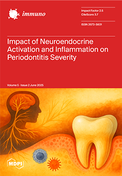
Cover Story (view full-size image): The interplay between neuroendocrine activation and inflammatory response is increasingly recognized as a contributing factor in periodontal disease. This study investigates salivary biomarkers—chromogranin A (CgA), alpha-amylase, interleukin-1β (IL-1β), and interleukin-6—in patients with different stages of periodontitis. Significantly elevated levels of CgA and IL-1β were observed in advanced cases, suggesting a potential link between stress-related biological signaling and periodontal inflammation. These findings may contribute to a better understanding of the biological mechanisms underlying periodontal disease progression. View this paper
  • Issues are regarded as officially published after their release is announced to the table of contents alert mailing list.
  • You may sign up for e-mail alerts to receive table of contents of newly released issues.
  • PDF is the official format for papers published in both, html and pdf forms. To view the papers in pdf format, click on the "PDF Full-text" link, and use the free Adobe Reader to open them.
Order results
Result details
Section
Select all
Export citation of selected articles as:
14 pages, 574 KiB  
Article
Impact of Sympathetic Nervous System Activation and Inflammatory Response on Periodontitis Severity
by Dimitar Dimitrov, Antoaneta Mlachkova, Marina Miteva, Dimitar Parvanov and Velitchka Dosseva-Panova
Immuno 2025, 5(2), 22; https://doi.org/10.3390/immuno5020022 - 5 Jun 2025
Viewed by 584
Abstract
The sympathetic nervous system (SNS) is crucial for stress response regulation and immune modulation. Prolonged SNS activation, often induced by stress exposure, disrupts immune homeostasis and intensifies inflammatory processes, contributing to periodontal disease progression. This study investigates the relationship between SNS activity and [...] Read more.
The sympathetic nervous system (SNS) is crucial for stress response regulation and immune modulation. Prolonged SNS activation, often induced by stress exposure, disrupts immune homeostasis and intensifies inflammatory processes, contributing to periodontal disease progression. This study investigates the relationship between SNS activity and periodontitis severity, utilizing salivary biomarkers chromogranin A (CgA) and alpha-amylase (sAA) alongside pro-inflammatory cytokines interleukin-1β (IL-1β) and interleukin-6 (IL-6). Saliva samples from 67 patients, categorized by periodontitis severity (Stages I/II and III/IV), were analyzed using enzyme-linked immunosorbent assay (ELISA). The results revealed significantly higher median levels of CgA (9.45 vs. 3.93 pmol/mL) and IL-1β (257.81 vs. 220.11 pg/mL) in patients with Stage III/IV periodontitis compared with those with Stage I/II, indicating heightened SNS activity and inflammatory response. Correlations between these biomarkers and clinical periodontal parameters, such as probing depth and clinical attachment loss, further support these findings. Despite elevated sAA levels in severe cases, statistical significance was not achieved. IL-6 levels also showed no significant variation across disease stages, although trends aligned with increased severity. This study highlights the interplay between SNA activation and periodontal inflammation, as evidenced by elevated salivary levels of CgA and IL-1β in patients with advanced periodontitis. By integrating neuroendocrine and inflammatory biomarkers into the diagnostic process, clinicians may be able to better identify patients at increased risk for periodontal breakdown and to consider adjunctive interventions such as stress management, thereby supporting more personalized approaches to periodontitis treatment. Full article
Show Figures

Figure 1

10 pages, 549 KiB  
Article
Complete Blood Count-Derived Biomarkers’ Association with Risk of PD-1 or PD-1/CTLA-4 Inhibitor-Induced Hypothyroidism in Patients with Solid Tumors
by Ketevan Lomidze, Nino Kikodze, Marine Gordeladze, Nino Charkviani and Tinatin Chikovani
Immuno 2025, 5(2), 21; https://doi.org/10.3390/immuno5020021 - 4 Jun 2025
Viewed by 346
Abstract
Background: A novel and highly effective strategy for tumor immunotherapy involves enhancing host immune responses against tumors through the blockade of checkpoint molecules. The most common toxicities associated with checkpoint blockade therapies include autoimmune damage to various organs. Purpose: This study aims to [...] Read more.
Background: A novel and highly effective strategy for tumor immunotherapy involves enhancing host immune responses against tumors through the blockade of checkpoint molecules. The most common toxicities associated with checkpoint blockade therapies include autoimmune damage to various organs. Purpose: This study aims to investigate hematological markers derived from complete blood counts (CBCs)—including the neutrophil-to-lymphocyte ratio (NLR), platelet-to-lymphocyte ratio (PLR), derived neutrophil-to-lymphocyte ratio (dNLR), white blood cell-to-hemoglobin ratio (WHR), neutrophils, lymphocytes, platelets, hemoglobin, red blood cell (RBC) count, neutrophil-to-RBC ratio (NRR), and neutrophil-to-hemoglobin ratio (NHR)—as potential prognostic biomarkers for the early identification of hypothyroidism in patients receiving PD-1 or PD-1/CTLA-4 immune checkpoint inhibitors. Materials and Methods: A prospective observational study was conducted on 44 patients with stage III-IV solid tumors treated with immune checkpoint (PD-1 or PD-1/CTLA-4) inhibitors. Thyroid function tests and CBC-derived biomarkers were collected at baseline, before immunotherapy. In the immunotherapy cohort, 15 of the 44 patients developed immune-related hypothyroidism, defined as overt autoimmune thyroiditis (TSH > 4.0, FT4 < 12, and anti-TPO antibodies > 30 IU/mL and/or anti-TG antibodies > 95 IU/mL) (Group 1). In comparison, 29 patients maintained normal thyroid function (Group 2). The control group comprised 14 age- and sex-matched healthy volunteers (Group 3). Statistical analyses were performed using analysis of variance (ANOVA) to compare blood parameters among the three groups (Group 1, Group 2, and Group 3) before treatment, with statistical significance set at a p-value < 0.05. Receiver operating characteristic (ROC) curve analysis was conducted to evaluate the diagnostic power of the potential prognostic biomarkers areas. The area under the curve (AUC), sensitivity, and specificity were calculated for the 44 immunotherapy patients. Results: The PLR was significantly higher (262.25 ± 162.95), while WBCs-neutrophils, the WHR, the NRR, the NHR, WBCs, neutrophils, and lymphocytes were lower (2.07 ± 0.66, 0.54 ± 0.19, 0.96 ± 0.28, 0.36 ± 0.14, 6.36 ± 2.07, 4.29 ± 1.55, and 1.23 ± 0.41, respectively) at baseline in Group 1 in comparison to Group 2. ROC curve analysis revealed that the areas under the curve (AUC) for WBCs, neutrophils, lymphocytes, WBCs-neutrophils, the PLR, the WHR, the NRR, and the NHR were 0.9, 0.87, 0.83, 0.85, 0.84, 0.92, 0.89, and 0.87, respectively. These values exceeded the threshold, indicating the high prognostic potential of each marker. Conclusions: Lower baseline levels of WBCs-neutrophils, the WHR, the NRR, the NHR, WBCs, neutrophils, and lymphocytes, along with a higher PLR, were associated with an increased risk of hypothyroidism in patients receiving PD-1 or PD-1/CTLA-4 inhibitors. These CBC-derived biomarkers represent simple, accessible, and potentially useful tools for predicting hypothyroidism in cancer patients undergoing immunotherapy. Further studies in bigger cohorts are needed to validate our findings. Full article
(This article belongs to the Section Cancer Immunology and Immunotherapy)
Show Figures

Figure 1

15 pages, 442 KiB  
Review
Relevance of Antibody-Dependent Enhancement in COVID-19
by Daniel Rodriguez-Pinto and María Sol Mendoza-Ruiz
Immuno 2025, 5(2), 20; https://doi.org/10.3390/immuno5020020 - 2 Jun 2025
Viewed by 320
Abstract
Antibody-dependent enhancement (ADE) is a well-established mechanism of pathology in several viral diseases, but its relevance in COVID-19 is not yet recognized. Although several studies in humans have shown an association between antibody responses and disease severity, long term studies addressing the presence [...] Read more.
Antibody-dependent enhancement (ADE) is a well-established mechanism of pathology in several viral diseases, but its relevance in COVID-19 is not yet recognized. Although several studies in humans have shown an association between antibody responses and disease severity, long term studies addressing the presence of antibodies before infection and their neutralization capacity are needed to establish ADE. Mechanistic studies have determined that the entry of SARS-CoV-2 into host cells can be mediated by immune complexes through Fcγ receptors or by favoring ACE2 conformation. However, the impact on viral replication is not clear. There is evidence for enhancing effects of immune complexes on Fcγ receptor-mediated effector mechanisms and cytokine secretion after modulation of cell signaling in immune cells, specially by antibodies with altered glycosylation, which points to ADE that can contribute to COVID-19 pathology. However, more studies are needed to determine the impact of antibodies both in naturally infected and vaccinated subjects, which can lead to their use as a prognostic marker and increase vaccine safety. Full article
(This article belongs to the Section Infectious Immunology and Vaccines)
Show Figures

Figure 1

21 pages, 946 KiB  
Review
Immunomodulatory and Anti-Inflammatory Properties of Honey and Bee Products
by Bashar Saad
Immuno 2025, 5(2), 19; https://doi.org/10.3390/immuno5020019 - 30 May 2025
Viewed by 629
Abstract
Honey and other bee products, including propolis, royal jelly, and bee pollen, are widely recognized for their medicinal properties. Among their numerous biological activities, their anti-inflammatory and immunomodulatory effects have gained significant attention in recent years. Immune and inflammatory disorders contribute significantly to [...] Read more.
Honey and other bee products, including propolis, royal jelly, and bee pollen, are widely recognized for their medicinal properties. Among their numerous biological activities, their anti-inflammatory and immunomodulatory effects have gained significant attention in recent years. Immune and inflammatory disorders contribute significantly to the development of chronic conditions, including cancer and diabetes. Bee-derived products, along with their bioactive compounds such as polyphenols, have shown promising therapeutic effects in modulating inflammatory mediators. Studies indicate that these products help regulate tumor necrosis factor-alpha (TNF-α), interleukin-1 beta (IL-1β), and interleukin-6 (IL-6), and interleukin-7 (IL-7) levels while reducing reactive oxygen species (ROS) production. Additionally, both in vitro and in vivo research, along with clinical studies, highlight their role in enhancing immune responses by activating B and T lymphocytes. This review explores the molecular mechanisms underlying these properties, emphasizing the role of bioactive compounds such as flavonoids, phenolic acids, and proteins in modulating immune responses and reducing inflammation. Evidence from in vitro, in vivo, and clinical studies suggests that honey and bee products influence cytokine production, regulate immune cell activity, and mitigate oxidative stress, making them potential therapeutic agents for inflammatory and immune-related disorders. To gather relevant information, databases such as Google Scholar, PubMed, and ScienceDirect were searched using various keyword combinations, including immunomodulatory, anti-inflammatory, bee products, honey, propolis, royal jelly, bee venom, and bee pollen. Given their anti-inflammatory, immune-protective, antioxidant, anti-apoptotic, and antimicrobial properties, bee products remain a subject of interest for further clinical evaluation. Full article
Show Figures

Figure 1

28 pages, 992 KiB  
Review
Efficacy of Intravenous Immunoglobulins and Other Immunotherapies in Neurological Disorders and Immunological Mechanisms Involved
by Angel Justiz-Vaillant, Sachin Soodeen, Odalis Asin-Milan, Julio Morales-Esquivel and Rodolfo Arozarena-Fundora
Immuno 2025, 5(2), 18; https://doi.org/10.3390/immuno5020018 - 26 May 2025
Viewed by 418
Abstract
This review aims to explore the role of immunotherapeutic strategies—primarily intravenous immunoglobulin (IVIG), plasma exchange (PLEX), and selected immunomodulatory agents—in the treatment of neurological and psychiatric disorders with suspected or confirmed autoimmune mechanisms. A central focus is placed on understanding the immunopathology of [...] Read more.
This review aims to explore the role of immunotherapeutic strategies—primarily intravenous immunoglobulin (IVIG), plasma exchange (PLEX), and selected immunomodulatory agents—in the treatment of neurological and psychiatric disorders with suspected or confirmed autoimmune mechanisms. A central focus is placed on understanding the immunopathology of these conditions through the identification and characterization of disease-associated autoantibodies. Disorders such as autoimmune encephalitis, myasthenia gravis, limbic epilepsy, neuropsychiatric systemic lupus erythematosus (NPSLE), and certain forms of schizophrenia have shown clinical responses to immunotherapy, suggesting an underlying autoimmune basis in a subset of patients. The review also highlights the diagnostic relevance of detecting autoantibodies targeting neuronal receptors, such as NMDA and AMPA receptors, or neuromuscular junction components, as biomarkers that guide therapeutic decisions. Furthermore, we synthesize findings from published randomized controlled trials (RCTs) that have validated the efficacy of IVIG and PLEX in specific diseases, such as Guillain–Barré syndrome, and myasthenia gravis. Emerging clinical evidence supports expanding these treatments to other conditions where autoimmunity is implicated. By integrating immunological insights with clinical trial data, this review offers a comprehensive perspective on how immunotherapies may be tailored to target autoimmune contributors to neuropsychiatric disease. Full article
Show Figures

Graphical abstract

9 pages, 831 KiB  
Brief Report
Recurrent SARS-CoV-2 Infection Is Linked to the TLR7 rs179008 Variant and Related to Diminished Baseline T Cell Immunity
by Camilla Natália Oliveira Santos, Priscila Lima dos Santos, Angela Maria da Silva and Lucas Sousa Magalhães
Immuno 2025, 5(2), 17; https://doi.org/10.3390/immuno5020017 - 16 May 2025
Viewed by 385
Abstract
Recurrent COVID-19, defined as two or more distinct episodes, may reflect an impaired immune response to SARS-CoV-2. In this case–control study, we compared three groups: individuals with recurrent COVID-19, those with a single episode, and SARS-CoV-2-naïve controls. We genotyped six immune-related SNPs, including [...] Read more.
Recurrent COVID-19, defined as two or more distinct episodes, may reflect an impaired immune response to SARS-CoV-2. In this case–control study, we compared three groups: individuals with recurrent COVID-19, those with a single episode, and SARS-CoV-2-naïve controls. We genotyped six immune-related SNPs, including TLR7 rs179008, and measured CD4+ and CD8+ T cell responses to SARS-CoV-2 antigens using flow cytometry. The T allele of TLR7 rs179008, previously linked to reduced receptor expression, was significantly overrepresented in the recurrent COVID-19 cohort. At baseline, frequencies of IFN-γ+, IL-2+, and TNF-α+ cells among CD4+ and CD8+ T cells did not differ between groups. However, stratification by the rs179008 genotype revealed that T allele carriers displayed diminished IFN-γ production in both CD4+ and CD8+ T cells and reduced IL-2 production in CD4+ T cells. Following vaccination, T cell responses were comparable across all genotypes. The T allele of TLR7 rs179008 is associated with recurrent COVID-19 and may contribute to impaired T cell-mediated immunity. Further studies are warranted to elucidate the mechanistic role of TLR7 variation in SARS-CoV-2 reinfection risk. Full article
(This article belongs to the Section Infectious Immunology and Vaccines)
Show Figures

Figure 1

12 pages, 1254 KiB  
Article
Pre- and Post-Transplant Anti-BKV IgG Responses and HLA Associations in BK Virus Reactivation Among Renal Transplant Recipients
by Deema Ibrahim Fallatah and Steve Christmas
Immuno 2025, 5(2), 16; https://doi.org/10.3390/immuno5020016 - 9 May 2025
Viewed by 331
Abstract
BK virus (BKV) reactivation is a significant complication in renal transplant recipients, often leading to BK viremia and BK virus-associated nephropathy (BKVAN), which can compromise graft survival. While the routine monitoring of BKV DNA in blood aids in early detection, identifying pre-transplant risk [...] Read more.
BK virus (BKV) reactivation is a significant complication in renal transplant recipients, often leading to BK viremia and BK virus-associated nephropathy (BKVAN), which can compromise graft survival. While the routine monitoring of BKV DNA in blood aids in early detection, identifying pre-transplant risk factors remains a challenge. This study investigates the role of pre- and post-transplant anti-BKV IgG levels and human leukocyte antigen (HLA) alleles in predicting BKV reactivation. The hospital-based cross-sectional study was conducted on 38 renal transplant recipients, stratified into viremic, non-viremic, and BKVAN groups. Anti-BKV IgG levels were measured pre-transplant, at viremia onset, and post-viremia using ELISA. BKV DNA was detected via qPCR, and HLA typing was performed using sequence-specific oligonucleotide probe (SSOP) hybridization. Statistical analyses included Kaplan–Meier survival curves and Cox regression models. Pre-transplant anti-BKV IgG seropositivity was higher in viremic (94%) and BKVAN (100%) patients than in non-viremic recipients (66.6%). Post-transplant IgG levels increased significantly in viremic recipients (p < 0.05). HLA-B44 and HLA-DR15 were significantly associated with increased BKV viremia risk (p = 0.02 and p = 0.01, respectively). Pre-transplant anti-BKV IgG levels and specific HLA alleles influence BKV reactivation risk. These findings highlight the potential for integrating serological and genetic screening into pre-transplant assessments to improve risk stratification and post-transplant monitoring strategies. Full article
Show Figures

Figure 1

21 pages, 2561 KiB  
Article
Indole-3-Carbinol Enhances Alternative Activation of Macrophages via AHR Pathway and Glucose Transporter Regulation
by Delara Omrani, Saeed Mohammadi, Moein Malekzadeh, Mohsen Saeidi, Fakhri Sadat Seyedhosseini, Ahmed Al-Harrasi and Yaghoub Yazdani
Immuno 2025, 5(2), 15; https://doi.org/10.3390/immuno5020015 - 2 May 2025
Viewed by 431
Abstract
Disruption in macrophage polarization is linked to inflammatory diseases and metabolic disorders. Our study aimed to investigate how AHR activation by I3C and TCDD could impact glucose transporters and macrophage phenotypes and functions in human macrophages. Human monocyte-derived macrophages (hMDMs) and THP-1 cell-derived [...] Read more.
Disruption in macrophage polarization is linked to inflammatory diseases and metabolic disorders. Our study aimed to investigate how AHR activation by I3C and TCDD could impact glucose transporters and macrophage phenotypes and functions in human macrophages. Human monocyte-derived macrophages (hMDMs) and THP-1 cell-derived macrophage-like cells were treated for 24 h with 100 ng/mL LPS, 100 nM TCDD, and 10 ng/µL I3C. CYP1A1 and CYP1B1 expression was significantly increased in the I3C and TCDD treatments, with CYP1B1 showing a higher fold change in I3C compared to TCDD. The AHRR expression was the highest in the TCDD group. For macrophage polarization, I3C significantly elevated CD163 expression while reducing CD16 and CD86, indicative of M2-like polarization. Additionally, I3C promoted ARG1 expression and reduced NOS2 levels, while TCDD increased NOS2. A cytokine analysis revealed I3C-induced upregulation of IL-10 and TGF-β, while TCDD significantly elevated TNF-α and IL-12. I3C upregulated glucose transporter genes (GLUT1, GLUT3, GLUT6), in contrast to the downregulation observed in TCDD-treated cells. Our findings demonstrated that I3C distinctly modulates AHR activation genes, macrophage polarization, cytokine expression, and glucose transporter levels in THP-1 cells compared to the TCDD and LPS treatments. Our findings suggest that I3C favors an anti-inflammatory M2-like macrophage polarization coupled with enhanced metabolic activity. Full article
(This article belongs to the Section Innate Immunity and Inflammation)
Show Figures

Figure 1

9 pages, 900 KiB  
Systematic Review
Turner Syndrome Increases the Risk of Psoriasis: A Systematic Review and Meta-Analysis
by Jirat Chenbhanich, Ben Ponvilawan and Patompong Ungprasert
Immuno 2025, 5(2), 14; https://doi.org/10.3390/immuno5020014 - 17 Apr 2025
Viewed by 818
Abstract
Aims: Patients with Turner syndrome (TS) may have a higher risk of psoriasis as suggested by some reports. Data on this association are still limited. We investigated the association between TS and the risk of prevalent and incident psoriasis by combining results from [...] Read more.
Aims: Patients with Turner syndrome (TS) may have a higher risk of psoriasis as suggested by some reports. Data on this association are still limited. We investigated the association between TS and the risk of prevalent and incident psoriasis by combining results from available studies using systematic reviews and meta-analysis techniques. Methods: Potentially eligible studies were identified from Medline and EMBASE databases from inception to December 2023 using a search strategy that comprised of terms for “Turner syndrome” and “psoriasis”. An eligible cohort study must comprise of two groups of participants—those with and without TS. It must report our outcome of interest—incidence and/or prevalence of psoriasis in each group. The pooled effect estimates were generated using the generic inverse variance method, which assigns weight to each study in reversal to its variance. Meta-analyses of the prevalent and incident psoriasis were conducted separately. Results: A total of 4919 articles were retrieved. After two rounds of independent review by two investigators, five cohort studies (two incident studies and three prevalent studies) met the eligibility criteria and were included in the meta-analyses. The meta-analyses found a significantly elevated risk of both incident and prevalent psoriasis in patients with TS compared to individuals without TS, with the pooled risk ratio of 5.58 (95% CI, 3.73–8.35; I2 0%) and 5.66 (95% CI, 1.52–21.03; I2 19%), respectively. Conclusions: An increased risk of both incident and prevalent psoriasis among patients with TS was demonstrated in this study. Full article
Show Figures

Figure 1

23 pages, 2184 KiB  
Review
Role of Inflammatory Mediators in Chronic Obstructive Pulmonary Disease Pathogenesis: Updates and Perspectives
by Pankush, Khushboo Bharti, Rohit Pandey, Namita Srivastava, Shashank Kashyap, Deepak Kumar, Lokender Kumar, Sunil K. Suman and Sanjay K. S. Patel
Immuno 2025, 5(2), 13; https://doi.org/10.3390/immuno5020013 - 15 Apr 2025
Viewed by 1742
Abstract
Chronic obstructive pulmonary disease (COPD) is a chronic, debilitating condition that affects the lungs and airways. It is characterized by persistent bronchitis, a condition exemplified by the inflammation of the bronchial tubes, the hypersecretion of mucus, emphysema, and the destruction of the airway [...] Read more.
Chronic obstructive pulmonary disease (COPD) is a chronic, debilitating condition that affects the lungs and airways. It is characterized by persistent bronchitis, a condition exemplified by the inflammation of the bronchial tubes, the hypersecretion of mucus, emphysema, and the destruction of the airway parenchyma. The combination of these conditions leads to persistent tissue damage, pulmonary fibrosis, and ongoing inflammation of the airways. The inflammatory response in COPD is a complex process that is orchestrated by a wide range of immune cells. These include lung epithelial cells, monocytes, macrophages, neutrophils, eosinophils, and T and B lymphocytes, among others. These cells work together to produce a wide range of inflammatory biomarkers that are involved in the pathogenesis of COPD. Some of the key inflammatory biomarkers that have been identified in COPD include a variety of cytokines, the C-reactive protein/serum albumin ratio, fibrinogen, soluble receptor for advanced glycation endproducts, club/clara cells in the lungs with a molecular weight of 16 kDa, surfactant protein D, adiponectin, reactive oxygen species, and proteases. This review aims to provide a comprehensive overview of the role of immune cells and key inflammatory biomarkers in the development and progression of COPD. It will delve into the intricacies of the inflammatory response in COPD, exploring the various cell types and biomarkers that are involved in this process. By understanding the underlying mechanisms that drive COPD, we can better develop targeted treatments that can help to alleviate the symptoms of COPD. Full article
Show Figures

Figure 1

Previous Issue
Next Issue
Back to TopTop