Brief Report

Recurrent SARS-CoV-2 Infection Is Linked to the TLR7 rs179008 Variant and Related to Diminished Baseline T Cell Immunity

by
Camilla Natália Oliveira Santos
1,2,
Priscila Lima dos Santos
1,2,
Angela Maria da Silva
1,2 and
Lucas Sousa Magalhães
1,3,*
1
Laboratory of Immunology and Molecular Biology, Universidade Federal de Sergipe, Aracaju 49060-108, Brazil
2
Health Sciences Graduate Program, Universidade Federal de Sergipe, Aracaju 49060-108, Brazil
3
Institute of Biological and Health Sciences, Universidade Federal de Alagoas, Maceió 57072-900, Brazil
*
Author to whom correspondence should be addressed.
Immuno 2025, 5(2), 17; https://doi.org/10.3390/immuno5020017
Submission received: 8 February 2025 / Revised: 8 May 2025 / Accepted: 13 May 2025 / Published: 16 May 2025
(This article belongs to the Section Infectious Immunology and Vaccines)

Abstract

Recurrent COVID-19, defined as two or more distinct episodes, may reflect an impaired immune response to SARS-CoV-2. In this case–control study, we compared three groups: individuals with recurrent COVID-19, those with a single episode, and SARS-CoV-2-naïve controls. We genotyped six immune-related SNPs, including TLR7 rs179008, and measured CD4+ and CD8+ T cell responses to SARS-CoV-2 antigens using flow cytometry. The T allele of TLR7 rs179008, previously linked to reduced receptor expression, was significantly overrepresented in the recurrent COVID-19 cohort. At baseline, frequencies of IFN-γ+, IL-2+, and TNF-α+ cells among CD4+ and CD8+ T cells did not differ between groups. However, stratification by the rs179008 genotype revealed that T allele carriers displayed diminished IFN-γ production in both CD4+ and CD8+ T cells and reduced IL-2 production in CD4+ T cells. Following vaccination, T cell responses were comparable across all genotypes. The T allele of TLR7 rs179008 is associated with recurrent COVID-19 and may contribute to impaired T cell-mediated immunity. Further studies are warranted to elucidate the mechanistic role of TLR7 variation in SARS-CoV-2 reinfection risk.

1. Introduction

Coronavirus disease 2019 (COVID-19) is a highly transmissible respiratory illness caused by the novel betacoronavirus SARS-CoV-2, first identified in Wuhan, China, in December 2019 [1]. Clinical manifestations range from asymptomatic infection to severe acute respiratory distress syndrome (ARDS) and multisystem complications [2]. SARS-CoV-2 gains entry via the ACE2 receptor on airway epithelial and other host cells, triggering an inflammatory cascade in which the balance between protective type I interferon (IFN-I) signaling and dysregulated cytokine release largely determines disease severity [3,4,5,6,7,8,9]. Toll-like receptors (TLRs) are central to innate viral recognition, with TLR7 sensing single-stranded RNA and orchestrating downstream IFN-I and T cell responses [10,11]. Genetic variation in TLR7 has been implicated in both life-threatening COVID-19 and differences in antiviral immunity, yet its role in susceptibility to multiple distinct SARS-CoV-2 episodes, recurrent COVID-19, remains unexplored.
Recurrent COVID-19, defined as two or more PCR-confirmed episodes separated by an asymptomatic interval, has been reported in a subset of patients and may reflect incomplete immune protection or viral persistence [12]. Previous work, including our own, has linked recurrent infection to demographic factors, blood type, and lower anti-SARS-CoV-2 antibody titers [13,14,15], but T cell functionality and host immunogenetics in this context are poorly understood.
In this study, we genotyped six immune-related SNPs (including TLR7 rs179008) and measured SARS-CoV-2-specific CD4+ and CD8+ T cell cytokine responses at baseline and after vaccination, comparing individuals with recurrent COVID-19 to single-episode and naïve controls. Our goal was to test whether the TLR7 rs179008 T allele predisposes to reinfection and to elucidate its impact on T cell-mediated immunity.

2. Materials and Methods

2.1. Ethical Aspects

The Ethical Committee of the Federal University of Sergipe approved this study (approval number 4.018.577). All participants, including COVID-19 patients and non-infected volunteers, were invited to join and provided written informed consent.

2.2. Study Design and Sampling

We designed a case series with a control group, assigning participants to three groups: individuals with two or more confirmed episodes of COVID-19, verified by clinical and laboratory evaluations (n = 20); individuals who had only one confirmed episode and no prior or subsequent disease at the time of the sample collection (n = 14); and volunteers with no history of COVID-19 (n = 12), confirmed by the absence of clinical symptoms and IgG anti-SARS-CoV-2. These group characteristics persisted for up to one year after sample collection. In the recurrent COVID-19 group, the mean age was 42.9 years, 95% were female, and 95% experienced symptoms during their disease episodes. The single COVID-19 group had a mean age of 42.0 years, 35% were female, and 71.4% were symptomatic. The control group had a mean age of 41.6 years, 92.3% were female, and, based on the inclusion criteria, had no symptomatic episodes.
Sampling was performed during 2020, and participant characteristics have been detailed previously [13,15]. Recurrent COVID-19 was defined by the reappearance of symptoms after a quarantine period of 14 days, complete clinical recovery, and at least 30 days without symptoms, in addition to the absence of SARS-CoV-2 as detected by qRT-PCR during both the initial and subsequent COVID-19 episodes. Initial classifications were based on clinical diagnoses, which were then confirmed through laboratory analyses, including qRT-PCR and IgG anti-SARS-CoV-2 tests. For the single COVID-19-episode group, only clinical criteria were used to rule out additional episodes, while in the non-infected group, the absence of a disease history was confirmed using IgG anti-SARS-CoV-2 testing alongside the lack of clinical symptoms. Participants were evaluated through interviews and had blood samples collected at two time points: first, between 30 and 90 days after the last COVID-19 episode, and second, 30 days following vaccination. Participants received CoronaVac, AstraZeneca (AZD1222, Cambridge, UK), or Pfizer (BNT162b2, New York, NY, USA) vaccines based on the Brazilian government’s distribution policy; vaccine type was not an inclusion or exclusion criterion. Consequently, vaccination was not matched across the three groups, although most participants in each group received CoronaVac (around 60%), followed by AstraZeneca (around 25%). Blood samples were processed according to the protocols outlined in the subsequent sections.
For SNP analyses, the case group comprised the Recurrent-COV cohort, while the control group included Single-COV and COVID-naïve participants. For the FACS analyses, the original three-group classification was used: Recurrent-COV, Single-COV, and Control.

2.3. Analysis of Single Nucleotide Polymorphisms

We performed genetic analyses on genomic DNA extracted from whole blood, focusing on the following single nucleotide polymorphisms (SNPs): rs2074192 (angiotensin-converting enzyme 2, ACE2), rs4073 (CXC motif chemokine ligand 8, CXCL8), rs4508917 (CXCL10), rs3132468 and rs3131639 (MHC class I polypeptide-related sequence B, MICB), and rs179008 (Toll-like receptor 7, TLR7), selected due to previous association to COVID-19 or other viral infectious diseases. All SNPs were identified using TaqMan probes and quantitative PCR, as previously explained and published [16,17].

2.4. Characterization of Immune Response

We performed flow cytometry on thawed peripheral blood mononuclear cells (PBMCs) following established protocols from our group [18]. Briefly, thawed cells were allowed to rest for 4 h and then stimulated for 18 h with 1 µg/mL of a truncated spike protein fragment containing 417 amino acids, as previously described [19]. Brefeldin A was added for the last 12 h to prevent cytokine release into the media. After stimulation, the cells were processed for flow cytometry and stained using a panel of antibodies to measure IFN-γ, IL-2, and TNF-α levels in CD4+ and CD8+ T cells. Data acquisition was performed on a FACSCanto II, with 30,000 cells acquired in the lymphocyte gate. Analysis was conducted using FlowJo (https://www.flowjo.com/).

3. Results and Discussion

As previously explained, we analyzed SNPs in five different genes: ACE2, CXCL8, CXCL10, MICB, and TLR7. First, the Hardy–Weinberg analysis showed that all groups were in equilibrium for the SNPs, except for the rs179008 SNP in TLR7 in the control group. This deviation could be due to the limited sample size and the SNP’s association with the disease [20]. Furthermore, it is important to note that TLR7 is located on the X chromosome, and the differing proportions of males and females between the recurrent COVID-19 (case) group and the single COVID-19 and control groups may introduce imbalance in Hardy–Weinberg equilibrium analyses [11]. Regarding disease association analyses, the rs179008 SNP in TLR7 was significantly associated with recurrent COVID-19 in two models of analysis (Table 1). Specifically, the heterozygous genotype (AT) was associated with recurrent disease episodes in both a codominant model (p = 0.017) and an overdominant model (p = 0.019), in which the heterozygous genotype was compared to both homozygous genotypes. No other tested SNPs showed a similar association with recurrent or single COVID-19 in this study population (Supplementary Table S1).
Toll-like receptor 7 (TLR7) is crucial to the innate immune response and is encoded on the X chromosome. TLR7 detects single-stranded viral RNA (ssRNA), thereby initiating signaling pathways that induce pro-inflammatory cytokines and type I interferons [21]. The rs179008 SNP (A > T) within TLR7 results in the substitution of glutamine for leucine, affecting both TLR7 function and expression. The T allele is associated with the lower TLR7 expression [22]. Previous studies have linked polymorphisms in the TLR7 gene to COVID-19 severity [23,24], as well as to HIV infection and its immune correlates [22,25]. Notably, this variant has been tied to diminished type I interferon production, reducing IFN levels in plasmacytoid dendritic cells even in heterozygous women [22]. As previously mentioned, the TLR7 gene is located on the X chromosome, and women exhibit higher TLR7 protein expression, which may underlie their enhanced antiviral responses [26]. Buschow et al. (2018) demonstrated that some molecules, such as CD40, exhibit similarly elevated expression in plasmacytoid dendritic cells from both males and females following TLR7 stimulation, whereas others, such as CD86, increase more markedly in females [27]. Similarly, in HTLV-1-infected patients, circulating type I IFN and TNF-α levels and TLR7 expression were comparable between A and T allele carriers in both males and females [28]. Also, the presence of hemizygous and homozygous for the TLR7 gene was previously used in association studies with similar response between these two characteristics [25,29]. Thus, we could infer that the T allele variant, regardless of sex, may contribute to impaired innate and T cell responses.
Indeed, TLR7 signaling has been shown to play multiple roles in the immune response. Rubtsova et al. (2016) demonstrated that TLR7 activation induces IFN-γ production in CD4 and CD8 T cells, which in turn enhances IgG production by activated B cells via antibody class switching, leading to improved antiviral immunity [30]. Complementarily, a previous study showed that TLR7 signaling augments CD8 T cell effector functions by boosting cellular metabolism, specifically glycolysis [31]. Conversely, TLR7 signaling has been shown to induce anergy in CD4 T cells, promoting a tolerance response in vitro during HIV infection, indicating that, in addition to its activating functions, TLR7 can also inhibit T cell responses [32]. Thus, TLR7 clearly contributes to the mounting immune response against viral pathogens, including SARS-CoV-2.
Building on our previous findings in recurrent COVID-19 patients and the newly identified association with a TLR7 variant, we evaluated T cell responses to SARS-CoV-2 antigens. As shown in Figure 1, the proportions of IFN-γ+, IL-2+, and TNF-α+ CD4+ and CD8+ T cells did not differ among the recurrent COVID-19, single-episode COVID-19, and control groups. Consistent with our earlier work demonstrating similar levels of multifunctional T cells across these groups, these data indicate that SARS-CoV-2-specific T cell responses are comparable between patients with recurrent and single-episode COVID-19.
Given this background, we aimed to investigate whether the TLR7 rs179008 SNP influences the immune response in COVID-19 patients, particularly with respect to T cell activation. In the absence of a specific TLR7 agonist, we instead evaluated SARS-CoV-2-specific responses in CD4 and CD8 T cells. We observed significant differences in IFN-γ responses in both CD4+ and CD8+ T cells when comparing the AT + TT genotypes to the AA genotype among patients with either recurrent or single episodes of COVID-19. Specifically, patients carrying the AT + TT genotypes exhibited lower levels of IFN-γ+ CD4+ and CD8+ T cells (Figure 2a). Additionally, lower levels of IL-2+ CD4+ T cells were observed in AT + TT genotype carriers compared to those with the AA genotype (Figure 2b). No differences were observed between genotype groups regarding IL-2+ CD8+ T cells or TNF-α+ T cells phenotype (Figure 2c). Additionally, we evaluated whether patient sex could act as a confounding factor. The analysis indicated similar proportions of men in both genotype groups, 24.1% in AA patients and 16.7% in AT + TT patients. No differences were observed in the frequencies of IFN-γ+, IL-2+, or TNF-α+ cells within CD4+ or CD8+ T cell populations across genotypes.
Additionally, we assessed whether the TLR7 genotype influenced responses within each study group by stratifying participants as AA or AT + TT. Although overall differences in the percentages of CD4+ and CD8+ T cells expressing IFN-γ, IL-2, and TNF-α were not statistically significant (Supplementary Figure S1), AA genotype carriers exhibited a trend toward higher levels of IFN-γ+ CD4+ and CD8+ T cells. This difference reached borderline significance in the recurrent COVID-19 group when comparing AA to AT + TT individuals (Supplementary Figure S1a).
Although studies on T cell responses in carriers of the rs179008 T allele are limited, previous data have shown reduced type I interferon production in such individuals in the context of HIV [22], HCV [29], and HTLV-1 [28]. Consistent with these findings, our results indicate an impaired IFN-γ response in T cells from patients harboring the TLR7 variant. The type I IFN response, triggered by TLR7 recognition of single-stranded RNA viruses, is crucial for viral control [33]. Moreover, TLR7 signaling enhances IFN-γ production in CD4+ and CD8+ T cells [30,31] and induces IFN-γ release by macrophages and dendritic cells [22,34]. A limitation of our study is that we did not measure TLR7 transcript levels directly; therefore, we hypothesize that reduced functional TLR7 contributes to COVID-19 recurrence, echoing previous reports linking TLR7 loss-of-function to severe disease outcomes [23,24,35]. Notably, our study included both sexes: men are hemizygous for TLR7, which may affect receptor expression and downstream signaling. Indeed, women may be more protected against SARS-CoV-2 due to biallelic TLR7 activation [36], whereas loss-of-function TLR7 variants in men correlate with poorer COVID-19 prognosis [37]. Interestingly, IFN-γ+ CD4+ and CD8+ T cell levels were marginally higher in AA genotype patients with recurrent COVID-19, suggesting that despite the potential immune dysfunction associated with multiple disease episodes [12], AA carriers mount a stronger memory response than AT/TT carriers, further implicating the T allele in impaired IFN-γ production.
Nonetheless, we investigated whether vaccination could normalize the IFN response in T cells from patients with the AT + TT genotypes. The results indicate that after vaccination, patients with the T genotype maintained IFN-γ response levels similar to those with the AA genotype (Supplementary Figure S2). Additionally, IL-2 and TNF-α levels were similar for both genotype groups following vaccination.
These findings indicate that a genotype associated with the altered TLR7 gene, which may impair initial viral RNA recognition and contribute to disease recurrence, patients carrying the T allele are still capable of mounting antigen-specific immune responses, likely via B-cell activation and antibody production. Moreover, vaccination in these individuals elicited a markedly enhanced interferon response. Thus, vaccination confers a robust protective immunity, especially in patients susceptible to COVID-19.
Notably, this brief report has several limitations. First, our analysis included both men and women for a gene located on the X chromosome, which may introduce sex-related biases. Second, the sample size was relatively small, limiting statistical power. Third, we were unable to directly assess TLR7 signaling in T cell activation. Also, it is important to clarify that circulating T cells do not reflect the entire T cell pool. Collectively, these factors constrain the interpretation of our findings.

4. Conclusions

In conclusion, our results demonstrate that patients with recurrent COVID-19 exhibit frequencies of IFN-γ+, IL-2+, and TNF-α+ CD4+ and CD8+ T cells comparable to those observed after a single disease episode. Moreover, the TLR7 rs179008 variant was significantly associated with recurrent COVID-19 and correlated with a partial impairment of both CD4+ and CD8+ T cell responses.

Supplementary Materials

The following supporting information can be downloaded at https://www.mdpi.com/article/10.3390/immuno5020017/s1. Figure S1: Evaluation of T cell memory in PBMCs stimulated with SARS-CoV-2 S1 protein for 18 h; Figure S2: Effect of vaccination on the response of T cells accordingly to SNP genotype. Table S1. Genotype frequencies of ACE2, CXCL8, MICB, and CXCL10 SNPs and its association with recurrent COVID-19.

Author Contributions

Conceptualization: C.N.O.S. and L.S.M.; Investigation: C.N.O.S., L.S.M. and P.L.d.S.; Formal analysis: C.N.O.S. and L.S.M.; Supervision: A.M.d.S.; Writing—original draft: C.N.O.S. and L.S.M.; Writing—review and editing: all authors. All authors have read and agreed to the published version of the manuscript.

Funding

This study was not supported by any funding agencies. L.S.M. received a postdoctoral fellowship from Conselho Nacional de Desenvolvimento Científico e Tecnológico (151365/2022-9).

Institutional Review Board Statement

This study was approved by the Research Ethical Committee of the Universidade Federal de Sergipe (advise number 4.018.577).

Informed Consent Statement

Written informed consent was obtained from all participants.

Data Availability Statement

Additional data are available from the corresponding author upon request.

Acknowledgments

The authors are grateful to all members of the Laboratório de Imunologia and Biologia Molecular, especially Roque de Almeida and Amélia de Jesus.

Conflicts of Interest

The authors have no relevant financial or non-financial interests to disclose.

References

  1. Jin, Y.; Yang, H.; Ji, W.; Wu, W.; Chen, S.; Zhang, W.; Duan, G. Virology, Epidemiology, Pathogenesis, and Control of COVID-19. Viruses 2020, 12, 372. [Google Scholar] [CrossRef] [PubMed]
  2. Castro, M.C.; Kim, S.; Barberia, L.; Ribeiro, A.F.; Gurzenda, S.; Ribeiro, K.B.; Abbott, E.; Blossom, J.; Rache, B.; Singer, B.H. Spatiotemporal Pattern of COVID-19 Spread in Brazil. Science 2021, 372, 821–826. [Google Scholar] [CrossRef] [PubMed]
  3. Chvatal-Medina, M.; Mendez-Cortina, Y.; Patiño, P.J.; Velilla, P.A.; Rugeles, M.T. Antibody Responses in COVID-19: A Review. Front. Immunol. 2021, 12, 633184. [Google Scholar] [CrossRef] [PubMed]
  4. Tay, M.Z.; Poh, C.M.; Rénia, L.; MacAry, P.A.; Ng, L.F.P. The Trinity of COVID-19: Immunity, Inflammation and Intervention. Nat. Rev. Immunol. 2020, 20, 363–374. [Google Scholar] [CrossRef]
  5. Parasher, A. COVID-19: Current Understanding of Its Pathophysiology, Clinical Presentation and Treatment. Postgrad. Med. J. 2021, 97, 312–320. [Google Scholar] [CrossRef]
  6. Ye, Q.; Wang, B.; Mao, J. The Pathogenesis and Treatment of the ‘Cytokine Storm’’ in COVID-19’. J. Infect. 2020, 80, 607–613. [Google Scholar] [CrossRef]
  7. Rha, M.S.; Shin, E.C. Activation or Exhaustion of CD8+ T Cells in Patients with COVID-19. Cell. Mol. Immunol. 2021, 18, 2325–2333. [Google Scholar] [CrossRef]
  8. Mathew, D.; Giles, J.R.; Baxter, A.E.; Oldridge, D.A.; Greenplate, A.R.; Wu, J.E.; Alanio, C.; Kuri-Cervantes, L.; Pampena, M.B.; D’Andrea, K.; et al. Deep Immune Profiling of COVID-19 Patients Reveals Distinct Immunotypes with Therapeutic Implications. Science 2020, 8511, eabc8511. [Google Scholar] [CrossRef]
  9. Möhlendick, B.; Schönfelder, K.; Breuckmann, K.; Elsner, C.; Babel, N.; Balfanz, P.; Dahl, E.; Dreher, M.; Fistera, D.; Herbstreit, F.; et al. ACE2 Polymorphism and Susceptibility for SARS-CoV-2 Infection and Severity of COVID-19. Pharmacogenet Genom. 2021, 31, 165–171. [Google Scholar] [CrossRef]
  10. Jung, H.E.; Lee, H.K. Current Understanding of the Innate Control of Toll-like Receptors in Response to SARS-CoV-2 Infection. Viruses 2021, 13, 2132. [Google Scholar] [CrossRef]
  11. Dai, J.; Wang, Y.; Wang, H.; Gao, Z.; Wang, Y.; Fang, M.; Shi, S.; Zhang, P.; Wang, H.; Su, Y.; et al. Toll-Like Receptor Signaling in Severe Acute Respiratory Syndrome Coronavirus 2-Induced Innate Immune Responses and the Potential Application Value of Toll-Like Receptor Immunomodulators in Patients with Coronavirus Disease 2019. Front. Microbiol. 2022, 13, 948770. [Google Scholar] [CrossRef] [PubMed]
  12. Azam, M.; Sulistiana, R.; Ratnawati, M.; Fibriana, A.I.; Bahrudin, U.; Widyaningrum, D.; Aljunid, S.M. Recurrent SARS-CoV-2 RNA Positivity after COVID-19: A Systematic Review and Meta-Analysis. Sci. Rep. 2020, 10, 20692. [Google Scholar] [CrossRef] [PubMed]
  13. Santos, C.N.O.; Caldas, G.C.; de Oliveira, F.A.; da Silva, A.M.; da Silva, J.S.; da Silva, R.L.L.; de Jesus, A.R.; Magalhães, L.S.; de Almeida, R.P. COVID-19 Recurrence Is Related to Disease-Early Profile T Cells While Detection of Anti-S1 IgG Is Related to Multifunctional T Cells. Med. Microbiol. Immunol. 2023, 212, 339–347. [Google Scholar] [CrossRef] [PubMed]
  14. De Castro, M.V.; Santos, K.S.; Apostolico, J.S.; Fernandes, E.R.; Almeida, R.R.; Levin, G.; Magawa, J.Y.; Nunes, J.P.S.; Bruni, M.; Yamamoto, M.M.; et al. Recurrence of COVID-19 Associated with Reduced T-Cell Responses in a Monozygotic Twin Pair. Open Biol. 2022, 12, 210240. [Google Scholar] [CrossRef]
  15. Adrielle dos Santos, L.; Filho, P.G.d.G.; Silva, A.M.F.; Santos, J.V.G.; Santos, D.S.; Aquino, M.M.; de Jesus, R.M.; Almeida, M.L.D.; da Silva, J.S.; Altmann, D.M.; et al. Recurrent COVID-19 Including Evidence of Reinfection and Enhanced Severity in Thirty Brazilian Healthcare Workers. J. Infect. 2021, 82, 399–406. [Google Scholar] [CrossRef]
  16. Santos, C.N.O.; Magalhães, L.S.; Fonseca, A.B.d.L.; Bispo, A.J.B.; Porto, R.L.S.; Alves, J.C.; dos Santos, C.A.; de Carvalho, J.V.; da Silva, A.M.; Teixeira, M.M.; et al. Association between Genetic Variants in TREM1, CXCL10, IL4, CXCL8 and TLR7 Genes with the Occurrence of Congenital Zika Syndrome and Severe Microcephaly. Sci. Rep. 2023, 13, 3466. [Google Scholar] [CrossRef]
  17. Franco, K.G.S.; de Amorim, F.J.R.; Santos, M.A.; Rollemberg, C.V.V.; de Oliveira, F.A.; França, A.V.C.; Santos, C.N.O.; Magalhães, L.S.; Cazzaniga, R.A.; de Lima, F.S.; et al. Association of IL-9, IL-10, and IL-17 Cytokines with Hepatic Fibrosis in Human Schistosoma mansoni Infection. Front. Immunol. 2021, 12, 779534. [Google Scholar] [CrossRef]
  18. Magalhães, L.S.; Melo, E.V.; Damascena, N.P.; Albuquerque, A.C.B.; Santos, C.N.O.; Rebouças, M.C.; Bezerra, M.D.O.; Louzada da Silva, R.; de Oliveira, F.A.; Santos, P.L.; et al. Use of N-Acetylcysteine as Treatment Adjuvant Regulates Immune Response in Visceral Leishmaniasis: Pilot Clinical Trial and in vitro Experiments. Front. Cell. Infect. Microbiol. 2022, 12, 1045668. [Google Scholar] [CrossRef]
  19. Bagno, F.F.; Sérgio, S.A.R.; Figueiredo, M.M.; Godoi, L.C.; Andrade, L.A.F.; Salazar, N.C.; Soares, C.P.; Aguiar, A.; Almeida, F.J.; da Silva, E.D.; et al. Development and Validation of an Enzyme-Linked Immunoassay Kit for Diagnosis and Surveillance of COVID-19. J. Clin. Virol. Plus 2022, 2, 100101. [Google Scholar] [CrossRef]
  20. Fardo, D.W.; Becker, K.D.; Bertram, L.; Tanzi, R.E.; Lange, C. Recovering Unused Information in Genome-Wide Association Studies: The Benefit of Analyzing SNPs out of Hardy-Weinberg Equilibrium. Eur. J. Hum. Genet. 2009, 17, 1676–1682. [Google Scholar] [CrossRef]
  21. Kawasaki, T.; Kawai, T. Toll-like Receptor Signaling Pathways. Front. Immunol. 2014, 5, 461. [Google Scholar] [CrossRef] [PubMed]
  22. Azar, P.; Mejía, J.E.; Cenac, C.; Shaiykova, A.; Youness, A.; Laffont, S.; Essat, A.; Izopet, J.; Passaes, C.; Müller-Trutwin, M.; et al. TLR7 Dosage Polymorphism Shapes Interferogenesis and HIV-1 Acute Viremia in Women. JCI Insight 2020, 5, 136047. [Google Scholar] [CrossRef] [PubMed]
  23. Naushad, S.M.; Mandadapu, G.; Ramaiah, M.J.; Almajhdi, F.N.; Hussain, T. The Role of TLR7 Agonists in Modulating COVID-19 Severity in Subjects with Loss-of-Function TLR7 Variants. Sci. Rep. 2023, 13, 13078. [Google Scholar] [CrossRef] [PubMed]
  24. Andrade, A.S.; Bentes, A.A.; Diniz, L.M.; Carvalho, S.H.; Kroon, E.G.; Campos, M.A. Association Between Single-Nucleotide Polymorphisms in Toll-like Receptor 3 (Tlr3), Tlr7, Tlr8 and Tirap Genes with Severe Symptoms in Children Presenting COVID-19. Viruses 2024, 17, 35. [Google Scholar] [CrossRef]
  25. Singh, H.; Samani, D.; Aggarwal, S. TLR7 Polymorphism (Rs179008 and Rs179009) in HIV-Infected Individual Naïve to ART. Mediat. Inflamm. 2020, 2020, 6702169. [Google Scholar] [CrossRef]
  26. Guéry, J.C. Sex Differences in Primary HIV Infection: Revisiting the Role of TLR7-Driven Type 1 IFN Production by Plasmacytoid Dendritic Cells in Women. Front. Immunol. 2021, 12, 729233. [Google Scholar] [CrossRef]
  27. Buschow, S.I.; Biesta, P.J.; Groothuismink, Z.M.A.; Erler, N.S.; Vanwolleghem, T.; Ho, E.; Najera, I.; Ait-Goughoulte, M.; de Knegt, R.J.; Boonstra, A.; et al. TLR7 Polymorphism, Sex and Chronic HBV Infection Influence Plasmacytoid DC Maturation by TLR7 Ligands. Antivir. Res. 2018, 157, 27–37. [Google Scholar] [CrossRef]
  28. Santana, E.G.M.; Ferreira, F.d.S.; Brito, W.R.d.S.; Lopes, F.T.; de Lima, A.C.R.; Neto, G.d.S.P.; Amoras, E.d.S.G.; Lima, S.S.; da Costa, C.A.; Souza, M.S.; et al. TLR7 Rs179008 (A/T) and TLR7 Rs3853839 (C/G) Polymorphisms Are Associated with Variations in IFN-α Levels in HTLV-1 Infection. Front. Immunol. 2024, 15, 1462352. [Google Scholar] [CrossRef]
  29. Askar, E.; Ramadori, G.; Mihm, S. Toll-like Receptor 7 Rs179008/Gln11Leu Gene Variants in Chronic Hepatitis C Virus Infection. J. Med. Virol. 2010, 82, 1859–1868. [Google Scholar] [CrossRef]
  30. Rubtsova, K.; Rubtsov, A.V.; Halemano, K.; Li, S.X.; Kappler, J.W.; Santiago, M.L.; Marrack, P. T Cell Production of IFNy in Response to TLR7/IL-12 Stimulates Optimal B Cell Responses to Viruses. PLoS ONE 2016, 11, e0166322. [Google Scholar] [CrossRef]
  31. Li, Q.; Yan, Y.; Liu, J.; Huang, X.; Zhang, X.; Kirschning, C.; Xu, H.C.; Lang, P.A.; Dittmer, U.; Zhang, E.; et al. Toll-Like Receptor 7 Activation Enhances CD8+ T Cell Effector Functions by Promoting Cellular Glycolysis. Front. Immunol. 2019, 10, 2191. [Google Scholar] [CrossRef] [PubMed]
  32. Dominguez-Villar, M.; Gautron, A.S.; De Marcken, M.; Keller, M.J.; Hafler, D.A. TLR7 Induces Anergy in Human CD4+ T Cells. Nat. Immunol. 2015, 16, 118–128. [Google Scholar] [CrossRef] [PubMed]
  33. Heil, F.; Hemmi, H.; Hochrein, H.; Ampenberger, F.; Kirschning, C.; Akira, S.; Lipford, G.; Wagner, H.; Bauer, S. Species-Specific Recognition of Single-Stranded RNA via Till-like Receptor 7 and 8. Science 2004, 303, 1526–1529. [Google Scholar] [CrossRef] [PubMed]
  34. Li, J.; Liu, L.; Xing, J.; Chen, D.; Fang, C.; Mo, F.; Gong, Y.; Tan, Z.; Liang, G.; Xiao, W.; et al. TLR7 Modulates Extramedullary Splenic Erythropoiesis in P. Yoelii NSM-Infected Mice through the Regulation of Iron Metabolism of Macrophages with IFN-γ. Front. Immunol. 2023, 14, 1123074. [Google Scholar] [CrossRef]
  35. Wang, C.; Khatun, M.S.; Ellsworth, C.R.; Chen, Z.; Islamuddin, M.; Nisperuza Vidal, A.K.; Afaque Alam, M.; Liu, S.; Mccombs, J.E.; Maness, N.J.; et al. Deficiency of Tlr7 and Irf7 in Mice Increases the Severity of COVID-19 through the Reduced Interferon Production. Commun. Biol. 2024, 7, 1162. [Google Scholar] [CrossRef]
  36. Viveiros, A.; Rasmuson, J.; Vu, J.; Mulvagh, S.L.; Yip, C.Y.Y.; Norris, C.M.; Oudit, G.Y. Sex Differences in COVID-19: Candidate Pathways, Genetics of ACE2, and Sex Hormones. Am. J. Physiol.-Heart Circ. Physiol. 2021, 320, H296–H304. [Google Scholar] [CrossRef]
  37. Fallerini, C.; Daga, S.; Mantovani, S.; Benetti, E.; Picchiotti, N.; Francisci, D.; Paciosi, F.; Schiaroli, E.; Baldassarri, M.; Fava, F.; et al. Association of Toll-like Receptor 7 Variants with Life-Threatening COVID-19 Disease in Males: Findings from a Nested Case-Control Study. eLife 2021, 10, e67569. [Google Scholar] [CrossRef]
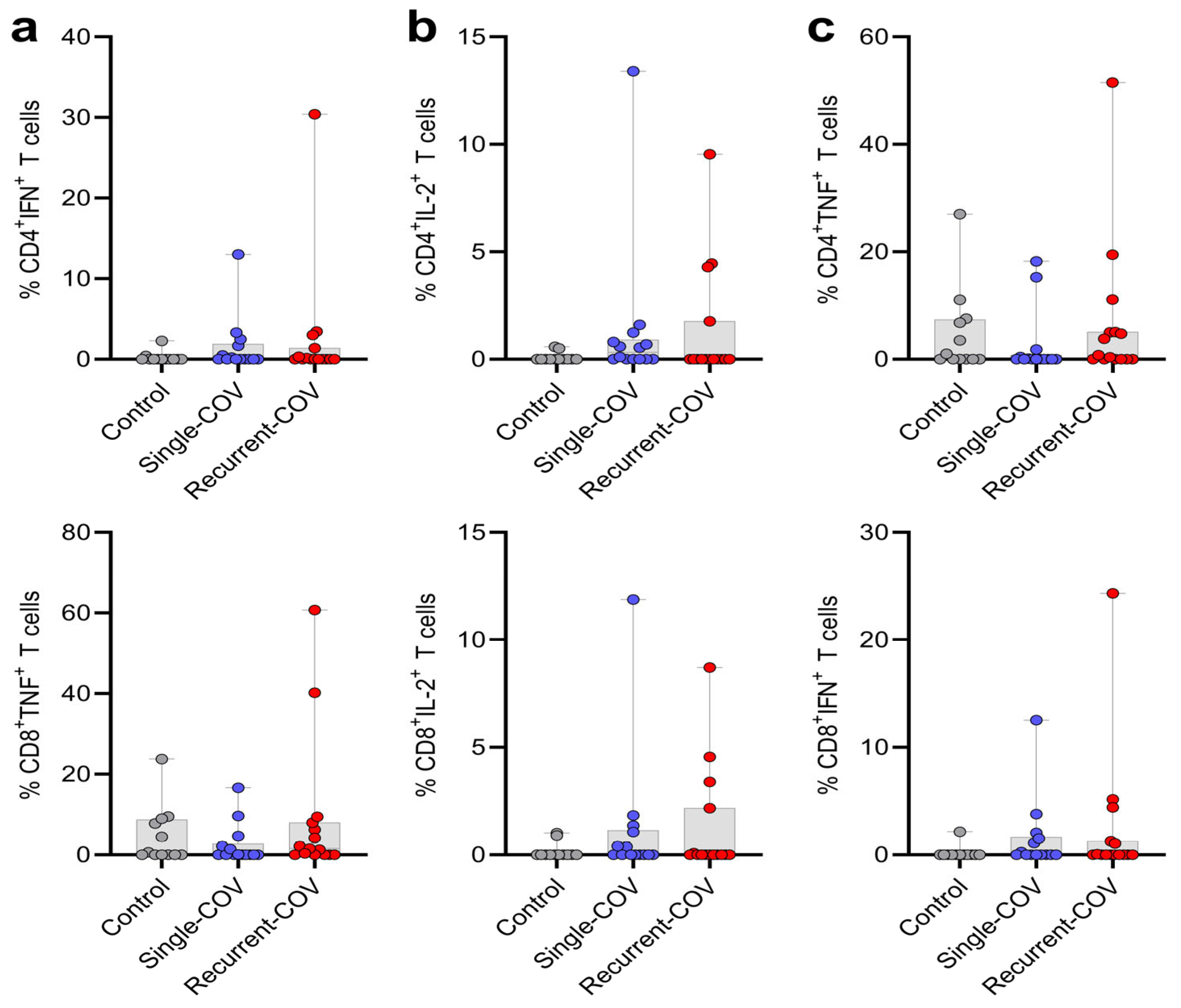
Figure 1. Cytokine production analysis in stimulated T cells (ac). PBMCs were analyzed by flow cytometry after 18 h of stimulation. T cells were examined based on the production of the cytokines IFN-γ, IL-2, and TNF-α. Comparisons were made according to the COVID-19 groups: Recurrent-COV, Single-COV, and Controls (without disease). Each dot represents a patient, while the bars indicate the minimum, maximum, and median values. Statistical comparisons were performed using the Kruskal–Wallis test followed by Dunn’s test.
Immuno 05 00017 g001
Figure 2. Cytokine production analysis in stimulated T cells (ac). PBMCs were analyzed by flow cytometry after 18 h of stimulation. T cells were examined based on the production of the cytokines IFN-γ, IL-2, and TNF-α. Comparisons were made according to the rs179008 genotype, including only patients with a history of COVID-19, regardless of the number of episodes (patients with single and recurrent COVID-19 were included, and the control group was excluded). Each dot represents a patient, while the bars indicate the minimum, maximum, and median values. Statistical comparisons were performed using the Mann–Whitney test. * p < 0.05.
Immuno 05 00017 g002
Table 1. Genotype frequencies of TLR7 rs179008 SNP and its association with recurrent COVID-19.
ModelGenotypeControl
n (%)
Case
n (%)
ORLower CIUpper CIp-ValueAIC
CodominantA/A or A20 (76.9)13 (65)1.0 0.0166359.8
A/T2 (7.7)7 (35)5.380.9630.06
T/T or T4 (15.4)00.00.00.0
DominantA/A or A20 (76.9)13 (65)1.0 0.374666.2
A/T-T/T or T6 (23.1)7 (35)1.790.496.55
RecessiveA/A or A-A/T22 (84.6)20 (100)1.0 0.121362.1
T/T or T4 (15.4)00.00.00.0
OverdominantA/A or A-T/T or T24 (92.3)13 (65)1.0 0.0192661.5
A/T2 (7.7)7 (35)6.461.1735.74
n, number of individuals; CI, confidence interval; AIC, Akaike information criterion. Case group: all individuals of the recurrent group. Control group: individuals from the single COVID-19 and the control groups. The males, hemizygous for the TRL7 gene, were considered together with females with homozygous genotypes. The association was analyzed through a univariate logistic regression analysis using the “SNPassoc” package in R Studio (2024.09).
Disclaimer/Publisher’s Note: The statements, opinions and data contained in all publications are solely those of the individual author(s) and contributor(s) and not of MDPI and/or the editor(s). MDPI and/or the editor(s) disclaim responsibility for any injury to people or property resulting from any ideas, methods, instructions or products referred to in the content.