Review

Efficacy of Intravenous Immunoglobulins and Other Immunotherapies in Neurological Disorders and Immunological Mechanisms Involved

by
Angel Justiz-Vaillant
1,*,
Sachin Soodeen
1,
Odalis Asin-Milan
2,
Julio Morales-Esquivel
3 and
Rodolfo Arozarena-Fundora
3,4
1
Department of Pathology/Microbiology and Pharmacology, University of the West Indies, St. Augustine Campus, St. Augustine 330912, Trinidad and Tobago
2
Independent Researcher, Laval, QC H7E 2Z8, Canada
3
Eric Williams Medical Sciences Complex, North Central Regional Health Authority, Champs Fleurs 330912, Trinidad and Tobago
4
Department of Clinical and Surgical Sciences, Faculty of Medical Sciences, University of the West Indies, St. Augustine 330912, Trinidad and Tobago
*
Author to whom correspondence should be addressed.
Immuno 2025, 5(2), 18; https://doi.org/10.3390/immuno5020018
Submission received: 24 April 2025 / Revised: 21 May 2025 / Accepted: 23 May 2025 / Published: 26 May 2025

Abstract

This review aims to explore the role of immunotherapeutic strategies—primarily intravenous immunoglobulin (IVIG), plasma exchange (PLEX), and selected immunomodulatory agents—in the treatment of neurological and psychiatric disorders with suspected or confirmed autoimmune mechanisms. A central focus is placed on understanding the immunopathology of these conditions through the identification and characterization of disease-associated autoantibodies. Disorders such as autoimmune encephalitis, myasthenia gravis, limbic epilepsy, neuropsychiatric systemic lupus erythematosus (NPSLE), and certain forms of schizophrenia have shown clinical responses to immunotherapy, suggesting an underlying autoimmune basis in a subset of patients. The review also highlights the diagnostic relevance of detecting autoantibodies targeting neuronal receptors, such as NMDA and AMPA receptors, or neuromuscular junction components, as biomarkers that guide therapeutic decisions. Furthermore, we synthesize findings from published randomized controlled trials (RCTs) that have validated the efficacy of IVIG and PLEX in specific diseases, such as Guillain–Barré syndrome, and myasthenia gravis. Emerging clinical evidence supports expanding these treatments to other conditions where autoimmunity is implicated. By integrating immunological insights with clinical trial data, this review offers a comprehensive perspective on how immunotherapies may be tailored to target autoimmune contributors to neuropsychiatric disease.

Graphical Abstract

1. Introduction

Neuropsychiatric systemic lupus erythematosus (NPSLE) and multiple sclerosis (MS) involve immune-mediated CNS injury linked to glutamatergic dysfunction. These conditions parallel other neuroimmune syndromes (e.g., anti-NMDA receptor encephalitis) and even models of schizophrenia, underscoring the relevance of glutamate receptor autoimmunity [1]. In NPSLE, autoantibody-mediated excitotoxicity is central: a subset of anti-dsDNA antibodies cross-reacts with NMDA receptor NR2A/B subunits. Such anti-NR2 antibodies are detected in a significant fraction (~30–50%) of SLE patients and are enriched in those with CNS involvement. When the blood–brain barrier is breached, these anti-NR2 IgG bind neuronal NMDA receptors and pathologically potentiate receptor activation [2]. The ensuing Ca2+ influx triggers excitotoxic neuronal apoptosis, correlating with seizures, psychosis, mood disturbances and cognitive decline in diffuse NPSLE. Anti-AMPA receptor autoantibodies, reported in other autoimmune encephalitides, may analogously perturb glutamatergic signaling in susceptible patients [1].
MS pathogenesis also entails glutamate excitotoxicity. Activated T cells and macrophages release excessive glutamate that overactivates AMPA/kainate and NMDA receptors on neurons and oligodendrocytes. Experimental autoimmune encephalomyelitis (EAE) models demonstrate that AMPA-mediated excitotoxicity damages myelin and axons. Pharmacologic blockade of AMPA receptors (e.g., with NBQX) preserved oligodendrocytes and ameliorated demyelination in EAE, despite unchanged inflammation [3]. Clinically, standard MS treatments (e.g., interferon-β, anti-CD20) reduce relapse frequency but do not directly prevent glutamate-mediated neurodegeneration. Moreover, glutamate is known to accumulate in MS lesions and CSF, linking excitotoxicity to disease pathology.

Therapeutic and Diagnostic Considerations

  • Immune modulation: Therapies that reduce pathogenic autoantibodies (high-dose corticosteroids, B-cell depletion with rituximab or BAFF inhibitors) or remove them (plasmapheresis, IVIg) are mainstays for CNS autoimmunity.
  • Glutamate antagonists: NMDA/AMPA receptor blockers (e.g., memantine or experimental AMPA antagonists) may confer neuroprotection; indeed, NBQX treatment reduced demyelination in an MS model [3].
  • Antibody screening: Serologic or CSF testing for anti-NMDA and anti-AMPA receptor autoantibodies can aid diagnosis and identify patients for targeted therapy.
These approaches aim to neutralize neurotoxic autoantibodies and interrupt the excitotoxic cascade, addressing both the immunologic trigger and downstream neuronal injury in NPSLE and MS [2,3]. Ongoing research explores novel CNS-targeted immunotherapies for these antibody-mediated pathways in NPSLE and MS.
Intravenous immunoglobulin (IVIG) and plasma exchange (PLEX) are vital immunotherapies in neuropsychiatric diseases where pathological autoantibodies contribute to disease mechanisms. IVIG acts by neutralizing autoantibodies, modulating Fc receptors, suppressing proinflammatory cytokines, and enhancing regulatory immune responses. PLEX, on the other hand, physically removes circulating autoantibodies and immune complexes from the bloodstream, offering rapid symptom relief. These therapies are particularly effective in conditions such as autoimmune encephalitis, myasthenia gravis, and neuropsychiatric lupus, where specific autoantibodies disrupt neuronal function. By eliminating or blocking these pathogenic antibodies, IVIG and PLEX help restore neural integrity and reduce cognitive, behavioral, or motor symptoms associated with autoimmune neuropsychiatric disorders [4,5].

2. Methods

To ensure a comprehensive and balanced narrative review, we conducted a targeted literature search using major databases including PubMed, Scopus, Web of Science, and Google Scholar. Keywords related to the central themes of the review were employed, and studies published in English between 2000 and 2025 were considered. Over 450 publications were initially screened based on relevance to the topic, novelty, scientific rigor, and contribution to current understanding. From these, we selected approximately 132 key publications that provided substantial evidence or novel insights aligned with the objectives of this review. Priority was given to peer-reviewed original research articles, systematic reviews, and high-impact clinical studies. Studies were excluded if they lacked relevance, scientific quality, or up-to-date findings.

2.1. The Biological Basis of Some Neuropsychiatric Diseases

The biological basis of depression in SLE has recently been corroborated. Biochemical and neurophysiological changes induced by cytokines have been demonstrated to contribute to the development of neuropsychiatric symptoms. Cytokines are known to cause mood swings and depression. The downregulation of the hypothalamic–pituitary–adrenal (HPA) axis correlates with neurophysiological changes involved in depression. Moreover, cerebro-reactive autoantibodies in the cerebrospinal fluid (CSF), such as anti-NMDA and anti-ribosomal P, can cause significant neuronal damage. This damage is pertinent to mood and behavior, leading to depressive symptoms [6]. Likewise, epilepsy is a complex and multifactorial condition. Increasing evidence suggests that the immune system plays a crucial role in neuronal excitability and epileptogenesis. Studies on epilepsy patients, including ex vivo investigations, have shown elevated levels of IL-1β, IL-2, IL-5, IL-6, and TNF-α following treatment with carbamazepine, valproic acid, and phenytoin [7].
Table 1 includes diseases well-established to be mediated by pathogenic autoantibodies that drive their immunopathogenesis. These conditions—such as autoimmune encephalitis, myasthenia gravis, schizophrenia (in specific autoimmune subsets), and limbic epilepsy—share a common feature: immune dysfunction involving autoantibody targeting of neuronal or neuromuscular components. The rationale for their inclusion lies in the strong evidence supporting an antibody-driven mechanism, with clinical improvements observed following immunosuppressive therapies such as corticosteroids, IVIG, or plasma exchange. These disorders exemplify how understanding autoantibody roles can guide diagnosis and treatment strategies, making them ideal models for studying autoimmunity and antibody-targeted therapeutic interventions.

2.2. Review of Neurological Immune-Related Adverse Events (n-irAEs)

Zammit and Seront (2024) [18] present a comprehensive and clinically focused review of neurological immune-related adverse events (n-irAEs) associated with immune checkpoint inhibitors (ICIs), including anti-CTLA-4, anti-PD-1, and anti-PD-L1 monoclonal antibodies. These agents have revolutionised cancer treatment by reinvigorating T cell responses against tumours but can inadvertently provoke immune-mediated damage to healthy tissues, including the nervous system [18].
The authors highlight that n-irAEs occur in approximately 1–5% of patients receiving ICIs, yet they represent some of the most severe and potentially life-threatening complications of immunotherapy. These events may affect both the central nervous system (CNS) and peripheral nervous system (PNS), manifesting as a diverse range of pathologies such as encephalitis, aseptic meningitis, transverse myelitis, cerebellar ataxia, optic neuritis, Guillain–Barré Syndrome (GBS), chronic inflammatory demyelinating polyneuropathy (CIDP), myasthenia gravis (MG), and neuromuscular junction disorders [18].
The immunopathogenesis of n-irAEs is not fully understood but is believed to involve dysregulated T cell activation, loss of peripheral tolerance, cytokine-mediated inflammation, and cross-reactivity with neuronal antigens. In some patients, autoantibodies commonly seen in classic autoimmune or paraneoplastic syndromes (e.g., anti-AChR, anti-NMDAR, anti-Hu) have been detected, suggesting overlapping mechanisms. Clinically, n-irAEs are challenging to diagnose due to their variable presentation and the need to differentiate them from cancer progression, infections, or metabolic complications. Early recognition is critical, as delayed diagnosis may result in irreversible neurological damage [18].
Treatment strategies include the immediate cessation of ICI therapy, followed by immunosuppression, usually beginning with high-dose corticosteroids. In moderate to severe cases, additional therapies such as intravenous immunoglobulin (IVIG), plasma exchange, or other immunosuppressive agents (e.g., rituximab, mycophenolate mofetil) may be required. The prognosis depends on the severity and speed of intervention; some patients recover fully, while others may suffer permanent deficits. Overall, this review emphasizes the importance of multidisciplinary management, prompt immunological workup, and early immunotherapy to improve outcomes in patients affected by ICI-induced neurological toxicities [18].

2.3. Cytokine Involvement in Neurological and Psychiatric Diseases Such as Guillain–Barré Syndrome and Schizophrenia

The involvement of cytokines in neuropsychiatric diseases suggests a pathogenic role for these molecules in the genesis of these conditions or as a result of interactions between the immune and/or endocrine systems and the brain. In summary, the interrelationship between the immune and central nervous systems is evident in various neuropsychiatric and neurological disorders. The presence of specific antibodies and cytokines underscores the potential autoimmune nature of these conditions. Screening for autoimmune markers and implementing immunotherapy may provide significant benefits in managing these disorders. Understanding the immune system’s role in these diseases offers a promising avenue for developing new therapeutic strategies and improving patient outcomes. The ongoing research in this field will continue to shed light on the complex mechanisms underlying these disorders and pave the way for more effective treatments. Table 2 shows the cytokine involvement in neuropsychiatric diseases such as Guillain–Barré syndrome and schizophrenia.

3. Immunotherapy Used in Neurological Disorders

3.1. The Use of Intravenous Immunoglobulin (IVIG) in Treating Neurological Disorders

Intravenous immunoglobulin (IVIG) is a treatment option used for various neurological disorders. It involves infusing a preparation of immunoglobulins—antibodies extracted from the blood of healthy donors—directly into a patient’s bloodstream. While IVIG demonstrated effectiveness for certain conditions, the exact mechanisms by which it works are not yet fully understood. This essay explores the potential ways IVIG might benefit neurological conditions and discusses why it is not recommended for some specific disorders. Table 3 lists the cases where intravenous immunoglobulin (IVIG) was used successfully.

3.2. Abstract Discussion

The therapeutic success of IVIG is influenced by disease pathophysiology. Conditions characterized by pathogenic circulating autoantibodies and B-cell dysregulation (e.g., Guillain–Barré syndrome, CIDP, myasthenia gravis) show consistent IVIG efficacy due to its ability to neutralize autoantibodies, inhibit Fc receptor-mediated damage, and modulate cytokine production. In contrast, disorders like MS and Rasmussen’s encephalitis involve complex T-cell–mediated or CNS-compartmentalized immune responses, where IVIG alone may be insufficient. The lack of success in some disorders may also reflect insufficient BBB penetration or a lack of antibody-driven pathology. Future RCTs and biomarker-driven stratification could help better define IVIG’s utility across the neuroimmunological disease spectrum.

3.3. How IVIG Works

Intravenous immunoglobulin (IVIG) has been successfully used in various neuroimmunological disorders due to its immunomodulatory effects, including Fc receptor blockade, complement inhibition, and modulation of autoantibody production. In Guillain–Barré syndrome (GBS), IVIG is a first-line therapy that significantly reduces disease progression by neutralizing pathogenic antibodies. In Chronic Inflammatory Demyelinating Polyneuropathy (CIDP) and Multifocal Motor Neuropathy (MMN), IVIG improves muscle strength and slows nerve damage. For Myasthenia Gravis and Lambert–Eaton Myasthenic Syndrome, IVIG is beneficial during myasthenic crises or when rapid immunosuppression is needed. Multiple sclerosis (MS) and Acute Disseminated Encephalomyelitis (ADEM) respond variably, with IVIG showing more efficacy in relapsing forms and pediatric cases. In Opsoclonus–myoclonus and Pediatric Autoimmune Neuropsychiatric Disorders Associated with Streptococcal Infections, IVIG reduces neuroinflammatory symptoms and abnormal movements. Although Diabetic Neuropathy is not primarily autoimmune, IVIG may help when inflammatory components are suspected. Rasmussen’s Encephalitis and Polymyositis may benefit from IVIG as an adjunct to steroids, especially in refractory cases. Overall, IVIG offers a broad-spectrum immune-regulating effect across these disorders, particularly where autoantibodies are involved, providing symptom control and functional recovery [25,26,27,28,29,30,31,32,33,34].
B cells produce antibodies, T cells are involved in directing the immune response, and antigen-presenting cells help other immune cells recognize and respond to foreign substances. By affecting these cells, IVIG may help reduce inappropriate immune reactions that can damage the nervous system. Another key function of IVIG is the downregulation of complement proteins, which are part of the immune system’s response to infection and injury [35,36,37]. When these proteins are overactive, they can contribute to inflammation and damage in neurological disorders. IVIG may help control this activity, thereby reducing inflammation and associated damage.
IVIG may also influence the NF-kB pathway and the degradation of IκB [35,36,37]. NF-kB is a crucial protein complex in regulating inflammation and immune responses. NF-kB activation can lead to increased inflammation. IVIG might help suppress this activation, thus reducing inflammation in the brain. Additionally, IVIG is believed to affect the idiotypic–anti-idiotypic network, which involves interactions between different antibodies [38]. This network helps regulate the immune response and may be disrupted in autoimmune disorders. IVIG could help restore immune balance by modulating this network.
IVIG has also been shown to promote the expansion of regulatory T cells [39]. These cells are important for maintaining immune tolerance and preventing autoimmune responses. IVIG might help the body regulate its immune response more effectively by increasing these cells’ numbers. Furthermore, IVIG can neutralize pathogenic autoantibodies [40,41]. These autoantibodies mistakenly target and damage the body’s own tissues in autoimmune disorders. IVIG can potentially reduce tissue damage and improve symptoms by neutralizing these harmful antibodies.
In addition to these effects, IVIG may support remyelination, which is the process of repairing damaged myelin, the protective sheath around nerve fibers. This can be crucial for conditions where myelin is damaged, such as multiple sclerosis. IVIG might also affect the release of cytokines and cytokine antagonists, which are molecules that help regulate inflammation and immune responses [42,43,44]. IVIG could help manage inflammation and promote healing in the nervous system by modulating these factors.

3.4. Conditions Treated with IVIG

IVIG has demonstrated effectiveness in several neurological disorders. For instance, it is used to treat conditions such as Guillain–Barré syndrome, a disorder where the immune system attacks the peripheral nerves, and chronic inflammatory demyelinating polyneuropathy (CIDP), a condition characterized by progressive weakness and sensory loss due to nerve damage. IVIG helps by modulating the immune response and reducing the inflammation that damages the nerves [35].

3.5. Conditions Where IVIG Is Not Recommended

Despite its benefits, IVIG is not suitable for every neurological disorder. For example, it is generally not recommended for conditions such as paraproteinemic neuropathy (IgM variant), intractable childhood epilepsy, inclusion body myositis, amyotrophic lateral sclerosis (ALS), adrenoleukodystrophy, autism, critical illness polyneuropathy, and POEMS syndrome [45].
Paraproteinemic neuropathy (IgM variant) is a condition where an abnormal protein in the blood causes nerve damage. Intractable childhood epilepsy involves severe and difficult-to-treat seizures in children. Inclusion body myositis is a muscle disorder that leads to progressive muscle weakness. ALS is a neurodegenerative disease that affects motor neurons, leading to muscle weakness and atrophy. Adrenoleukodystrophy is a genetic disorder affecting the brain and adrenal glands. Autism is a developmental disorder affecting communication and behavior. Critical illness polyneuropathy occurs in patients who have been critically ill and involves nerve damage. POEMS syndrome is a rare condition characterized by polyneuropathy, organomegaly (enlarged organs), endocrinopathy (hormonal imbalances), edema (swelling), M-protein (an abnormal protein), and skin abnormalities. In these conditions, IVIG may not be effective due to the specific nature of the disease processes involved. For example, the disease mechanisms in ALS and inclusion body myositis may not be primarily immune-mediated, making IVIG less useful. Similarly, IVIG’s benefits may be limited in conditions such as autism, where the underlying pathophysiology is not primarily autoimmune or inflammatory. In summary, intravenous immunoglobulin (IVIG) is a valuable treatment for various neurological disorders, primarily due to its ability to modulate the immune response and reduce inflammation. Its effectiveness is attributed to several mechanisms, including immunosuppression, the downregulation of complement proteins, and the promotion of regulatory T cells. However, IVIG is not recommended for all neurological conditions and dermatological disorders [34,35,36,37,38,39,40,41,42,43,44].

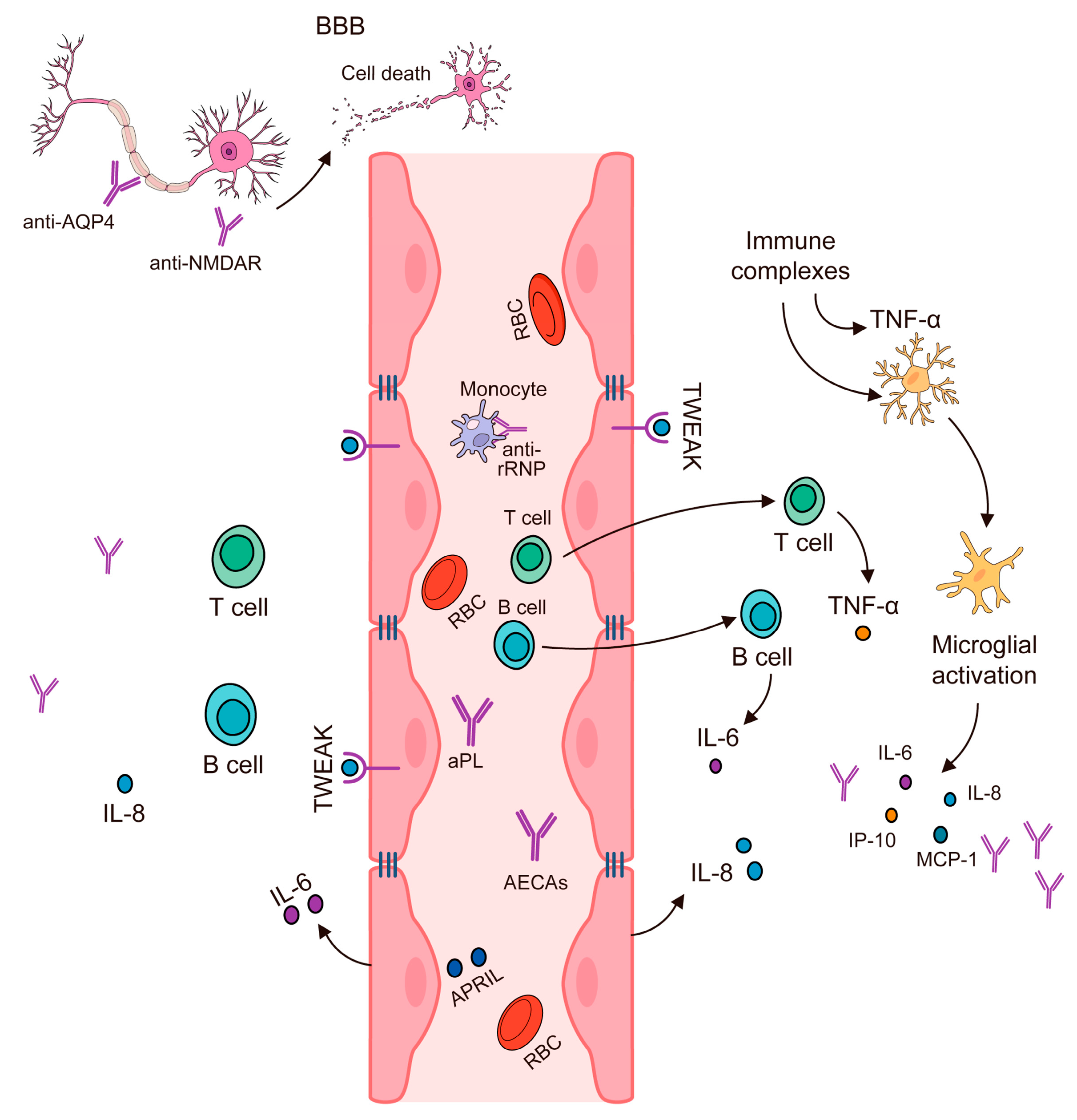
4. Disruption of the Blood–Brain Barrier

The blood–brain barrier (BBB) is an intricate and highly selective structure that plays a pivotal role in maintaining the brain’s homeostasis. This barrier comprises a layer of endothelial cells that line the capillaries throughout the brain, forming a protective shield between the bloodstream and the central nervous system (CNS). Its primary function is to regulate the movement of substances between the blood and the brain, ensuring that the neural environment remains stable and conducive to optimal neuronal function. Under normal physiological conditions, the BBB effectively restricts the passage of large-molecular-weight substances, such as proteins and other potentially harmful agents, from entering the brain [44].
This selective permeability is crucial for safeguarding the brain from toxins, pathogens, and fluctuations in blood composition that could disrupt neural activities. By limiting the entry of these substances, the BBB helps to maintain the brain’s delicate chemical balance, which is essential for proper neuronal function and overall cognitive health. The BBB’s integrity is maintained by tight junctions between endothelial cells, which form a nearly impermeable barrier. These tight junctions prevent the diffusion of large molecules and most cells across the endothelial layer while allowing the selective transport of essential nutrients and ions through specialized transport mechanisms [44].
This controlled permeability is vital for protecting the brain from potential damage and ensuring that only necessary substances, such as glucose and amino acids, can enter the CNS. Disruption of the BBB can have severe consequences for brain health. Conditions such as multiple sclerosis (MS) and neuropsychiatric systemic lupus erythematosus (NPSLE) are associated with BBB breakdown, which allows immune cells and autoantibodies to infiltrate the brain (Figure 1). This infiltration can lead to inflammation, tissue damage, and a range of neurological symptoms [45,46].
Understanding the mechanisms that regulate BBB function and its disruption is crucial for developing effective treatments for various neurological and neurodegenerative disorders. In summary, the BBB is a fundamental component of brain protection, maintaining a stable environment for neural function and preventing the entry of potentially harmful substances. Its role in preserving brain homeostasis underscores its importance in both healthy and diseased states, highlighting the need for ongoing research into its mechanisms and potential therapeutic interventions [44].
A breach in the BBB can have significant implications for brain health. When the BBB is compromised, circulating antibodies and other immune components may infiltrate the brain. This infiltration is particularly problematic in the context of autoimmune disorders. For instance, in neuropsychiatric systemic lupus erythematosus (NSLE), a condition characterized by systemic autoimmune activity, antibodies that cross-react with neurological tissues can enter the brain. Once inside, these antibodies can interact with neuronal structures, leading to tissue damage and potentially contributing to neurotoxicity. The presence of autoantibodies and proinflammatory cytokines within the brain can initiate and exacerbate inflammatory responses. This inflammation is further intensified by the actions of T-helper 1 (TH1) and T-helper 17 (TH17) lymphocytes, which are known to amplify inflammatory reactions and contribute to neurodegeneration [46]. Multiple sclerosis (MS) provides a clear example of how BBB disruption can lead to neurological damage. In MS, the BBB is disrupted, allowing myelin-specific lymphocytes to penetrate the central nervous system (CNS) [47].
These lymphocytes then target and induce demyelination, a process where the protective myelin sheath surrounding nerve fibers is damaged. This demyelination impairs the conduction of electrical signals along the affected nerves, leading to a range of neurological symptoms. The presence of gadolinium (gd)-enhancing lesions in magnetic resonance (MR) imaging is a hallmark of this disease, reflecting areas of active inflammation and BBB breakdown. When the BBB is compromised, immune and accessory cells that typically do not have access to the brain can breach this immunological sanctuary. The brain, normally considered an immunologically privileged organ, becomes exposed to these immune components, which can infiltrate the extracellular spaces and cause significant neuronal damage and neurotoxicity. Autoantibodies that specifically target neuronal cells or other CNS components can lead to various forms of neuroinflammation and tissue damage. Several autoimmune diseases are associated with such pathological processes. For example, in multiple sclerosis, the breakdown of the BBB allows for the infiltration of myelin-specific lymphocytes that attack and damage the myelin sheaths, resulting in impaired nerve signal conduction. Neuromyelitis optica (NMO) is another autoimmune condition where autoantibodies against aquaporin-4, a water channel protein, can cross the compromised BBB and attack astrocytes, leading to severe demyelination and neuronal damage. Guillain–Barré syndrome (GBS) is a peripheral nerve disorder where molecular mimicry between bacterial or viral antigens and peripheral nerve components leads to an autoimmune attack on the myelin sheaths. Chronic inflammatory demyelinating polyneuropathy (CIDP) also involves a similar autoimmune attack on the peripheral nerves, causing progressive weakness and sensory impairment due to demyelination. The common feature in all these conditions is the breach of the BBB, which allows autoimmune factors to enter the CNS and cause damage. The ensuing inflammation and demyelination impair nerve function, leading to a range of neurological deficits. The damage to myelin disrupts the normal conduction of electrical impulses along the nerves, which can manifest as motor and sensory symptoms, depending on the specific nerves affected [47,48,49]. Table 4 shows an overview of experimental therapies and their mechanisms of action in neurological autoimmune disorders.

5. Cytokines, Antibodies, and Autoimmune Inflammation

In a study, age-dependent differences in cytokine responses were observed in mice infected with the Powassan virus (POWV). In 50-week-old mice, the infection triggered the expression of Th1-type cytokines (IFNγ, IL-2, IL-12, IL-4, TNFα, and IL-6), indicating a proinflammatory, neurodegenerative response. Conversely, 10-week-old mice exhibited Th2-type cytokines (IL-10, TGFβ, and IL-4), which are associated with a neuroprotective response. These findings highlight potential targets for therapeutic intervention [62].
Another study explored the role of STAT4 in dendritic cell (DC) function, particularly in the context of experimental autoimmune encephalomyelitis (EAE), a model for multiple sclerosis. STAT4, which mediates the effects of proinflammatory cytokines such as IL-12 and IL-23, is crucial for DC function. Mice lacking STAT4 in CD11c-expressing cells showed reduced T cell activation and central nervous system inflammation. Restoring EAE susceptibility through the transfer of wild-type DCs, but not STAT4- or IL-23R-deficient DCs, indicates that STAT4 is essential for DC-mediated inflammation. Single-cell RNA-sequencing identified STAT4-dependent gene signatures in DCs, aligning with patterns seen in multiple sclerosis (MS) patients [63].
Another study compared tocilizumab and conventional immunotherapy for treating refractory acetylcholine receptor antibody-positive (AChR-Ab+) generalized myasthenia gravis (gMG). It was conducted at a single center in China and included 34 patients, with 20 receiving tocilizumab and 14 on conventional therapy. Tocilizumab treatment resulted in a more significant reduction in the MG activities of daily living (MG-ADL) score at week 4, and these improvements were sustained through week 24. The tocilizumab group showed a higher proportion of patients achieving notable reductions in both their MG-ADL and QMG scores. Tocilizumab was also found to be safe, with no severe or unexpected adverse effects reported. These results suggest that tocilizumab is a promising and effective treatment for refractory AChR-Ab+ gMG [64].
Multiple sclerosis (MS) is a chronic inflammatory disease of the central nervous system (CNS), affecting an estimated 2.8 million people globally [65]. The condition is thought to result from aberrant adaptive immune responses targeting the myelin sheaths, though its exact causes remain unclear. Genetic factors, particularly the HLA-DR*15:01 allele, are strongly linked to MS, suggesting a role for antigen presentation and CD4+ T cell responses [66,67]. Despite this, no consistent autoantigenic targets have been identified. Moreover, while myelin-specific T cells are present in MS patients, their frequency and activity vary [68]. Epstein–Barr virus (EBV), a γ-herpesvirus known for causing infectious mononucleosis (IM) and for its association with B cell tumors, has been implicated in MS pathogenesis. Evidence shows that a history of IM increases the MS risk, and EBV seroconversion often precedes disease onset [69,70,71]. Elevated levels of EBNA1 IgG, an EBV latent-cycle antigen, are linked to a significantly higher MS risk [72,73]. Moreover, antibodies against EBNA1 have been detected in the oligoclonal bands of MS patients and show cross-reactivity with human proteins, suggesting a direct role in neuroimmune mechanisms [74,75,76,77]. A study aimed to expand the understanding of EBV-related immune responses by analyzing antibody responses to various EBV antigens, including EBNA1, other components of the EBNA complex, and the viral-capsid antigen (VCA), in plasma from individuals with relapsing–remitting MS (RRMS), healthy controls, and those with recent IM. It also investigated EBV-specific T cell responses, particularly cross-reactivity with CNS autoantigens [78,79,80,81]. The research may clarify the role of EBV in MS and identify potential targets for therapeutic intervention.
In autoimmune central nervous system (CNS) disorders, specific antibodies are associated with different clinical manifestations.
Limbic encephalitis is a condition that features personality changes, memory deficits, seizures, and psychosis, with MRI changes in the mesial temporal lobes. Antineuronal nuclear antibody (ANNA)-1 and gamma-aminobutyric acid-B receptor (GABAB-R) antibodies are linked to small-cell lung cancer (SCLC) and limbic encephalitis [82,83]. Leucine-rich glioma inactivated 1 (LGI1) antibodies are associated with autoimmune limbic encephalitis and faciobrachial dystonic seizures (FBDSs), often with rare tumors [84,85]. Other phenotypic descriptions include autoimmune encephalitis: Autoimmune encephalitis is a group of immune-mediated brain inflammations caused by autoantibodies targeting neuronal surface antigens or intracellular proteins. It presents with a wide spectrum of neurological and psychiatric symptoms, often with a subacute onset (Table 5 and Table 6 [86,87,88]). Retrograde amnesia refers to the loss of memories formed before the onset of a neurological insult, often affecting personal experiences (episodic memory), and is commonly seen in conditions like LGI1 or CASPR2-associated autoimmune encephalitis. In contrast, anterograde amnesia is the inability to form new long-term memories after the event, typically resulting from damage to the hippocampus or related structures, as seen in NMDA receptor encephalitis. While retrograde amnesia affects past recall, anterograde amnesia impairs the creation of new memories, with the latter often being more disabling and persistent [86].
Brainstem encephalitis is characterized by oculomotor abnormalities and cranial nerve dysfunction. The associated antibodies include Kelch-like protein 11 (KLHL-11), ANNA-2 (anti-Ri), and IgLON5 IgG [89]. Bickerstaff’s brainstem encephalitis, linked with ganglioside Q1B (GQ1B) IgG, features multiple cranial nerve abnormalities [90]. IgLON5 autoimmunity may present with sleep disorders, and severe cases can lead to cardiorespiratory dysfunction [91,92,93,94].
Extralimbic encephalitis is characterized by symptoms localized in non-limbic structures, and anti-GABAA-R antibodies are linked to seizures and multifocal cerebral lesions [95]. Glial fibrillary acidic protein (GFAP) astrocytopathy, affecting deep periventricular white matter, is associated with meningism, parkinsonism, and other CNS symptoms [96,97]. ANNA-1 antibodies can present with extralimbic symptoms, and N-methyl-d-aspartate receptor (NMDA-R) antibodies lead to psychiatric symptoms, psychosis, seizures, and autonomic instability [98,99].
Rapidly progressive cerebellar ataxia is a syndrome that often involves a gait disorder and limb ataxia with a rapid onset. Antibodies include Purkinje cytoplasmic antibody type 1 (PCA-1; anti-Yo) and glutamic acid decarboxylase (GAD) 65 IgG. Immune therapies may result in stabilization or improvement, with poorer outcomes for antibodies against intracellular antigens, though GAD65 often responds well [100,101].
Autoimmune myelopathy involves ascending sensorimotor deficits and may show sensory levels and upper motor neuron signs. Key antibodies include myelin oligodendrocyte glycoprotein (MOG) and aquaporin-4 (AQP4). Optic neuritis and longitudinally extensive spinal cord lesions are common [102,103,104,105,106]. Autoimmune paraneoplastic myelopathies, such as those with amphiphysin and collapsin-responsive mediator protein (CRMP)-5 IgG, may show tract-specific lesions [107].
Opsoclonus–myoclonus syndrome (OMS) involves arrhythmic eye movements and myoclonus. In children, it is associated with neuroblastoma, while in adults, ANNA-2 IgG is most common [108]. Other antibodies such as NMDA-R, GABAB, GAD65, and dipeptidyl-peptidase-like protein (DPPX) are less frequent [109].

6. New Approaches in the Treatment of Neurological Disorders

Chimeric antigen receptor (CAR)-T cell therapy targeting B cell maturation antigen (BCMA) shows potential in treating neuromyelitis optica spectrum disorder (NMOSD), a neurological autoimmune condition. A study analyzed cerebrospinal fluid (CSF) and blood samples from NMOSD patients treated with anti-BCMA CAR-T cells using single-cell multi-omics sequencing. The results highlighted the dominant role of proliferating CD8⁺ CAR-T cells with cytotoxic-like profiles in counteracting autoimmunity. These engineered cells demonstrated enhanced chemotactic properties, enabling them to cross the blood–CSF barrier effectively, where they depleted plasmablasts and plasma cells, contributing to the reduction in neuroinflammation. A subset of CAR-T cells expressing CD44, indicative of an early memory phenotype, was associated with prolonged cell persistence in patients. CAR-T cells from NMOSD patients showed reduced cytotoxic characteristics compared with those used in blood cancers. These findings advance our understanding of CAR-T cell behavior in autoimmune neurological settings and may inform therapeutic improvements [110].
CAR-T cell therapy is an emerging immunotherapy that shows promise in the treatment of neuroimmune diseases. This approach modifies a patient’s T cells to target and eliminate specific immune cells involved in the disease process. Initial studies have shown encouraging results in conditions such as neuromyelitis optica spectrum disorder, where CAR-T cells have been able to cross the blood–brain barrier, reduce inflammation in the central nervous system, and eliminate harmful B cell populations. These findings suggest potential clinical benefits, including symptom relief and reduced disease activity. Despite these advances, the long-term implications of CAR-T cell therapy in neuroimmune disorders remain uncertain. Potential risks, such as cytokine release syndrome, immune system suppression, and unintended neurological effects, require further investigation. Additionally, questions around the ideal timing for treatment, long-term persistence of CAR-T cells in the nervous system, and patient selection criteria need to be addressed. More prospective studies with extended follow-up are necessary to determine the full safety and effectiveness of this therapy. In summary, CAR-T cell therapy may represent a new direction in managing neuroimmune diseases, but its routine use will depend on robust clinical evidence and continued monitoring of outcomes [111,112].

7. Control Randomized Trials in Neurological Diseases

Table 7 shows several randomized trials in neurological diseases [113,114,115,116].

8. Limitations of IVIG Therapy in Autoimmune Neurological Diseases

Intravenous immunoglobulin (IVIG) therapy is used as an immunomodulatory treatment for various autoimmune neurological diseases. It has been applied in conditions ranging from central nervous system disorders (e.g., multiple sclerosis, autoimmune encephalitis) to peripheral neuropathies (e.g., Guillain–Barré Syndrome, chronic inflammatory demyelinating polyneuropathy) and neuromuscular junction disorders (e.g., Myasthenia Gravis). Despite its broad use, IVIG therapy faces significant therapeutic limitations (related to its efficacy, safety, cost, and patient factors) and methodological limitations in the literature supporting its use. This report provides a detailed overview of these limitations, with a focus on specific diseases—including multiple sclerosis (MS), neuropsychiatric systemic lupus erythematosus (NPSLE), autoimmune encephalitis (and associated “limbic” autoimmune epilepsy), Myasthenia Gravis (MG), and Guillain–Barré Syndrome (GBS)—while highlighting general issues that impact IVIG therapy across all autoimmune neurological conditions.

8.1. Efficacy and Therapeutic Limitations in Specific Diseases

8.1.1. Multiple Sclerosis (MS)

IVIG is not a first-line therapy in MS, and its efficacy in this disease is limited. Some early trials suggested that IVIG could modestly reduce relapse rates in relapsing–remitting MS (RRMS), but there is no evidence that IVIG slows the long-term progression of disability [117]. In secondary progressive MS, controlled trials showed no beneficial effect of IVIG on disease progression or MRI lesions [117]. Given the advent of numerous disease-modifying therapies with proven efficacy from large trials, clinical guidelines have largely relegated IVIG to exceptional use in MS [118]. For example, the Association of British Neurologists and other bodies advise that IVIG should not be used routinely in MS, reserving it only for special circumstances such as severe relapses in patients who cannot receive standard disease-modifying treatment [117]. Even in those scenarios, the evidence supporting IVIG is considered low-grade (e.g., Category 2a, “probable benefit”) [118]. One niche use investigated is prophylaxis of post-partum MS relapses, but a meta-analysis could not clearly establish a therapeutic benefit of IVIG in preventing postpartum relapses [119]. Given the uncertain efficacy and high cost, this practice remains controversial [119]. In summary, MS exemplifies a disease where IVIG’s therapeutic role is very limited: it may confer a modest effect on relapse frequency, but this must be weighed against its cost and the availability of more effective therapies, and thus it is rarely utilized except when other options are contraindicated [117,118].

8.1.2. Neuropsychiatric Systemic Lupus Erythematosus (NPS.LE)

IVIG is occasionally used as an immunotherapy in severe NPSLE, but its use is constrained by both scant evidence and specific clinical contexts. No large randomized controlled trials (RCTs) have evaluated IVIG in NPSLE, and the available evidence consists mainly of case reports and small series [120]. These uncontrolled observations suggest that IVIG can sometimes induce improvement in serious neuropsychiatric lupus manifestations (e.g., seizures, psychosis, neuropathies), especially when conventional therapies have failed [120]. For instance, in one series of 11 NPSLE patients treated with IVIG, 8 showed at least partial remission of neuropsychiatric symptoms while 3 did not respond [120]. IVIG is thus regarded as a salvage or adjunct therapy in NPSLE—current practice reserves it for severe, refractory cases that do not respond to high-dose corticosteroids and cyclophosphamide, or for situations where standard immunosuppression is contraindicated (such as concurrent infection or pregnancy) [121]. The therapeutic limitations are clear: efficacy is uncertain (some patients improve, others do not), and IVIG is not part of first-line NPSLE treatment. Furthermore, because lupus patients often require long-term immunosuppression, the use of IVIG (an expensive, short-acting therapy) is not practical except as a temporary measure. Overall, methodological limitations in the literature (lack of RCTs and small sample sizes) make it difficult to draw firm conclusions on IVIG’s efficacy in NPSLE, and future trials are needed to establish its true value and optimal use in this condition [120].

8.1.3. Autoimmune Encephalitis (And Limbic Autoimmune Epilepsy)

Autoimmune encephalitides (such as anti-NMDA receptor encephalitis, LGI1/CASPR2 antibody encephalitis, and others) are typically treated with immunotherapies, including IVIG. In clinical practice, IVIG is often part of no large RCTs (along with high-dose steroids and/or plasma exchange) for acute autoimmune encephalitis, based on consensus and observational evidence [122]. Many reports suggest that initiating immunotherapy early (including IVIG) is associated with better neurological outcomes compared to no treatment. However, the therapeutic efficacy of IVIG alone in these syndromes is hard to ascertain. There are no large RCTs isolating IVIG’s effect in most autoimmune encephalitides, and patients are usually treated with combinations of therapies, which confounds understanding of the specific contribution of IVIG [123]. In disorders like anti-NMDA receptor encephalitis, a substantial fraction of patients do improve with first-line immunotherapy (often including IVIG), but many require escalation to second-line agents (e.g., rituximab or cyclophosphamide) if there is no response to initial treatment. This indicates a limitation: IVIG (with steroids) is not universally effective, and a subset of patients will fail to improve until more potent or prolonged immunosuppression is given. Certain forms of autoimmune encephalitis respond very poorly to IVIG—for example, IgLON5 antibody encephalitis is noted to have a poor response to immunotherapies (IVIG or others) and a high mortality, highlighting that IVIG offers little benefit in some variants [123]. Even in more responsive subtypes, relapses can occur, and the optimal duration of IVIG therapy is unclear. In summary, while IVIG is a recommended acute treatment for autoimmune encephalitis, its therapeutic limitations include variable efficacy (depending on the antibody syndrome) and the frequent need for additional therapies.

8.1.4. Autoimmune “Limbic” Epilepsy (LGI1/CASPR2 Encephalitis) Deserves Special Mention

This form of autoimmune encephalitis often presents with refractory seizures of limbic origin, and immunotherapy is critical for seizure control and preventing cognitive decline. Historically, there was only anecdotal evidence or expert opinion supporting IVIG in such autoimmune epilepsies. Recently, the first double-blind placebo-controlled trial in LGI1/CASPR2 antibody encephalitis was performed, providing level I evidence (albeit in a small sample) that IVIG can significantly reduce seizure frequency [124]. In that study (17 patients, terminated early due to slow enrollment), 75% of patients on IVIG responded (achieved ≥50% seizure reduction) versus 22% on placebo, and two IVIG-treated patients became completely seizure-free. This is a promising therapeutic result, confirming that immunotherapy (including IVIG) can substantially outperform symptomatic anti-seizure drugs in autoimmune epilepsy. Nonetheless, the trial’s small size underscores a methodological limitation—evidence remains limited. Many patients in clinical practice still face challenges getting IVIG approved for autoimmune epilepsy due to the prior lack of robust data [124]. Thus, while IVIG appears efficacious for limbic autoimmune epilepsy, especially in LGI1 encephalitis, the limitations are the very small evidence base and the recognition that even with treatment, not all patients achieve full seizure remission.

8.1.5. Myasthenia Gravis (MG)

IVIG is an established therapy in Myasthenia Gravis, primarily used for acute exacerbations or myasthenic crises, and occasionally as a maintenance therapy in patients with refractory disease. Its therapeutic benefit in MG is generally short-term. Clinical trials have demonstrated that IVIG can produce transient improvement in muscle strength during MG worsening, but it often shows no clear advantage over plasmapheresis (plasma exchange), an alternative rapid immunomodulatory treatment [125]. For example, randomized trials in moderate to severe MG exacerbations found no significant difference in efficacy between IVIG and plasma exchange on standardized strength scores after two weeks [125]. IVIG also failed to show superiority over corticosteroids in a small trial, and a high-dose (2 g/kg) regimen was not more effective than a 1 g/kg regimen in another study [125]. These trials were relatively small, and some were underpowered or had design limitations, yet collectively they indicate that IVIG’s impact on acute MG symptoms is roughly equivalent to plasma exchange and only modestly better than placebo (one placebo-controlled RCT showed a borderline short-term improvement) [125]. In chronic MG, the limitations are even more pronounced: there is insufficient evidence from RCTs to confirm that periodic IVIG is effective as a long-term therapy [125]. Long-term IVIG monotherapy has been tried in select patients and can help some individuals, but these observations come from uncontrolled studies [125]. Moreover, using IVIG as a maintenance treatment requires regular infusions (e.g., monthly), incurring high costs and potential cumulative risks. Given these factors, IVIG is generally reserved for bridging therapy (while slower-acting treatments take effect) or for patients in crisis, rather than as a continual immunosuppressive strategy in MG. In sum, therapeutic limitations in MG include IVIG’s equivalence to alternative therapies (it is not more efficacious than plasma exchange, and thus is chosen based on practicality or tolerance rather than superior efficacy) and its transient effect. Additionally, the emergence of newer targeted therapies for refractory MG (such as complement inhibitors and neonatal Fc receptor blockers) may further limit IVIG’s role in the future, especially considering the costs and resources required for IVIG.

8.1.6. Guillain–Barré Syndrome (GBS)

GBS is one of the few autoimmune neurological diseases where IVIG’s efficacy is well-established by high-quality evidence. IVIG (2 g/kg total over 2–5 days) is considered standard first-line therapy for acute GBS, shown to hasten recovery from paralysis to the same extent as plasma exchange [126]. Multiple RCTs in the 1990s demonstrated that IVIG started within two weeks of GBS onset accelerates improvement in muscle strength and reduces the time to walking recovery, with a magnitude of benefit comparable to that of plasma exchange [126]. Thus, from a therapeutic standpoint, IVIG is effective in many GBS patients (Class I evidence). However, important limitations remain. IVIG is not universally effective or sufficient for full recovery in all GBS cases. Approximately 20% of GBS patients are still unable to walk unaided at 6 months after treatment, and about 25% still require mechanical ventilation during the acute phase despite IVIG. These outcomes highlight that while IVIG improves the overall prognosis, it often does not prevent significant residual disability in severe cases [127]. There has been interest in whether giving a second course of IVIG to the worst prognostic patients might improve outcomes since a subset of patients show inadequate serum IgG level rise after the first course and recover more slowly [127]. However, a recent double-blind trial (the SID-GBS study) found no benefit from a second IVIG dose in GBS patients with a poor prognosis—the additional dose did not significantly improve muscle strength or disability outcomes at 4 weeks [127]. Moreover, giving a second IVIG treatment doubled the risk of serious adverse events in that study [128]. Thus, clinicians are left with the limitation that the standard single IVIG course, while helpful, may be “not sufficiently effective in many GBS patients”, yet intensification of IVIG therapy does not clearly help and may cause harm [128]. In practice, if patients continue to deteriorate despite IVIG, supportively waiting or considering plasma exchange (if not used initially) is carried out, but no proven augmentation strategy exists. Another practical limitation is that IVIG and plasma exchange are essentially equivalent options in GBS [4], so IVIG’s utility in GBS is not unique—the choice between them often depends on resource availability, contraindications, or convenience. In resource-limited settings, the high cost of IVIG may make plasma exchange the preferred modality. In summary, GBS illustrates both the strengths and limits of IVIG: it is efficacious to a point (substantially improving outcomes relative to no treatment), but it does not cure every patient, and optimizing its use (e.g., timing, dosing) remains an area of active investigation.

8.2. Safety and Practical Constraints of IVIG Therapy

Beyond disease-specific efficacy, IVIG therapy has general therapeutic limitations related to safety, tolerability, cost, and patient-specific factors that apply across all autoimmune neurological diseases:

8.2.1. Adverse Effects and Safety Concerns

IVIG is generally considered safe, especially compared to long-term immunosuppressants, but it is not without risks. Mild infusion-related side effects are common: up to 5–20% of patients experience headaches, fever, chills, flushing, myalgias or nausea during or shortly after infusion [129]. These are usually self-limited. However, IVIG can also cause more serious adverse effects in some patients. One significant risk is thromboembolism: IVIG can increase blood viscosity and has been associated with an increased risk of blood clots (e.g., deep vein thrombosis, stroke, myocardial infarction), particularly in the elderly or those with pre-existing cardiovascular risk factors [123]. Thrombotic events have been reported to occur even with recommended dosing, so patients with risk factors (advanced age, hypercoagulable states, prolonged immobilization, etc.) should be monitored carefully during and after infusions [130]. Another important risk is acute kidney injury. IVIG products (especially those stabilized with sucrose in the past) can cause osmotic nephrosis; acute renal failure has been observed, usually in patients with risk factors such as baseline renal impairment, diabetes, dehydration, or concurrent nephrotoxic drugs [130]. Because of this, IVIG must be used cautiously in patients with kidney disease, and renal function should be monitored. Aseptic meningitis syndrome (AMS) is a rare but notable complication: patients can develop severe headaches, neck stiffness, photophobia, and CSF pleocytosis a day or two after IVIG infusion [130]. Although aseptic meningitis from IVIG is reversible (symptoms typically resolve within several days of stopping IVIG), it can be alarming and may necessitate discontinuation. Severe hypersensitivity reactions can also occur. In particular, patients with IgA deficiency are at risk for anaphylactic reactions to IVIG, because they may have anti-IgA antibodies that react to IgA present in the IVIG product [130]. For such patients, special IgA-depleted IVIG preparations or alternative therapies must be used to avoid life-threatening allergies. Even in patients with normal IgA, rare cases of anaphylaxis or angioedema have been reported [130]. Overall, while IVIG is often well-tolerated, clinicians must be vigilant about these safety issues. Pre-medication and slow infusion rates can mitigate mild reactions, but the risks of thrombosis and renal failure require patient selection and monitoring. These safety limitations may contraindicate IVIG in certain patients (e.g., those with recent thrombotic events or severe renal insufficiency) or necessitate prophylactic measures (such as hydration, aspirin prophylaxis, or using low-sugar formulations). Compared to plasma exchange, IVIG tends to be safer in terms of hemodynamic instability or infection risk, yet the above adverse effects are an important counterbalance to its immunotherapeutic benefits [125].

8.2.2. Cost and Availability Challenges

High cost and limited supply represent major practical limitations of IVIG therapy. IVIG is a blood product derived from pooled human plasma donations; its manufacturing is complex and expensive. A single treatment course (total dose 2 g/kg) for an average adult can cost many thousands of dollars (often in the order of USD 10,000–20,000 or more, depending on region and dose) [119]. For example, the use of IVIG to prevent postpartum MS relapses was estimated to cost in excess of USD 15,000 per patient [119], a cost that must be weighed against uncertain benefits. Chronic use of IVIG (as in CIDP or repeated MG treatments) can incur cumulative costs reaching six figures annually per patient. Such expense can strain healthcare systems and insurers, meaning access to IVIG may be restricted. Many countries have implemented strict criteria or authorization processes for IVIG use in autoimmune diseases, to ensure that this scarce resource is reserved for indications with the best evidence or highest need [118]. Indeed, availability can be a limiting factor: because IVIG relies on plasma donors, periodic shortages occur, especially in regions with fewer donors or budget constraints [4]. These supply challenges are exacerbated by the expanding use of IVIG in multiple medical fields (neurology, immunology, hematology, etc.) [4]. In less-developed healthcare settings, plasma exchange may be far cheaper than imported IVIG, leading clinicians to favor the former despite IVIG’s convenience. Even in wealthy countries, hospitals often ration IVIG or require documentation that alternative treatments failed before approving IVIG for off-label uses. The high cost also ties into insurance approval: as noted in the context of autoimmune epilepsy, the lack of prior evidence made it difficult to obtain insurance coverage for IVIG, forcing reliance on steroids despite their drawbacks [4] Logistically, IVIG infusions also demand resources: infusion centers, nursing time, cold storage, etc. Widespread use in common diseases like MS would carry “enormous resource implications”, as one analysis pointed out [117]. Therefore, cost and availability not only limit individual patient access but also influence treatment guidelines, often relegating IVIG to second- or third-line status in diseases where cheaper therapies exist. This economic and supply barrier is a fundamental limitation that any consideration of IVIG must account for.

8.2.3. Patient-Specific and Logistic Factors

Certain patient-specific factors can limit the use or efficacy of IVIG. IgA deficiency, as mentioned, is one such factor due to the risk of anaphylaxis [131]. Patients must be screened if suspected, and if IgA deficiency is confirmed, either avoid IVIG or use IgA-depleted formulations. Volume and infusion time can be problematic for some patients—a full dose of IVIG requires infusion over hours (sometimes on successive days). Patients with poor venous access, cardiac failure (who might not tolerate fluid load or the transient increase in plasma viscosity), or who live far from infusion centers may find IVIG less feasible. In contrast to oral therapies, the intravenous route is less convenient and can affect quality of life if frequent infusions are needed. There is also inter-patient variability in response. Some individuals do not respond to IVIG for unclear reasons (possibly due to differences in disease pathophysiology or pharmacokinetics). For example, in GBS, patients who had a smaller increment in serum IgG level after IVIG tended to recover more slowly [127]; whether this reflects suboptimal dosing or inherent differences is not fully understood, but it means not every patient gains the hoped-for benefit. Repeated IVIG administrations can theoretically lead to attenuated responses over time, perhaps via homeostatic mechanisms, although true “tachyphylaxis” to IVIG is not well documented. Interactions with lab testing can be another consideration: IVIG contains a plethora of antibodies and can cause false positives in certain assays (for instance, it can interfere with serological tests or antibody titers), complicating the interpretation of follow-up tests in autoimmune diseases [132]. Finally, if a patient has anti-human antibodies (from prior IVIG or other exposures), infusion reactions can be more frequent. All these patient and logistical issues mean that IVIG therapy must be individualized—some patients are not good candidates, and for others, the inconvenience or risks might not justify the modest benefit. This reality tempers the enthusiasm for IVIG as a one-size-fits-all therapy in autoimmune neurology.

8.3. Methodological Limitations in IVIG Research

When interpreting the literature on IVIG in autoimmune neurological diseases, it is crucial to recognize significant methodological limitations that affect the quality of evidence available. These limitations include the paucity of randomized trials, small sample sizes, heterogeneous study designs, and inconsistent outcome measures, all of which make it challenging to form definitive conclusions and guidelines.
  • Scarcity of High-Quality Trials: For many autoimmune neurological conditions, there is a lack of large RCTs evaluating IVIG. Ethically and logistically, conducting placebo-controlled trials can be difficult in life-threatening or rare diseases, so the evidence often comes from lower levels. In NPSLE, for example, trials are “scarce and most of the data are extracted from case series and case reports”, with virtually no RCTs to guide therapy [129]. Similarly, in autoimmune encephalitis, no randomized controlled treatment trials have been available up to recent years [123]—the field has relied on observational studies and expert consensus. The first-ever RCT in autoimmune epilepsy (LGI1/CASPR2 encephalitis) had only 17 patients, illustrating how rare such trials have been [124]. The absence of robust trials means that many purported benefits of IVIG (or lack thereof) rest on uncontrolled observations that are prone to bias. Publication bias is a concern: positive case reports are more likely to be published than negative ones, potentially overstating IVIG efficacy in the literature. Without controlled comparisons, it is hard to determine how much improvement in a given study was due to IVIG versus the natural disease course or concurrent treatments. This limitation is widely acknowledged, and experts consistently call for more rigorous studies—for instance, a review in NPSLE explicitly concludes that “future RCTs are needed” to establish the efficacy, optimal dose, and duration of IVIG [129]. Until more trials are performed, the confidence in IVIG’s effectiveness for many indications remains limited by the quality of evidence.
  • Small Sample Sizes and Power: Even when RCTs or controlled studies have been conducted, they often involve small sample sizes, reducing statistical power. Many trials in rare neuroimmunological conditions have been underpowered to detect anything but very large effects. For example, several MG trials had between 12 and 84 participants and were unable to detect moderate differences between IVIG and comparators, partly due to limited enrolment [125]. Landmark trials in MS with IVIG were relatively small (on the order of a few dozen patients per arm), and meta-analyses had to combine data from just a handful of studies [117]. The IVIG in postpartum MS meta-analysis included only 380 total treated patients across all studies [119]—a modest number considering the question spanned multiple trials. Small studies increase the margin of error and make results less reliable (a single outlier patient can sway outcomes). They also often cannot rigorously assess subgroups or rare adverse events. Null results in underpowered studies cannot be taken as proof of no effect—for instance, a trial of 15 MG patients found no difference between IVIG and placebo at 6 weeks [125], but such a small study could easily miss a true benefit. Thus, the literature may contain false negatives (or false positives) due to sample size limitations. Combining data via systematic reviews helps but can be hampered by study heterogeneity. Overall, the evidence base for IVIG in many of these disorders is built on relatively small cohorts, and this is a key methodological shortcoming.
  • Heterogeneity and Inconsistent Outcome Measures: Another challenge is the inconsistency in outcome measures and study designs across published studies. Different trials often use different endpoints to define “response” to IVIG. For example, in MS some studies focused on annual relapse rate, others on MRI lesions, and others on disability progression—outcomes that do not always align, leading to mixed conclusions (IVIG appeared to reduce relapses in RRMS but showed no effect on disability in SPMS) [117]. In NPSLE and autoimmune encephalitis case series, outcomes are typically reported in subjective terms (“improved” vs. “not improved”), without standardized scales. An evidence review by NHS England highlighted that most studies in autoimmune encephalitis did not include a precise definition of patient outcomes, and only a few used a common scale like the modified Rankin Scale [123]. This lack of uniform outcome metrics makes it hard to compare results across studies or perform meta-analyses. Additionally, non-standardized treatment protocols contribute to heterogeneity. Dosing of IVIG (e.g., 0.4 g/kg for 5 days vs. 2 g/kg over 2 days) and timing relative to disease onset vary between studies. According to the same NHS review, “a standardised protocol for the use of IVIG was lacking” in the literature, with many studies not detailing the sequence of therapies used [123]. Some patients received IVIG as first-line, others after steroids or plasma exchange failure, etc., introducing variability. Such heterogeneity in methodologies and patient populations (different disease severities, diagnostic criteria, concomitant treatments) is a serious limitation—it precludes definitive conclusions about efficacy and makes it difficult to generalize results. What works in one context (say, IVIG after steroid failure in one series) might not in another, yet the data are often pooled together. Consistency in study design is improving somewhat (for instance, MG trials now often use the Quantitative Myasthenia Gravis score as a standard outcome), but for many autoimmune neurologic diseases, the literature remains a patchwork of disparate reports. In summary, the lack of standard outcomes and the heterogeneity of study conditions undermine the strength of evidence regarding IVIG.
  • Biases and Confounding in Observational Studies: Given the reliance on case series and open-label studies, methodological biases are a major concern. Many reports are retrospective, meaning they rely on chart review and are subject to selection bias (e.g., a clinician might publish on the 5 patients who responded to IVIG, while not reporting on 5 others who did not). Confounding by indication is another issue: sicker patients are more likely to receive treatments like IVIG (especially second-line), which can skew outcomes. As an example, one analysis of autoimmune encephalitis noted that patients who required second-line therapies tended to have worse initial severity, complicating any comparison of those who received only first-line (IVIG/steroids) vs. those who escalated to rituximab [123]. Without randomization, it is difficult to disentangle whether IVIG was truly ineffective in those severe cases or whether their poor outcomes were due to the disease’s aggressiveness. Lack of blinding in open-label studies can also inflate perceived benefits due to placebo effect or observer bias. Some MG studies that were unblinded reported subjective improvement with IVIG, but when tested in blinded trials, the differences narrowed or vanished [125]. Furthermore, outcome reporting bias may exist: studies may emphasize whichever endpoints showed a favorable trend. All these potential biases mean that the current literature likely paints an overly optimistic picture of IVIG in some conditions while also leaving certain risks underreported. High-quality RCTs are the antidote to these biases, but as noted, they are scarce. Until more rigorous data are available, any conclusions about IVIG’s efficacy in conditions like NPSLE or autoimmune encephalitis must be made cautiously, and with the understanding that the evidence is low level (levels 3–4 in the hierarchy) [123].

Summary

In summary, the methodological limitations in existing studies of IVIG include: the dearth of randomized controlled trials for many diseases, the small and underpowered nature of available trials, inconsistent and non-standard outcome measures across studies, and the heavy reliance on observational data with potential biases [123]. These limitations constrain our ability to fully judge the therapeutic value of IVIG. They also highlight why clinical guidelines often give IVIG a relatively weak recommendation or advise use only after other proven therapies—the evidence base does not support stronger endorsement in many cases. Ongoing and future research, including larger multicenter trials and standardized registries, will be essential to overcome these gaps and better define when IVIG is truly beneficial versus when it is simply being used out of theoretical rationale or desperation.
IVIG therapy occupies a unique niche in the management of autoimmune neurological diseases: it offers a broad immunomodulatory approach that can be life-saving or disability-sparing in certain contexts (such as GBS, severe MG, or acute autoimmune encephalitis), yet it also comes with significant limitations. Therapeutically, IVIG is not a cure-all—its efficacy varies widely by disease. In some disorders (GBS, CIDP, some forms of autoimmune encephalitis), it is highly effective, whereas in others (MS, many cases of NPSLE, and certain refractory autoimmune syndromes) its benefits are marginal or unproven. Even where effective, IVIG often provides temporary relief rather than long-term remission, necessitating additional therapies. Its safety profile, while favorable in many patients, includes important risks like thrombosis, renal failure, and infusion reactions that must be managed. Practical issues of cost and availability further limit its use, ensuring that IVIG is employed judiciously and not frivolously in most healthcare systems. On top of these therapeutic considerations, the evidence base supporting IVIG is fraught with methodological challenges—a lack of large, high-quality trials in many conditions leaves uncertainties regarding when IVIG is truly indicated. Small studies and inconsistent methodologies mean that for several syndromes clinicians must extrapolate from limited data or analogies.
Inclusion of IVIG in a treatment regimen, therefore, requires a careful, individualized decision: is there reasonable evidence for benefit in this specific condition and patient? Are the potential gains worth the costs and risks? In scenarios like a myasthenic crisis or severe GBS, the answer is usually yes (IVIG can be justified as a standard therapy). In contrast, for something like progressive MS or mild NPSLE, IVIG would be hard to justify given scant evidence and high expense. Researchers and clinicians are actively working to address the gaps in knowledge—for example, new trials in autoimmune encephalitis and MG are being conducted, and efforts to standardize outcome measures are underway. Over time, these will hopefully clarify the role of IVIG. Until then, physicians must remain cognizant of the limitations described above. IVIG should neither be reflexively used in every autoimmune neurological disorder nor dismissed outright; instead, its use should be guided by the best available evidence, the severity of illness, patient factors, and pragmatic considerations. By understanding its limitations—in efficacy, safety, and evidence—we can deploy IVIG more effectively and safely in the care of patients with autoimmune neurological diseases [124].

9. Conclusions

Intravenous immunoglobulin (IVIG) represents a major therapeutic advance for autoimmune and inflammatory neurological disorders by modulating immune responses, neutralising autoantibodies, and promoting remyelination. However, it is not effective in neurodegenerative or non-immune conditions like ALS or autism due to differing pathophysiologies. Emerging research highlights age-dependent immune responses and key inflammatory mediators such as STAT4 in MS, offering novel targets. Alternative therapies like tocilizumab show promise in myasthenia gravis. MS, influenced by HLA-DR*15:01 and EBV, remains a major global burden. Distinct antibody profiles define autoimmune CNS disorders, and innovative approaches like CAR-T cell therapy may further advance neuroimmunological treatment landscapes.

Author Contributions

This manuscript was conceptualized by A.J.-V., and planning and discussions were conducted by A.J.-V., S.S., O.A.-M., J.M.-E. and R.A.-F. All authors participated in writing the initial draft of this manuscript. All authors have read and agreed to the published version of the manuscript.

Funding

This research received no external funding.

Institutional Review Board Statement

Not applicable.

Informed Consent Statement

Not applicable.

Data Availability Statement

The dataset supporting the findings of this study is included within this manuscript and its referenced sources, ensuring comprehensive access to the relevant data for further examination and analysis.

Conflicts of Interest

The authors declare no conflicts of interest.

References

  1. Wang, M.; Wang, Z.; Zhang, S.; Wu, Y.; Zhang, L.; Zhao, J.; Wang, Q.; Tian, X.; Li, M.; Zeng, X. Progress in the pathogenesis and treatment of neuropsychiatric systemic lupus erythematosus. J. Clin. Med. 2022, 11, 4955. [Google Scholar] [CrossRef]
  2. Taiwo, R.O.; Goldberg, H.S.; Ilouz, N.; Singh, P.K.; Shekh-Ahmad, T.; Levite, M. Enigmatic intractable Epilepsy patients have antibodies that bind glutamate receptor peptides, kill neurons, damage the brain, and cause Generalized Tonic Clonic Seizures. J. Neural Transm. 2025, 132, 663–688. [Google Scholar] [CrossRef]
  3. Kao, Y.C.; Lin, M.I.; Weng, W.C.; Lee, W.T. Neuropsychiatric disorders due to limbic encephalitis: Immunologic aspect. Int. J. Mol. Sci. 2020, 22, 389. [Google Scholar] [CrossRef]
  4. Pavlekovics, M.; Engh, M.A.; Lugosi, K.; Szabo, L.; Hegyi, P.; Terebessy, T.; Csukly, G.; Molnar, Z.; Illes, Z.; Lovas, G. Plasma Exchange versus Intravenous Immunoglobulin in Worsening Myasthenia Gravis: A Systematic Review and Meta-Analysis with Special Attention to Faster Relapse Control. Biomedicines 2023, 11, 3180. [Google Scholar] [CrossRef]
  5. Dargahi, N.; Katsara, M.; Tselios, T.; Androutsou, M.E.; de Courten, M.; Matsoukas, J.; Apostolopoulos, V. Multiple Sclerosis: Immunopathology and Treatment Update. Brain Sci. 2017, 7, 78. [Google Scholar] [CrossRef]
  6. Appenzeller, S.; Andrade, S.D.O.; Bombini, M.F.; Sepresse, S.R.; Reis, F.; França, M.C., Jr. Neuropsychiatric manifestations in primary Sjogren syndrome. Expert Rev. Clin. Immunol. 2022, 18, 1071–1081. [Google Scholar] [CrossRef]
  7. Dima, A.; Caraiola, S.; Delcea, C.; Ionescu, R.A.; Jurcut, C.; Badea, C. Self-reported disease severity in women with systemic lupus erythematosus. Rheumatol. Int. 2019, 39, 533–539. [Google Scholar] [CrossRef]
  8. Gremke, N.; Printz, M.; Möller, L.; Ehrenberg, C.; Kostev, K.; Kalder, M. Association between anti-seizure medication and the risk of lower urinary tract infection in patients with epilepsy. Epilepsy Behav. 2022, 135, 108910. [Google Scholar] [CrossRef]
  9. Kamyshna, I.I.; Pavlovych, L.B.; Kamyshnyi, A.M. Prediction of the cognitive impairment development in patients with autoimmune thyroiditis and hypothyroidism. Endocr. Regul. 2022, 56, 178–189. [Google Scholar] [CrossRef]
  10. Manocchio, N.; Magro, V.M.; Massaro, L.; Sorbino, A.; Ljoka, C.; Foti, C. Hashimoto’s Encephalopathy: Clinical Features, Therapeutic Strategies, and Rehabilitation Approaches. Biomedicines 2025, 13, 726. [Google Scholar] [CrossRef]
  11. Shojima, Y.; Nishioka, K.; Watanabe, M.; Jo, T.; Tanaka, K.; Takashima, H.; Nodayes, K.; Okuma, Y.; Urabe, T.; Yokoyama, K.; et al. Clinical characterization of definite autoimmune limbic encephalitis: A 30-case series. Intern. Med. 2019, 58, 3369–3378. [Google Scholar] [CrossRef]
  12. Mangnus, T.J.; Dirckx, M.; Huygen, F.J. Different types of pain in complex regional pain syndrome require a personalized treatment strategy. J. Pain Res. 2023, 16, 4379–4391. [Google Scholar] [CrossRef]
  13. Dziadkowiak, E.; Moreira, H.; Buska-Mach, K.; Szmyrka, M.; Budrewicz, S.; Barg, E.; Janik, M.; Pokryszko-Dragan, A. Occult Autoimmune Background for Epilepsy—The Preliminary Study on Antibodies Against Neuronal Surface Antigens. Front. Neurol. 2021, 12, 660126. [Google Scholar] [CrossRef]
  14. Murashko, A.A.; Pavlov, K.A.; Pavlova, O.V.; Gurina, O.I.; Shmukler, A. Antibodies against N-Methyl D-aspartate receptor in psychotic disorders: A systematic review. Neuropsychobiology 2022, 81, 1–18. [Google Scholar] [CrossRef]
  15. Kitanosono, H.; Motomura, M.; Tomita, H.; Iwanaga, H.; Iwanaga, N.; Irioka, T.; Shiraishi, H.; Tsujino, A. Paraneoplastic cerebellar degeneration with lambert-eaton myasthenic syndrome: A report of an effectively treated case and systematic review of Japanese cases. Brain Nerve. Shinkei Kenkyu No Shinpo 2019, 71, 167–174. [Google Scholar]
  16. Takamori, M. Myasthenia gravis: From the viewpoint of pathogenicity focusing on acetylcholine receptor clustering, trans-synaptic homeostasis and synaptic stability. Front. Mol. Neurosci. 2020, 13, 86. [Google Scholar] [CrossRef]
  17. Hébert, J.; Muccilli, A.; Wennberg, R.A.; Tang-Wai, D.F. Autoimmune encephalitis and autoantibodies: A review of clinical implications. J. Appl. Lab. Med. 2022, 7, 81–98. [Google Scholar] [CrossRef]
  18. Zammit, F.; Seront, E. Neurological Adverse Events Related to Immune Checkpoint Inhibitors: A Practical Review. Pharmaceuticals 2024, 17, 501. [Google Scholar] [CrossRef]
  19. Justiz-Vaillant, A.; Soodeen, S.; Gopaul, D.; Arozarena-Fundora, R.; Thompson, R.; Unakal, C.; Akpaka, P.E. Tackling Infectious Diseases in the Caribbean and South America: Epidemiological Insights, Antibiotic Resistance, Associated Infectious Diseases in Immunological Disorders, Global Infection Response, and Experimental Anti-Idiotypic Vaccine Candidates Against Microorganisms of Public Health Importance. Microorganisms 2025, 13, 282. [Google Scholar]
  20. Ferrazzano, G.; Crisafulli, S.G.; Baione, V.; Tartaglia, M.; Cortese, A.; Frontoni, M.; Altieri, M.; Pauri, F.; Millefiorini, E.; Conte, A. Early diagnosis of secondary progressive multiple sclerosis: Focus on fluid and neurophysiological biomarkers. J. Neurol. 2021, 268, 3626–3645. [Google Scholar] [CrossRef]
  21. Li, H.; Liu, S.; Han, J.; Li, S.; Gao, X.; Wang, M.; Zhu, J.; Jin, T. Role of toll-like receptors in neuroimmune diseases: Therapeutic targets and problems. Front. Immunol. 2021, 12, 777606. [Google Scholar] [CrossRef]
  22. Bryll, A.; Skrzypek, J.; Krzyściak, W.; Szelągowska, M.; Śmierciak, N.; Kozicz, T.; Popiela, T. Oxidative-antioxidant imbalance and impaired glucose metabolism in schizophrenia. Biomolecules 2020, 10, 384. [Google Scholar] [CrossRef]
  23. Gagné, A.M.; Moreau, I.; St-Amour, I.; Marquet, P.; Maziade, M. Retinal function anomalies in young offspring at genetic risk of schizophrenia and mood disorder: The meaning for the illness pathophysiology. Schizophr. Res. 2020, 219, 19–24. [Google Scholar] [CrossRef]
  24. Slavov, G. Changes in serum cytokine profile and deficit severity in patients with relapsing-remitting multiple sclerosis. Folia Medica 2023, 65, 625–630. [Google Scholar] [CrossRef]
  25. Velikova, T.; Sekulovski, M.; Bogdanova, S.; Vasilev, G.; Peshevska-Sekulovska, M.; Miteva, D.; Georgiev, T. Intravenous immunoglobulins as immunomodulators in autoimmune diseases and reproductive medicine. Antibodies 2023, 12, 20. [Google Scholar] [CrossRef]
  26. Conti, F.; Moratti, M.; Leonardi, L.; Catelli, A.; Bortolamedi, E.; Filice, E.; Fetta, A.; Fabi, M.; Facchini, E.; Cantarini, M.E.; et al. Anti-inflammatory and immunomodulatory effect of high-dose immunoglobulins in children: From approved indications to off-label use. Cells 2023, 12, 2417. [Google Scholar] [CrossRef]
  27. Manganotti, P.; Garascia, G.; Furlanis, G.; Buoite Stella, A. Efficacy of intravenous immunoglobulin (IVIg) on COVID-19-related neurological disorders over the last 2 years: An up-to-date narrative review. Front. Neurosci. 2023, 17, 1159929. [Google Scholar] [CrossRef]
  28. Bayry, J.; Ahmed, E.A.; Toscano-Rivero, D.; Vonniessen, N.; Genest, G.; Cohen, C.G.; Dembele, M.; Kaveri, S.V.; Mazer, B.D. Intravenous immunoglobulin: Mechanism of action in autoimmune and inflammatory conditions. J. Allergy Clin. Immunol. Pract. 2023, 11, 1688–1697. [Google Scholar] [CrossRef]
  29. Shock, A.; Humphreys, D.; Nimmerjahn, F. Dissecting the mechanism of action of intravenous immunoglobulin in human autoimmune disease: Lessons from therapeutic modalities targeting Fcγ receptors. J. Allergy Clin. Immunol. 2020, 146, 492–500. [Google Scholar] [CrossRef]
  30. Ashton, C.; Paramalingam, S.; Stevenson, B.; Brusch, A.; Needham, M. Idiopathic inflammatory myopathies: A review. Intern. Med. J. 2021, 51, 845–852. [Google Scholar] [CrossRef]
  31. Zeng, R.; Glaubitz, S.; Schmidt, J. Antibody therapies in autoimmune inflammatory myopathies: Promising treatment options. Neurotherapeutics 2022, 19, 911–921. [Google Scholar] [CrossRef]
  32. Gandiga, P.C.; Ghetie, D.; Anderson, E.; Aggrawal, R. Intravenous immunoglobulin in idiopathic inflammatory myopathies: A practical guide for clinical use. Curr. Rheumatol. Rep. 2023, 25, 152–168. [Google Scholar] [CrossRef]
  33. Sanchis, P.; Fernández-Gayol, O.; Comes, G.; Escrig, A.; Giralt, M.; Palmiter, R.D.; Hidalgo, J. Interleukin-6 derived from the central nervous system may influence the pathogenesis of experimental autoimmune encephalomyelitis in a cell-dependent manner. Cells 2020, 9, 330. [Google Scholar] [CrossRef]
  34. Danieli, M.G.; Antonelli, E.; Auria, S.; Buti, E.; Shoenfeld, Y. Low-dose intravenous immunoglobulin (IVIg) in different immune-mediated conditions. Autoimmun. Rev. 2023, 22, 103451. [Google Scholar] [CrossRef]
  35. Gillespie, E.R.; Ruitenberg, M.J. Neuroinflammation after SCI: Current insights and therapeutic potential of intravenous immunoglobulin. J. Neurotrauma 2022, 39, 320–332. [Google Scholar] [CrossRef]
  36. Mroué, M.; Bessaguet, F.; Nizou, A.; Richard, L.; Sturtz, F.; Magy, L.; Bourthoumieu, S.; Danigo, A.; Demiot, C. Neuroprotective Effect of Polyvalent Immunoglobulins on Mouse Models of Chemotherapy-Induced Peripheral Neuropathy. Pharmaceutics 2024, 16, 139. [Google Scholar] [CrossRef]
  37. Ren, X.; Zhang, M.; Zhang, X.; Zhao, P.; Zhai, W. Can low-dose intravenous immunoglobulin be an alternative to high-dose intravenous immunoglobulin in the treatment of children with newly diagnosed immune thrombocytopenia: A systematic review and meta-analysis. BMC Pediatr. 2024, 24, 199. [Google Scholar] [CrossRef]
  38. Hoffmann, J.H.; Enk, A.H. High-dose intravenous immunoglobulin in skin autoimmune disease. Front. Immunol. 2019, 10, 1090. [Google Scholar] [CrossRef]
  39. Nadig, P.L.; Joshi, V.; Pilania, R.K.; Kumrah, R.; Kabeerdoss, J.; Sharma, S.; Suri, D.; Rawat, A.; Singh, S. Intravenous immunoglobulin in Kawasaki disease—Evolution and pathogenic mechanisms. Diagnostics 2023, 13, 2338. [Google Scholar] [CrossRef]
  40. de Carvalho, J.F.; Skare, T.L. Rituximab combined with intravenous immunoglobulin in autoimmune diseases: A systematic review. Adv. Rheumatol. 2025, 65, 19. [Google Scholar] [CrossRef]
  41. N’kaoua, E.; Attarian, S.; Delmont, E.; Campana-Salort, E.; Verschueren, A.; Grapperon, A.-M.; Mestivier, E.; Roche, M. Immunoglobulin shortage: Practice modifications and clinical outcomes in a reference centre. Rev. Neurol. 2022, 178, 616–623. [Google Scholar] [CrossRef] [PubMed]
  42. Barthel, C.; Musquer, M.; Veyrac, G.; Bernier, C. Delayed eczematous skin reaction as an adverse drug reaction to immunoglobulin infusions: A case series. In Annales de Dermatologie et de Vénéréologie; Elsevier Masson: Amsterdam, The Netherlands, 2022; Volume 149, pp. 264–270. [Google Scholar]
  43. Ozen, S.; Esenboga, S. Alternative Therapies for Cytokine Storm Syndromes. In Cytokine Storm Syndromes; Cron, R.Q., Behrens, E.M., Eds.; Springer: Cham, Switzerland, 2019; pp. 581–593. [Google Scholar]
  44. Kadry, H.; Noorani, B.; Cucullo, L. A blood–brain barrier overview on structure, function, impairment, and biomarkers of integrity. Fluids Barriers CNS 2020, 17, 69. [Google Scholar] [CrossRef] [PubMed]
  45. Justiz-Vaillant, A.; Gopaul, D.; Soodeen, S.; Unakal, C.; Thompson, R.; Pooransingh, S.; Arozarena-Fundora, R.; Asin-Milan, O.; Akpaka, P.E. Advancements in Immunology and Microbiology Research: A Comprehensive Exploration of Key Areas. Microorganisms 2024, 12, 1672. [Google Scholar] [CrossRef] [PubMed]
  46. Justiz-Vaillant, A.A.; Gopaul, D.; Soodeen, S.; Arozarena-Fundora, R.; Barbosa, O.A.; Unakal, C.; Thompson, R.; Pandit, B.; Umakanthan, S.; Akpaka, P.E. Neuropsychiatric Systemic Lupus Erythematosus: Molecules Involved in Its Imunopathogenesis, Clinical Features, and Treatment. Molecules 2024, 29, 747. [Google Scholar] [CrossRef]
  47. Wesselingh, R.; Butzkueven, H.; Buzzard, K.; Tarlinton, D.; O’Brien, T.J.; Monif, M. Innate immunity in the central nervous system: A missing piece of the autoimmune encephalitis puzzle? Front. Immunol. 2019, 10, 2066. [Google Scholar] [CrossRef]
  48. Radetz, A.; Groppa, S. White Matter Pathology. In Translational Methods for Multiple Sclerosis Research; Springer: New York, NY, USA, 2021; pp. 29–46. [Google Scholar]
  49. Dhaiban, S.; Al-Ani, M.; Elemam, N.M.; Al-Aawad, M.H.; Al-Rawi, Z.; Maghazachi, A.A. Role of peripheral immune cells in multiple sclerosis and experimental autoimmune encephalomyelitis. Science 2021, 3, 12. [Google Scholar] [CrossRef]
  50. Bordet, R.; Camu, W.; De Seze, J.; Laplaud, D.A.; Ouallet, J.C.; Thouvenot, E. Mechanism of action of s1p receptor modulators in multiple sclerosis: The double requirement. Rev. Neurol. 2020, 176, 100–112. [Google Scholar] [CrossRef]
  51. Yang, T.; Tian, X.; Chen, C.; Ma, L.; Zhou, S.; Li, M.; Wu, Y.; Zhou, Y.; Cui, Y. The efficacy and safety of fingolimod in patients with relapsing multiple sclerosis: A meta-analysis. Br. J. Clin. Pharmacol. 2020, 86, 637–645. [Google Scholar] [CrossRef]
  52. Stascheit, F.; Li, L.; Mai, K.; Baum, K.; Siebert, E.; Ruprecht, K. Delayed onset hypophysitis after therapy with daclizumab for multiple sclerosis–A report of two cases. J. Neuroimmunol. 2021, 351, 577469. [Google Scholar] [CrossRef]
  53. Kim, W.; Patsopoulos, N.A. Genetics and functional genomics of multiple sclerosis. In Seminars in Immunopathology; Springer: Berlin/Heidelberg, Germany, 2022; Volume 44, pp. 63–79. [Google Scholar]
  54. Cohan, S.L.; Lucassen, E.B.; Romba, M.C.; Linch, S.N. Daclizumab: Mechanisms of action, therapeutic efficacy, adverse events and its uncovering the potential role of innate immune system recruitment as a treatment strategy for relapsing multiple sclerosis. Biomedicines 2019, 7, 18. [Google Scholar] [CrossRef]
  55. Rothhammer, V.; Kenison, J.E.; Li, Z.; Tjon, E.; Takenaka, M.C.; Chao, C.C.; de Lima, K.A.; Borucki, D.M.; Kaye, J.; Quintana, F.J. Aryl hydrocarbon receptor activation in astrocytes by laquinimod ameliorates autoimmune inflammation in the CNS. Neurol. Neuroimmunol. Neuroinflam. 2021, 8, e946. [Google Scholar] [CrossRef]
  56. Biernacki, T.; Sandi, D.; Bencsik, K.; Vécsei, L. Kynurenines in the Pathogenesis of Multiple Sclerosis: Therapeutic Perspectives. Cells 2020, 9, 1564. [Google Scholar] [CrossRef] [PubMed]
  57. Comi, G.; Dadon, Y.; Sasson, N.; Steinerman, J.R.; Knappertz, V.; Vollmer, T.L.; Boyko, A.; Vermersch, P.; Ziemssen, T.; Montalban, X.; et al. CONCERTO: A randomized, placebo-controlled trial of oral laquinimod in relapsing-remitting multiple sclerosis. Mult. Scler. J. 2022, 28, 608–619. [Google Scholar] [CrossRef] [PubMed]
  58. Ruetsch-Chelli, C.; Bresch, S.; Seitz-Polski, B.; Rosenthal, A.; Desnuelle, C.; Cohen, M.; Brglez, V.; Ticchioni, M.; Lebrun-Frenay, C. Memory B cells predict relapse in rituximab-treated myasthenia gravis. Neurotherapeutics 2021, 18, 938–948. [Google Scholar] [CrossRef] [PubMed]
  59. Mantegazza, R.; Antozzi, C. When myasthenia gravis is deemed refractory: Clinical signposts and treatment strategies. Ther. Adv. Neurol. Disord. 2018, 11, 1756285617749134. [Google Scholar] [CrossRef]
  60. Chan, A.M.; Baehring, J.M. Paraneoplastic neurological syndromes: A single institution 10-year case series. J. Neurooncol. 2019, 141, 431–439. [Google Scholar] [CrossRef]
  61. Rosenfeld, M.R.; Dalmau, J. Paraneoplastic neurologic syndromes. Neurol. Clin. 2018, 36, 675–685. [Google Scholar] [CrossRef]
  62. Mladinich, M.C.; Himmler, G.E.; Conde, J.N.; Gorbunova, E.E.; Schutt, W.R.; Sarkar, S.; Tsirka, S.-A.E.; Kim, H.K.; Mackow, E.R. Age-dependent Powassan virus lethality is linked to glial cell activation and divergent neuroinflammatory cytokine responses in a murine model. J. Virol. 2024, 98, e0056024. [Google Scholar] [CrossRef]
  63. Alakhras, N.S.; Zhang, W.; Barros, N.; Sharma, A.; Ropa, J.; Priya, R.; Yang, X.F.; Kaplan, M.H. An IL-23-STAT4 pathway is required for the proinflammatory function of classical dendritic cells during CNS inflammation. Proc. Natl. Acad. Sci. USA 2024, 121, e2400153121. [Google Scholar] [CrossRef]
  64. Ruan, Z.; Tang, Y.; Gao, T.; Li, C.; Guo, R.; Sun, C.; Huang, X.; Li, Z.; Chang, T. Efficacy and safety of tocilizumab in patients with refractory generalized myasthenia gravis. CNS Neurosci. Ther. 2024, 30, e14793. [Google Scholar] [CrossRef]
  65. Walton, C.; King, R.; Rechtman, L.; Kaye, W.; Leray, E.; Marrie, R.A.; Robertson, N.; La Rocca, N.; Uitdehaag, B.; Van Der Mei, I.; et al. Rising prevalence of multiple sclerosis worldwide: Insights from the Atlas of MS, third edition. Mult. Scler. J. 2020, 26, 1816–1821. [Google Scholar] [CrossRef]
  66. Heitmann, H.; Andlauer, T.F.; Korn, T.; Mühlau, M.; Henningsen, P.; Hemmer, B.; Ploner, M. Fatigue, depression, and pain in multiple sclerosis: How neuroinflammation translates into dysfunctional reward processing and anhedonic symptoms. Mult. Scler. J. 2022, 28, 1020–1027. [Google Scholar] [CrossRef]
  67. Zarghami, A.; Li, Y.; Claflin, S.B.; van der Mei, I.; Taylor, B.V. Role of environmental factors in multiple sclerosis. Expert Rev. Neurother. 2021, 21, 1389–1408. [Google Scholar] [CrossRef]
  68. Cruciani, C.; Puthenparampil, M.; Tomas-Ojer, P.; Jelcic, I.; Docampo, M.J.; Planas, R.; Manogaran, P.; Opfer, R.; Wicki, C.; Reindl, M.; et al. T-cell specificity influences disease heterogeneity in multiple sclerosis. Neurol. Neuroimmunol. Neuroinflamm. 2021, 8, e1075. [Google Scholar] [CrossRef]
  69. Long, H.M.; Meckiff, B.J.; Taylor, G.S. The T-cell response to Epstein-Barr virus–new tricks from an old dog. Front. Immunol. 2019, 10, 2193. [Google Scholar] [CrossRef]
  70. Bjornevik, K.; Münz, C.; Cohen, J.I.; Ascherio, A. Epstein–Barr virus as a leading cause of multiple sclerosis: Mechanisms and implications. Nat. Rev. Neurol. 2023, 19, 160–171. [Google Scholar] [CrossRef]
  71. Deeba, E.; Koptides, D.; Gaglia, E.; Constantinou, A.; Lambrianides, A.; Pantzaris, M.; Krashias, G.; Christodoulou, C. Evaluation of Epstein-Barr virus-specific antibodies in Cypriot multiple sclerosis patients. Mol. Immunol. 2019, 105, 270–275. [Google Scholar] [CrossRef]
  72. Bjornevik, K.; Cortese, M.; Healy, B.C.; Kuhle, J.; Mina, M.J.; Leng, Y.; Elledge, S.J.; Niebuhr, D.W.; Scher, A.I.; Munger, K.L.; et al. Longitudinal analysis reveals high prevalence of Epstein-Barr virus associated with multiple sclerosis. Science 2022, 375, 296–301. [Google Scholar] [CrossRef]
  73. Huang, J.; Tengvall, K.; Lima, I.B.; Hedström, A.K.; Butt, J.; Brenner, N.; Gyllenberg, A.; Stridh, P.; Khademi, M.; Ernberg, I.; et al. Genetics of immune response to Epstein-Barr virus: Prospects for multiple sclerosis pathogenesis. Brain 2024, 147, 3573–3582. [Google Scholar] [CrossRef]
  74. Jog, N.R.; McClain, M.T.; Heinlen, L.D.; Gross, T.; Towner, R.; Guthridge, J.M.; Axtell, R.C.; Pardo, G.; Harley, J.B.; James, J.A. Epstein Barr virus nuclear antigen 1 (EBNA-1) peptides recognized by adult multiple sclerosis patient sera induce neurologic symptoms in a murine model. J. Autoimmun. 2020, 106, 102332. [Google Scholar] [CrossRef]
  75. Robinson, W.H.; Steinman, L. Epstein-Barr virus and multiple sclerosis. Science 2022, 375, 264–265. [Google Scholar] [CrossRef]
  76. Tengvall, K.; Huang, J.; Hellström, C.; Kammer, P.; Biström, M.; Ayoglu, B.; Bomfim, I.L.; Stridh, P.; Butt, J.; Brenner, N.; et al. Molecular mimicry between Anoctamin 2 and Epstein-Barr virus nuclear antigen 1 associates with multiple sclerosis risk. Proc. Natl. Acad. Sci. USA 2019, 116, 16955–16960. [Google Scholar] [CrossRef]
  77. Wang, Z.; Kennedy, P.G.; Dupree, C.; Wang, M.; Lee, C.; Pointon, T.; Langford, T.D.; Graner, M.W.; Yu, X. Antibodies from Multiple Sclerosis Brain Identified Epstein-Barr Virus Nuclear Antigen 1 & 2 Epitopes which Are Recognized by Oligoclonal Bands. J. Neuroimmune Pharmacol. 2021, 16, 567–580. [Google Scholar]
  78. Neves, M.; Marinho-Dias, J.; Ribeiro, J.; Sousa, H. Epstein-Barr virus strains and variations: Geographic or disease-specific variants? J. Med. Virol. 2017, 89, 373–387. [Google Scholar] [CrossRef]
  79. Thomas, O.G.; Bronge, M.; Tengvall, K.; Akpinar, B.; Nilsson, O.B.; Holmgren, E.; Hessa, T.; Gafvelin, G.; Khademi, M.; Alfredsson, L.; et al. Cross-reactive EBNA1 immunity targets alpha-crystallin B and is associated with multiple sclerosis. Sci. Adv. 2023, 9, eadg3032. [Google Scholar] [CrossRef]
  80. Lanz, T.V.; Brewer, R.C.; Ho, P.P.; Moon, J.-S.; Jude, K.M.; Fernandez, D.; Fernandes, R.A.; Gomez, A.M.; Nadj, G.-S.; Bartley, C.M.; et al. Clonally expanded B cells in multiple sclerosis bind EBV EBNA1 and GlialCAM. Nature 2022, 603, 321–327. [Google Scholar] [CrossRef]
  81. Telford, M.; Hughes, D.A.; Juan, D.; Stoneking, M.; Navarro, A.; Santpere, G. Expanding the geographic characterisation of Epstein–Barr virus variation through gene-based approaches. Microorganisms 2020, 8, 1686. [Google Scholar] [CrossRef]
  82. Romero, C.; Quijada, A.; Abudinén, G.; Céspedes, C.; Aguilera, L. Opercular myoclonic-anarthric status (OMASE) secondary to anti-Hu paraneoplastic neurological syndrome. Epilepsy Behav. Rep. 2024, 27, 100703. [Google Scholar] [CrossRef]
  83. Zhu, F.; Shan, W.; Lv, R.; Li, Z.; Wang, Q. Clinical characteristics of anti-GABA-B receptor encephalitis. Front. Neurol. 2020, 11, 403. [Google Scholar] [CrossRef]
  84. Tsang-Shan, C.; Ming-Chi, L.; Huang, H.Y.I.; Chin-Wei, H. Immunity, Ion Channels and Epilepsy. Int. J. Mol. Sci. 2022, 23, 6446. [Google Scholar]
  85. Gilligan, M.; McGuigan, C.; McKeon, A. Paraneoplastic neurologic disorders. Curr. Neurol. Neurosci. Rep. 2023, 23, 67–82. [Google Scholar] [CrossRef]
  86. Malvaso, A.; Cerne, D.; Bernini, S.; Bottiroli, S.; Marchioni, E.; Businaro, P.; Masciocchi, S.; Morandi, C.; Scaranzin, S.; Mobilia, E.M.; et al. Retrograde Amnesia in LGI1 and CASPR2 Limbic Encephalitis: Two Case Reports and a Systematic Literature Review. Eur. J. Neurol. 2025, 32, e70113. [Google Scholar] [CrossRef]
  87. Ricken, G.; Schwaiger, C.; De Simoni, D.; Pichler, V.; Lang, J.; Glatter, S.; Macher, S.; Rommer, P.S.; Scholze, P.; Kubista, H.; et al. Detection methods for autoantibodies in suspected autoimmune encephalitis. Front. Neurol. 2018, 9, 841. [Google Scholar] [CrossRef]
  88. Tanaka, K.; Kawamura, M.; Sakimura, K.; Kato, N. Significance of Autoantibodies in Autoimmune Encephalitis in Relation to Antigen Localization: An Outline of Frequently Reported Autoantibodies with a Non-Systematic Review. Int. J. Mol. Sci. 2020, 21, 4941. [Google Scholar] [CrossRef]
  89. Orozco, E.; Valencia-Sanchez, C.; Britton, J.; Dubey, D.; Flanagan, E.P.; Lopez-Chiriboga, A.S.; Zalewski, N.; Zekeridou, A.; Pittock, S.J.; McKeon, A. Autoimmune encephalitis criteria in clinical practice. Neurol. Clin. Pract. 2023, 13, e200151. [Google Scholar] [CrossRef]
  90. Abide, Z.; Nasr, K.S.; Kaddouri, S.; Edderai, M.; Elfenni, J.; Salaheddine, T. Bickerstaff brainstem encephalitis: A case report. Radiol. Case Rep. 2023, 18, 2704–2706. [Google Scholar] [CrossRef]
  91. Orozco, E.; Guo, Y.; Chen, J.J.; Dubey, D.; Howell, B.; Moutvic, M.; Louis, E.K.S.; McKeon, A. Clinical reasoning: A 43-year-old man with subacute onset of vision disturbances, jaw spasms, and balance and sleep difficulties. Neurology 2022, 99, 387–392. [Google Scholar] [CrossRef]
  92. Tisavipat, N.; Chang, B.K.; Ali, F.; Pittock, S.J.; Kammeyer, R.; Declusin, A.; Cohn, S.J.; Flanagan, E.P. Subacute horizontal diplopia, jaw dystonia, and laryngospasm. Neurol. Neuroimmunol. Neuroinflamm. 2023, 10, e200128. [Google Scholar] [CrossRef]
  93. Lana-Peixoto, M.A.; Talim, N. Neuromyelitis optica spectrum disorder and anti-MOG syndromes. Biomedicines 2019, 7, 42. [Google Scholar] [CrossRef]
  94. Blattner, M.S.; Day, G.S. Sleep disturbances in patients with autoimmune encephalitis. Curr. Neurol. Neurosci. Rep. 2020, 20, 28. [Google Scholar] [CrossRef]
  95. O’Connor, K.; Waters, P.; Komorowski, L.; Zekeridou, A.; Guo, C.Y.; Mgbachi, V.C.; Probst, C.; Mindorf, S.; Teegen, B.; Gelfand, J.M.; et al. GABA(A) receptor autoimmunity: A multicenter experience. Neurol. Neuroimmunol. Neuroinflamm. 2019, 6, e552. [Google Scholar] [CrossRef]
  96. Gravier-Dumonceau, A.; Ameli, R.; Rogemond, V.; Ruiz, A.; Joubert, B.; Muniz-Castrillo, S.; Vogrig, A.; Picard, G.; Ambati, A.; Benaiteau, M.; et al. Glial fibrillary acidic protein autoimmunity: A French cohort study. Neurology 2022, 98, e653–e668. [Google Scholar] [CrossRef]
  97. Flanagan, E.P.; Hinson, S.R.; Lennon, V.A.; Fang, B.; Aksamit, A.J.; Morris, P.P.; Basal, E.; Honorat, J.A.; Alfugham, N.B.; Linnoila, J.J.; et al. Glial fibrillary acidic protein immunoglobulin G as biomarker of autoimmune astrocytopathy: Analysis of 102 patients. Ann. Neurol. 2017, 81, 298–309. [Google Scholar] [CrossRef]
  98. Guasp, M.; Dalmau, J. Encephalitis associated with antibodies against the NMDA receptor. Med. Clin. 2018, 151, 71–79. [Google Scholar] [CrossRef]
  99. Budhram, A.; Sharma, M.; Young, G.B. Seizures in anti-hu-associated extra-limbic encephalitis: Characterization of a unique disease manifestation. Epilepsia 2022, 63, e172–e177. [Google Scholar] [CrossRef]
  100. Liu, M.; Ren, H.; Wang, L.; Fan, S.; Bai, L.; Guan, H. Prognostic and relapsing factors of primary autoimmune cerebellar ataxia: A prospective cohort study. J. Neurol. 2024, 271, 1072–1079. [Google Scholar] [CrossRef]
  101. Hadjivassiliou, M.; Graus, F.; Honnorat, J.; Jarius, S.; Titulaer, M.; Manto, M.; Hoggard, N.; Sarrigiannis, P.; Mitoma, H. Diagnostic criteria for primary autoimmune cerebellar ataxia-guidelines from an international task force on immune-mediated cerebellar ataxias. Cerebellum 2020, 19, 605–610. [Google Scholar] [CrossRef]
  102. Banwell, B.; Bennett, J.L.; Marignier, R.; Kim, H.J.; Brilot, F.; Flanagan, E.P.; Ramanathan, S.; Waters, P.; Tenembaum, S.; Graves, J.S.; et al. Diagnosis of myelin oligodendrocyte glycoprotein antibody-associated disease: International MOGAD panel proposed criteria. Lancet Neurol. 2023, 22, 268–282. [Google Scholar] [CrossRef]
  103. Valencia-Sanchez, C.; Flanagan, E.P. Uncommon inflammatory/immune-related myelopathies. J. Neuroimmunol. 2021, 361, 577750. [Google Scholar] [CrossRef]
  104. Chiriboga, S.L.; Flanagan, E.P. Myelitis and other autoimmune myelopathies. Contin. Lifelong Learn. Neurol. 2021, 27, 62–92. [Google Scholar] [CrossRef]
  105. Banks, S.A.; Morris, P.P.; Chen, J.J.; Pittock, S.J.; Sechi, E.; Kunchok, A.; Tillema, J.-M.; Fryer, J.P.; Weinshenker, B.G.; Krecke, K.N.; et al. Brainstem and cerebellar involvement in MOG-IgG-associated disorder versus aquaporin-4-IgG and MS. J. Neurol. Neurosurg. Psychiatry 2021, 92, 384–390. [Google Scholar] [CrossRef]
  106. Ciron, J.; Cobo-Calvo, A.; Audoin, B.; Bourre, B.; Brassat, D.; Cohen, M.; Collongues, N.; Deschamps, R.; Durand-Dubief, F.; Laplaud, D.; et al. Frequency and characteristics of short versus longitudinally extensive myelitis in adults with MOG antibodies: A retrospective multicentric study. Mult. Scler. J. 2020, 26, 936–944. [Google Scholar] [CrossRef]
  107. Cacciaguerra, L.; Sechi, E.; Rocca, M.A.; Filippi, M.; Pittock, S.J.; Flanagan, E.P. Neuroimaging features in inflammatory myelopathies: A review. Front. Neurol. 2022, 13, 993645. [Google Scholar] [CrossRef]
  108. McKeon, A.; Lesnick, C.; Vorasoot, N.; Buckley, M.W.; Dasari, S.; Flanagan, E.P.; Gilligan, M.; Lafrance-Corey, R.; Miske, R.; Pittock, S.J.; et al. Utility of protein microarrays for detection of classified and novel antibodies in autoimmune neurologic disease. Neurol. Neuroimmunol. Neuroinflam. 2023, 10, e200145. [Google Scholar] [CrossRef]
  109. Fonseca, E.; Varas, R.; Godoy-Santín, J.; Valenzuela, R.; Sandoval, P. Opsoclonus-myoclonus syndrome associated with anti Kelch-like protein-11 antibodies in a young female patient without cancer. J. Neuroimmunol. 2021, 355, 577570. [Google Scholar] [CrossRef]
  110. Qin, C.; Zhang, M.; Mou, D.P.; Zhou, L.Q.; Dong, M.H.; Huang, L.; Wang, W.; Cai, S.B.; You, Y.F.; Shang, K.; et al. Single-cell analysis of anti-BCMA CAR T cell therapy in patients with central nervous system autoimmunity. Sci. Immunol. 2024, 9, eadj9730. [Google Scholar] [CrossRef]
  111. Vukovic, J.; Abazovic, D.; Vucetic, D.; Medenica, S. CAR-engineered T cell therapy as an emerging strategy for treating autoimmune diseases. Front Med. 2024, 11, 1447147. [Google Scholar] [CrossRef]
  112. Li, Y.R.; Lyu, Z.; Chen, Y.; Fang, Y.; Yang, L. Frontiers in CAR-T cell therapy for autoimmune diseases. Trends Pharmacol. Sci. 2024, 45, 839–857. [Google Scholar] [CrossRef]
  113. Bien, C.G.; Tiemeier, H.; Sassen, R.; Kuczaty, S.; Urbach, H.; von Lehe, M.; Becker, A.J.; Bast, T.; Herkenrath, P.; Karenfort, M.; et al. Rasmussen encephalitis: Incidence and course under randomized therapy with tacrolimus or intravenous immunoglobulins. Epilepsia 2013, 54, 543–550. [Google Scholar] [CrossRef]
  114. Hommes, O.R.; Sørensen, P.S.; Fazekas, F.; Enriquez, M.M.; Koelmel, H.W.; Fernandez, O.; Pozzilli, C.; O’Connor, P. Intravenous immunoglobulin in secondary progressive multiple sclerosis: Randomised placebo-controlled trial. Lancet 2004, 364, 1149–1156. [Google Scholar] [CrossRef]
  115. Sandoglobulin Guillain-Barre Syndrome Trial Group. Randomised trial of plasma exchange, intravenous immunoglobulin, and combined treatments in Guillain-Barré syndrome. Lancet 1997, 349, 225–230. [Google Scholar] [CrossRef]
  116. Wolfe, G.I.; Barohn, R.J.; Foster, B.M.; Jackson, C.E.; Kissel, J.T.; Day, J.W.; Thornton, C.A.; Nations, S.P.; Bryan, W.W.; Amato, A.; et al. Randomized, controlled trial of intravenous immunoglobulin in myasthenia gravis. Muscle Nerve Off. J. Am. Assoc. Electrodiagn. Med. 2002, 26, 549–552. [Google Scholar] [CrossRef] [PubMed]
  117. Gray, O.; McDonnell, G.V.; Forbes, R.B. Intravenous immunoglobulins for multiple sclerosis. Cochrane Database Syst Rev. 2003, 2003, CD002936. [Google Scholar] [CrossRef] [PubMed]
  118. Multiple Sclerosis—(MS) [Relapsing Remitting Multiple Sclerosis (RRMS)]. Available online: https://www.criteria.blood.gov.au/MedicalCondition/View/2553#:~:text=Justification%20for%20Evidence%20Category%20While,For%20more%20information%20see (accessed on 20 May 2025).
  119. Fragoso, Y.D.; Adoni, T.; Anacleto, A.; Barreira, A.A.; Brooks, J.B.; Carneiro, T.; Damasceno, A.; Ferreira, M.L.B.; Gonçalves, M.V.; Gonçalves, M.V.; et al. There is no benefit in the use of postnatal intravenous immunoglobulin for the prevention of relapses of multiple sclerosis: Findings from a systematic review and meta-analysis. Arq. Neuro-Psiquiatr. 2018, 76, 407–412. [Google Scholar]
  120. Guo, Y.; Tian, X.; Wang, X.; Xiao, Z. Adverse Effects of Immunoglobulin Therapy. Front Immunol 2018, 9, 1299. [Google Scholar] [CrossRef]
  121. Toubi, E.; Kessel, A.; Shoenfeld, Y. High-dose intravenous immunoglobulins: An option in the treatment of systemic lupus erythematosus. Hum. Immunol. 2005, 66, 395–402. [Google Scholar] [CrossRef]
  122. IVIG (Intravenous Immunoglobulin). Available online: https://www.fepblue.org/-/media/PDFs/Medical-Policies/2024/March/Mar-2024-Pharmacy-Policies/Remove---Replace/520003-IVIG-intravenous-immunoglobulin.pdf#:~:text=There%20are%20various%20types%20of,15 (accessed on 19 May 2025).
  123. Turnkey Clinical Evidence Review Team on Behalf of NHS England Specialised Commissionnig. Intravenous Immunoglobulins for Autoimmune Encephalitis. Available online: https://www.engage.england.nhs.uk/consultation/clinical-commissioning-wave8/user_uploads/f06x05-aie-evidence-rev.pdf#:~:text=The%20review%20does%20not%20include,DPPX (accessed on 20 May 2025).
  124. Otto, A.M. First Autoimmune Epilepsy RCT Supports IVIG Therapy. Available online: https://www.mdedge9-ma1.mdedge.com/neurology/article/214674/epilepsy-seizures/first-autoimmune-epilepsy-rct-supports-ivig-therapy#:~:text=Although%20the%20numbers%20of%20enrolled,MBBS%2C%20from%20the%20Mayo%20Clinic (accessed on 20 May 2025).
  125. Gajdos, P.; Chevret, S.; Toyka, K.V. Intravenous immunoglobulin for myasthenia gravis. Cochrane Database Syst. Rev. 2012, 12, CD002277. [Google Scholar] [CrossRef]
  126. Hughes, R.A.; Swan, A.V.; van Doorn, P.A. Intravenous immunoglobulin for Guillain-Barré syndrome. Cochrane Database Syst. Rev. 2014, 2014, CD002063. [Google Scholar] [CrossRef]
  127. van Doorn, P.A.; Kuitwaard, K.; Walgaard, C.; van Koningsveld, R.; Ruts, L.; Jacobs, B.C. IVIG treatment and prognosis in Guillain-Barré syndrome. J. Clin. Immunol. 2010, 30 (Suppl. S1), S74–S78. [Google Scholar] [CrossRef]
  128. Tilburg, S.J.V.; Huizinga, R.; Kuitwaard, K.; Sassen, S.D.; Walgaard, C.; van Doorn, P.A.; Jacobs, B.C.; Koch, B.C. If it does not help, it might hurt: Pharmacodynamics of a second IVIG course in Guillain-Barre syndrome. Ann. Clin. Transl. Neurol. 2025, 12, 966–975. [Google Scholar] [CrossRef]
  129. Magro-Checa , C.; Zirkzee, E.J.; Huizinga, T.W.; Steup-Beekman, G.M. Management of Neuropsychiatric Systemic Lupus Erythematosus: Current Approaches and Future Perspectives. Drugs 2016, 76, 459–483. [Google Scholar] [CrossRef]
  130. IVIG (Intravenous Immunoglobulin). Available online: https://www.fepblue.org/-/media/PDFs/Medical-Policies/2024/March/Mar-2024-Pharmacy-Policies/Remove---Replace/520003-IVIG-intravenous-immunoglobulin.pdf#:~:text=Immune%20globulin%20use%20is%20associated,administering%20the%20medication%2C%20practitioners (accessed on 20 May 2025).
  131. Arumugham, V.B.; Rayi, A. Intravenous Immunoglobulin (IVIG); StatPearls Publishing: Treasure Island, FL, USA, 2025. [Google Scholar]
  132. Mahadeen, A.Z.; Carlson, A.K.; Cohen, J.A.; Galioto, R.; Abbatemarco, J.R.; Kunchok, A. Review of the Longitudinal Management of Autoimmune Encephalitis, Potential Biomarkers, and Novel Therapeutics. Neurol. Clin. Pr. 2024, 14, e200306. [Google Scholar] [CrossRef]
Figure 1. The blood–brain barrier (BBB) and the immune system play a role in the immunopathogenesis of NPSLE [46].
Immuno 05 00018 g001
Table 1. Presence of autoantibodies in neurological and psychiatric disorders.
DiseaseAutoantibodyReference
Cognitive and affective dysfunctions in autoimmune thyroiditisAnti-thyroid peroxidase Ab; anti-central nervous system Ab[8]
Hashimoto’s encephalopathy (HE)Anti-α-enolase Ab;
anti-thyroid peroxidase Ab
[8,9]
Limbic encephalitis–multiple sclerosisAnti-N-methyl D-aspartate-type glutamate receptor Ab[1,10]
Complex regional syndromeAnti-nuclear Ab (ANA); anti-neuronal Ab[11]
Idiopathic and symptomatic epilepsiesNeurotropic Abs to NF-200, GFAP, MBP, and S100β, and to receptors of neuromediators (glutamate, GABA, dopamine, serotonin, and choline receptors)[12]
SchizophreniaAutoantibodies against glutamate, dopamine, acetylcholine, and serotonin receptors, and antineuronal antibodies against synaptic biomolecules[13]
Lambert–Eaton myasthenic syndromeAutoantibodies against P/Q-type voltage-gated calcium channels[14]
Myasthenia gravisAutoantibodies to acetylcholine
receptor
[15]
Autoimmune encephalitisAnti-N-methyl-D-aspartate receptor antibody[16]
Multiple sclerosisAnti-Oligoclonal bands (OCBs) antibodies[17]
Table 2. Cytokine involvement in neurological and psychiatric diseases.
DiseaseCytokines InvolvedReference
Neuropsychiatric systemic lupus erythematosusElevated interleukin (IL)-17, IL-2, interferon-gamma (IFN-γ), IL-5, basic
fibroblast growth factor (FGF), and IL-15 levels
[19]
Relapsing–remitting multiple sclerosisElevated IL-17 and INF-gamma and decreased transforming growth factor-beta (TGF-beta 1) levels[20]
Guillain–Barré syndromeElevated TNFα and IL-10 levels[21]
SchizophreniaIncreased interleukin (IL)-1, IL-6, and TGF-β appear to be state markers, whereas IL-12, interferon-gamma (IFN-γ), TNF-α, and soluble IL-2 receptor appear to be trait markers[22,23]
Multiple sclerosis (MS)IL-17 plays an important role in the inflammatory phase of relapsing–remitting MS[24]
Table 3. Clinical outcomes of IVIG in neurological disorders.
DiseaseIVIG OutcomeEvidence Source
Guillain–Barré syndromeSuccessful[25,26,27,28,29]
Chronic inflammatory demyelinating polyneuropathy (CIDP)Successful[25,26,27,28,29]
Multifocal motor neuropathySuccessful[26,27]
Myasthenia gravisSuccessful[26,27,28]
Acute disseminated encephalomyelitis (ADEM)Successful[27]
Diabetic neuropathyLimited/Off-label use[27]
Lambert–Eaton myasthenic syndromeSuccessful[27]
Opsoclonus–myoclonusSuccessful[27]
Pediatric autoimmune neuropsychiatric disorders (PANDAS)Successful/Case-based[27]
PolymyositisSuccessful[27,30,31,32]
Rasmussen’s encephalitisLimited/Experimental[27]
Multiple sclerosis (MS)Mixed/Experimental[26,27,33]
Table 4. An overview of experimental therapies and their mechanisms of action in neurological autoimmune disorders. These therapies aim to address the underlying autoimmune processes, restore BBB integrity, and mitigate inflammation and neurotoxicity.
DiseaseDrug UsedMechanism of ActionReferences
Multiple sclerosisOral fingolimodInhibits egress of lymphocytes from lymph nodes and their recirculation[50,51]
Multiple sclerosisDaclizumabHumanized neutralizing monoclonal antibody against the α-chain of the interleukin-2 receptor[52,53,54]
Experimental autoimmune encephalomyelitisLaquinimodModulates adaptive T cell immune responses via its effects on cells of the innate immune system and may not directly influence T cells [55,56,57]
Myasthenia gravisRituximabA chimeric IgG k monoclonal antibody that targets CD20 on B cells[58,59]
Guillain–Barré syndromePlasma exchangeDepletes pathogenic autoantibodies[60]
Paraneoplastic neurological disordersIVIG; plasma exchangeImmunomodulator that depletes auto-Abs[61]
Table 5. Common symptoms and signs of autoimmune encephalitis [86].
Category
CognitiveMemory loss, confusion, disorientation, impaired attention/concentration
PsychiatricAnxiety, depression, psychosis, hallucinations, agitation, paranoia
SeizuresFocal or generalised seizures, status epilepticus
Movement disordersDyskinesias, chorea, dystonia, catatonia, myoclonus
Speech disturbanceAphasia, mutism, echolalia
Autonomic dysfunctionCardiac arrhythmias, blood pressure fluctuations, hyperthermia, urinary retention
Sleep abnormalitiesHypersomnia, insomnia, disrupted circadian rhythm
ConsciousnessLethargy, stupor, coma
Table 6. Associated autoantibodies and target antigens [87,88].
AutoantibodyTarget AntigenClinical Association
Anti-NMDARNMDA receptor (NR1 subunit)Young women, ovarian teratoma; psychosis, seizures
Anti-AMPARAMPA receptor (GluR1, GluR2 subunits)Limbic encephalitis, memory loss, seizures
Anti-LGI1Leucine-rich glioma-inactivated protein 1Elderly males; faciobrachial dystonic seizures (FBDS)
Anti-CASPR2Contactin-associated protein-like 2Limbic encephalitis, Morvan’s syndrome
Anti-GABA_A-RGABA-A receptorRefractory seizures, encephalopathy
Anti-GABA_B-RGABA-B receptorSeizures, associated with small cell lung cancer
Anti-GlyRGlycine receptorStiff-person spectrum, brainstem encephalitis
Anti-DPPXDipeptidyl-peptidase-like protein 6Diarrhoea, weight loss, encephalopathy
Anti-GFAPGlial fibrillary acidic proteinMeningoencephalomyelitis, optic involvement
Anti-Hu (ANNA-1)Neuronal nuclear antigenParaneoplastic, small cell lung cancer
Anti-Ma2Ma2/Ta proteinTesticular cancer; diencephalic/brainstem involvement
Table 7. Randomized trials in neurological diseases.
DiseasesClinical Randomized TrialResultsReferences
Rasmussen’s encephalitis (RE)Germany-wide, patients with suspected recent-onset RE were recruited and if eligible randomized to tacrolimus or intravenous immunoglobulins (IVIGs).Treatment with tacrolimus or IVIG may slow down tissue and function loss and prevent development of intractable epilepsy.[113]
Multiple sclerosis“318 patients with clinically definite secondary progressive multiple sclerosis (mean age 44 years [SD 7]) were randomly assigned IVIG 1 g/kg per month (n = 159) or an equivalent volume of placebo (albumin 0.1%; n = 159) for 27 months”.“Treatment with IVIG in this study did not show any clinical benefit and therefore cannot be recommended for patients with secondary progressive multiple sclerosis”.[114]
Severe Guillain–Barré syndromePatients with severe neuropathy onset within 14 days were randomly assigned to plasma exchange, IVIG, or both. Treatments were administered over 8–13 days, with clinical outcomes monitored for 48 weeks post-intervention.“In treatment of severe Guillain-Barré syndrome during the first 2 weeks after onset of neuropathic symptoms, PE and IVIg had equivalent efficacy. The combination of PE with IVIg did not confer a significant advantage”.[115]
Acute exacerbation of myasthenia gravis“Randomized double-blind placebo-controlled multicenter trial designed to demonstrate superiority of the 2 g/kg dose over the 1 g/kg dose of IVIG, conducted between 13 November 1996, and 26 October 2002”.“This trial found no significant superiority of 2 g/kg over 1 g/kg of IVIG in the treatment of myasthenia gravis exacerbation”.[116]
Disclaimer/Publisher’s Note: The statements, opinions and data contained in all publications are solely those of the individual author(s) and contributor(s) and not of MDPI and/or the editor(s). MDPI and/or the editor(s) disclaim responsibility for any injury to people or property resulting from any ideas, methods, instructions or products referred to in the content.

