Review

Merging Neuroscience and Engineering Through Regenerative Peripheral Nerve Interfaces

by
Melanie J. Wang
,
Theodore A. Kung
,
Alison K. Snyder-Warwick
and
Paul S. Cederna
*
Department of Surgery, Section of Plastic Surgery, University of Michigan, Ann Arbor, MI 48109, USA
*
Author to whom correspondence should be addressed.
Prosthesis 2025, 7(4), 97; https://doi.org/10.3390/prosthesis7040097 (registering DOI)
Submission received: 22 April 2025 / Revised: 31 July 2025 / Accepted: 1 August 2025 / Published: 6 August 2025

Abstract

Approximately 185,000 people in the United states experience limb loss each year. There is a need for an intuitive neural interface that can offer high-fidelity control signals to optimize the advanced functionality of prosthetic devices. Regenerative peripheral nerve interface (RPNI) is a pioneering advancement in neuroengineering that combines surgical techniques with biocompatible materials to create an interface for individuals with limb loss. RPNIs are surgically constructed from autologous muscle grafts that are neurotized by the residual peripheral nerves of an individual with limb loss. RPNIs amplify neural signals and demonstrate long term stability. In this narrative review, the terms “Regenerative Peripheral Nerve Interface (RPNI)” and “RPNI surgery” are used interchangeably to refer to the same surgical and biological construct. This narrative review specifically focuses on RPNIs as a targeted approach to enhance prosthetic control through surgically created nerve–muscle interfaces. This area of research offers a promising solution to overcome the limitations of existing prosthetic control systems and could help improve the quality of life for people suffering from limb loss. It allows for multi-channel control and bidirectional communication, while enhancing the functionality of prosthetics through improved sensory feedback. RPNI surgery holds significant promise for improving the quality of life for individuals with limb loss by providing a more intuitive and responsive prosthetic experience.

1. Introduction

Neuroengineering strives to create ways to interface with the nervous system to improve the lives of those facing neurological difficulties by enhancing functionality and quality of life. A promising aspect of this field involves the advancement of peripheral nerve interfaces for people with limb loss. Losing a limb is a challenge for numerous Americans each year; approximately 185,000 new cases are reported annually in the US. Despite the progress, there is still a gap in creating a seamless neural interface that can offer high-fidelity control signals to maximize the potential of neuroprosthetic devices. Current prosthetic systems utilize nerve or muscle electrodes, each with their own set of advantages and limitations based on factors such as size, shape, electrode site, quantity, and placement. These existing interfaces create challenges in delivering real-time, long-term, and interference-free signal fidelity necessary for optimal prosthetic control. Regenerative peripheral nerve interface (RPNI) surgery overcomes these challenges by utilizing the natural regenerative abilities of the body to establish a biologically stable and sensitive biotic–abiotic interface between the remaining limb and prosthetic devices. The RPNI is a biologic construct formed by surgical implantation of a transected peripheral nerve or nerve fascicle into a free muscle graft within the residual limb (Figure 1) [1,2]. The axons sprout, elongate, and ultimately reinnervate the muscle graft. The RPNI amplifies the microvolt level efferent motor action potentials in the peripheral nerve to millivolt amplitude EMG signals from the RPNI muscle that correspond directly to the nerve action potentials [3]. In addition, basic science experiments in animal models and clinical studies in humans have demonstrated the long-term stability of RPNI and real-time control capabilities in signal transmission [4]. Exploring this field of research holds significance as it provides an opportunity to address the constraints observed in existing interfaces. This review explores the developments in prosthetic control systems and discusses the challenges they face, as well as the advancements in RPNI surgery to overcome these obstacles. The main goal is to shed light on recent advances in the field of neuroprosthetics and emphasize how RPNI surgery plays a pivotal role in bridging the gap between neuroprosthetics and engineering to improve functional restoration through prosthetic rehabilitation. For clarity, throughout this manuscript, “Regenerative Peripheral Nerve Interface” and “RPNI surgery” are used synonymously. This narrative review specifically focuses on the anatomy, physiology, and surgical techniques critical to RPNI creation, highlighting its unique role among peripheral nerve interfaces in advancing neuroprosthetic control. Unlike general reviews on neural interfaces, this manuscript consolidates foundational scientific evidence and evolving clinical applications demonstrating how RPNIs enables long-term, stable, high-fidelity peripheral nerve interfaces to improve functional outcomes in prosthetic users. By critically synthesizing experimental, translational, and early clinical findings, this review identifies RPNIs as a pivotal advancement bridging neuroscience and engineering for prosthetic rehabilitation, providing a pathway toward seamless motor control and sensory feedback for individuals with limb loss. While other surgical strategies such as Targeted Muscle Reinnervation (TMR) have also shown excellent results in prosthetic control, this manuscript delineates how RPNIs uniquely address the limitations of current interfacing methods, offering a biologically stable and scalable solution in neuroprosthetic integration.

2. Basic Science: The Catalyst for RPNI Progress

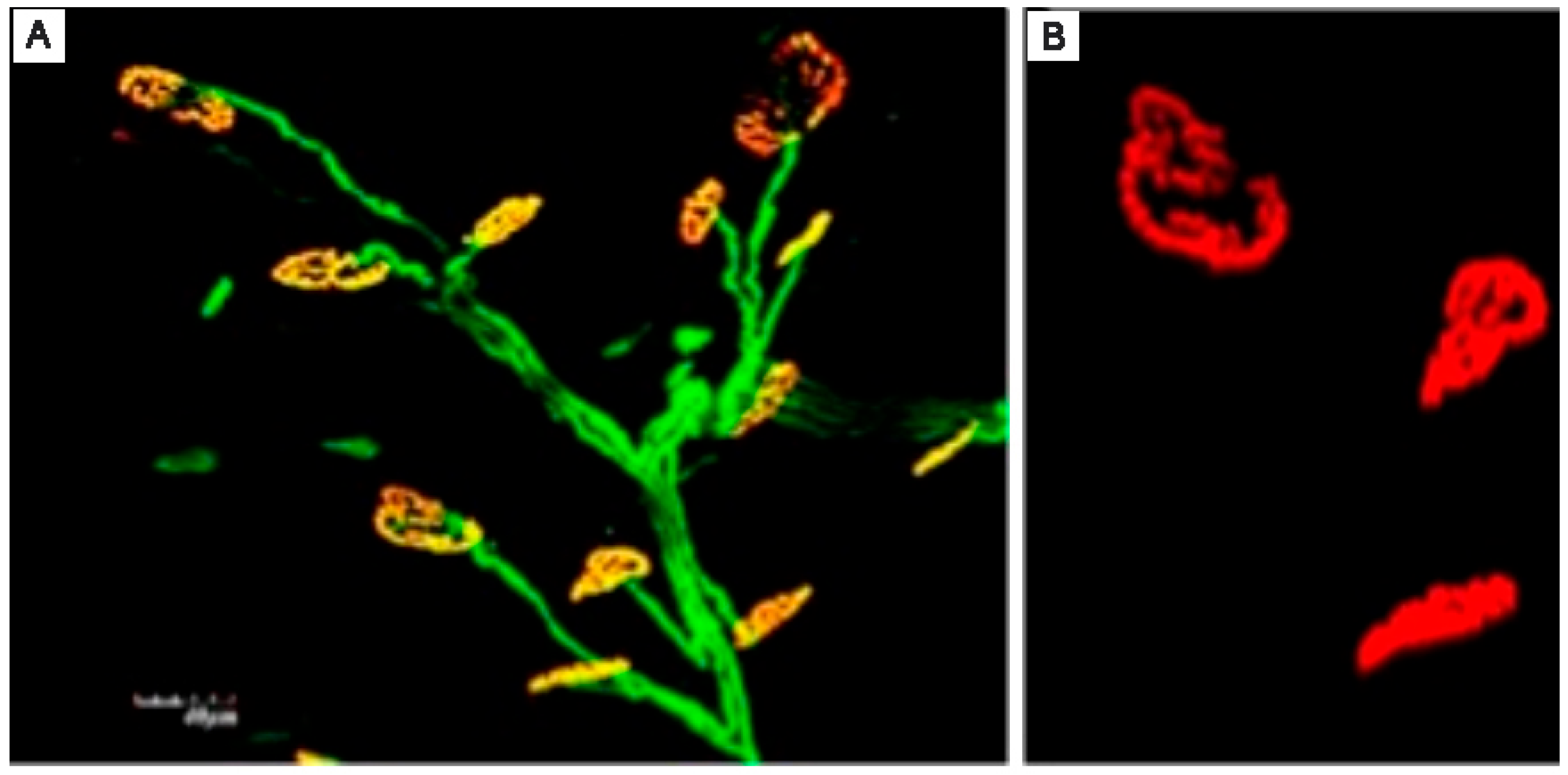
Extensive basic science research was performed on RPNIs prior to any human clinical trials. Animal experiments were performed utilizing the extensor digitorum longus (EDL) or soleus muscle as the free muscle graft, and the peroneal or tibial nerve of the rat hindlimb, to demonstrate feasibility and effectiveness of the approach. Stainless steel and electroconductive polymer coated electrodes (3,4-ethylenedioxythiophene) (PEDOT) were inserted into the RPNI to record the efferent motor action potentials and determine the degree of signal amplification facilitated with the RPNI [6,7]. A series of experiments were performed to evaluate the physiologic effects of utilizing a free skeletal muscle graft for the RPNI including assessments of muscle fiber regeneration and revascularization over time. In addition, the process of synaptogenesis and muscle reinnervation was investigated with a number of robust basic science experiments [8]. Histologic evaluations were also performed to anatomically evaluate axonal sprouting, axonal elongation, and muscle reinnervation in the RPNI constructs (Figure 2).
Physiologically, the quality of reinnervation of the RPNIs was evaluated by measuring the compound muscle action potentials (CMAP) and determining the resultant signal-to-noise (SNR) [10]. Through this series of experiments, RPNIs were clearly shown to convert small efferent motor action potentials with low SNR to large RPNI signals with high SNR. The motor control signal recordings from RPNI during rat ambulation similarly demonstrated favorable SNR. In addition, there were no significant issues regarding crosstalk with adjacent muscles or signal interference from adjacent muscles. The recorded EMG signals from the RPNIs aligned with hind limb kinematics and exhibited gait-entrained periodicity, indicating successful transduction of centrally generated motor commands [11,12]. Signal stability was maintained up to 7 months post-surgery without any deterioration in signal amplitude or SNR [13]. Follow up studies substantiated the biological stability of the RPNI construct and histological evidence of axonal regeneration and synaptogenesis within the interface.
The following series of experiments were in a more advanced animal model to determine if RPNIs are safe and can be used for volitional control of a prosthetic device. The model selected for these investigations was the nonhuman primate (NHP) [13,14]. In these experiments, RPNIs were created in the upper limb of the NHP. The RPNIs produced high-amplitude EMG signals for highly specific functions over a period of 20 months post-implantation. Researchers were able to detect finger movements with a success rate exceeding 96% [9]. The NHPs successfully completed assigned finger movement tasks using a combination of physical finger exercises and RPNI-based movement classifications. The findings indicated that RPNIs can generate SNRs with minimal interference from adjacent muscles over long durations of time. This finding affirmed the dependability and stability of RPNIs as a peripheral nerve interface. The tests demonstrated the capability to record functionally specific EMG signals, which hints at the potential for controlling individual finger movements in patients.

3. RPNI for the Prevention and Treatment of Neuroma Pain

One important area of RPNI research is dedicated to the prevention of neuroma formation following nerve injuries [15,16]. Painful neuromas are a common complication that arises after nerve injury [17]. Traditional approaches for treating neuromas included nerve ligation, cauterizing the end of the nerve, capping the nerve, or burying the nerve ending in muscle or bone [18,19]. Without offering a proper target for reinnervation for the regenerating axons, these approaches lead to relatively high neuroma recurrence rates. RPNIs are promising for neuroma treatment or prevention because they provide denervated muscle fibers as physiologic targets for reinnervation, reducing the number of erratic axons that could contribute to neuroma formation. Basic science studies in both rats and NHPs demonstrated that RPNIs prevent neuroma formation. Clinical studies have shown similar results to those seen in animal models. Kubiak et al. [20] compared patients undergoing RPNI surgery prophylactically at the time of amputation compared to those undergoing traditional nerve management procedures. This study compared 45 patients who underwent RPNI implantation at the time of primary amputation with 45 control patients who underwent standard amputation without RPNI. The results showed that none of the RPNI patients developed symptomatic neuromas, compared to 13.3% of the control group (p = 0.026). This study underscores the effectiveness of RPNIs for neuroma prevention following amputation. Various studies have consistently demonstrated that RPNI offers an effective strategy for neuroma treatment and prevention by presenting a functional target for regenerating axons. This approach improves pain management for patients with limb loss and consequently can improve comfort in their socket to enhance prosthetic rehabilitation. A recent study [21] on pediatric lower limb amputation patients further corroborates these findings. In this study, 25 patients who underwent RPNI surgery at the time of amputation were compared to 19 control patients who did not receive RPNI surgery. The results were striking: RPNI patients had a significantly lower incidence of post-amputation pain compared to control patients (p < 0.001), including lower rates of symptomatic neuroma (p < 0.001) and phantom limb associated pain (p < 0.01). Control patients had a nearly five-fold higher risk of developing chronic post-amputation pain compared to RPNI patients (RR = 4.7, p < 0.0001). Throughout the entire follow-up period, RPNI patients showed significantly lower objective pain scores (p = 0.007). Moreover, RPNI patients had significantly lower chronic narcotic usage (1.7 MME/day) compared to the control group (16.4 MME/day) (p < 0.01), with control patients having a roughly four-fold higher risk of developing reliance on chronic narcotic use (RR = 4.4, p < 0.001) compared to RPNI patients. These findings underscore the effectiveness of RPNI in preventing neuroma development and reducing post-amputation pain, not only in adults but also in pediatric patients. The significant reductions in pain scores and narcotic usage further emphasize the potential of RPNI surgery to improve pain management and quality of life for patients undergoing amputation.

4. Bridging Neuroscience and Engineering Through RPNI Surgery

RPNIs provide a seamless biotic–abiotic interface between the human nervous system and prosthetic devices. This pioneering approach merges surgical procedures with biocompatible materials and sophisticated signal processing to develop a user friendly interface for individuals with limb loss. The core of RPNI technology lies in its surgical methodology. RPNI allows for multi-channel control by leveraging the fascicular organization of peripheral nerves. The fascicular selectivity of RPNIs allows a wide range of possibilities including the creation of multiple independent RPNIs, increasing DOFs, and allowing for more complex movements involving multiple joints and muscles [1,4]. RPNIs have been successfully employed for control of various types of prosthetic systems, including upper and lower limb prostheses, as well as exoskeleton devices. Their adaptability makes them suitable for prosthetic applications where efferent motor control is desired. One of the groundbreaking aspects of RPNIs is the ability to establish bidirectional communication resembling the feedback loops found in natural sensorimotor systems [8].
The integration of electrodes improves the performance of RPNIs. The electrodes are placed either on the RPNI muscle surface (epimysial) or within the RPNI muscle itself (intramuscular). They facilitate recording and stimulation of neural activity [4]. The selection of type and placement is influenced by the signal specificity desired where intramuscular electrodes are noted for their ability for high fidelity control of individual finger movements. Specialized electrode designs and materials are being continuously evaluated to enhance performance and longevity of the interface connection with the peripheral nervous system. Many innovative and novel materials and electrode configurations have been designed to reduce iatrogenic nerve injury during electrode placement, reduce electrode biofouling over time, and enhance the long term stability of the interface [6,8,22]. Choosing the right electrode is an ongoing area of research and development where scientists and engineers are working closely together to enhance the reliability and stability of an RPNI.
Signal processing is important in converting raw electrical signals that are recorded from the peripheral nervous system into meaningful control commands for prosthetic devices. Studies show the use of sophisticated algorithms to interpret these signals and detect patterns linked to specific movements or intentions. These signal processing control strategies are customized to every user’s body structure/anatomy and control preferences in order to provide a personalized and natural prosthetic experience. There are several challenges when incorporating RPNIs into functional prosthetic systems. One of the major obstacles is creating miniaturized and biocompatible electronics that can seamlessly fit into a prosthetic limb. These neural control systems must be able to rectify, amplify, filter, and process RPNI signals for conversion into prosthetic control motor commands. These motor commands then need to be converted into electrical signals to control the prosthetic’s actuators. At the same time, the entire system needs to have minimal power consumption to enhance battery life of the device and be completely biocompatible and safe [23,24].
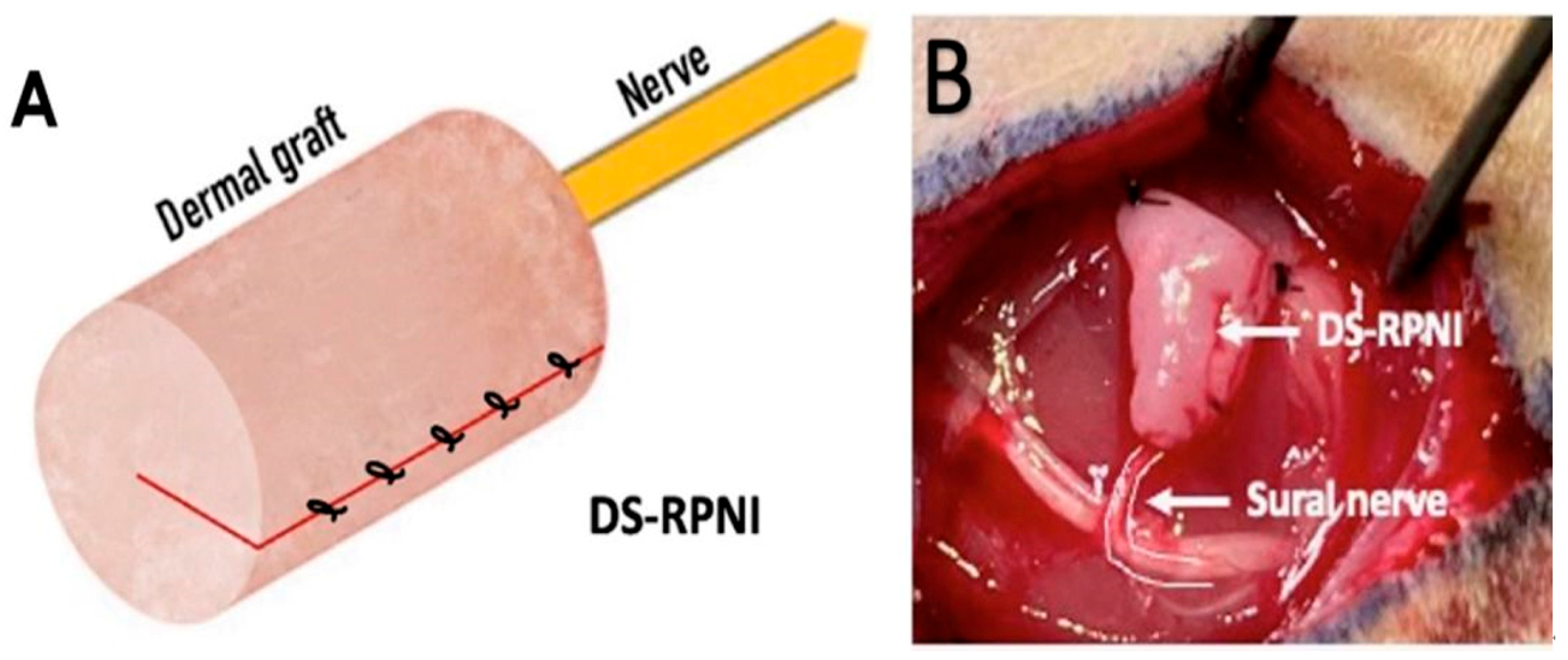
Sensory feedback plays an important role in human movement and interaction with the environment. It allows us to perceive the world around us, modulate our movements, and experience a sense of embodiment. Although advances have been made in providing artificial sensory feedback, current prosthetic devices still lack the ability to deliver naturalistic, somatotopically accurate, and stable sensory experiences comparable to a biological limb. Research has shown that RPNI stimulation has promise in generating meaningful and somatotopically accurate sensory feedback which is stable over several months [22,25]. One study [9] implanted RPNIs on the median and ulnar nerves of participants with upper limb amputations. Electrical stimulation of these RPNIs resulted in primitive proprioceptive (awareness of phantom limb movement) and cutaneous (touch, pressure, and temperature) sensations in the residual limb. The development of specialized RPNIs further expands the scope of sensory feedback applications; Dermal Sensory Regenerative Peripheral Nerve Interface (DS-RPNI) is an example of this innovative strategy to enhance the quality of sensory feedback [26]. DS-RPNIs are created by implanting sensory nerves or nerve fascicles into dermal grafts (Figure 3). Experiments in rats showed that DS-RPNIs survived and were successfully reinnervated by regenerated sensory afferent fibers. Upon stimulation of the DS-RPNIs, compound sensory nerve action potentials (CSNAPs) could be recorded proximally for both mechanical and electrical stimulation. This suggests that DS-RPNIs can effectively transduce sensory stimuli into neural signals. This discovery offers a promising approach to restoring naturalistic touch sensations in prosthetic users. Additionally, this sensory feedback plays a role in creating a deeper sense of attachment to the prosthetic limb making it feel more integrated into the body like a genuine extension. As a result, individuals may experience psychological advantages such as heightened satisfaction, self-assurance and acceptance of their prosthesis. Finally, research has indicated that sensory feedback can help alleviate phantom limb pain—a frequent and debilitating issue for people with limb loss [27,28,29]. Although the exact reasons for this pain relief remain unclear, it is thought that the sensory signals from RPNIs may assist in reshaping how the brain perceives the limb thus lessening the disparity between what the brain anticipates and the real sensory input.

5. Future Research Direction

Future research in regenerative peripheral nerve interfaces (RPNIs) holds immense potential to revolutionize prosthetic function, user embodiment, and restoration of naturalistic sensory feedback. At the forefront are Composite Regenerative Peripheral Nerve Interfaces (C-RPNIs). These biologic constructs which are composed of a piece of autogenous, de-epithelialized, full thickness skin is placed on a free skeletal muscle graft with a mixed motor/sensory nerve placed in the middle for neurotization. The C-RPNI is reinnervated preferentially by different axons based on the end organ present. The skin is reinnervated by cutaneous sensory afferents in the mixed nerve that can be used to provide light touch, temperature, and pinprick sensation from the prosthetic device. The muscle is reinnervated by efferent motor axons to provide signals for motor control of the prosthesis. The muscle is also reinnervated by sensory afferents that supply sensory feedback about prosthetic position through reinnervation of the Golgi tendon organs and the muscle spindles in the muscle portion of the C-RPNI [31,32]. These constructs provide high-fidelity signals for volitional control while simultaneously offering physiologic sensory feedback, a critical step toward true prosthetic embodiment. Complementing these biologic advances are significant technological developments in electrode design, with researchers focusing on creating smaller, more flexible, and higher-resolution neural interfaces. These next-generation electrodes aim to enhance signal fidelity, minimize tissue damage, and improve long-term biocompatibility. The ultimate goal is the development of fully implantable neural prosthetic systems that can operate seamlessly in real-world environments, providing users with a more natural and intuitive prosthetic experience. Machine learning algorithms and advanced signal processing techniques are being explored to decode neural signals with greater precision, translating user intent into refined prosthetic movements in real time [13,33]. Such algorithms can adapt to user-specific neural patterns and preferences, enhancing intuitive control and reducing cognitive load during prosthetic use. The development of biomimetic stimulation strategies is another critical direction, aiming to recreate the nuanced, spatiotemporal sensory experiences of natural limb interactions. Advanced computational models now emulate dynamic touch, pressure, and proprioception, enabling prosthetic devices to deliver somatotopically accurate, stable, and meaningful sensory feedback [34,35]. Importantly, studies investigating closed-loop systems using RPNIs have demonstrated the feasibility of combining efferent motor control with afferent sensory feedback to significantly improve prosthetic performance and user satisfaction. These systems may also contribute to the reduction in phantom limb pain by recalibrating cortical representations through physiologically meaningful sensory input [36]. Overall, future research should focus on integrating these biological and engineering advancements to develop fully implantable, low-power, and user-friendly neural prosthetic systems. Such systems have the potential to restore not only mobility but also a sense of embodiment, agency, and quality of life to individuals with limb loss, marking a transformative step in the field of neuroprosthetics.

6. Conclusions

RPNIs represent a transformative advancement in the field of neuroprosthetics, offering a biologically stable, high-fidelity interface for prosthetic control while concurrently addressing post-amputation neuroma pain and phantom limb pain. This innovative approach leverages the body’s natural regenerative capacity to enable intuitive, multi-channel prosthetic control and meaningful sensory feedback, addressing key limitations of previous interfacing methods. The evidence from foundational science to clinical application demonstrates that RPNIs provide stable, interference-resistant signals suitable for real-world prosthetic use, with long-term durability and high signal-to-noise ratios. Clinically, RPNI surgery not only facilitates advanced prosthetic function but also significantly reduces symptomatic neuroma formation and chronic pain, enhancing patient comfort and prosthetic acceptance.
Looking forward, the integration of RPNIs with advanced electrode technologies and machine learning algorithms holds the potential to achieve true prosthetic embodiment, with seamless bidirectional communication for motor control and naturalistic sensory feedback. RPNIs stand at the intersection of engineering and neuroscience, offering a scalable and effective solution for restoring function and improving quality of life for individuals with limb loss. As research and clinical adoption continue to grow, RPNIs are positioned to become a cornerstone technology in the next generation of neuroprosthetic systems.

Author Contributions

M.J.W.: conceptualization, data curation, formal analysis, investigation, methodology, project administration, resources, supervision, validation, writing—original draft, writing—review and editing. T.A.K.: conceptualization, supervision, writing—original draft, writing—review and editing. A.K.S.-W.: writing—review and editing. P.S.C.: conceptualization, supervision, writing—original draft, writing—review and editing. All authors have read and agreed to the published version of the manuscript.

Funding

This research received no external funding.

Institutional Review Board Statement

Not applicable.

Informed Consent Statement

Not applicable.

Conflicts of Interest

Paul S. Cederna, MD, serves as President of Blue Arbor Technologies, Inc., a company that designs, manufactures, and produces neural prosthetic control systems. None of the Blue Arbor Technology products will be discussed in this article. No funding has been received from any source for this article. Alison K. Snyder Warwick, MD, has no disclosures relevant to this work. She has received research funding from Checkpoint Surgical. Theodore A. Kung, MD, serves as Chief Medical Officer of Blue Arbor Technologies, Inc., a company that designs, manufactures, and produces neural prosthetic control systems. None of the Blue Arbor Technology products will be discussed in this article. No funding has been received from any source for this article. Melanie J. Wang, MD has no disclosures.

References

  1. Kubiak, C.A.; Adidharma, W.; Kung, T.A.; Kemp, S.W.P.; Cederna, P.S.; Vemuri, C. Decreasing Postamputation Pain with the Regenerative Peripheral Nerve Interface (RPNI). Ann. Vasc. Surg. 2022, 79, 421–426. [Google Scholar] [CrossRef] [PubMed]
  2. Leach, G.A.; Dean, R.A.; Kumar, N.G.; Tsai, C.; Chiarappa, F.E.; Cederna, P.S.; Kung, T.A.; Reid, C.M. Regenerative Peripheral Nerve Interface Surgery: Anatomic and Technical Guide. Plast. Reconstr. Surg. Glob. Open 2023, 11, e5127. [Google Scholar] [CrossRef] [PubMed] [PubMed Central]
  3. Adewole, D.O.; Serruya, M.D.; Harris, J.P.; Burrell, J.C.; Petrov, D.; Chen, H.I.; Wolf, J.A.; Cullen, D.K. The Evolution of Neuroprosthetic Interfaces. Crit. Rev. Biomed. Eng. 2016, 44, 123–152. [Google Scholar] [CrossRef] [PubMed] [PubMed Central]
  4. Burke, K.L.; Dumanian, G.A.; Cederna, P.S. Emergence of Nerve Interfaces with Robotic Applications. In Landmark Papers in Plastic Surgery; Khajuria, A., Hong, J.P., Neligan, P., Eds.; Springer: Cham, Switzerland, 2014. [Google Scholar] [CrossRef]
  5. Pettersen, E.; Sassu, P.; Pedrini, F.A.; Granberg, H.; Reinholdt, C.; Breyer, J.M.; Roche, A.; Hart, A.; Ladak, A.; Power, H.A.; et al. Regenerative Peripheral Nerve Interface: Surgical Protocol for a Randomized Controlled Trial in Postamputation Pain. J. Vis. Exp. 2024, 205, e66378. [Google Scholar] [CrossRef] [PubMed]
  6. Frost, C.M.; Ursu, D.C.; Flattery, S.M.; Nedic, A.; Hassett, C.A.; Moon, J.D.; Buchanan, P.J.; Brent Gillespie, R.; Kung, T.A.; Kemp, S.W.P.; et al. Regenerative peripheral nerve interfaces for real-time, proportional control of a Neuroprosthetic hand. J. Neuroeng. Rehabil. 2018, 15, 108. [Google Scholar] [CrossRef] [PubMed] [PubMed Central]
  7. Ursu, D.; Nedic, A.; Urbanchek, M.; Cederna, P.; Gillespie, R.B. Adjacent regenerative peripheral nerve interfaces produce phase-antagonist signals during voluntary walking in rats. J. Neuroeng. Rehabil. 2017, 14, 33. [Google Scholar] [CrossRef] [PubMed] [PubMed Central]
  8. Gates, D.H.; Gonzalez, M.; Kung, T.A.; Chestek, C.A. Toward the use of muscle reinnveration for chronic bi-directional control of prostheses to improve functional outcomes of end users. Curr. Opin. Biomed. Eng. 2023, 28, 100497. [Google Scholar] [CrossRef]
  9. Burke, K.L.; Kung, T.A.; Hooper, R.C.; Kemp, S.W.P.; Cederna, P.S. Regenerative peripheral nerve interfaces (RPNIs): Current status and future direction. Plast. Aesthetic Res. 2022, 9, 48. [Google Scholar] [CrossRef]
  10. Ursu, D.C.; Urbanchek, M.G.; Nedic, A.; Cederna, P.S.; Gillespie, R.B. In vivo characterization of regenerative peripheral nerve interface function. J. Neural. Eng. 2016, 13, 026012. [Google Scholar] [CrossRef] [PubMed]
  11. Urbanchek, M.G.; Kung, T.A.; Frost, C.M.; Martin, D.C.; Larkin, L.M.; Wollstein, A.; Cederna, P.S. Development of a Regenerative Peripheral Nerve Interface for Control of a Neuroprosthetic Limb. Biomed. Res. Int. 2016, 2016, 5726730. [Google Scholar] [CrossRef] [PubMed] [PubMed Central]
  12. Kubiak, C.A.; Svientek, S.R.; Dehdashtian, A.; Lawera, N.G.; Nadarajan, V.; Bratley, J.V.; Kung, T.A.; Cederna, P.S.; Kemp, S.W.P. Physiologic signaling and viability of the muscle cuff regenerative peripheral nerve interface (MC-RPNI) for intact peripheral nerves. J. Neural. Eng. 2021, 18, 0460d5. [Google Scholar] [CrossRef] [PubMed]
  13. Irwin, Z.T.; Schroeder, K.E.; Vu, P.P.; Tat, D.M.; Bullard, A.J.; Woo, S.L.; Sando, I.C.; Urbanchek, M.G.; Cederna, P.S.; Chestek, C.A. Chronic recording of hand prosthesis control signals via a regenerative peripheral nerve interface in a rhesus macaque. J. Neural. Eng. 2016, 13, 046007. [Google Scholar] [CrossRef] [PubMed]
  14. Vu, P.P.; Irwin, Z.T.; Bullard, A.J.; Ambani, S.W.; Sando, I.C.; Urbanchek, M.G.; Cederna, P.S.; Chestek, C.A. Closed-Loop Continuous Hand Control via Chronic Recording of Regenerative Peripheral Nerve Interfaces. IEEE Trans. Neural. Syst. Rehabil. Eng. 2018, 26, 515–526. [Google Scholar] [CrossRef] [PubMed]
  15. Lee, J.C.; Kubiak, C.A.; Best, C.S.W.; Hamill, J.B.; Ki, J.; Kim, H.M.; Roth, R.S.; Kozlow, J.H.; Tinney, M.J.; Geisser, M.E.; et al. Regenerative Peripheral Nerve Interface Surgery to Treat Chronic Postamputation Pain: A Prospective Study in Major Lower Limb Amputation Patients. Ann. Surg. Open. 2025, 6, e535. [Google Scholar] [CrossRef] [PubMed] [PubMed Central]
  16. Ganesh Kumar, N.; Cederna, P.S.; Kung, T.A. Regenerative Peripheral Nerve Interfaces for Treatment of Symptomatic Neuromas. In Contemporary Neuroma Management; Eberlin, K.R., Ducic, I., Moore, A., Cederna, P.S., Valerio, I.L., Dumanian, G.A., Eds.; Springer: Cham, Switzerland, 2024. [Google Scholar] [CrossRef]
  17. Hwang, C.D.; Hoftiezer, Y.A.J.; Raasveld, F.V.; Gomez-Eslava, B.; van der Heijden, E.P.A.; Jayakar, S.; Black, B.J.; Johnston, B.R.; Wainger, B.J.; Renthal, W.; et al. Biology and pathophysiology of symptomatic neuromas. Pain 2024, 165, 550–564. [Google Scholar] [CrossRef] [PubMed]
  18. Chou, J.; Liston, J.M.; DeGeorge, B.R. Traditional Neuroma Management Strategies: A Systematic Review. Ann. Plast. Surg. 2023, 90 (Suppl. 4), S350–S355. [Google Scholar] [CrossRef] [PubMed]
  19. Pettersen, E.; Sassu, P.; Reinholdt, C.; Dahm, P.; Rolfson, O.; Björkman, A.; Innocenti, M.; Pedrini, F.A.; Breyer, J.M.; Roche, A.; et al. Surgical treatments for postamputation pain: Study protocol for an international, double-blind, randomised controlled trial. Trials 2023, 24, 304. [Google Scholar] [CrossRef] [PubMed] [PubMed Central]
  20. Kubiak, C.A.; Kemp, S.W.P.; Cederna, P.S.; Kung, T.A. Prophylactic Regenerative Peripheral Nerve Interfaces to Prevent Postamputation Pain. Plast. Reconstr. Surg. 2019, 144, 421e–430e. [Google Scholar] [CrossRef] [PubMed]
  21. Mohanty, A.; Cederna, P.; Kemp, S.; Kung, T. Prophylactic Regenerative Peripheral Nerve Interface (RPNI) Surgery in Pediatric Lower Limb Amputation Patients. Ann. Surg. 2025, 282, 346–351. [Google Scholar] [CrossRef]
  22. Gonzalez, M.A.; Nwokeabia, C.; Vaskov, A.K.; Vu, P.P.; Lu, C.W.; Patil, P.G.; Cederna, P.S.; Chestek, C.A.; Gates, D.H. Electrical Stimulation of Regenerative Peripheral Nerve Interfaces (RPNIs) Induces Referred Sensations in People With Upper Limb Loss. IEEE Trans. Neural Syst. Rehabil. Eng. 2024, 32, 339–349. [Google Scholar] [CrossRef] [PubMed] [PubMed Central]
  23. Irwin, Z.T. Restoring Fine Motor Skills Through Neural Interface Technology. Ph.D. Thesis, The University of Michigan, Ann Arbor, MI, USA, 2016. [Google Scholar]
  24. Vu, P. Restoring Fine Motor Prosthetic Hand Control via Peripheral Neural Technology. Ph.D. Thesis, The University of Michigan, Ann Arbor, MI, USA, 2019. [Google Scholar]
  25. Vu, P.P.; Lu, C.W.; Vaskov, A.K.; Gates, D.H.; Gillespie, R.B.; Kemp, S.W.P.; Patil, P.G.; Chestek, C.A.; Cederna, P.S.; Kung, T.A. Restoration of Proprioceptive and Cutaneous Sensation Using Regenerative Peripheral Nerve Interfaces in Humans with Upper Limb Amputations. Plast Reconstr Surg. 2022, 149, 1149e–1154e. [Google Scholar] [CrossRef] [PubMed] [PubMed Central]
  26. Sando, I.C.; Adidharma, W.; Nedic, A.; Ursu, D.C.; Mays, E.A.; Hu, Y.; Kubiak, C.A.; Sugg, K.B.; Kung, T.A.; Cederna, P.S.; et al. Dermal Sensory Regenerative Peripheral Nerve Interface for Reestablishing Sensory Nerve Feedback in Peripheral Afferents in the Rat. Plast. Reconstr. Surg. 2023, 151, 804e–813e. [Google Scholar] [CrossRef] [PubMed]
  27. Dietrich, C.; Nehrdich, S.; Seifert, S.; Blume, K.R.; Miltner, W.H.R.; Hofmann, G.O.; Weiss, T. Leg Prosthesis With Somatosensory Feedback Reduces Phantom Limb Pain and Increases Functionality. Front. Neurol. 2018, 9, 270. [Google Scholar] [CrossRef]
  28. Nanivadekar, A.C.; Bose, R.; Petersen, B.A.; Okorokova, E.V.; Sarma, D.; Madonna, T.J.; Barra, B.; Farooqui, J.; Dalrymple, A.D.; Levy, I.; et al. Restoration of sensory feedback from the foot and reduction of phantom limb pain via closed-loop spinal cord stimulation. Nat. Biomed. Eng. 2023, 8, 992–1003. [Google Scholar] [CrossRef]
  29. Culp, C.J.; Abdi, S. Current Understanding of Phantom Pain and its Treatment. Pain Physician 2022, 25, E941–E957. [Google Scholar] [PubMed]
  30. Berberoglu, I.; Burke, K.L.; Cederna, P.S.; Kemp, S.W.P. Regenerative peripheral nerve interfaces (RPNIs): An overview of innovative surgical approaches. Plast. Aesthet. Res. 2024, 11, 14. [Google Scholar] [CrossRef]
  31. Svientek, S.R.; Ursu, D.C.; Cederna, P.S.; Kemp, S.W.P. Fabrication of the Composite Regenerative Peripheral Nerve Interface (C-RPNI) in the Adult Rat. J. Vis. Exp. 2020, 156, e60841. [Google Scholar] [CrossRef] [PubMed]
  32. Kubiak, C.A.; Ursu, D.C.; Moon, J.D.; Patil, P.G.; Kung, T.A.; Cederna, P.S.; Kemp, S.W.P. Abstract 36: Viability and Signal Transduction with the Composite Regenerative Peripheral Nerve Interface (C-RPNI). Plast. Reconstr. Surg. Glob. Open 2019, 7 (Suppl. 4), 26–27. [Google Scholar] [CrossRef] [PubMed Central]
  33. Vaskov, A.K.; Vu, P.P.; North, N.; Davis, A.J.; Kung, T.A.; Gates, D.H.; Cederna, P.S.; Chestek, C.A. Surgically Implanted Electrodes Enable Real-Time Finger and Grasp Pattern Recognition for Prosthetic Hands. IEEE Trans. Robot. 2022, 38, 2841–2857. [Google Scholar] [CrossRef] [PubMed] [PubMed Central]
  34. Graczyk, E.L.; Schiefer, M.A.; Saal, H.P.; Delhaye, B.P.; Bensmaia, S.J.; Tyler, D.J. The neural basis of perceived intensity in natural and artificial touch. Sci. Transl. Med. 2016, 8, 362ra142. [Google Scholar] [CrossRef] [PubMed] [PubMed Central]
  35. George, J.A.; Kluger, D.T.; Davis, T.S.; Wendelken, S.M.; Okorokova, E.V.; He, Q.; Duncan, C.C.; Hutchinson, D.T.; Thumser, Z.C.; Beckler, D.T.; et al. Biomimetic sensory feedback through peripheral nerve stimulation improves dexterous use of a bionic hand. Sci. Robot. 2019, 4, eaax2352. [Google Scholar] [CrossRef] [PubMed]
  36. Ortiz-Catalan, M.; Guðmundsdóttir, R.A.; Kristoffersen, M.B.; Zepeda-Echavarria, A.; Caine-Winterberger, K.; Kulbacka-Ortiz, K.; Widehammar, C.; Eriksson, K.; Stockselius, A.; Ragnö, C.; et al. Phantom motor execution facilitated by machine learning and augmented reality as treatment for phantom limb pain: A single group, clinical trial in patients with chronic intractable phantom limb pain. Lancet 2016, 388, 2885–2894. [Google Scholar] [CrossRef] [PubMed]
Figure 1. Schematic diagram of a Regenerative Peripheral Nerve Interface construct [5].
Prosthesis 07 00097 g001
Figure 2. Histological demonstration of axon growth and formation of neuromuscular junctions within the RPNI. (A) Regenerating axons are highlighted in green through immunostaining with anti-neurofilament 200. (B) Neuromuscular junctions are visualized in red using fluorescently labeled alpha-bungarotoxin [9].
Prosthesis 07 00097 g002
Figure 3. (A) Schematic diagram of a DS-RPNI construct. (B) A DS-RPNI in a rat [30].
Prosthesis 07 00097 g003
Disclaimer/Publisher’s Note: The statements, opinions and data contained in all publications are solely those of the individual author(s) and contributor(s) and not of MDPI and/or the editor(s). MDPI and/or the editor(s) disclaim responsibility for any injury to people or property resulting from any ideas, methods, instructions or products referred to in the content.