Article

Microleakage Study of a Bulk Fill over an Uncured Adhesive System

by
Mariana Vaz Pinto
1,
Salomé Pires
2,3,4,5,
Carlos Miguel Marto
1,2,3,4,5,6,
Inês Amaro
1,3,4,5,
Ana Coelho
1,3,4,5,
José Sousa
1,
Manuel Marques Ferreira
3,4,5,7,
Maria Filomena Botelho
2,3,4,5,
Eunice Carrilho
1,3,4,5,
Ana Margarida Abrantes
2,3,4,5 and
Anabela Baptista Paula
1,3,4,5,*
1
Institute of Integrated Clinical Practice, Faculty of Medicine, University of Coimbra, 3000-354 Coimbra, Portugal
2
Institute of Biophysics, Faculty of Medicine, University of Coimbra, 3000-354 Coimbra, Portugal
3
Institute for Clinical and Biomedical Research (iCBR), Area of Environment Genetics and Oncobiology (CIMAGO), Faculty of Medicine, University of Coimbra, 3000-354 Coimbra, Portugal
4
Clinical Academic Center of Coimbra (CACC), 3000-354 Coimbra, Portugal
5
Center for Innovative Biomedicine and Biotecnhology (CIBB), 3000-354 Coimbra, Portugal
6
Institute of Experimental Pathology, Faculty of Medicine, University of Coimbra, 3000-354 Coimbra, Portugal
7
Institute of Endodontics, Faculty of Medicine, University of Coimbra, 3000-354 Coimbra, Portugal
*
Author to whom correspondence should be addressed.
J. Compos. Sci. 2023, 7(1), 40; https://doi.org/10.3390/jcs7010040
Submission received: 3 September 2022 / Revised: 7 October 2022 / Accepted: 6 January 2023 / Published: 13 January 2023
(This article belongs to the Section Composites Applications)

Abstract

:
This study aimed to evaluate and compare the microleakage of composite resin restorations under a total-etch adhesive system applied with two different techniques, with and without cure. Cavities were made on the buccal surface of twenty-six intact teeth and subsequently restored with bulk-fill flow composite resin (SDR®® flow+) and conventional resin (Spectra™ ST HV). Two experimental groups were created, one where the total-etch adhesive (Prime & Bond Active®) was cured and another where the adhesive was not cured before placing the flowable resin. Two control groups were also created, negative and positive. After the restorations were finished the, teeth were submerged in a solution of sodium pertechnetate (99mTcNaO4) for 3 h to evaluate the microleakage. The results showed more infiltration of radioisotopes characterized by the highest total count values of microleakage in group 1 (cured adhesive) and in the positive control. Group 2 (noncured adhesive) and the negative control showed lower values. We therefore considered that these groups were similar since their p-value was less than 0.05, with no statistically significant difference. Group 4 showed a statistically significant increase in relation to group 3 (p = 0.027). We concluded that the method of bulk-fill composite resins with noncured adhesive agents can have a positive effect on the longevity of bond strengths, with reduced microleakage.

1. Introduction

Contemporary dentistry cannot be performed without the use of adhesive systems. They can be considered revolutionary since they enable previously inconceivable clinical maneuvers. Adhesive systems provide immediate bond strength, bonding to tooth structure without needing a retentive cavity [1].
The main objective of adhesive procedures is to create and maintain a firm adhesive–tooth structure interface that is stable for several years and that provides retentive strength, marginal sealing, and, consequently, clinical durability [2].
Regardless of the technique used in dentin adhesion, it is based on the formation of the “hybrid layer” (CH) which is constituted by a structure of demineralized collagen fibers reinforced by a resin matrix [3].
Nakabayashi et al. were the first to demonstrate that after etching the dentin with phosphoric acid, the collagen is exposed, allowing the impregnation of the resin ingredients to form a new structure, the hybrid layer, a chemical and mechanical union between dentin and resin [4].
To better understand these processes, it is important to know the structure and composition of dentin. It is a mineralized collagen matrix that contains approximately 30–50% organic material and approximately 20% water. Its composition varies in different areas of the tooth, depending on its proximity to the pulp tissue and whether the matrix is demineralized or has been affected by caries [1].
The success and durability of resin restorations are closely dependent on the discoveries that are made in bonding processes [5]. To simplify the bonding procedure, user-friendly adhesive systems such as two-step etch-and-rinse, one-step self-etch, and universal adhesives have been created over time [6].
Despite the facility of use, these adhesives prevent the great penetration of resin monomers into the demineralized dentin, leaving residual water in the interfibrillar spaces, which activates protease enzymes [7] and, consequently, reduces the durability of the dentin adhesive interface [8].
This happens because, during the etching process and subsequent wetting and drying, 50% of the dentin’s organic material is dissolved, removed and replaced by the washing water, which combined with the preexisting 20% of water, results in a new volume of 70% water. In the resin infiltration stage, the 70% water ideally should be replaced by 70% resin monomers [9]. However, this replacement is never ideal [10], which results in incomplete infiltration of the resin into the dentin tubules, creating water-rich and resin-poor sites in the polymerized hybrid layer, making them more susceptible to microleakage [11].
Excess water in the hybrid layer avoids an optimal polymerization of the adhesive resin monomers [12] and leads to the separation of phases and plasticization of the resin adhesive interface over time [13]. This results in the hybrid layer degradation and, therefore, the failure of the adhesive interface [14].
The destruction of the hybrid layer involves hydrolysis of the adhesive resin that once has penetrated the demineralized dentin matrix [15]. This will lead to micro gap development that is easily invaded by pathogens and begins a cascade of events that culminate in the failure of the composite resin restoration [14].
To improve the dentin adhesive bond interface and consequently reduce microleakage, several techniques have been tested, such as wet adhesion with ethanol [16], the application of multiple adhesive layers [17], the application of an extra hydrophobic binding layer [18], the sonic application of the binding agents [19], and the co-cure technique, which will be the basis of the present study, where the adhesive is polymerized simultaneously with the composite resin [20]. Nevertheless, there is some controversy and little scientific evidence regarding which technique is considered the best for sealing the dentin tissue and creating a more durable hybrid layer.
In this study, a technique involving the application of fluid composite bulk-fill resin over a nonpolymerized adhesive system was evaluated. The objective is to evaluate the microleakage of composite resin restorations when this technique is applied and compare it with the conventional technique.

2. Materials and Methods

2.1. Sample Collection

Twenty-six noncarious human molars or premolars (extracted for orthodontic reasons) were stored in 0.9% normal saline at 5 °C for no more than 4 months after extraction. This study was submitted to the Ethics Committee of the Faculty of Medicine of the University of Coimbra and was approved under the number CE-077/2019.

2.2. Sample Preparation

Class V cavities were prepared on the buccal surface of each tooth. A transparent resin mold was made to draw the cavities on the surface of each tooth. Each cavity had the following dimensions: 4 mm mesiodistal, 3 mm occlusal–gingival, and 3 mm deep. Inner line angles were kept at 90 degrees. The cavity margins were in enamel (Figure 1A).
The burs FG 835/010 drills (Proclinic, 34/09, Nyon, Switzerland) were used to create the cavities and were replaced after every 5 preparations.

2.3. Study Groups

The twenty-six specimens were randomly distributed into four groups. Twenty samples were used for the study groups and six samples for the control groups, three for the positive control group, and three for the negative control group.
Group 1: experimental group
SDR®® Flow+ composite resin (A1 shade) (Dentsply-Sirona, 78467, Konstanz, Germany) and Spectra ST HV composite resin (A1 shade) (Dentsply-Sirona, 78467, Konstanz, Germany) were used to restore the class V cavities of 10 samples. The enamel was conditioned for 30 s with 35% phosphoric acid and washed immediately after with an air/water jet for 30 s. A total-etch, Prime & Bond Universal adhesive (Dentsply-Sirona, 78467, Konstanz, Germany) was used according to the manufacturer’s instructions. The adhesive was consistently dried with gentle airflow before being light cured for 20 s using the Blue Phase™ G2 curing light (Ivoclar Vivadent, 5VDC, Liechtenstein, Austria). After the adhesive was placed, the composite resin SDR®® Flow+ was placed in the cavity through an increment of 2 mm and light-cured for 20 s. Then, the Spectra™ ST HV composite resin was applied with an increment of 1 mm to the buccal surface and light-cured in the same way for 20 s using the BluePhase™ G2 light curing light (Ivoclar Vivadent, 5VDC, Liechtenstein, Austria).
Restorations were polished using the Enhance®® System (Dentsply-Sirona, Konstanz, Germany).
The description of the procedures mentioned above can be seen in Figure 1, from B to I.
Group 2: experimental group
Enamel and dentin were conditioned in the same way as described for group 1. A total-etch Prime & Bond Universal adhesive (Dentsply-Sirona, 78467, Konstanz, Germany) was applied in the same way as in group 1, but there was no polymerization after drying. As in the previous group, the composite resin SDR®® Flow+ (color A1) (Dentsply-Sirona, 78467, Konstanz, Germany) was used in a single layer of 2 mm and the resin Spectra ST™ HV composite (color A1) (Dentsply-Sirona, 78467, Konstanz, Germany) in a 1 mm layer, to restore the class V cavities of 10 samples. Light curing was carried out between the two layers of composite with the BluePhase™ G2 light curing light (Ivoclar Vivadent, 5VDC, Liechtenstein, Austria)
The restorations were polished using the same disc described above.
Group 3: negative control group
Enamel and dentin were conditioned in the same way as described in the previous groups. A total-etch bonding agent, Prime & Bond Universal (Dentsply-Sirona, 78467, Konstanz, Germany) was used, applied in the same way as in group 1, and cured after application. The composite resins SDR®® Flow+ were used (color A1) (Dentsply-Sirona, 78467, Konstanz, Germany) in a single layer of 2 mm and Spectra ST™ HV composite (color A1) (Dentsply-Sirona, 78467, Konstanz, Germany) was used in a single layer of 1 mm, to restore 3-sample class V cavities. Light curing was carried out between the two layers of composite resin with the BluePhase™ G2 light curing light (Ivoclar Vivadent, 5VDC, Liechtenstein, Austria).
The restorations were polished using the same disc system.
Group 4: positive control group
In this group, no intervention was performed, and the class V cavities of 3 samples remained intact, without any restoration.
There was only one operator performing all restorative procedures. Samples were stored in distilled water at 37 °C for one week.

2.4. Nuclear Medicine Technique for Microleakage Assessment

Specimens of groups 1, 2, and 4 were covered with two layers of nail varnish (Essie, New York, NY, USA, 16S201) up to 2 mm from the margins around the restorations.
Group 3 (negative control) was covered by two layers of varnish over the entire surface, including the area of the restored cavity.
The specimens from all groups were submerged in a sodium pertechnetate (99mTc-NaO4) solution for 3 h.
Technetium-99m is an artificial element obtained by the radioactive decay of molybdenum, which is a radioactive metallic element belonging to the transition metals with an atomic radius of 135.8 pm. It has a half-life of 6.04 h. Its decay occurs through isometric transition and emission and 140.5 keV of gamma radiation. fterward, the varnish was completely removed.
The radioactivity emitted by the samples was detected by a gamma camera (GE Millennium MG, Milwaukee, WI, USA) and controlled by a computer (GenieAcq, GE, Milwaukee, WI, USA).
A static image was obtained for each specimen for two minutes. The images had a matrix dimension of 512 × 512 and a zoom of 1.33. The regions of interest (ROIs) of each image were drawn over the teeth to obtain total, maximum, and average counts using a specified software (XelerisTM, GE, Milwaukee, WI, USA). The total counts obtained from each image were used to quantify infiltration.

2.5. Statistical Analysis
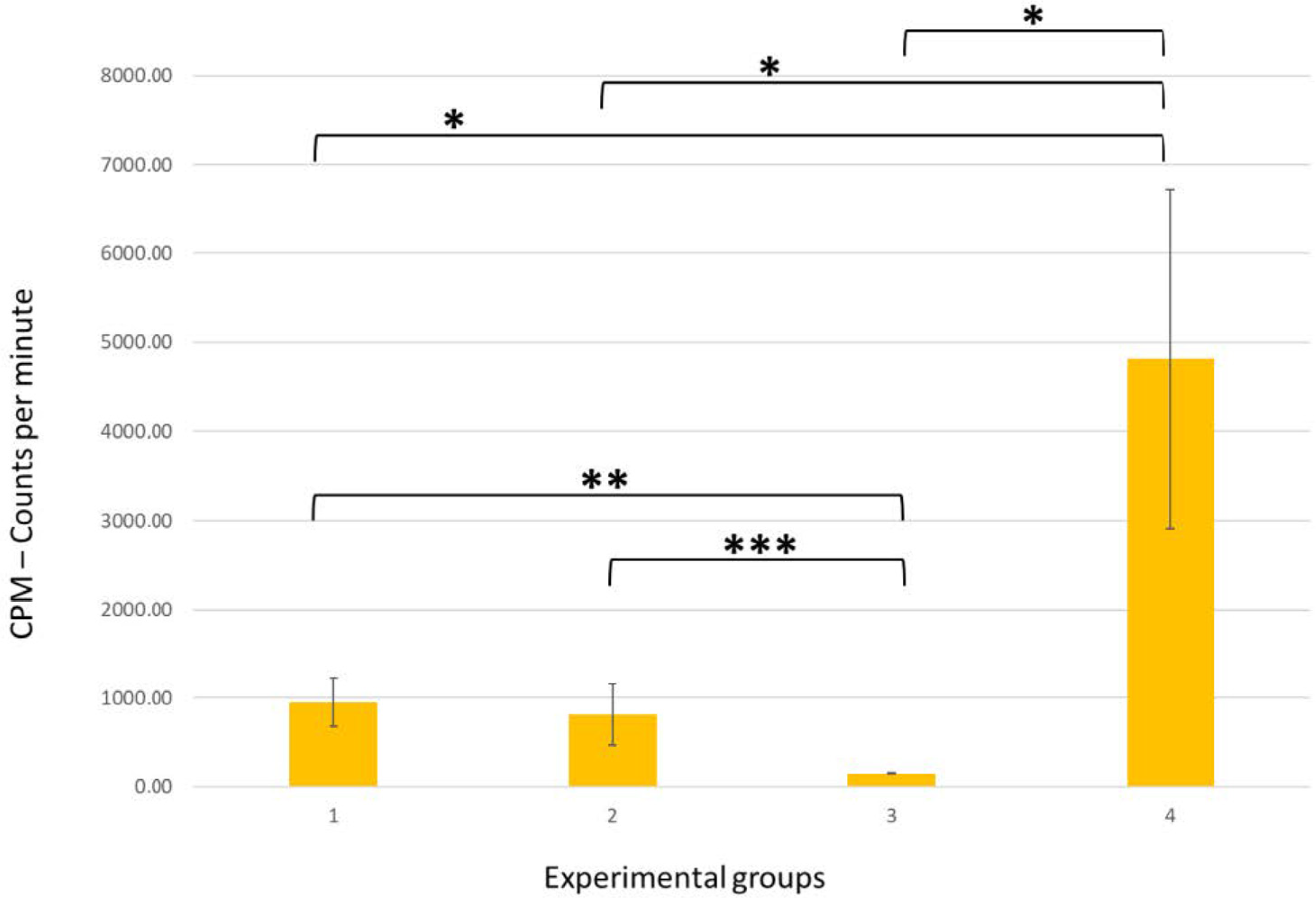
The sample was characterized through the presentation of the average with standard deviation. The different groups were compared using the Kruskal–Wallis test and for multiple comparisons using the Games–Howell test. The option for nonparametric tests was based on the lack of adjustment of the sample quantitative distributions to normal distributions, and this assumption was evaluated by the Shapiro–Wilk test. The analysis was performed using SPSS version 2, and analyzed at a significance level of 5%.

3. Results

The obtained results are presented in Figure 2.
The highest radioisotope infiltration characterized by the highest microleakage values (counts per minute) was observed in the positive control group (group 4, whose cavities were not restored), followed by group 1 (where the adhesive system was cured).
Group 2 (where the adhesive was not cured) and the negative control group (group 3, cavities restored and completely isolated with polish) showed lower infiltration values.
The greater the infiltration of the radioisotope was, the higher the microleakage values of the restoration.
The mean value of infiltration in group 1 was 955.8, while in group 2, this value was slightly lower, corresponding to 816.9 without statistical significance. We, therefore, consider that these groups are similar since their p-value is less than 0.05.
The positive control group, whose teeth were not restored, presented the highest microleakage value, with the average value of 4817.2 counts per minute. In the negative control group, group 3, the cavities were restored in the same way as that in group 1 and isolated with polish over its entire surface, which resulted in the lowest microleakage values, with the average value of radioactivity of 151.2.
This way, group 4 showed an increase compared to group 3 that was statistically significant (p = 0.027).
Between groups 3 and 1, there was a statistical difference since the p-value was 0.006. However, the groups with a smaller statistical difference were group 3 and group 2 (p = 0.001).
There was also a statistically significant difference between group 4 and group 1, with a value of p = 0.044.
Between group 4 and group 2, there was no difference (p = 0.050).

4. Discussion

The present experimental study was designed to evaluate the microleakage of restorations on a noncured adhesive system. Submersion of samples in sodium pertechnetate solution (99mTcNaO4) was used.
Currently, the most common methods to evaluate microleakage are the methylene blue staining method and the bacterial infiltration method. Therefore, these methods present some limitations. The blue dye staining method is semiquantitative and provides us with a subjective assessment of the results, requiring a specialized operator. Furthermore, it is a technique that requires the destruction of the samples [21]. The bacterial infiltration method is believed to be more clinically and biologically relevant when compared to the dye penetration method [22]. However, it is generally qualitative and can only be applied to specimens that show antimicrobial activity for the type of bacteria being infiltrated [23].
The method used, which uses radioactive isotopes such as 99mTcNaO4, has advantages since it is quantitative and does not require the destruction of the sample, diverging from the methods presented above. It is also able to detect infiltration per minute and detects infiltration even if it exists in very small concentrations [24].
Higher values of radioactivity correspond to higher values of microleakage in areas of the adhesive interface that allowed technetium penetration.
In this study, positive and negative control groups were used, allowing the validation of the results, establishing a minimum and maximum value, and thus validating the methodology. Control groups were valid as their radioactivity values matched the highest and lowest scores. In the negative control group (group 3), there was a lower value of radioactivity, which means a lower value of microleakage. This happened because it was the group completely covered by varnish and, consequently, the most isolated, since the radiation penetrated the least.
The current technique mixes a fluid bulk-fill resin with a noncured adhesive system, and the cavity is then restored with a conventional composite resin. Studies that evaluated polymerization shrinkage showed that bulk-fill resins have less polymerization shrinkage than do conventional composite resins. The bulk-fill flow composite resin used in this study includes a polymerization modulator that interacts with the camphorquinone photo initiator to provide a greater elasticity module and, in this way, a better dissipation of shrinkage stress during cure. This flow has been widely used and applied in a single increment up to 4 mm thick [25]. The low infiltration values, observed in the experimental groups of this study, are a result of the low shrinkage of this type of bulk-fill resins.
Concerning hybrid layer formation, recent studies have shown that adhesive systems cannot completely replace the water in the dentin interfibrillar spaces [26], and this remaining water causes the plastification of the adhesive resins and the hydrolysis of the exposed collagen fibers, affecting the survival of the hybrid layer [27]. The greater and better the penetration of the adhesive is into the dentinal tubules, the greater the replacement of water by resin monomers, the stabler the hybrid layer, and the stronger the adhesive interface [2].
In the co-cure technique where the adhesive system is not cured, a slow chemical polymerization is obtained, creating a longer working time for the penetration of the adhesive in the dentinal tubules and reducing the formation of gaps [5]. Champman et al. tried the noncured adhesive technique and the co-cure technique but evaluated and compared shear bond strength. Three self-etching adhesive agents were applied, either light-cured before application of the composite resin or co-cured together with it. The results contradicted our study since it was the precured adhesives that presented the highest bond strength to dentin. In the same study, concerning bonding to enamel, they considered that the polymerization technique has no significant effect on bond strength [20].
In our study, the results did not present statistically significant differences for groups 1 (cured adhesive) and 2 (noncured adhesive). Nevertheless, in the group where the technique of noncure of the adhesive was tested, lower values of microleakage were reached when compared to the group where the adhesive was cured. This confirms that this is a valid technique that can be useful in certain clinical situations.
Another study by McMurphy et al. studied the impact of light-curing adhesive agents on the bond strength of sealants in extracted teeth. This was performed exclusively on enamel. The results were contrary to our study since the addition of nonpolymerized adhesive systems before the placement of the sealant weakened the microtensile bond strength [28].
In addition to the technique presented, others have emerged to improve the strength of the adhesive–dentin interface.
One of them is the Ethanol Wet Bonding Technique (EWBT), where the tooth surface is filled with ethanol and gently dried before the application of the adhesive. The ethanol is used to upgrade the penetration of the resin in the collagen fibrils and dentinal tubules, helping the water evaporation. It is believed that it can chemically dehydrate the demineralized collagen matrix and expand the interfibrillar spaces through the shrinkage of collagen fibrils. However, the results showed that this technique did not affect the postoperative hyper sensibility of composite resin restorations, having no impact on better adhesion [16].
Another study tried the application of multiple adhesive layers. A double layer of the adhesive was applied, without light-curing the first layer. The results showed that the improvements in adhesive bond strength are dependent on the adhesive used. iBond SE, Clearfil S(3) Bond, and XenoV(+) were the ones that presented higher values of micro tensile bond strength in the dual application the technique as compared to the single application technique [17].
Serrano et al. compared the sonic application of the adhesive with manual application and concluded that there were no differences in terms of bond strength [19].
Our study is preliminary, so it has some limitations. First, it used a small sample (low n value). Therefore, it would be an advantage to do a new study with a larger sample size.
Furthermore, the evaluation of microleakage with the quantitative method of radioisotopes could be combined with a qualitative method, which would provide a more complete evaluation of the infiltration [29].

5. Conclusions

The present study suggests that the application of bulk-fill composite resins with noncured adhesive agents has a positive effect on the strength and longevity of bond strengths with reduced microleakage and is a beneficial technique to some clinical situations.
Bearing in mind that these differences are not statistically significant, it is up to the professional to choose the most convenient technique for each clinical situation.

Author Contributions

Conceptualization, M.V.P., S.P., M.F.B., E.C., A.M.A. and A.B.P.; Data curation, S.P., I.A., J.S. and A.M.A.; Formal analysis, C.M.M., A.C., M.M.F. and A.B.P.; Funding acquisition, M.M.F., M.F.B. and E.C.; Investigation, M.V.P., S.P., A.M.A. and A.B.P.; Methodology, M.V.P., A.M.A. and A.B.P.; Project administration, M.M.F., M.F.B. and E.C.; Resources, C.M.M., I.A. and A.B.P.; Software, C.M.M., A.C. and J.S.; Supervision, E.C.; Validation, I.A. and J.S.; Visualization, A.C. and M.F.B.; Writing—original draft, M.V.P.; Writing—review & editing, S.P., E.C., A.M.A. and A.B.P. All authors have read and agreed to the published version of the manuscript.

Funding

Institute of Clinical and Biomedical Research.

Institutional Review Board Statement

The study was conducted in accordance with the Declaration of Helsinki and approved by the Ethics Committee of the Faculty of Medicine of the University of Coimbra and was approved under the number CE-077/2019.

Informed Consent Statement

Informed consent was obtained from all subjects involved in the study.

Data Availability Statement

The data presented in this study are available on request from the corresponding author.

Acknowledgments

All authors are grateful to Dentsply Sirona, Konstanz, Germany, for the donation of the Prime & Bond Universal, SDR®® Flow+, Spectra™ ST HV nanohybrid composite and other materials used in this study.

Conflicts of Interest

The authors declare no conflict of interest.

References

  1. Breschi, L.; Maravic, T.; Cunha, S.R.; Comba, A.; Cadenaro, M.; Tjaderhane, L.; Pashley, D.H.; Tay, F.R.; Mazzoni, A. Dentin bonding systems: From dentin collagen structure to bond preservation and clinical applications. Dent. Mater. 2018, 34, 78–96. [Google Scholar] [CrossRef] [PubMed] [Green Version]
  2. Tjaderhane, L. Dentin bonding: Can we make it last? Oper. Dent. 2015, 40, 4–18. [Google Scholar] [CrossRef] [PubMed] [Green Version]
  3. Nakabayashi, N.; Nakamura, M.; Yasuda, N. Hybrid layer as a dentin-bonding mechanism. J. Esthet. Dent. 1991, 3, 133–138. [Google Scholar] [CrossRef]
  4. Nakabayashi, N.; Kojima, K.; Masuhara, E. The promotion of adhesion by the infiltration of monomers into tooth substrates. J Biomed. Mater. Res. 1982, 16, 265–273. [Google Scholar] [CrossRef]
  5. Al-Nabulsi, M.; Daud, A.; Yiu, C.; Omar, H.; Sauro, S.; Fawzy, A.; Daood, U. Co-Blend Application Mode of Bulk Fill Composite Resin. Materials 2019, 12, 2504. [Google Scholar] [CrossRef] [Green Version]
  6. Tay, F.R.; Pashley, D.H. Have dentin adhesives become too hydrophilic? J. Can. Dent. Assoc. 2003, 69, 726–731. [Google Scholar] [PubMed]
  7. Sano, H.; Shono, T.; Takatsu, T.; Hosoda, H. Microporous dentin zone beneath resin-impregnated layer. Oper. Dent. 1994, 19, 59–64. [Google Scholar] [PubMed]
  8. Pashley, D.H.; Tay, F.R.; Yiu, C.; Hashimoto, M.; Breschi, L.; Carvalho, R.M.; Ito, S. Collagen degradation by host-derived enzymes during aging. J. Dent. Res. 2004, 83, 216–221. [Google Scholar] [CrossRef] [PubMed]
  9. Pashley, D.H.; Tay, F.R.; Breschi, L.; Tjaderhane, L.; Carvalho, R.M.; Carrilho, M.; Tezvergil-Mutluay, A. State of the art etch-and-rinse adhesives. Dent. Mater. 2011, 27, 1–16. [Google Scholar] [CrossRef] [Green Version]
  10. Pashley, D.H.; Ciucchi, B.; Sano, H.; Horner, J.A. Permeability of dentin to adhesive agents. Quintessence Int. 1993, 24, 618–631. [Google Scholar]
  11. Tay, F.R.; Hashimoto, M.; Pashley, D.H.; Peters, M.C.; Lai, S.C.; Yiu, C.K.; Cheong, C. Aging affects two modes of nanoleakage expression in bonded dentin. J. Dent. Res. 2003, 82, 537–541. [Google Scholar] [CrossRef] [PubMed]
  12. Cadenaro, M.; Antoniolli, F.; Sauro, S.; Tay, F.R.; Di Lenarda, R.; Prati, C.; Biasotto, M.; Contardo, L.; Breschi, L. Degree of conversion and permeability of dental adhesives. Eur. J. Oral Sci. 2005, 113, 525–530. [Google Scholar] [CrossRef] [PubMed]
  13. Spencer, P.; Wang, Y. Adhesive phase separation at the dentin interface under wet bonding conditions. J. Biomed. Mater. Res. 2002, 62, 447–456. [Google Scholar] [CrossRef] [PubMed]
  14. Frassetto, A.; Breschi, L.; Turco, G.; Marchesi, G.; Di Lenarda, R.; Tay, F.R.; Pashley, D.H.; Cadenaro, M. Mechanisms of degradation of the hybrid layer in adhesive dentistry and therapeutic agents to improve bond durability—A literature review. Dent. Mater. 2016, 32, e41–e53. [Google Scholar] [CrossRef]
  15. Hashimoto, M. A review—Micromorphological evidence of degradation in resin-dentin bonds and potential preventional solutions. J. Biomed. Mater. Res. B Appl. Biomater. 2010, 92, 268–280. [Google Scholar] [CrossRef]
  16. Boruziniat, A.; Alizadeh, S.; Gifani, M.; Cehreli, Z.C.; Khazaei, Y. The effect of ethanol wet bonding technique on postoperative hypersensitivity of Class II composite restorations: A randomized trial. Dent. Res. J. 2021, 18, 97. [Google Scholar]
  17. Taschner, M.; Kummerling, M.; Lohbauer, U.; Breschi, L.; Petschelt, A.; Frankenberger, R. Effect of double-layer application on dentin bond durability of one-step self-etch adhesives. Oper. Dent. 2014, 39, 416–426. [Google Scholar] [CrossRef] [Green Version]
  18. King, N.M.; Tay, F.R.; Pashley, D.H.; Hashimoto, M.; Ito, S.; Brackett, W.W.; Garcia-Godoy, F.; Sunico, M. Conversion of one-step to two-step self-etch adhesives for improved efficacy and extended application. Am. J. Dent. 2005, 18, 126–134. [Google Scholar]
  19. Mena-Serrano, A.; Costa, T.R.; Patzlaff, R.T.; Loguercio, A.D.; Reis, A. Effect of sonic application mode on the resin-dentin bond strength and dentin permeability of self-etching systems. J. Adhes. Dent. 2014, 16, 435–440. [Google Scholar]
  20. Chapman, J.L.; Burgess, J.O.; Holst, S.; Sadan, A.; Blatz, M.B. Precuring of self-etching bonding agents and its effect on bond strength of resin composite to dentin and enamel. Quintessence Int. 2007, 38, 637–641. [Google Scholar]
  21. Camps, J.; Pashley, D. Reliability of the dye penetration studies. J. Endod. 2003, 29, 592–604. [Google Scholar] [CrossRef]
  22. Jafari, F.; Jafari, S. Importance and methodologies of endodontic microleakage studies: A systematic review. J. Clin. Exp. Dent. 2017, 9, e812–e819. [Google Scholar] [CrossRef] [Green Version]
  23. Jafari, F.; Rahimi, S.; Shahi, S.; Jafari, S. Endodontic microleakage studies: Correlation among different methods, clinical relevance, and potential laboratory errors. Minerva Stomatol. 2017, 66, 169–177. [Google Scholar] [CrossRef] [PubMed]
  24. Pereira, I.R.; Carvalho, C.; Paulo, S.; Martinho, J.P.; Coelho, A.S.; Paula, A.B.; Marto, C.M.; Carrilho, E.; Botelho, M.F.; Abrantes, A.M.; et al. Apical Sealing Ability of Two Calcium Silicate-Based Sealers Using a Radioactive Isotope Method: An In Vitro Apexification Model. Materials 2021, 14, 6456. [Google Scholar] [CrossRef] [PubMed]
  25. Chesterman, J.; Jowett, A.; Gallacher, A.; Nixon, P. Bulk-fill resin-based composite restorative materials: A review. Br. Dent. J. 2017, 222, 337–344. [Google Scholar] [CrossRef] [PubMed]
  26. Kim, J.; Arola, D.D.; Gu, L.; Kim, Y.K.; Mai, S.; Liu, Y.; Pashley, D.H.; Tay, F.R. Functional biomimetic analogs help remineralize apatite-depleted demineralized resin-infiltrated dentin via a bottom-up approach. Acta. Biomater. 2010, 6, 2740–2750. [Google Scholar] [CrossRef] [Green Version]
  27. De Munck, J.; Van Landuyt, K.; Peumans, M.; Poitevin, A.; Lambrechts, P.; Braem, M.; Van Meerbeek, B. A critical review of the durability of adhesion to tooth tissue: Methods and results. J. Dent. Res. 2005, 84, 118–132. [Google Scholar] [CrossRef]
  28. McMurphy, A.; Xu, X.; Fournier, S.; Cehreli, Z.C.; Sherman, K.; Tremmel, C.; Qingzhao, Y.; Townsend, J. Effect of Cured Versus Uncured Adhesive Inclusion on the Microtensile Bond Strength of Sealants. J. Dent. Child. 2017, 84, 58–64. [Google Scholar]
  29. Mazzi, U. Technetium-99m Pharmaceuticals: Preparation and Quality Control in Nuclear Medicine, 1st ed.; Zolle, I., Ed.; Springer: Berlin, Germany, 2007; p. 95. [Google Scholar]
Figure 1. (A) Preparation of the cavity of standard dimensions on the buccal face of one of the samples. (B) Acid etching of enamel with 35% orthophosphoric acid for 30 s. (C) Acid etching of dentin with 35% orthophosphoric acid for 15 s. (D) Washing with air/water jet for 30 s. (E) Placement of Prime & Bond Universal adhesive system throughout the prepared cavity. (F) Placement of a 2 mm increment of SDR®® flow+ resin under the adhesive layer. (G) Light curing of the SDR®® flow+ resin layer. (H) Final 1 mm increment placement of Spectra ST HV resin. (I) Polishing of the restoration.
Jcs 07 00040 g001
Figure 2. Visual representation of mean values and standard deviation of the total counts in each group and the significance level between groups: * p < 0.05; ** p < 0.01; *** p < 0.001; ns—not significant; Y-axis—CPM—the measurement count of the radioactive isotope per minute; X-axis—groups—1 and 2 (experimental groups), 3 (negative control group), and 4 (positive control group).
Jcs 07 00040 g002
Disclaimer/Publisher’s Note: The statements, opinions and data contained in all publications are solely those of the individual author(s) and contributor(s) and not of MDPI and/or the editor(s). MDPI and/or the editor(s) disclaim responsibility for any injury to people or property resulting from any ideas, methods, instructions or products referred to in the content.

