You are currently viewing a new version of our website. To view the old version click .
Fishes
  • Article
  • Open Access

2 November 2025

First Record of Urceolaria carmenae n. sp. (Ciliophora, Peritrichia, Mobilida) Infesting the Gills of Octopus bimaculatus Verrill from the Gulf of California, Mexico

and
1
Instituto de Investigaciones Oceanológicas, Universidad Autónoma de Baja California, Carretera Transpeninsular Ensenada-Tijuana 3917, Fraccionamiento Playitas, Ensenada 22860, Mexico
2
HUN-REN Veterinary Medical Research Institute, 1143 Budapest, Hungary
*
Author to whom correspondence should be addressed.
Fishes2025, 10(11), 553;https://doi.org/10.3390/fishes10110553 
(registering DOI)
This article belongs to the Special Issue Biology and Culture of Marine Invertebrates

Abstract

Ciliate infestations in aquatic organisms are commonly associated with aquaculture, yet their impact on natural ecosystems remains largely understudied. This study describes a mobilid peritrich species infesting the gills of Octopus bimaculatus from the Gulf of California, Mexico. All 76 examined hosts (100%) exhibited infestation, with a mean intensity of 687 ± 228 (279–1077) urceolariid cells per gill. The ciliate cells displayed morphological traits consistent with those of the genus Urceolaria: turban-shaped cells measuring 44.2 ± 13.2 (31.3–88.6) µm in diameter; an adhesive disc of 36.5 ± 10.7 (29.2–74.6) μm in diameter; 18–19 plates measuring 11.0 ± 0.86 (9–12) µm in length; and 166–169 radial pins. Phylogenetic analysis of 18S rDNA sequences placed this species within the genus Urceolaria, a sister group to Urceolaria urechi and Urceolaria serpularum, with a genetic distance of 1.0% with respect to the previously described species. Combined morphological and molecular data support the description of a new species, Urceolaria carmenae n. sp. This is the first record of a mobilid peritrich in cephalopod mollusks, thereby enhancing our understanding of the diversity of ciliates among marine invertebrates in their natural habitats.
Key Contribution:
First record of a mobilid peritrich ciliate infesting a cephalopod host. Description of a new species, Urceolaria carmenae n. sp., based on morphological and molecular data. A 100% prevalence of infestation observed in the gills of Octopus bimaculatus from the Gulf of California. First 18S rDNA sequence provided for a mobilid ciliate from an octopus host.

1. Introduction

The study of cephalopod symbionts remains limited, which may be attributed to the challenges in obtaining specimens for examination []. A variety of protozoans and metazoans have been documented in marine environments, among which, protozoan ciliates (phylum Ciliophora) are a common associated symbiont in cephalopods []. Members of the order Apostomatida Chatton & Lwoff, 1928, family of Opalinopsidae Hartog, 1906, have been reported in the digestive tract and renal appendages, while ciliates of the order Rhynchodida Chatton & Lwoff, 1939, family Ancistrocomidae Chatton and Lwoff, 1939, have been reported in the gills and skin []. However, ciliates belonging to the order Mobilida Kahl, 1933 which constitute a diverse and vast group of unicellular ectosymbionts that belong to the subclass Peritrichia Stein, 1859 [,] in cephalopods have yet to be documented. The order under consideration comprises five valid families: Leiotrochidae Johnston, 1938, Polycyclidae Poljansky, 1951, Trichodinopsidae Kent, 1881, Trichodinidae Claus, 1951; and Urceolariidae Dujardin, 1840. The validity of the last two families is well-established, while the approval and phylogenetic relationships of the rest have been the subject of debate [,,,,]. Members of Urceolariidae are distinguished by their parasitic and commensal relationships with mollusks, with important ecological and economic implications [,]. The genus Urceolaria Stein, 1867 (Urceolariidae) is associated with freshwater platyhelminthes (class Rhabditophora), marine annelids (class Polychaeta), and mollusks (class Gastropoda) [,,]. Some urceolariid species that infest the phylum Mollusca have been reported as harmless commensals that inhabit the mantle cavity, gills, and digestive tract [,,]. Nevertheless, an increase in the number of ciliates can potentially compromise the physiological condition of the host and trigger a switch from commensal to pathogen [,]. To date, only seven Urceolaria species have been identified through molecular data analysis: Urceolaria korschelti Zick 1928 (host Lepidochitona cinerea Linnaeus), Urceolaria mitra Von Sieb (host Polycelis tenuis Ijima), Urceolaria parakorschelti Irwin, Sabetrasekh & Lynn, 2017 (host Lottia pelta Rathke), Urceolaria urechi Noble, 1940, Urceolaria serpularum (Fabre-Domergue, 1888) Haider 1964 (host Serpula sp.), Urceolaria clepsydra Martinez, Leander and Park 2025, (host Cucumaria miniata Brandt) and Urceolaria bratalia Martinez, Leander and Park 2025 (host Terebratalia transversa Sowerbi) [,,,,,,,,].
Octopus bimaculatus is a merobenthic species with a life expectancy of 1.5 to 2 years. It is a carnivorous species throughout its life cycle, feeding primarily of crustaceans and small bivalves [,]. The species is distinguished by the presence of a pair of blue ocelli on the dorsal mantle, composed of broken chain-like links with distinct spokes radiating towards the outer dark spots surrounding each blue ring [,]. The geographical distribution of O. bimaculatus extends from southern California (USA) to the Baja California peninsula (Mexico), including the Gulf of California [,], inhabiting rocky substrates from 0 up to 50 m in depth where the octopus fishery mainly targets this species [].

2. Materials and Methods

2.1. Sample Collection

Between 2020 and 2022, a total of 76 specimens of O. bimaculatus were collected from the artisanal fishery operating in Bahia de Los Angeles, Baja California, Mexico (29°00″ N, 113°30″ W) during the regular fishing season []. All hosts specimens were obtained as follows: December (n = 15), 2020; January (n = 10), February (n = 8) April (n = 9), and June (n = 10) of 2021; and January (n = 10), and February (n = 14) of 2022. Non-living specimens were collected between 2020–2021 and transported on ice to the Instituto de Investigaciones Oceanológicas (IIO) to the Universidad Autónoma de Baja California. These specimens were dissected and checked for parasites immediately. In contrast, live octopuses collected in January and February 2022 were used for complementary biological research on the species. Transportation and captive maintenance followed Colunga-Ramírez et al. (2023) []. The average total body weight and average dorsal mantle length of females were 870 ± 337 (389–1665) g and 159 ± 132 (90–930) mm respectively; and 159 ± 132 (90–930) g and 170 ± 249 (105–1620) mm for males, respectively. To this end, live octopuses were humanely euthanized by anesthetic overdose of 1.12% MgCl2 and 1% ethanol for dissection according to guidelines for cephalopod welfare [,].
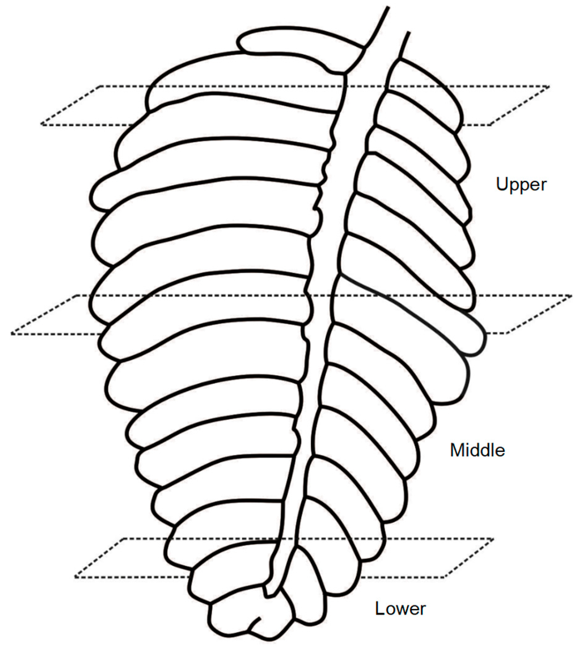
Gill scrapings were examined under a light microscope to detect urceolariid cells. To assess the intensity of infestation by urceolariid ciliates the right and left gills of 15 octopuses were examined. Each gill was sectioned in three segments: upper, middle, and lower sections (Figure 1). Each section was placed between two clean glass slides and observed under the Primo Star microscope (Carl Zeiss, Oberkochen, Germany) at 10× magnification. Mean intensity of infestation was determined by counting five random fields on each slide per segment, while prevalence was determined by the presence-absence of ciliates for all octopuses according to Bush et al. (1997) []. The results were tabulated and presented as mean ± standard deviation followed by minimum and maximum values.
Figure 1. Line drawing of a gill from Octopus bimaculatus. The drawing displays three transverse sections (upper, middle and lower) showing the representative sections used to assess the intensity of infestation by Urceolaria carmenae n. sp.

2.2. Morphological Characterization

The urceolariid cells were repeatedly rinsed with filtered and sterile seawater to eliminate excess gill mucus. Then, approximately 20–30 urceolariid cells per gill were transferred into a microcentrifuge tube. To emphasize the structural details of urceolariid cells, a series of slides were dried and stained with Giemsa or impregnated with 2% silver nitrate (AgNO3) following the Klein method []. Additional samples were stored at −20 °C for subsequent molecular analysis.
The infested gills were fixed in Davidson solution for 24 h, dehydrated in a graded ethanol series and embedded in paraffin wax blocks. The blocks were cut into 4–5 µm sections using a microtome and stained with hematoxylin and eosin (H&E) [].
Photomicrographs of fresh, Giemsa-stained, silver-impregnated urceolariid cells and histological slides were taken using a Primo Star microscope (Carl Zeiss, Oberkochen, Germany) equipped with a Motic 1080 camera and Motic Images Plus v. 3.0.19.113b software (Motic, Xiamen, China). Measurements were performed using ImageJ 1.54m software, available online: https://imagej.net/ij/ (accessed on 2 November 2024). All measurements are given in micrometers (μm) and expressed as the mean, followed by the ±standard deviation (SD) and the range in parentheses.

2.3. Molecular Characterization

Genomic DNA was isolated from the collected ciliates using the PureLink Genomic DNA Mini Kit (Invitrogen, Waltham, MA, USA), following the instructions of the manufacturer. The small subunit (SSU) 18S rRNA gene sequences were amplified by polymerase chain reaction (PCR) using GoTaq Green Master Mix (Promega, Madison, WI, USA) in a final volume of 25 µL (12.5 µL GoTaq Green Master Mix, 2×, 1 µL of upstream primer, 1 µL of downstream primer, 1 µL of DNA template (50 ng/µL) and 9.5 µL of nuclease-free water) and forward and reverse primers 82F (5′-GAAACTGCGAATGGCTC-3′ and LSUR (5′-GTTAGTTTCTTTTCCTCCGC-3′ [], respectively. The DNA concentration of all extracts was quantified using a NanoDrop Spectrophotometer (Thermo Scientific, Wilmington, DE, USA) prior to PCR setup. PCR was performed by a MaxyGene II Thermal cycler (Corning) with the following parameters: initial denaturation at 94 °C for 10 min, followed by 5-step cycling of denaturation at 94 °C for one minute, alignment at 56 °C for 2 min and extension at 72 °C for 2 min, and followed by 35 cycles as the previous parameters, but the temperature of the alignment period increased to 62 °C, and a final extension step at 72 °C for 10 min []. Amplicons were evaluated by electrophoresis on 1% agarose gel electrophoresis, stained with SyBr Safe (Invitrogen), and visualized on an UV transilluminator. Direct sequencing of PCR products was carried out by the Sanger sequencing method in an ABI 3730-XL sequencer (Macrogen Inc., Seoul, Republic of Korea).

2.4. Phylogenetic Analyses

The chromatograms were checked and edited in Chromas software v. 2.6.6 (Technelysium Pty Ltd., South Brisbane, QLD, Australia) and assembled in MEGA 11.0.13 software []. A total of 37 sequences of the 18S rDNA gene were obtained from the GenBank database. Ophryoglena catenula (U17355), Ichthyophthirius multifiliis (KJ690572), and Tetrahymena corlissi (OR858827) were designated as an outgroup. Alignment was performed with ClustalW in MEGA 11.013 []. The alignment was then manually optimized for the best fit to the consensus structure, taking particular note of the length of our sequence. According to the Akaike Information Criterion (AIC) in MEGA 11.013 [], the general time-reversible model with gamma-distributed rate and invariant sites (GTR + G + I) was the most plausible evolution model. The phylogenetic relationships were reconstructed under Bayesian Inference (BI) and Maximum Likelihood (ML) criteria. Bayesian reconstruction was inferred with MrBayes 3.2.7a. using two parallel analyses of Metropolis-Coupled Markov Chain Monte Carlo (MCMCMC) for 2,000,000 generations with sampling every 100 generations, and the first 25% of trees were discarded []. The ML analysis was conducted in MEGA 11.013. Node support was evaluated by non-parametric bootstrapping, and 1000 replicates were performed. Bootstrap (BS) ≥ 70% and posterior probabilities (PP) ≥ 0.85 values were considered strongly supportive for clades. The resulting ML and BI trees were visualized using MEGA11.013 and FigTree v. 1.4.4, respectively. Both trees were edited in CorelDRAW Graphics Suite 2019 v.21.3.0.755 (Corel Corporation, Ottawa, ON, Canada). The genetic distance of sequences was calculated by the pairwise method with the p-distance model in MEGA 11.013 [].

3. Result

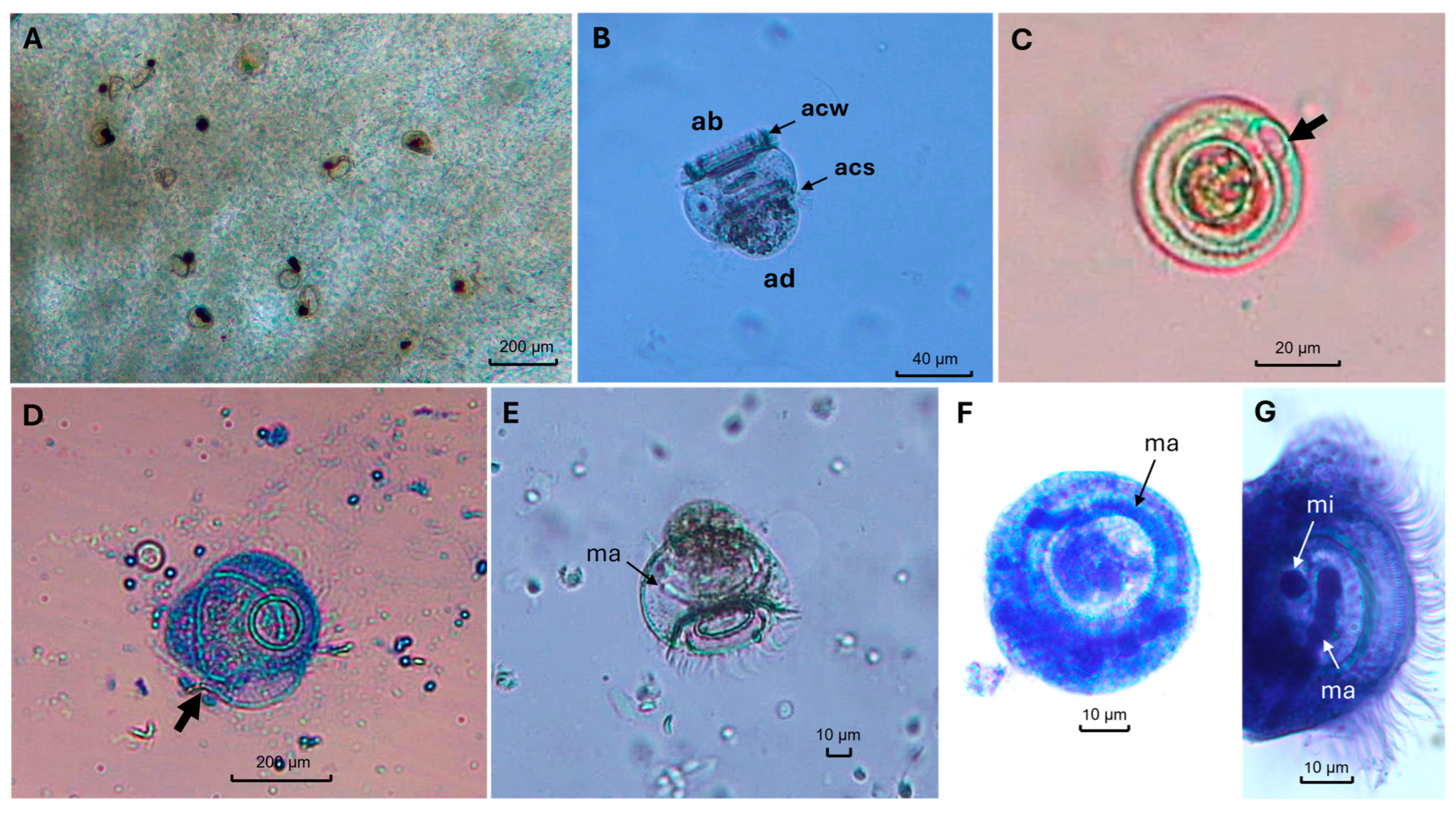
Urceolariid ciliates were detected in the gills of all specimens of O. bimaculatus analyzed (76/76). The mean intensity of ciliates, based on counts from both gills of 15 octopi, was 687 ± 228 (279–1077) ciliates per host (Table 1). All ciliates were morphologically identified as mobilids based on the presence of an adhesive disk, a ring of smooth plates, and an adhesive disk on the aboral pole (Figure 2). Examination of histological sections confirmed the presence of urceolariid ciliates in the gill epithelium and provided additional morphological detail of the ciliate in situ (Figure 3A). During this examination, basophilic inclusions resembling Rickettsia-like organisms (RLOs) were also incidentally observed in the gill tissue. A marked increase in mucus production, visible as a thick, cloudy layer covering the gill filaments, was observed in all infested octopuses and provided the medium in which the ciliates were embedded. No tissue damage was associated with the host gills because of the infestation of Urceolaria carmenae n. sp.
Table 1. Comparative morphological data of Urceolaria carmenae n. sp. from the gills of Octopus bimaculatus in the Gulf of California, and closely related Urceolaria species. Data are shown as mean ± standard deviation (SD), followed by minimum and maximum in parentheses. All measurements are expressed in micrometers (μm).
Figure 2. Urceolaria carmenae n. sp. collected from the gills of Octopus bimaculatus. (A) Fresh smash preparation of the gills showing the infestation of Urceolaria carmenae n. sp. (B) Fresh unstained specimen in lateral view, showing the aboral pole (ab) with its adhesive disc and the opposite adoral pole (ad). The adoral ciliary spiral (acs) and aboral ciliary wreath (acw) are indicated. The dark pigmentation is the result of incidental ink retention from the host’s ink sac. (C) View of oral end showing the infundibulum (black arrow). (D) Giemsa staining shows a complete-turned ciliature making more than 360° turn, indicating the incision where the adoral ciliary spiral begins (black arrow). (E) Lateral view of a fresh ciliate, macronucleus (ma). (F,G) Giemsa staining of ciliates showing the macronucleus (ma) and micronucleus (mi).
Figure 3. Stained Urceolaria carmenae n. sp. collected from the gills of Octopus bimaculatus. (A) Morphology of the urceolariid in a histological slide of octopus gills (H&E staining). (B) After wet silver nitrate staining. (C) After Giemsa staining. Note the 19 skeletal plates (black arrow) and the radial pins of the adoral disk; white arrows indicate the distance between 10 radial pins (166 total); cilia (ci).
Description of Urceolaria carmenae n. sp. (Figure 2, Figure 3 and Figure 4)
Figure 4. Bayesian inference cladogram of Urceolaria carmenae n. sp. (present study) based on the 18S rDNA gene sequences and related Mobilida species. Ophryoglena catenula (U17355), Ichthyophthirius multifiliis (KJ690572), and Tetrahymena corlissi (OR858827) were designated as outgroup taxa. Clade A includes Urceolaria species infesting marine invertebrate hosts, while Clade B contains Urceolaria species associated with freshwater invertebrate hosts. Posterior probabilities (PP)/maximum likelihood bootstrap (BS) values are shown above the nodes. Value node numbers lower than PP < 0.85 and BS < 70% are represented with dashes. Horizontal distance is proportional to hypothesized evolutionary change, and the scale bars represent the number of nucleotide substitutions per site.
Urceolariid ciliates exhibited short and cylindrical bodies (turban-shaped) (Figure 2A–C). Multi-lobed mass, with a micronucleus and macronucleus (Figure 2B). In the lateral view, oral (or) and aboral (ab) regions are similar in size, with the presence of the adoral ciliary spiral (acs), and aboral ciliary wreath (acw) (Figure 2C). The measurements indicated a mean body diameter of 44.2 ± 13.2 (31.3–88.6) µm, with a complete adoral spiral making more than 360° turn (Figure 2C,D), adhesive disk diameter of 36.5 ± 10.7 µm (29.2–74.6) µm, plate ring diameter 28.5 ± 7.1 µm (24.1–54.6) µm, central zone diameter 22.4 ± 4.1 (17.2–37.6) µm, plate length 11.0 ± 0.86 (9–12) µm; with 18–19 skeletal smooth plate overlapping like roof tiles; and 166–169 radial pins (Figure 3A–C). Border membrane of 1.6 ± 0.4 (1–2) µm, total body height 67.0 ± 14.2 (43.0–86.1) µm, body width 58.9 ± 12.1 (43.0–75.3) µm, body height 29.4 ± 7.5 (18.0–42.3) µm (Table 1; Figure 2 and Figure 3).
Taxonomic summary
Phylum: Ciliophora, Doflein, 1901
Class: Oligohymenophorea, de Puytorac, 1974
Subclass: Peritrichia, Stein, 1859
Order: Mobilida, Kahl, 1933
Family: Urceolariidae, Dujardin, 1840
Genus: Urceolaria Stein, 1867
Type host: Octopus bimaculatus Verrill, 1883 (Octopoda: Octopodidae)
Site of infestation: Gills
Prevalence: 100% (76/76)
Type locality: Bahia de Los Angeles, Baja California, Mexico (29°00″ N, 113°30″ W).
Type material: Silver-stained preparation was deposited in the Colección de Invertebrados of the Universidad Nacional Autónoma de México (pending number assignment)
DNA sequence: pending number assignment
Zoobank Registration LSID: urn:lsid:zoobank.org:pub:B1FEE798-D218-48D5-B804-EA91DEEEB4A8
Mean intensity and range: 687 ± 228 (279–1077) urceolariid cells per octopus specimen
Reference sequences: 18S rDNA gene with GenBank accession number (pending number assignment)
Etymology: The specific epithet derives from Professor María del Carmen Gómez del Prado Rosas (Professor of Parasitology in Universidad Autonóma de Baja California Sur, Mexico) in honor of her lifetime contribution to teaching and research on parasitology of wild animals in the Mexican Northwest.
Remarks: According to the morphology of the ciliate described here, in particular, the shape and organization of its plates, it was placed within the genus Urceolaria. In addition, its 18S rDNA gene sequence unambiguously places it with 100% statistical support in a clade with other mobilid peritrichs assigned to the genus Urceolaria.
The morphological characterization supported a consistent genus-level assignment to Urceolaria and revealed metric differences (e.g., cell size) from congeners such as U. urechi. However, a definitive species diagnosis based solely on morphology remained inconclusive. This was due to the nuclear apparatus—a key taxonomic character—which could not be clearly visualized or measured reliably in our prepared specimens. Consequently, the molecular phylogenetic analysis of the 18S rDNA gene was indispensable to provide a robust, quantitative assessment and confirm the genetic distinctness of the proposed new species.
Molecular and phylogenetic analyses
A partial fragment of the 18S rDNA gene, comprising 1987 bp, was obtained. The sequence was placed within the genus Urceolaria (Mobilida: Trichodinidae) based on phylogenetic analyses produced by BI and ML, which resulted in comparable tree topologies. Both trees showed similar topologies (Figure 4). The Urceolaria spp. clade was recovered as a monophyletic group in both phylogenetic analyses (PP = 0.86; BS = 95%). Within this clade, three well-supported subgroups were identified for the urceolariids, and our newly obtained sequence belongs to one of these clades, grouped as a sister taxon to the congeners Urceolaria (=Leiotrocha) serpularum (JQ663867) and Urceolaria urechi (FJ499388). This assignment was supported by robust values (PP = 1; BS = 99%) (Figure 4). Moreover, these three sequences are nested with U. clepsydra and U. korschelti (PP = 1; BS = 100%) (Figure 4). The genetic analysis revealed 90.6–98.8% sequence similarity (≥1.0% genetic distance) with their congeners (Table 2).
Table 2. Genetic matrix for the 18S rDNA sequences from Urceolaria species. Genetic distances (p-distance) are below the diagonal (%), while those above indicate sequence similarities (%).

4. Discussion

4.1. Morphological Diagnosis and Taxonomic Distinctness

Mobilids are distinguished by having a complex adhesive disk reinforced by a ring of skeletal plates on the aboral body pole. This sophisticated adhesive apparatus enables them to attach to animate or living substrates from a diverse array of aquatic organisms and terrestrial animals with aquatic ancestry [,]. All reliable studies on urceolariids have been reported in mollusks. However, this constitutes the first confirmed report of a urceolariid ciliate in a cephalopod, herein described as Urceolaria carmenae n. sp., found infesting the gills of O. bimaculatus.
Research on Urceolaria species remains limited; consequently, detailed morphological data are still lacking (Table 1). According to the morphology, Urceolaria carmenae n. sp. is only similar to Urceolaria urechi in the turban body shape and the number of skeletal plates (18–19 vs. 18–25, respectively) (Table 1). However, Urceolaria carmenae n. sp. is smaller than Urceolaria urechi in the body diameter (44 vs. 58, respectively) and body height (29 vs. 40, respectively).
A detailed morphometric characterization of the nuclear apparatus (macronucleus and micronucleus) of U. carmenae n. sp. could not be reliably established in this study. The nuclear material in our specimens was not consistently visible or optimally oriented for accurate measurement across individuals. Providing robust quantitative data from the available images would be methodologically unsound. Therefore, a definitive description of the nuclear structure and its division pattern remains an important goal for future research. Such work should involve specific staining techniques (e.g., DAPI, Feulgen) applied to freshly collected specimens to allow accurate visualization and morphometry analysis.

4.2. Phylogenetic Affiliation and Evolutionary Relationships

The phylogenetic analyses of 18S rDNA and the percentage of genetic divergence (≥1%) support that Urceolaria carmenae n. sp. is a new species (Figure 4, Table 2). Both Bayesian and ML analyses grouped Urceolaria carmenae n. sp. in a monophyletic clade with the Urceolaria species (Figure 4). The clade of Urceolaria is divided into two clades (PP = 0.74; BS = 96%); clade A includes Urceolaria species infesting the marine invertebrate species, U. bratalia, U. carmenae n. sp., U. clepsydra, U. korschelti, U. parakorschelti, U. serpularum, and U. urechi. Clade B contains the sequences of U. mitra, the unique species that has been associated with freshwater invertebrates, the flatworms Platyhelminthes, Dugesia gonocephala Duges, 1830, and Polycelis tenuis Ijima, 1884 [,]. Mainly, U. carmenae n. sp. is a sister group with high support (PP = 1; BS = 99%) values with U. urechi and U. serpularum sequences annotated by [] and [], respectively. The latest Urceolaria species were described as inhabiting marine invertebrates of the phylum Annelida, Class Polychaeta. Urceolaria urechi inhabiting Urechis unicinctus (fat innkeeper worm), and U. serpularum in the Serpula sp. (plume worm) gills, both collected on the coast of China [,].
In 2025, Martinez et al. [] reported two new species collected in British Columbia, Canada. The first of these, U. clepsydra, was found in the sea cucumbers, C. miniata and Eupentacta quinquesemita; the second, U. bratalia, in the lamp shells, Terebratalia transversa. In their phylogenetic analysis, U. clepsydra is a sister to U. urechi and U. serpularum, sharing 97.3% of genetic similarity, which could indicate that U. carmenae n. sp. and U. clepsydra could be phylogenetically closely related. Although morphologically, U. carmenae n. sp., is more related to U. bratalia. However, the sequences of U. clepsydra and U. bratalia are not yet available in GenBank and, therefore, are not included in the present study. The above emphasized that molecular phylogenetic analyses are key to elucidating the phylogenetic placement of unknown members of the genus Urceolaria.

4.3. Ecological Context and Host-Symbiont Dynamics

In this study, the prevalence of infestation by Urceolaria carmenae n. sp. was 100% in the octopuses analyzed between 2020 to 2022. The precise causes of such events are unclear, but might be attributable to an unidentified environmental driver. The use of protozoan communities as sensitive bioindicators of environmental conditions is well established in aquatic ecosystems []. This precedent raises the possibility that population dynamics of Urceolaria carmenae n. sp. could serve as a sentinel for subtle changes in local oceanographic conditions (e.g., temperature, productivity). Monitoring its abundance and prevalence could thus be a valuable line of future ecological research.
The life cycle of peritrichous ciliates is completed in a single host by binary fission or conjugation. This allows the rapid propagation of ciliates within host populations, especially under conditions of stress or suboptimal environmental conditions, high stocking densities, rising water temperatures, and excessive amounts of organic matter in the water [,]. For several years, our group has been conducting its research on the parasite fauna of O. bimaculatus in the Mexican Pacific Ocean and the Gulf of California, Mexico. Thus far, our findings have demonstrated that O. bimaculatus is typically found in association with the dicyemid Dicyemennea abelis McConnaughey, 1949, the Apicomplexa, Aggregata polibraxiona Colunga-Ramírez, Martínez-Aquino, Flores-López, Gestal, Azevedo, and Castellanos-Martínez 2021, trematodes of Lecithochirium sp., and the marine leech, Cochimibdella mexicana Fernando Ruiz-Escobar, Graciela E. Colunga-Ramírez, Alejandro Oceguera-Figueroa, Sheila Castellanos-Martínez 2025 [,,,]. Nevertheless, the present study constitutes the first record of ciliates in the gills of cephalopods and, moreover, the first event in which infestation of ciliates in the gills of cephalopods is accounted. Although most mobilids are classified as commensals, they feed on particles from the surfaces of organisms, such as gills or skin. However, in cases where they occur in large numbers on healthy organisms, their constant attachment and movement, caused by the adhesive disc, damage the epithelium and consequently cause irritation that can compromise the host’s health and welfare. In such cases, mobilids behave like harmful parasites, feeding on disrupted cells and bacterial proliferation []. To date, the cause and consequence of infestation in O. bimaculatus are uncertain.
An intriguing finding was the co-occurrence of U. carmenae n. sp. with basophilic, intracytoplasmic inclusions resembling Rickettsia-like organisms (RLOs) in the gill epithelium of the same host individuals. It is crucial to note that our study design, based on field samples, cannot determine a causal or temporal relationship between these two agents; we cannot ascertain whether one facilitates the other or if both simply occur in the same host in response to an underlying condition. This precise ecological interaction represents a compelling target for future research. Ultimately, this study underscores the need for continued health surveillance of wild octopus populations, which should include evaluating the impact of symbionts like urceolariids and identifying the environmental drivers of their fluctuating prevalence.

5. Conclusions and Future Directions

This study establishes the first record of a mobilid ciliate infesting a cephalopod host, Octopus bimaculatus from the Gulf of California, and confirms through integrative taxonomy that it represents a new species, herein described as Urceolaria carmenae n. sp. A key finding was the absence of specific damage to the gill tissue despite high infestation loads, suggesting a commensal relationship under the conditions observed. However, given the documented potential of heavy mobilid infestations to cause pathology in other hosts, this dynamic warrants careful monitoring. Future studies are strongly encouraged to evaluate the pathological implications of this association, particularly under scenarios of environmental stress, to ascertain its true impact on the health of wild and cultured octopus populations.

Author Contributions

Data curation, G.E.C.-R.; Funding acquisition, S.C.-M.; Investigation, S.C.-M. and G.E.C.-R.; Methodology, S.C.-M. and G.E.C.-R.; Resources, S.C.-M.; Supervision, S.C.-M.; Writing—original draft, G.E.C.-R.; Writing—review & editing, S.C.-M. All authors have read and agreed to the published version of the manuscript.

Funding

This study was supported by the Secretariat of Science, Humanities, Technology, and Innovation, Mexico (SECIHTI, Mexico; project number: 286347).

Institutional Review Board Statement

Ethical review and approval were waived for this study due to two main reasons: 1. Regulatory Framework: Cephalopods, including the species Octopus bimaculatus, are not classified as laboratory animals under the Mexican Official Standard that regulates animal experimentation (NOM-062-ZOO-1999 (“Especificaciones técnicas para la producción, cuidado y uso de los animales de laboratorio”). Therefore, formal ethical committee approval is not mandated by national law for studies involving these organisms. 2. Source of Specimens: The octopuses used in this study were not collected directly by the authors. They were purchased from a licensed fishing cooperative, “Cooperativa Buzos de Bahía”, which operates under a valid commercial fishing permit (Permiso de Pesca Comercial) issued by the relevant Mexican authority (CONAPESCA). The animals were harvested as part of their routine, legal fishing activities independent of this research project.

Data Availability Statement

The original contributions presented in this study are included in the article. Further inquiries can be directed to the corresponding author.

Acknowledgments

We are deeply grateful to the anonymous reviewers for their meticulous evaluation and constructive suggestions, which were instrumental in strengthening this work. We also extend our warm thanks to Alma G. Islas-Ortega for their invaluable assistance in reviewing several figures of ciliates to support the morphological description.

Conflicts of Interest

The authors declare that they have no competing interests.

References

  1. Tedesco, P.; Bevilacqua, S.; Fiorito, G.; Terlizzi, A. Global patterns of parasite diversity in cephalopods. Sci. Rep. 2020, 10, 11303. [Google Scholar] [CrossRef]
  2. Souidenne, D.; Furuya, H. Protist (Ciliates) and related diseases. In Handbook of Pathogens and Diseases in Cephalopods; Gestal, C., Pascual, S., Guerra, Á., Fiorito, G., Vieites, J.M., Eds.; Springer International Publishing: Cham, Switzerland, 2019; pp. 153–158. ISBN 978-3-030-11330-8. [Google Scholar]
  3. Zhan, Z.; Xu, K.; Warren, A.; Gong, Y. Reconsideration of phylogenetic relationships of the subclass Peritrichia (Ciliophora, Oligohymenophorea) based on small subunit ribosomal RNA gene sequences, with the establishment of a new subclass Mobilia Kahl, 1933. J. Eukaryot. Microbiol. 2009, 56, 552–558. [Google Scholar] [CrossRef]
  4. Lynn, D.H. The ciliate taxa, including families and genera. In The Ciliated Protozoa: Characterization, Classification, and Guide to the Literature; Springer: Dordrecht, The Netherlands, 2008; pp. 339–440. ISBN 978-1-4020-8239-9. [Google Scholar]
  5. Corliss, J.O. The Ciliated Protozoa: Characterization, Classification and Guide to the Literature, 2nd ed.; Pergamon: Oxford, UK, 2016; ISBN 978-1-4831-5417-6. [Google Scholar]
  6. Xu, K.; Song, W.; Warren, A. Observations on trichodinid ectoparasites (Ciliophora: Peritricha) from the gills of maricultured molluscs in China, with descriptions of three new species of Trichodina Ehrenberg, 1838. Syst. Parasitol. 2000, 45, 17–24. [Google Scholar] [CrossRef]
  7. Zhan, Z.; Xu, K.; Dunthorn, M. Evaluating molecular support for and against the monophyly of the Peritrichia and phylogenetic relationships within the Mobilida (Ciliophora, Oligohymenophorea). Zool. Scr. 2013, 42, 213–226. [Google Scholar] [CrossRef]
  8. Tang, F.; Zhang, Y.; Zhao, Y. Morphological and molecular identification of the new species, Trichodina pseudoheterodentata sp. n. (Ciliophora, Mobilida, Trichodinidae) from the channel catfish, Ictalurus punctatus, in Chongqing China. J. Eukaryot. Microbiol. 2017, 64, 45–55. [Google Scholar] [CrossRef]
  9. Irwin, N.A.T.; Sabetrasekh, M.; Lynn, D.H. Diversification and phylogenetics of mobilid peritrichs (Ciliophora) with description of Urceolaria parakorschelti sp. nov. Protist 2017, 168, 481–493. [Google Scholar] [CrossRef]
  10. Van As, L.L.; Basson, L.; Van As, J. Mobiline peritrichs (Ciliophora) collected from the gills of African limpets. Acta Protozool. 2017, 56, 245–254. [Google Scholar] [CrossRef]
  11. Xu, K.; Song, W. Two trichodinid ectoparasites from marine molluscs in the Yellow Sea, Off China, with the description of Trichodina caecellae n. sp. (Protozoa: Ciliophora: Peritrichia). Syst. Parasitol. 2007, 69, 1–11. [Google Scholar] [CrossRef]
  12. Hirshfield, H. The morphology of Urceolaria karyolobia, sp. nov., Trichodina tegula sp. nov., and Scyphidia ubiquita sp. nov., three new ciliates from Southern California limpets and turbans. J. Morphol. 1949, 85, 1–33. [Google Scholar] [CrossRef]
  13. Richards, C.S. Urceolaria viridis n. sp., a ciliate (Peritrichida, Mobilina) with elongate symbiotic green algae. J. Protozool. 1971, 18, 410–413. [Google Scholar] [CrossRef]
  14. Rataj, M.; Vd’ačný, P. Cryptic host-driven speciation of mobilid ciliates epibiotic on freshwater planarians. Mol. Phylogenetics Evol. 2021, 161, 107174. [Google Scholar] [CrossRef]
  15. Hausmann, K.; Bradbury, P.C. Ciliates: Cells as Organisms; Gustav Fischer Verlag: Stuttgart, Germany, 1996; ISBN 978-1-56081-432-0. [Google Scholar]
  16. Noble, G.A. Trichodina urechi n. sp., an entozoic ciliate from the echiuroid worm, Urechis caupo. J. Parasitol. 1940, 26, 387–405. [Google Scholar] [CrossRef]
  17. Haider, G. Monographie der Familie Urceolariidae (Ciliata, Peritricha, Mobilina) mit besonderer berucksichtigung der imsu¨ ddeutschen raum vorkommenden arten. Parasitol. Schriftenr. 1964, 17, 1–251. [Google Scholar]
  18. Irwin, N.A.T.; Lynn, D.H. Molecular phylogeny of mobilid and sessilid ciliates symbiotic in Eastern Pacific limpets (Mollusca: Patellogastropoda). J. Eukaryot. Microbiol. 2015, 62, 543–552. [Google Scholar] [CrossRef]
  19. Martinez, G.; Leander, B.S.; Park, E. Morphology and molecular phylogeny of endosymbiotic ciliates (Peritrichia, Mobilida) of marine invertebrates with descriptions of two novel species Urceolaria clepsydra n. sp. and Urceolaria bratalia n. sp. J. Eukaryot. Microbiol. 2025, 72, e70003. [Google Scholar] [CrossRef]
  20. Zick, K. Urceolaria korschelti sp. n., eine neue marine Urceolariide nebst einem Oberblick fiber die Urceolarinen. Z. Wiss. Zool. 1928, 132, 356–403. [Google Scholar]
  21. Jereb, P.; Roper, C.F.E.; Norman, M.D.; Finn, J.K. Cephalopods of the world. In An Annotated and Illustrated Catalogue of Cephalopod Species Known to Date: Octopods and Vampire Squids; FAO: Rome, Italy, 2016; Volume 3, ISBN 978-92-5-107989-8. [Google Scholar]
  22. Domínguez-Contreras, J.F.; Munguia-Vega, A.; Ceballos-Váazquez, B.P.; Arellano-Martínez, M.; García-Rodríguez, F.J.; Culver, M.; Reyes-Bonilla, H. Life histories predict genetic diversity and population structure within three species of octopus targeted by small-scale fisheries in Northwest Mexico. PeerJ 2018, 6, 1–25. [Google Scholar] [CrossRef]
  23. Díaz-Santana-Iturrios, M.; Salinas-Zavala, C.A.; García-Rodríguez, F.J.; Granados-Amores, J. Taxonomic assessment of species of the genus Octopus from the northeastern Pacific via morphological, molecular and morphometric analyses. PeerJ 2019, 7, e8118. [Google Scholar] [CrossRef]
  24. DOF Acuerdo por el que se Modifica el Similar por el que se Establece la Veda Temporal y Tallas Mínimas de Captura para la Pesca de las Especies de Pulpo en Bahía de Los Ángeles, Baja California, Publicado El 1 de Junio de 2016, para Considerar como Zona de Veda la Reserva de La Biosfera Bahía de Los Ángeles, Canales de Ballenas y de Salsipuedes, Respecto del Pulpo Café (Octopus bimaculatus) y Pulpo Verde (Octopus hubbsorum). Available online: https://dof.gob.mx/nota_detalle.php?codigo=5503748&fecha=07/11/2017#gsc.tab=0 (accessed on 18 September 2025).
  25. Colunga-Ramírez, G.E.; Del Rio-Zaragoza, O.B.; Castellanos-Martínez, S. Unusual formation of air bubbles in the arms of the california two-spot octopus, Octopus bimaculatus Verrill, 1883 during handling in captivity. Mar. Biol. 2023, 170, 143. [Google Scholar] [CrossRef]
  26. Shomrat, T.; Zarrella, I.; Fiorito, G.; Hochner, B. The octopus vertical lobe modulates short-term learning rate and uses LTP to acquire long-term memory. Curr. Biol. 2008, 18, 337–342. [Google Scholar] [CrossRef]
  27. Fiorito, G.; Affuso, A.; Basil, J.; Cole, A.; De Girolamo, P.; D’Angelo, L.; Dickel, L.; Gestal, C.; Grasso, F.; Kuba, M.; et al. Guidelines for the care and welfare of cephalopods in research –a consensus based on an initiative by CephRes, FELASA and the Boyd group. Lab. Anim. 2015, 49, 1–90. [Google Scholar] [CrossRef]
  28. Bush, A.O.; Lafferty, K.D.; Lotz, J.M.; Shostak, A.W. Parasitology meets ecology on its own terms: Margolis et al. Revisited. J. Parasitol. 1997, 83, 575–583. [Google Scholar] [CrossRef]
  29. Klein, B.M. The “Dry” silver method and its proper use. J. Protozoo. 1958, 5, 99–103. [Google Scholar] [CrossRef]
  30. Culling, C.F.A.; Allison, R.T.; Barr, W.T.; Culling, C.F.A. Cellular Pathology Technique, 4th ed.; Butterworths: London, UK; Boston, MA, USA, 1985; ISBN 978-0-407-72903-2. [Google Scholar]
  31. Bourland, W.A.; Strüder-Kypke, M.M. Agolohymena aspidocauda nov. gen., nov. spec., a histophagous freshwater tetrahymenid ciliate in the family Deltopylidae (Ciliophora, Hymenostomatia), from Idaho (northwest U.S.A.): Morphology, ontogenesis and molecular phylogeny. Eur. J. Protistol. 2010, 46, 221–242. [Google Scholar] [CrossRef]
  32. Tang, F.; Zhao, Y.; Warren, A. Phylogenetic analyses of Trichodinids (Ciliophora, Oligohymenophora) inferred from 18S rRNA gene sequence data. Curr. Microbiol. 2012, 30, 306–313. [Google Scholar] [CrossRef]
  33. Tamura, K.; Stecher, G.; Kumar, S. MEGA11: Molecular evolutionary genetics analysis version 11. Mol. Biol. Evol. 2021, 38, 3022–3027. [Google Scholar] [CrossRef]
  34. Ronquist, F.; Teslenko, M.; van der Mark, P.; Ayres, D.L.; Darling, A.; Höhna, S.; Larget, B.; Liu, L.; Suchard, M.A.; Huelsenbeck, J.P. MrBayes 3.2: Efficient Bayesian phylogenetic inference and model choice across a large model space. Syst. Biol. 2012, 61, 539–542. [Google Scholar] [CrossRef]
  35. Lom, J.; Dyková, I. Protozoan Parasites of Fishes; Elsevier Science: Amsterdam, The Netherlands, 1992; ISBN 978-0-444-89434-2. [Google Scholar]
  36. Reynoldson, T.B. The dispersal of Urceolaria mitra (Peritricha) epizoic on flatworms. J. Anim. Ecol. 1951, 20, 123. [Google Scholar] [CrossRef]
  37. Rataj, M.; Vd’ačný, P. Living morphology and molecular phylogeny of Oligohymenophorean ciliates associated with freshwater turbellarians. Dis. Aquat. Org. 2019, 134, 147–166. [Google Scholar] [CrossRef]
  38. Xu, H.; Song, W.; Warren, A.; Al-Rasheid, K.A.S.; Al-Farraj, S.A.; Gong, J.; Hu, X. Planktonic protist communities in a semi-enclosed mariculture pond: Structural variation and correlation with environmental conditions. J. Mar. Biol. Assoc. 2008, 88, 1353–1362. [Google Scholar] [CrossRef]
  39. Martins, M.L.; Cardoso, L.; Marchiori, N.; Benites de Pádua, S. Protozoan infections in farmed fish from Brazil: Diagnosis and pathogenesis. Rev. Bras. Parasitol. Vet. 2015, 24, 1–20. [Google Scholar] [CrossRef]
  40. Ruiz-Escobar, F.; Colunga-Ramírez, G.E.; Oceguera-Figueroa, A.; Castellanos-Martínez, S. Cochimibdella mexicana n. gen. n. sp. (Hirudinida: Piscicolidae), from Octopus bimaculatus Verrill (Cephalopoda: Octopodidae) in the Gulf of California, Mexico. Syst. Parasitol. 2025, 102, 23. [Google Scholar] [CrossRef]
  41. Chan-Martin, A.D.J.; Castellanos-Martínez, S.; Aguirre-Macedo, M.L.; Martínez-Aquino, A. Immature trematodes of Lecithochirium sp. (Digenea: Hemiuridae) in the California two-spot octopus (Octopus bimaculatus) from Mexico. Parasitol. Res. 2022, 121, 2651–2660. [Google Scholar] [CrossRef]
  42. Colunga-Ramírez, G.E.; Martínez-Aquino, A.; Flores-López, C.A.; Gestal, C.; Azevedo, C.; Castellanos-Martínez, S. Aggregata polibraxiona n. sp. (Apicomplexa: Aggregatidae) from Octopus bimaculatus Verrill, 1883 (Mollusca: Cephalopoda) from the Gulf of California, Mexico. Eur. J. Protistol. 2021, 81, 125825. [Google Scholar] [CrossRef]
  43. McConnaughey, B.H. Mesozoa of the family Dicyemidae from California. Univ. Calif. publ. Zool. 1949, 55, 1–34. [Google Scholar]
Disclaimer/Publisher’s Note: The statements, opinions and data contained in all publications are solely those of the individual author(s) and contributor(s) and not of MDPI and/or the editor(s). MDPI and/or the editor(s) disclaim responsibility for any injury to people or property resulting from any ideas, methods, instructions or products referred to in the content.

Article Metrics

Citations

Article Access Statistics

Multiple requests from the same IP address are counted as one view.